Pediatr Radiol (1993) 23:45%462
Pediatric
Radiology
9 Springer-Verlag 1993Chronic intoxication by ethane-l-hydroxy-l,l-diphosphonate (EHDP)
in a child with myositis ossificans progressiva
U. E. Pazzaglia 1, G. Beluffi 2, A. Ravelli 3, G. Zatti 1, A. Martini 3
1 Clinica Ortopedica, Ospedale F. Del Ponte, Varese, Italy
2 Servizio di Radiodiagnostica, Sezione Radiologia Pediatrica, Pavia, Italy
3 Clinica Pediatrica dell'Universit~ di Pavia, IRCCS Policlinico S. Matteo, Pavia, Italy Received: 4 February 1993/Accepted: 25 March 1993
Abstract.
A c h i l d w i t h m y o s i t i s ossificans p r o g r e s s i v a was t r e a t e d for 8 y e a r s w i t h e t h a n e - l - h y d r o x y - l , l - d i p h o s p h o - n a t e ( E H D P ) 3 0 - 4 0 m g / k g p e r day. L a t t e r l y h e c o m - p l a i n e d o f s e v e r e , p r o g r e s s i v e b o n e a n d j o i n t p a i n w h i c h m a d e s t a n d i n g a n d w a l k i n g a l m o s t i m p o s s i b l e . A r a d i o - g r a p h i c s k e l e t a l s u r v e y s h o w e d diffuse r i c k e t - l i k e lesions. W i t h d r a w a l o f E H D P t h e r a p y p r o d u c e d s u b s t a n t i a l im- p r o v e m e n t in his g e n e r a l c o n d i t i o n as w e l l as in t h e r a d i o - g r a p h i c a p p e a r a n c e o f t h e b o n e s . M u l t i p l e e x o s t o s e s w e r e o b s e r v e d in this c a s e and, p a r t i c u l a r l y t h o s e a r o u n d t h e k n e e s , p r e s e n t e d a p e c u l i a r m o r p h o l o g y . T h i s s u p p o r t s t h e t h e o r y t h a t e x o s t o s e s o r i g i n a t e f r o m a d e f e c t o f m e t a - p h y s e a l m o d e l l i n g . E t h a n e - l - h y d r o x y - l , l - d i p h o s p h o n a t e ( E H D P ) was t h e first d i p h o s p h o n a t e to b e u s e d in clinical p r a c t i c e t o i n h i b i t o s t e o c l a s t i c r e s o r p t i o n a n d t h e p r o c e s s o f c a l c i f i c a t i o n [1]. I n this c h i l d w i t h m y o s i t i s ossificans p r o g r e s s i v a it h a d b e e n a d m i n i s t e r e d o v e r a l o n g p e r i o d to c o u n t e r a c t t h e p r o g r e s s i v e o s s i f i c a t i o n c a u s e d b y t h e d i s e a s e . T h e clinical a n d r a d i o g r a p h i c e v o l u t i o n o f t h e c a s e i l l u s t r a t e t h e side effects o f t h i s t h e r a p y in m y o s i t i s ossificans p r o g r e s s i v a a n d t h e c a r e w h i c h is n e c e s s a r y w h e n b o n e r e m o d e l l i n g in- h i b i t o r s a r e u s e d in c h i l d r e n .Case report
In April 1983, a 5-year-old boy complained of pain in the right knee which was followed by the appearance of a small hard nodule over the lateral aspect of the joint. No congenital or hereditary diseases were present in the family history and no relevant disease had been reported in the first 5 years of life. Shortly afterwards a localized swelling appeared in the right sternocleidomastoid muscle. Initially the lesion was hot and tender, but after a few days the inflammation subsided, leaving a smaller ossified lesion in a retracted muscle. After 5 months the child was found to have torticollis. A muscle bi-
Correspondence to: U. E. Pazzaglia, Clinica Ortopedica, II Facolt5 di Medicina e Chirurgia dell'Universith di Pavia, Ospedale E Del Ponte, 1-21100 Varese, Italy
opsy performed elsewhere did not show any relevant pathological change. Treatment with steroid was started and continued for sev- eral months.
One and half years later a large soft tissue swelling appeared in the shoulder girdle and followed a course similar to the lesion in the sternodeidomastoid muscle. The child was admitted to another hos- pital 1 month later. Laboratory tests showed that inflammatory pa- rameters, serum calcium and phosphate, alkaline phosphatase and muscle enzymes were normal. Electromyography was also normal. A muscle biopsy was consistent with myositis ossificans progressiva. Therapy was started with ethane-l-hydroxyd,l-diphosphonate (EHDP) 30 mg/kg per day. Over the following months new lesions occurred periodically and spread diffusely m the cervical and dorsal regions and sacrum. Limitation of movement developed in the major joints of the limbs, with the exception of the knees.
Two and half years after the appearance of the first signs, diffuse rigidity and widespread caldficafion of soft tissue were present de- spite continuous therapy with EHDR Repeated laboratory tests showed a normal level of alkaline phosphatase. The dose of EHDP was increased to 40 mg/kg per day and low-dose prednisone, piroxi- cam and androgens were added. Physiotherapy was performed throughout this time in an attempt to prevent joint contractures. In spite of the therapy ossification of muscles, tendons and fascias pro- gressed with increasing limitation of movement and contractures of joints in the following 3 years.
The patient was first admitted to the Paediatric Clinic of the University of Pavia at the age of 13 years, after having taken EHDP continuously for 8 years. Physical examination showed severely re- duced mobility of shoulders, elbows, wrists, hips and ankles; the neck was flexed and ankylosed; the dorsal spine had a severe ky- phoscoliosis and widespread subcutaneous calcifications were pre- sent in the right scapular region. Muscles were diffusely wasted. A short, valgus big toe was present in both feet. The child referred severe pain to joints and bones; walking and standing were almost impossible. The results of routine laboratory tests were normal, as were those on serum muscle enzymes. Bone metabolism parame- ters were as follows: calcium 9,8 mg/dl [normal range (NR) 9-11]; phosphate 5.0 mg/dl (NR 3-5). Alkaline phosphatase 1942 (in- creased) fundal examination was normal. Functional tests of respir- ation revealed severe restrictive changes. Radiography showed frank ricket-like lesions (see below); therefore therapy with diphos- phonates was withdrawn.
Two months later the patient's general condition had improved, the pain had disappeared and he could walk without help. Stiffness and joint contractures were unchanged, but the absence of pain had improved his ability in daily activities and he moved more confident- ly. Results of laboratory tests were as follows: calcium 9.3 mg/dl (NR %11); phosphate 6.8 mg/dl (NR 3-5); alkaline phosphatase 1849 (in-
460
creased); osteocalcin 36.3mg/ml (NR 2.0-8.5); vitamin D3 5.2 mg/ml (NR 16.0-74.0); procollagen I > 500 ng/ml (NR 50.0- 170.0); procollagen II1200 U/ml (NR 0.3-0.8).
Radiographic findings
Skeletal radiographs performed at different times were available: 1. When the child was 12 years old after 7 years of therapy with
diphosphonates
2. At 13 years, when the decision to withdraw therapy was taken 3. Three months after withdrawal of diphosphonates
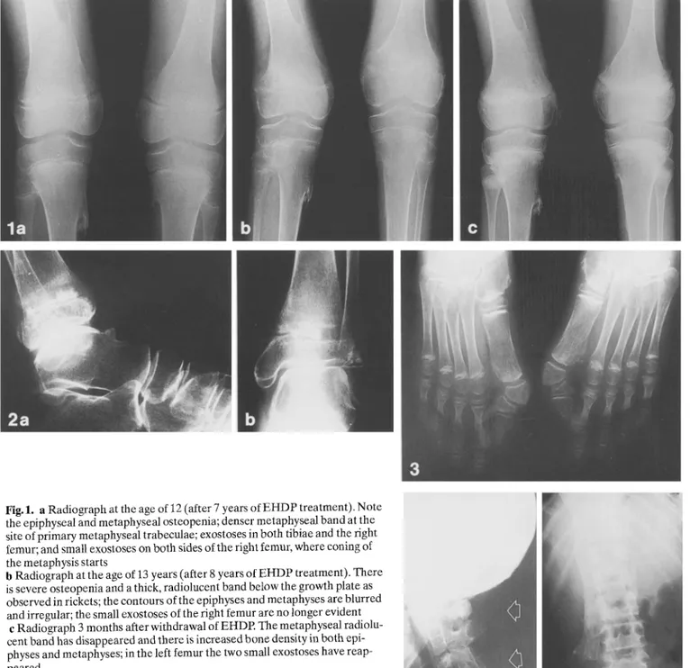
1. At 12 years epiphyseal and metaphyseal osteopenia of the proximal tibia and distal femur was evident. The growth plate was regular and of normal thickness. A thin denser band was present in the upper part of the metaphysis just below the growth plate (corre- sponding to the area of primary metaphyseal trabeculae). The proxi- mal tibial metaphyses on the medial side showed bony beaks similar to exostoses; a similar formation was present in the right distal fem- oral metaphysis. Small exostoses were present on both sides of the left distal femoral metaphysis (Fig. i a).
2. At 13 years more severe osteopenia was evident in the grow- ing metaphyses and there was, furthermore, a very broad band of radiolucency (osteoid) between the growth plate and metaphysis such as is usually seen in severe rickets. Within the metaphyseal
Fig. 1. a Radiograph at the age of 12 (after 7 years of EHDP treatment). Note the epiphyseal and metaphyseal osteopenia; denser metaphyseal band at the site of primary metaphyseal trabeculae; exostoses in both tibiae and the right femur; and small exostoses on both sides of the right femur, where coning of the metaphysis starts
b Radiograph at the age of 13 years (after 8 years of EHDP treatment). There is severe osteopenia and a thick, radiolucent band below the growth plate as observed in rickets; the contours of the epiphyses and metaphyses are blurred and irregular; the small exostoses of the right femur are no longer evident
c Radiograph 3 months after withdrawal of EHDP. The metaphyseal radiolu- cent band has disappeared and there is increased bone density in both epi- physes and metaphyses; in the left femur the two small exostoses have reap- peared
Fig.2a, b. Radiographs at the age of 13 years (after 8 years of EHDP treat- ment). There is osteopenia and ricket-like changes of the distal tibial meta- physis; inside the thick radiolucent band an irregular and dense calcification is present; note the exostosis of the tarsal bone
band of osteoid dense areas of calcification were present (Fig. lb); the contours of the epiphyseal ossification centres and the borders of the metaphyses were blurred and irregular. Cloaking of tibial and fibular shafts was also evident. Both proximal tibial metaphyses had a cylindrical shape with the lower angle corresponding to the base of the exostoses. The small exostoses in the left femur were no longer evident (Fig. I b). Similar changes were evident in both proximal metaphyses of the humeri, distal radii, proximal and distal femora, chondrosternal junctions and vertebral end plates; they were not present in the metaphyses of the tubular bones of the hands and feet. Osteopenia was also evident in the shafts of the long bones, the carpal and tarsal bones (Fig.2) and the epiphyses of long bones. The skull, facial bones, clavicles, ribs and pelvic bones showed mineral density within the normal range. A taIar beak was present in both talar bones (Fig. 2). The first metatarsals were short and stubby and the proximal phalanges were divided into two large triangular bones resulting in valgus deviation (Fig. 3). Other find- ings consistent with myositis ossificans progressiva were the wide- spread calcification of the nuchal ligament (Fig.4 a), dorsal and lumbar paravertebral muscles (Fig. 4 b), and interspinous and inter- transverse ligaments of the cervical spine. The apophyseal joints between C2 and C7 were fused (Fig.4a). The seventh and the eighth left ribs showed a sharp angulation in their second portion and were fused in a large, irregular bony structure abutting below the muscles.
3. At follow-up 3 months after withdrawal of diphosphonates a dramatic disappearance of the rnetaphyseal osteoid border was evi- dent. In its place bone denser than normal was detectable and the zone of provisional calcification was sharp and well defined. Both proximal tibial epiphyses presented a double bony contour, instead of the blurred and irregular border previously observed. Above the left distal femoral metaphysis the two small exostoses were again evident (Fig. 1 c). Osteopenia was also no longer present in the other bones.
Discussion
Skeletal malformations and abnormalities of the big toes are present in 95 % of cases of myositis ossificans pro- gressiva [2] and are therefore of great diagnostic value. Four subtypes of malformations of the big toe have been described [3]: those observed in the case r e p o r t e d here fit type 1, but with the peculiar feature (previously unre- ported) that the single proximal phalanx was divided into two large triangular bones.
E H D P has been used in the treatment of myositis ossi- ficans progressiva to prevent mineralization of areas of ac- tive myositis or remineralization after surgical removal of ectopic ossifications [4-6]. Controversial findings regard- ing the efficacy of this therapy have been r e p o r t e d [2, 7] and variability of results has been ascribed to the activity of the disease and to the plasma level of diphosphonate, which in turn depends on the oral dosage and intestinal absorption [8]. Doses which vary from a minimum of 5 mg/kg per day to a maximum of 40 mg/kg per day have
Fig.3. Radiograph at the age of 13 years (after 8 years of EHDP treatment). Note the dense metaphyseal band of the first metatarsal and the widening of the growth plate; the proximal phalanx is formed by two triangular bones and the toe is deviated in valgus
Fig.4a, b. Radiographs at the age of 13 years (after 8years of EHDP treatment), a Ossification of the nuchal ligament
(arrows)
and interspinous and intertransverse ligaments of the cervical spine. b Ossification of the dorsal and lumbar paravertebral muscles
461 been used [7]. Complications of diphosphonate therapy have been reported by Smith et al. [8] in a boy treated with E H D P 10 mg/kg per day for 2 years and by Rogers et al. [9] in a 3-year-old boy treated with 37 mg/kg per day. The former showed widened growth plates, dense metaphyses and pathological decalcification, while bone pain and muscle weakness were absent. T h e latter complained of hypotonia, weakness and a shuffling, unsteady gait, and radiographs showed generalized osteopenia, transverse radiolucent bands in the metaphyses of the long bones and widening of the growth plates. Weiss et al. [10], in a n o t h e r patient, d o c u m e n t e d a decreased bone turnover by radio- isotope measurement.
By comparison, o t h e r diphosphonates, namely dichlo- ro-methylene diphosphonate and the nitrogen-containing diphosphonates, are known to be potent inhibitors of bone resorption but are less effective inhibitors of min- eralization. The effects of the latter on the growing skele- ton are band-like metaphyseal sclerosis and concentric epiphyseal and apophyseal sclerosis associated with meta- physeal undertubulation of long bones [11].
Our patient, after a very long period of treatment with 3 0 - 4 0 m g / k g per day E H D R developed osteopenia, widening of the growth plates and a dense metaphyseal band as r e p o r t e d by Smith et al. [8]. These features pro- gressed to more severe changes of the metaphyses, as des cribed in the case of Rogers et al. [2]. These radiographic lesions were accompanied by weakness, hypotonia and difficulty of gait. T h e r e is little doubt that all these are con- sequences of high-dose E H D P therapy, since withdrawal of therapy was followed by disappearance of all symptoms and normalization of the radiographic appearances.
The variable radiographic features secondary to E H D P treatment can be explained by the mechanism of action of the drug, as shown in animal studies [12]. T h e dense metaphyseal band, due to arrested resorption of primary metaphyseal trabeculae, together with the cylin- drical shape of the proximal tibial metaphyses (failure of conization), indicate an arrest of the remodelling process. This is followed by complete inhibition of calcification, with the transverse band of radiolucency corresponding to accumulation in the metaphyses of a mass of osteoid tissue. The block on calcification is maintained as long as the plasma level of E H D P remains above a threshold value [12]. If this falls, as a consequence of irregularities in the oral intake or intestinal absorption, focal calcification of the osteoid mass occurs, as was evident in our case.
Exostoses were another peculiar feature of our case. It has already been observed that bony beaks, more fre- quent around the knee, are often associated with myositis ossificans progressiva treated with bone remodelling in- hibitors [3]. In the case reported here the multiple exos- toses have a peculiar morphology, especially those on the medial side of both tibiae, which supports the view that their formation is due to the arrest of metaphyseal modell- ing. M o r e o v e r radiographic monitoring of the knees after E H D P withdrawal showed the appearance of two small, new exostoses in the left femoral metaphysis where struc- tural anomalies were more severe. This finding supports the theory of the origin of exostoses formulated in a study on multiple exostosis disease [13].
462
T h e o s t e o p e n i a o b s e r v e d in all the r e p o r t e d cases can be i n t e r p r e t e d as o s t e o m a l a c i a a n d histological c o n f i r m a - tion of this i n t e r p r e t a t i o n is given b y case 3 of Smith et al. [8]. It has b e e n stated that c o m p l i c a t i o n s of E H D P treat- m e n t in growing children are different f r o m rickets [9]. T h e m e c h a n i s m of i n t e r f e r e n c e of E H D P with vitamin D m e t a b o l i s m remains obscure; however, it c a n n o t be over- e m p h a s i z e d that the arrest of b o n e r e m o d e l l i n g and in- hibition of calcification are b o t h c o m m o n aspects of rick- ets and h i g h - d o s e E H D P therapy. M o r e o v e r the w e a k - ness, h y p o t o n i a and difficulty of gait p r e s e n t in o u r case, as well as in that r e p o r t e d by R o g e r s et al. [9] s u p p o r t the view of a relative disturbance of calcium and p h o s p h a t e homeostasis.
T h e results of l a b o r a t o r y tests in o u r case s u p p o r t the hypothesis o f an i n t e r f e r e n c e by E H D P with vitamin D metabolism, since the value of the latter was m u c h lower than normal.
T h e o c c u r r e n c e of these severe complications in chil- dren t r e a t e d for myositis ossificans progressiva casts seri- ous d o u b t on E H D P therapy. It is n o t simply a q u e s t i o n of o v e r d o s e , b e c a u s e to p r e v e n t mineralization of foci of active myositis it is n e c e s s a r y to r e a c h p l a s m a levels of E H D P at which the active g r o w t h plate cartilage and m e t a p h y s e s are also affected.
References
1. Francis MD, Russell RGG, Fleisch H (1969) Diphosphonates in- hibit formation of calcium phosphate crystals in vitro and patho- logical calcifcation in vivo. Science 165:1264-1266
2. Rogers JC, Geho WB (1979) Fibrodysplasia ossificans progres- siva: a survey of forty-two cases. J Bone Joint Surg [Am] 61: 909- 914
3. Connor JM, Evans DAP (1982) Fibrodysplasia ossificans pro- gressiva: the clinical features and natural history of 34 patients. J Bone Joint Surg [Br] 64:76-83
4. B asset CAL, Donath A, Maragno K Preisig R, Fleisch H, Francis MD (1969) Diphosphonates in the treatment of myositis ossif- cans. Lancet II: 845
5. Fleisch H, Bonjour JP (1973) Diphosphonate treatment in bone disease. N Engl J Med 289:1419-1427
6. Russell RGG, Smith R, Bishop MC, Price DA, Squire CM (1972) Treatment of myositis ossificans progressiva with a diphospho- hate. Lancet I: 10-12
7. Bruni L, Giammonia R Tozzi MC, Camparcola D, Scopinaro E Imperato C (1990) Fibrodysplasia ossificans progressiva: an ll- year-old boy treated with a diphosphonate. Acta Paediatr Scand 79:994-998
8. Smith R, Russell RGG, Woods CG (1976) Myositis ossifcans progressiva: clinical features of eight patients and their response to treatment. J Bone Joint Surg [Br] 58:48-57
9. Rogers JG, Dorst JR Geho WB (1977) Use and complications of high-dose disodium etidronate therapy in fibrodysplasia ossifi- cans progressiva. J Pediatr 91:1011-1014
10. Weiss IW, Fisher L, Phang JM (1971) Diphosphonate therapy in a patient with myositis ossificans progressiva. Ann Intern Med 74:933 936
11. Meerten EL van, Kroon HM, Papapoulus SE (1992) Epi- and metaphyseal changes in children caused by administration of bis- phosphonates. Radiology 184:24%254
12. Schenk R, Merz WA, Muhlbauer R, Russell RGG, Fleisch H (1973) Effect of ethane-l-Hydroxy-l,l-diphosphonate (EHDP) and dichloromethylene diphosphonate (C12 MDP) on the calcifi- cation and resorption of cartilage and bone in the tibial epiphysis and metaphysis of rats. Calcif Tissue Res 11:196-215
13. Pazzaglia UE, Pedrotti L, Beluffi G, Monaf6 V, Savasta S (1990) Radiographic findings in hereditary multiple exostoses and a new theory of the pathogenesis of exostoses. Pediatr Radiol 20: 594-597