doi: 10.3389/fnins.2016.00373
Edited by: Yuri Bozzi, University of Trento, Italy Reviewed by: Suhash Chakraborty, Hindustan Aeronautics Limited Hospital, India Roberto Canitano, University Hospital of Siena, Italy *Correspondence: Elliot Murphy [email protected]
†
These authors have contributed equally to this work.
Specialty section: This article was submitted to Child and Adolescent Psychiatry, a section of the journal Frontiers in Neuroscience Received: 01 April 2016 Accepted: 02 August 2016 Published: 29 August 2016 Citation: Benítez-Burraco A, Lattanzi W and Murphy E (2016) Language Impairments in ASD Resulting from a Failed Domestication of the Human Brain. Front. Neurosci. 10:373. doi: 10.3389/fnins.2016.00373
Language Impairments in ASD
Resulting from a Failed
Domestication of the Human Brain
Antonio Benítez-Burraco
1, Wanda Lattanzi
2 †and Elliot Murphy
3*
†1Department of Philology, University of Huelva, Huelva, Spain,2Institute of Anatomy and Cell Biology, Università Cattolica del
Sacro Cuore, Rome, Italy,3Division of Psychology and Language Sciences, University College London, London, UK
Autism spectrum disorders (ASD) are pervasive neurodevelopmental disorders entailing
social and cognitive deficits, including marked problems with language. Numerous genes
have been associated with ASD, but it is unclear how language deficits arise from gene
mutation or dysregulation. It is also unclear why ASD shows such high prevalence within
human populations. Interestingly, the emergence of a modern faculty of language has
been hypothesized to be linked to changes in the human brain/skull, but also to the
process of self-domestication of the human species. It is our intention to show that people
with ASD exhibit less marked domesticated traits at the morphological, physiological, and
behavioral levels. We also discuss many ASD candidates represented among the genes
known to be involved in the “domestication syndrome” (the constellation of traits exhibited
by domesticated mammals, which seemingly results from the hypofunction of the neural
crest) and among the set of genes involved in language function closely connected to
them. Moreover, many of these genes show altered expression profiles in the brain of
autists. In addition, some candidates for domestication and language-readiness show
the same expression profile in people with ASD and chimps in different brain areas
involved in language processing. Similarities regarding the brain oscillatory behavior of
these areas can be expected too. We conclude that ASD may represent an abnormal
ontogenetic itinerary for the human faculty of language resulting in part from changes
in genes important for the “domestication syndrome” and, ultimately, from the normal
functioning of the neural crest.
Keywords: autism, domestication, language evolution, neural oscillations, language deficits
INTRODUCTION
Autism spectrum disorders (ASD) are pervasive neurodevelopmental conditions characterized by
several and severe cognitive and social deficits, including language and communication problems,
repetitive and stereotypical behavior, and problems with social interaction (
Bailey et al., 1996
). In
DSM-V, language deficits are no longer explicitly postulated as a central feature of ASD because
they are subsumed in its distinctive communication problems. Nevertheless, it is clear that ASD
entails a typical language profile and language developmental path (reviewed in
Benítez-Burraco
and Murphy, 2016
; see also
Tager-Flusberg et al., 2005; Tager-Flusberg, 2006; Eigsti et al., 2007;
Bourguignon et al., 2012
). Because of the masking effect of a variable IQ, and the variable degree of
functionality exhibited by ASD patients, it is difficult to
hypothesize a core language deficit in this condition. The
impairment of the oromotor function has been claimed to
account for expressive language problems in some autistic
subjects (
Belmonte et al., 2013
). Comprehension problems
seemingly result from other underlying deficit(s), including a
reduced effect of semantic priming (
Preissler, 2008
), problems
with phonological processing (
Lindgren et al., 2009
), or
impairment of procedural memory (
Walenski et al., 2006
).
At the neural level, ASD entails atypical development, wiring
and interconnection of areas involved in language processing
(
Stefanatos and Baron, 2011; Bourguignon et al., 2012
). Not
surprisingly, functional differences in language processing tasks
of ASD compared with unaffected subjects have been attested
as well (
Courchesne and Pierce, 2005; Scott-Van Zeeland
et al., 2010a,b
). For instance, microstructural anomalies and
reduced lateralization patterns have been observed in the
arcuate fasciculus of ASD patients (
Fletcher et al., 2010
),
suggesting that a constraint on the integrative processes during
development may contribute to language impairment in this
condition (
Schipul et al., 2011
). We also wish to highlight
both increased and decreased intra- and inter-hemispheric
connectivity (
Hahamy et al., 2015
), and abnormal responses to
linguistic stimuli (reviewed in
Stefanatos and Baron, 2011
, pp
259–262). Intriguingly, the ASD phenotype is characterized by
increased intrinsic functional connectivity during the first years
of life (the time window where language is acquired) and reduced
connectivity in adolescent and adult states (
Uddin et al., 2013
).
In spite of this growing body of neurobiological data, a
comprehensive view of language processing in the ASD brain
is still lacking. Specifically, ASD studies need to move beyond
simplistic models of language processing and focus instead
on how collections of brain areas jointly engaged in specific,
impaired cognitive operations (see
Fedorenko and
Thompson-Schill, 2014
, for a general discussion). This is a real challenge,
provided that abnormal brain profiles are not expected to easily
map on to anomalous categories or computations of linguistic
theories (see
Poeppel, 2012; Murphy, 2016a
, for discussion).
We have recently proposed a translational theory of language
deficits in ASD as amounting to abnormal patterns of brain
rhythms (
Benítez-Burraco and Murphy, 2016
); although a
clarification and empirical validation of this hypothesis is still
pending.
Finally, we wish to emphasize that ASD has been associated
with sequence variants, copy number variation (CNVs), and/or
changes in the expression patterns of an extensive number of
genes (
Geschwind and State, 2015
). Despite the remarkable
genetic heterogeneity, it is noteworthy that all these genes
tend to converge on specific pathways and neural mechanisms,
functionally relevant in this condition and expected to account
for its associated deficits (
Willsey and State, 2015
). Specifically,
several candidates for language impairment in ASD have been
proposed, including MET, CTTNBP2, EN2, NBEA, HRAS, and
PTEN (
Comings et al., 1996; Naqvi et al., 2000; Cheung et al.,
2001; Castermans et al., 2003; Benayed et al., 2005; Campbell
et al., 2006
). Nonetheless, the gap between genes and language
deficits in ASD still remains open (see
Jeste and Geschwind, 2014
,
for a general discussion, and
Benítez-Burraco and Murphy, 2016
,
for a specific discussion on candidates for language dysfunction
in ASD).
The aim of this paper is to contribute to the bridging of
the gap between the genetic backdrop and language deficits
observed in ASD. To this end, we will primarily focus on language
evolution. There exists a strong, deep link between evolution
and (abnormal) development. Recently-evolved neural networks
seem to be more sensitive to damage because of their lower levels
of resilience (
Toro et al., 2010
). As a consequence, aspects of
brain development and function that are preferably impaired
in modern populations are expected to be involved in recently
evolved, human-specific cognitive abilities. Some comprehensive
accounts of the human condition set against the cognitive
profiles of other primates have been recently put forth (
Seed and
Tomasello, 2010; Platt et al., 2016
). Comparative genomics also
provides valuable information about the sources of the observed
differences and similarities in the human genome (
Rogers and
Gibbs, 2014; Franchini and Pollard, 2015
). Likewise, we are
beginning to achieve an advanced understanding of the genetic
changes that occurred after our split from extinct hominins
(
Pääbo, 2014; Zhou et al., 2015
). We expect that the same
factors that prompted the transition from an ape-like cognition
to our specific mode of cognition are involved in the etiology of
cognitive disorders involving language deficits and, particularly,
of ASD (see
Benítez-Burraco, 2016a
, for a general discussion).
In what follows, the focus is placed on one aspect of
this evolutionary process: the self-domestication of the human
species. At present, we have a decent understanding of how our
language-readiness (that is, our species-specific ability to learn
and use language) may have evolved. Accordingly, among the
changes brought about by human evolution, one very relevant
aspect is the ability to transcend (better than other species) the
signature limits of core knowledge systems and thus go beyond
modular boundaries (
Mithen, 1996; Spelke, 2003; Carruthers,
2006; Hauser, 2009; Boeckx, 2011; Wynn and Coolidge, 2011
).
As hypothesized in
Boeckx and Benítez-Burraco (2014a)
, our
language-readiness boils down to this enhanced cognitive ability,
but also to its embedding inside cognitive systems responsible
for interpretation (thought) and externalization (speech). This
language-readiness was seemingly brought about by specific
changes in the skull/brain developmental path (resulting in a
more globular brain), which entailed new patterns of
long-distance connections among distributed neurons and, ultimately,
new patterns of brain rhythmicity, including an adequate degree
and pattern of cortical inhibition. Interestingly, brain rhythms
are heritable components of brain function (
Linkenkaer-Hansen
et al., 2007; Hall et al., 2011
) and have been linked to
computational primitives of language (
Murphy, 2015a,b, 2016a
),
allowing for a good explanation (and not just a description) of
linguistic computation (and of language deficits) at the brain
level, and specifically, for a satisfactory mapping of language
deficits to neural dysfunction and its genetic basis in ASD
(
Benítez-Burraco and Murphy, 2016
). We have found many
candidates for ASD among the genes known to be involved in the
emergence of language-readiness (
Benítez-Burraco and Boeckx,
2015
).
At the same time, the emergence of modern-like languages
(and perhaps of core aspects of language too) was seemingly
favored by changes in the cultural niche of our ancestors.
The archeological record shows that cognitive modernity
(encompassing language-readiness) did not automatically entail
behavioral modernity (seemingly resulting from using
fully-fledged languages), which only appeared long after the emergence
of anatomically-modern humans (AMHs) together with changes
in human cultural dynamics. Current linguistic research has
shown that aspects of linguistic complexity (including core
aspects of grammatical knowledge) correlate with aspects of
social complexity (
Wray and Grace, 2007; Lupyan and Dale,
2010
). Moreover, core properties of human languages (like
duality of patterning) can develop in response to environmental
pressure, as research into emergent sign languages has nicely
illustrated, implying that they cannot be regarded as part
of the biological endowment (see
Benítez-Burraco, 2016b
, for
discussion). Importantly, language acquisition by the child
demands a prolonged socialization window that enables her to
receive the proper amount of triggering stimuli and to interact
with other conspecifics. All this means that the intrinsic cognitive
machinery may be not enough for granting the acquisition of a
successful tool for linguistic cognition and that the environment
has to be of the right kind too (see
Sterelny, 2011
on behavioral
modernity set against cognitive modernity).
It has been hypothesized that the social conditions (or the
cultural niche) that facilitated the enhancement of linguistic
structure through a cultural mechanism were brought about
by a process of human self-domestication (see
Thomas,
2014
, for details, and
Hare and Tomasello, 2005; Deacon,
2009
, on relaxed selective pressures resulting from
self-domestication as explanations of the emergence of key aspects of
behavioral modernity). Different factors may have contributed to
human self-domestication, from adaptation to the human-made
environment to selection against aggression to sexual selection.
We have hypothesized (
Benítez-Burraco et al., in press
) that
the very changes that brought about our globular skull/brain
and our language-readiness may have also fuelled the emergence
of a (self-domesticated) phenotype in the human species.
Accordingly, we have found numerous links between the
candidates for globularization and language-readiness, and genes
important for the development and function of neural crest cells
(NCC). Indeed, the hypofunction of the neural crest (NC) has
been claimed to account for the constellation of distinctive traits
observed in domestic mammals (the “domestication syndrome”)
(
Wilkins et al., 2014
).
Because of the deep link between evolution and development,
we expect that examining the signatures of the domesticated
phenotype in people with ASD contributes to a better
understanding of etiology of ASD, and specifically, of language
deficits in this condition. In a recent paper
Reser (2014)
found similarities between autism and species of solitary
mammals. Although the focus was put on behavior, the author
suggests that future research will benefit from investigating
the neurobiological, genetic and epigenetic causes of these
similarities. Here we try to push research in this direction.
The paper is structured as follows. First, we provide a general
account of the domesticated traits that are absent or attenuated
in ASD. Then we move to the genes and focus on candidates
for ASD that are found among the set of genes involved in
the domestication syndrome and the evolution of
language-readiness, as characterized in
Benítez-Burraco et al. (in press)
,
showing that they exhibit a distinctive expression profile in
the brain of autists. Finally, we compare the ASD phenotype
with wild primates, focusing on the expression profile of these
genes, but also on oscillatory signatures of areas important for
language processing, considering that language impairment in
ASD can be interpreted as an “oscillopathic” condition (see
Benítez-Burraco and Murphy, 2016
). We will conclude that ASD
(and language deficits in ASD) can be viewed as an abnormal
ontogenetic itinerary for the human faculty of language, resulting
in part from changes in genes important for the domestication
syndrome and seemingly from changes in the normal functioning
of the NC.
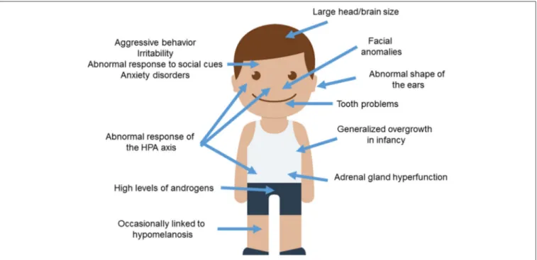
DOMESTIC TRAITS IN THE ASD
PHENOTYPE
Wilkins et al. (2014)
provide a comprehensive summary of traits
known to be modified in domesticated mammals, many of them
concerning the cranial region. These include changes in ear
size and shape, changes in the orofacial area (including shorter
snouts and smaller jaws), changes in dentition (particularly,
smaller teeth), and a reduced brain capacity (specifically, of
components of the forebrain such as the amygdala or parts of
the limbic system). Other distinctive traits commonly found
in domesticated strains are depigmentation, neoteny, shorter
reproductive cycles, and increased docility, which is thought to
result from adrenal size reduction and adrenal hypofunction
as well as from reduced levels of stress hormones (including
adrenocorticoids, adrenocorticotropic hormone, cortisol, and
corticosterone). This delayed adrenal maturation also involves
a hypofunction of the sympathetic nervous system and an
increase of the duration of the immaturity of the
hypothalamic-pituitary-adrenal system (the HPA axis), which provides the
animal with a longer socialization window. According to
Wilkins
et al. (2014)
, the multiple phenotypic traits that characterize
the domestication syndrome emerge as unselected by-products
from a developmental reduction in NCC inputs, resulting
from selection for tameness. Interestingly, compared to extinct
hominins, AMHs exhibit a number of domesticated traits,
including reduced brains (at least during the last 50,000 years),
changes in dentition, reduction of aggressiveness, and retention
of juvenile characteristics (see
Thomas, 2014
, for details).
Intriguingly, most of these features are generally attenuated in
ASD (Figure 1).
To begin with, ASD subjects show significant differences
with healthy controls regarding minor physical anomalies,
particularly in the craniofacial region (assumed to result from
deviations during fetal development and suggested to constitute
external markers of atypical brain growth) (
Tripi et al., 2008;
Manouilenko et al., 2014
). Specifically, in adults the abnormal
shape of the ears is robustly associated with autistic traits, with
FIGURE 1 | Anomalous presentation of domesticated traits in people with ASD. Main clinical features observed in ASD patients and concerning the domestication syndrome are shown. The child diagram was gathered from Iconfinder output (available at http://www.iconfinder.com/icons/525448/ boy_child_kid_male_man_person_white_icon).
higher scores correlating with poorer functioning (
Manouilenko
et al., 2014
). Regarding the changes in the orofacial region,
prepubertal boys with ASD show significant differences in
facial morphology compared to typically developing (TD) boys
(
Aldridge et al., 2011
). This distinctive facial phenotype is
more pronounced in subjects with severe symptoms, significant
cognitive impairment, and language regression (
Obafemi-Ajayi
et al., 2015
). Concerning tooth peculiarities, children with ASD
show greater abnormalities in dentition, including missing teeth,
diastemas, or reverse overjets (
Luppanapornlarp et al., 2010
).
With respect to brain size, head circumference is significantly
larger in people with ASD, with nearly 15% suffering from
macrocephaly. Higher brain volumes correlate with lower
functioning abilities; indeed nearly 9% ASD individuals exhibit
brain overgrowth (
Sacco et al., 2015
). It is worth noticing that
higher head circumference and brain size values are observed
only during early childhood (
Fukumoto et al., 2008; Courchesne
et al., 2011
, although see
Raznahan et al., 2013
), particularly
when ASD is presented with regression (
Nordahl et al., 2011
).
Typically, early brain overgrowth is followed by a decrease in
structural volumes (
Courchesne et al., 2011
). Although brain
overgrowth may result from a dysregulation of the overall
systemic growth (see below), it is thought to impact on cognition.
This is believed to occur as a result of the reduced networking
efficiency among widespread regions of the cortex, due to
the increased long-distance connections (
Lewis et al., 2013
).
Specifically, people with ASD show increased volumes of the
amygdala (
Mosconi et al., 2009; Murphy et al., 2012
), which
correlate with the severity of their social and communication
impairments (
Schumann et al., 2009
). In the TD population,
higher amygdala volumes are associated with poorer language
abilities in infancy (
Ortiz-Mantilla et al., 2010
).
Regarding
the
behavioral
traits
associated
with
the
domestication syndrome, we wish to highlight that aggressive
behaviors are frequent in children with ASD (with about 25%
of them having scores in the clinical range), and correlate with
lower cognitive outcomes (
Hill et al., 2014
). Children with ASD
display more reactive than proactive aggression attitudes (
Farmer
et al., 2015
). Likewise, irritability is also commonly observed
in affected individuals (
Mikita et al., 2015
). Additionally, ASD
is commonly found to be comorbid with generalized anxiety
disorder (
Hollocks et al., 2014; Bitsika et al., 2015
). Several studies
have been carried out to learn more about the physiological
basis of this anomalous response to the social environment.
Interestingly, higher serum cortisol responses are usually found
in children with ASD, particularly after stressor stimulation,
when prolonged duration and recovery of cortisol elevation
is also observed (
Spratt et al., 2012
). Moreover, children with
ASD show a distinctive diurnal rhythm of cortisol compared to
their TD peers; this involves elevated cortisol levels at the end
of the day and dampened linear decline across the day in some
children (
Tomarken et al., 2015
). Dysregulation of the diurnal
rhythm as a whole has been found in low functioning ASD
(
Taylor and Corbett, 2014
). Also, anxiety symptoms correlate
with high cortisol levels in ASD pediatric patients (
Bitsika
et al., 2015
). Plasma levels of adrenocorticotropic hormone are
also significantly higher in children with ASD, and correlate
positively with the severity of the symptoms (
Hamza et al.,
2010
). The HPA axis in ASD responds in a more sluggish way
to physiological or physical manipulation. Accordingly,
Taylor
and Corbett (2014)
found hyper-responsiveness of the HPA axis
when unpleasant stimuli or relatively benign social situations
are involved, whereas they observed hypo-responsiveness in
conditions involving social evaluative threat. On the whole, the
HPA axis may be more reactive to stress in social anxiety disorder
and ASD (
Spratt et al., 2012; Jacobson, 2014
). Because children
with autism and anxiety disorders show a blunted cortisol
response to psychosocial stress, and given that reduced cortisol
responsiveness is significantly related to increased anxiety
symptoms,
Hollocks et al. (2014)
suggested that a non-adaptive
physiological response to psychosocial stress may exist in ASD.
Finally, it is worth considering some other traits commonly
observed in domesticated mammals: neoteny, alterations of
reproductive cycles, and pigmentation changes. Regarding
neotenic features, it is noteworthy that children with ASD
exhibit an early generalized overgrowth (
van Daalen et al.,
2007; Fukumoto et al., 2008; Chawarska et al., 2011
). Typically,
boys with ASD show increased body size at birth and during
infancy, with postnatal overgrowth correlating with lower
adaptive functioning, greater severity of social deficits, and
poorer verbal skills (
Chawarska et al., 2011; Campbell et al.,
2014
). Interestingly, higher levels of androgens are found in
children and adolescents with ASD. This correlates with the
severity of autistic traits and might account for the precocious
puberty also reported in this condition (
El-Baz et al., 2014
).
These findings emphasize the role of elevated pre- and postnatal
testosterone levels in the liability for ASD (see
Hauth et al.,
2014
). Testosterone significantly affects brain development,
particularly targeting the hypothalamus, the amygdala and the
hippocampus, impacting on aspects of memory consolidation
(
Filová et al., 2013
). High perinatal testosterone concentration
negatively correlates with early vocabulary development in TD
boys (
Hollier et al., 2013
). Interestingly, children with elevated
androgen levels due to congenital adrenal hyperplasia show
atypical patterns of brain asymmetry in the perisylvian areas,
and language/learning disabilities (
Plante et al., 1996
). Less
data on reproductive functions in females is available, due
to the lower prevalence of ASD among women. Nevertheless,
women with ASD reported significantly more irregular menstrual
cycles and dysmenorrhea (
Ingudomnukul et al., 2007; Hamilton
et al., 2011
). Likewise, an increase in premenstrual syndrome
has been observed in women with ASD (
Obaydi and Puri,
2008; Hamilton et al., 2011
), who are more likely to exhibit
behavioral issues related to the onset of periods (
Burke et al.,
2009
). In addition, delayed age of menarche seems to correlate
with the severity of autistic traits (
Hergüner and Hergüner,
2016
). These findings lend support to the androgen theory of
ASD, according to which elevated levels of testosterone during
fetal development may contribute to the development of ASD.
Finally, concerning changes in pigmentation, it is of interest that
hypomelanotic diseases usually entail autistic symptoms, as is
commonly observed in hypomelanosis of Ito (OMIM#300337;
Akefeldt and Gillberg, 1991; von Aster et al., 1997;
Gómez-Lado et al., 2004
). It has been hypothesized that the comorbidity
between hypomelanosis and ASD may result from a deficiency
in vitamin D (
Eyles, 2010; Bakare et al., 2011
). In fact, core
symptoms of ASD improve after vitamin D supplementation (
Jia
et al., 2015
). Interestingly, core candidates for the globularization
of the AMH skull/brain and the evolution of language-readiness
are involved in vitamin D homeostasis and function (see
Benítez-Burraco and Boeckx, 2015
, for details).
As noted above, regardless of the different selectionist
scenarios that may account for the traits commonly found in
domesticated mammals, a role for NC hypofunction during
embryonic development has been proposed (see
Wilkins et al.,
2014
, for details). No comprehensive view of the role (if any) of
the NC in the aetiopathogenesis of ASD has been provided to
date. Still, it is important to note that neurocristopathies (that is,
conditions resulting from NC defects) commonly involve autistic
features. For instance, in CHARGE syndrome (OMIM#214800)
autistic traits coexist with developmental abnormalities affecting
endocrine, reproductive, urinary and digestive systems, along
with skeletal and craniofacial features (
Fernell et al., 1999
). Given
this background, we will now examine whether candidates for
ASD are overrepresented among the genes believed to play a
central role in NC development and function, with a special
emphasis on those that interact with genes important for the
globularization of the AMH skull/brain.
ASD AND THE GENETICS OF THE
DOMESTICATION SYNDROME
In order to improve our characterization of the domesticated
traits in ASD, it is of interest to assess whether candidate genes
for this condition (with a particular emphasis on language
disabilities) are overrepresented among, or are functionally
related to, candidates for domestication. We have relied on
an extended list of candidates, which includes the core set
of genes proposed by
Wilkins et al. (2014)
, plus a subset
of the genes involved in the globularization of the AMH
skull/brain and the emergence of language-readiness that are
functionally related to them, through direct interaction, and/or
that play a role in the development and function of the NC
(see
Benítez-Burraco et al., in press
, for details). Our list
also comprises NC-related genes known to play a key role
in craniofacial development and/or disorders. As noted above,
most of the domesticated traits result from the modification of
the cranial region and many of the ASD distinctive features
concern the skull, face and brain. Moreover, as reasoned in
Boeckx and Benítez-Burraco (2014a,b)
, we expect that our
language-readiness resulted from changes in the development
of the skull/brain, but also from the refinement of the
externalization devices, specifically, the orofacial region: As also
noted above, the impairment of oromotor function has been
hypothesized to account for some language deficits in ASD.
Table 1
provides a full list and a schematic characterization of
these genes.
When we tried to identify ASD-candidates among this
extended list of genes via PubMed (http://www.ncbi.nlm.nih.gov/
pubmed), we found out that nearly 25% of them have been
suggested to play a role in the aetiopathogenesis of ASD. If we
also consider genes that we found differentially expressed in
postmortem brain tissues isolated from patients (as discussed
TABLE 1 | List of putative candidate genes for domestication and ASD.
Gene symbol
Gene name Domesticationa
Language-readinessb NCCc Craniofaciald Brain rhythmicitye ASD Candidatef Differentially expressedg
ALX1 Aristaless-like homeobox protein 1 + +
ALX3 Aristaless-like homeobox protein 3 + +
ALX4 Aristaless-like homeobox protein 4 + +
AXIN2 Axin 2 + + +
BAZ1B Bromodomain adjacent to zinc finger domain 1B
+ +
BMP2 Bone morphogenetic protein 2 + + + +
BMP7 Bone morphogenetic protein 7 + + + +
CDC42 Cell division cycle 42 + + +
CHD7 Chromodomain helicase DNA binding protein 7
+ + + +
CITED2 Cbp/p300 interacting transactivator with Glu/Asp rich carboxy-terminal domain 2
+ + + CTNNB1 Catenin Beta 1 + + + + DLX1 Distal-less homeobox 1 + + + DLX2 Distal-less homeobox 2 + + + DLX5 Distal-less homeobox 5 + + + + + + DLX6 Distal-less homeobox 6 + + + + + EDN1 Endothelin 1 + + + EDN3 Endothelin 3 + + +
EDNRA Endothelin receptor type A + + +
EDNRB Endothelin receptor type B + + +
ERF ETS2 repressor factor + + +
FGF7 Fibroblast growth factor 7 + +
FGF8 Fibroblast growth factor 8 + + + +
FGFR1 Fibroblast growth factor receptor 1 + + + +
FGFR2 Fibroblast growth factor receptor 2 + + +
FOXD3 Forkhead box D3 + + +
FOXP2 Forkhead box P2 + +
FREM1 FRAS1 related extracellular matrix 1 + +
GDNF Glial-derived neurotrophic factor + +
GLI3 GLI family zinc finger 3 + + + +
GRHL3 Grainyhead like transcription factor 3 + +
GSC Goosecoid homeobox + +
HES1 Hes family bHLH transcription factor 1 + + + +
HOXA2 Homeobox A2 + + +
HSH2D Hematopoietic SH2 domain containing + +
KIT KIT proto-oncogene receptor tyrosine kinase
+ + + +
MAGOH Mago homolog, exon junction complex core component
+ + +
MITF Microphthalmia-associated transcription factor
+ + +
MSX1 Msh homeobox 1 + + + +
MSX2 Msh homeobox 2 + + +
NCAM1 Neural cell adhesion molecule 1 + + + +
NODAL Nodal growth differentiation factor + + +
NOG Noggin + +
NTN1 Netrin 1 + + +
TABLE 1 | Continued
Gene symbol
Gene name Domesticationa
Language-readinessb NCCc Craniofaciald Brain rhythmicitye ASD Candidatef Differentially expressedg
PAX3 Paired box 3 + + +
PAX6 Paired box 6 + + + + +
PAX7 Paired box 7 + +
POLR1A Polymerase (RNA) I subunit A + +
POU3F2 POU class 3 homeobox 2 + + + +
PQBP1 Polyglutamine binding protein 1 + + +
PTCH1 Patched 1 + + + +
RET Ret proto-oncogene + + +
ROBO1 Roundabout guidance receptor 1 + + +
ROBO2 Roundabout guidance receptor 2 + + +
RUNX2 Runt related transcription factor 2 + + + +
SATB2 Special AT-rich sequence binding-homeobox 2
+ + +
SHH Sonic hedgehog + + + +
SIX2 Sine oculis-related homeobox 2 + +
SLIT1 Slit guidance ligand 1 + +
SLIT2 Slit guidance ligand 2 + + +
SOX2 Sex determining region Y-box 2 + + + +
SOX9 Sex determining region Y-box 9 + + + + + +
SOX10 Sex determining region Y-box 10 + + + +
SPECC1L Sperm antigen with calponin homology and coiled-coil domains 1-like
+ + +
TCF12 Trascription factor 12 + + +
TCOF1 Treacle ribosome biogenesis factor 1 + +
VCAN Versican + + +
ZIC1 Zinc finger protein family member 1 + + +
aCore candidates for the “domestication syndrome” according toWilkins et al. (2014)(bold italicized tags) plus language-readiness genes functionally interacting with them according
toBenítez-Burraco et al. (in press)(regular tags).
bGenes highlighted as candidates for globularization of the AMH skull/brain and the emergence of language-readiness according toBoeckx and Benítez-Burraco (2014a,b)and Benítez-Burraco and Boeckx (2015).
cInvolved in neural crest (NC) development and function.
dInvolved in craniofacial development and/or found mutated in craniofacial syndromes. eInvolved in brain oscillation and rhythmicity.
fCandidate for ASD as resulting from genomic studies (pathogenic SNPs, association studies, CNVs, functional studies, etc.). gDifferentially expressed in postmortem brain tissues of ASD-vs.-control individuals (see text for details).
in the subsequent section), the percentage rises above 50%.
Interestingly, some of these genes are thought to be involved
in brain rhythmicity (see Table 1), plausibly contributing to
the oscillopathic signature of the ASD brain during language
processing.
We expect that the genes we highlight here are functionally
interconnected and map on to specific pathways, signaling
cascades, or aspects of brain development and function, of
interest for language processing and the aetiopathology of ASD.
In silico analyses offer promising insights. Accordingly, String 10
(http://www.string-db.org) predicts quite robust links between
most of these genes (Figure 2). Likewise, ontology analyses by
Panther (http://www.pantherdb.org) suggest that they might play
biological functions important for ASD and be part of signaling
pathways known to be impaired in this condition (Table 2).
Candidate Genes: A Functional
Characterization
Some of
Wilkins et al.’s (2014)
original candidates for the
domestication syndrome are candidates for ASD. KIT mutations
have been found in patients featuring ASD symptoms (
Kilsby
et al., 2013
). KIT is a tyrosine kinase receptor (
Kasamatsu
et al., 2008
), which acts as a key developmental regulator in
the NC-derived processes of hematopoiesis, melanogenesis, and
gametogenesis (
Rothschild et al., 2003
). In rats mutations of Kit
impair hippocampal synaptic potentiation and spatial learning
and memory (
Katafuchi et al., 2000
). Likewise, whole-genome
sequencing analyses have identified deleterious variants of CHD7
in ASD probands (
Jiang et al., 2013
). CHD7 is known to be the
main candidate for CHARGE syndrome (
Vissers et al., 2004;
Lalani et al., 2006
), mentioned above. Interestingly, CHARGE
FIGURE 2 | Functional links among candidates for domestication and ASD. The chart (drawn using String 10.0 license-free software, http://string-db.org/) shows the network of known and predicted interactions among all genes listed in Table 1. The colored nodes symbolize proteins: small nodes are proteins with unknown 3D structure, while large nodes are those with known 3D structures. The color of the edges represent different kind of known protein-protein associations. Green: activation, red: inhibition, dark blue: binding, light blue: phenotype, dark purple: catalysis, light purple: posttranslational modification, black: reaction, yellow:
FIGURE 2 | Continued
transcriptional regulation. Edges ending in an arrow symbolize positive effects, edges ending in a bar symbolize negative effects, whereas edges ending in a circle symbolize unspecified effects. Gray edges symbolize predicted links based on literature search ((co-mention in PubMed abstracts). Stronger associations between proteins are represented by thicker lines. The medium confidence value was 0.0400 (a 40% probability that a predicted link exists between two enzymes in the same metabolic map in the KEGG database: http://www.genome.jp/kegg/pathway.html). String 10 predicts associations between proteins that derive from a limited set of databases: genomic context, high-throughput experiments, conserved coexpression, and the knowledge previously gained from text mining (Szklarczyk et al., 2015). This is why the figure does not represent a fully connected graph (evidence for additional links are provided in the main text). Importantly, the diagram only represents the potential connectivity between the involved proteins, which has to be mapped onto particular biochemical networks, signaling pathways, cellular properties, aspects of neuronal function, or cell-types of interest that can be confidently related to aspects of language development and function.
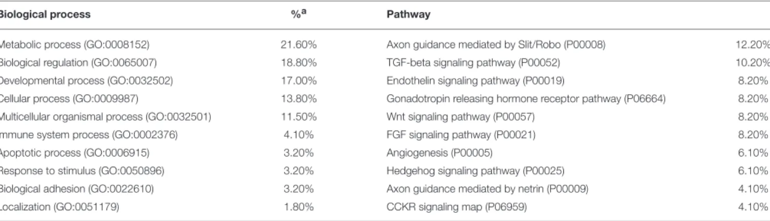
TABLE 2 | GO classifications of candidates for domestication and ASD.
Biological process %a Pathway
Metabolic process (GO:0008152) 21.60% Axon guidance mediated by Slit/Robo (P00008) 12.20% Biological regulation (GO:0065007) 18.80% TGF-beta signaling pathway (P00052) 10.20% Developmental process (GO:0032502) 17.00% Endothelin signaling pathway (P00019) 8.20% Cellular process (GO:0009987) 13.80% Gonadotropin releasing hormone receptor pathway (P06664) 8.20% Multicellular organismal process (GO:0032501) 11.50% Wnt signaling pathway (P00057) 8.20%
Immune system process (GO:0002376) 4.10% FGF signaling pathway (P00021) 8.20%
Apoptotic process (GO:0006915) 3.20% Angiogenesis (P00005) 6.10%
Response to stimulus (GO:0050896) 3.20% Hedgehog signaling pathway (P00025) 6.10% Biological adhesion (GO:0022610) 3.20% Axon guidance mediated by netrin (P00009) 4.10%
Localization (GO:0051179) 1.80% CCKR signaling map (P06959) 4.10%
aNumbers refer to percent of gene hit against total of process or pathway hits. Only the top 10 functions, filted after Bonferroni post-hoc correction, have been included.
syndrome also involves microcephaly, face asymmetry, cleft
lip/palate, along with variable degrees of intellectual disability
(
Pisano et al., 2014; Hale et al., 2016
, for review). Changes in
the expression pattern of CDH7 can also result in behavioral
anomalies resembling the autistic phenotype. Accordingly, in
utero exposure to heavy metals in mice increases autism-like
behavioral phenotypes in adult animals through inducing the
hypomethylation of Chd7 (
Hill et al., 2015
). FOXD3, encoding a
transcription factor, is downregulated by DISC1 (
Drerup et al.,
2009
), a robust candidate for schizophrenia that has been also
associated to ASD (
Williams et al., 2009; Zheng et al., 2011;
Kanduri et al., 2016
). FOXD3 maps within one of the
present-day human-specific differentially-methylated genomic regions
(DMRs) (
Gokhman et al., 2014
). Interestingly, loss of Disc1
results in abnormal NCC migration and differentiation (
Drerup
et al., 2009
). Also, DISC1 downregulates SOX10, another NC
gene, involved in the maintenance of precursor NCC pools,
in the timing of NCC migration onset, and in the induction
of their differentiation; it is also implicated in oligodendrocyte
differentiation (
Hattori et al., 2014
). In turn, SOX10 interacts
with PAX3, another core candidate proposed by
Wilkins et al.
(2014)
, and with POU3F2 (
Smit et al., 2000
). Sequence and
CNVs affecting POU3F2 have been found in subjects with ASD,
and in individuals with different developmental and language
delays (
Huang et al., 2005; Lin et al., 2011
). POU3F2 is a
known interactor of FOXP2, the renowned “language gene”
(
Maricic et al., 2013
). AMHs bear a derived allele of the
binding site which is less efficient in activating transcription
than the Neanderthal/Denisovan counterpart (
Maricic et al.,
2013
). Likewise, POU3F2 has been associated with human
accelerated conserved non-coding sequences (haCNSs) (
Miller
et al., 2014
). Also, it interacts with PQBP1, which has been linked
to intellectual disability (
Wang et al., 2013
) and developmental
delay and microcephaly (
Li et al., 2013
). Also SOX9, considered
a master regulator of craniofacial development and related to
several congenital skeletal malformations (
Mansour et al., 2002;
Gordon et al., 2009; Lee and Saint-Jeannet, 2011
), is found among
the candidates for ASD.
Accordingly, gene and miRNA expression profiling using
cell-line derived total RNA has revealed SOX9 as one of the
genes dysregulated in ASD (
Ghahramani Seno et al., 2011
). As
discussed in detail by
Benítez-Burraco et al. (in press)
SOX9
interacts with BMP2, BMP7, DLX2, and HES1. All of them
are core components of the network believed important for
globularization and language-readiness (reviewed in
Boeckx and
Benítez-Burraco, 2014a
). In addition, all of them are involved
in NCC development and migration, and in the patterning of
NC-derived tissues (
Mallo, 2001; Gajavelli et al., 2004; Correia
et al., 2007; Glejzer et al., 2011; Ishii et al., 2012
). BMP2
is a key osteogenic regulator, which has been associated to
craniosynostosis (
Justice et al., 2012; Lattanzi et al., 2013
). BMP2,
BMP7, and DLX2 act upstream SOX9 (
Sperber et al., 2008;
Li et al., 2013
). In turn, SOX9 mediates the retinoic
acid-induced expression of HES1, known also to be involved in
language function, craniofacial development, and neuron growth
and interconnection (reviewed in
Boeckx and Benítez-Burraco,
2014b
). Importantly, retinoic acid also regulates the expression of
other genes that are relevant for language, like FOXP2 (
Devanna
et al., 2014
), or for globularization, like ASCL1 (see
Benítez-Burraco and Boeckx, 2015
, for details). Retinoic acid has proven
to be important for brain plasticity (
Luo et al., 2009
), and memory
and learning processes (
Etchamendy et al., 2003; Jiang et al.,
2012
). Recent whole-exome sequencing analyses have linked
retinoic acid regulation pathways to ASD (
Moreno-Ramos et al.,
2015
). In neuronal cells reduced levels of RORA downregulate
multiple transcriptional targets that are significantly enriched
in biological functions negatively impacted in ASD and which
include known ASD-associated genes, like A2BP1, CYP19A1,
ITPR1, NLGN1, and NTRK2 (
Sarachana and Hu, 2013a
). RORA
itself is downregulated in postmortem prefrontal cortex and
cerebellum of subjects with ASD (
Nguyen et al., 2010
). RORA
is differentially regulated in them by masculine and feminine
hormones: Whereas it is under negative feedback regulation by
androgens, it is under positive regulation by estrogens (
Sarachana
et al., 2011; Sarachana and Hu, 2013b
). In certain regions of
the brain this sexually dimorphic expression is also found in
several of RORA’s targets and this correlation is much higher in
the cortex of males (
Hu et al., 2015
). Perhaps not surprisingly,
synthetic RORα/γ agonist improve autistic symptoms in animal
models of the disease, particularly, repetitive behavior (
Wang
et al., 2016
).
We wish to highlight two other genes thought to be involved
in the changes that brought about modern language that are also
candidates for ASD and interact with core candidates for the
domestication syndrome as posited by Wilkins et al. The first
one is DLX5, involved in crucial aspects of NC development
(
McLarren et al., 2003; Ruest et al., 2003
), but also of skull
and brain development (
Kraus and Lufkin, 2006; Wang et al.,
2010
). Accordingly, it plays a role in thalamic development
(
Jones and Rubenstein, 2004
) and contributes to regulate the
migration and differentiation of precursors of GABA-expressing
neurons in the forebrain (
Cobos et al., 2006
). DLX5 is a candidate
for ASD (
Nakashima et al., 2010
), due to an ultraconserved
cis-regulatory element (
Poitras et al., 2010
), which is bound
by GTF2I, encoded by one of the genes commonly deleted in
Williams-Beuren syndrome (OMIM#194050) and a candidate
for ASD too (
Malenfant et al., 2012
). Additionally, DLX5 is
regulated by MECP2 (
Miyano et al., 2008
), encoded by the
main candidate for Rett syndrome (OMIM#312750), a condition
entailing problems for motor coordination, autistic behavior, and
language regression (
Uchino et al., 2001; Veenstra-VanderWeele
and Cook, 2004
). Interestingly, Dlx5/6(±) mice exhibit abnormal
pattern of γ rhythms resulting from alterations in GABAergic
interneurons, particularly in fast-spiking interneurons (
Cho
et al., 2015
). In addition, DLX5 interacts with key candidates
for language evolution, in particular, with RUNX2 and FOXP2
(see
Boeckx and Benítez-Burraco, 2014a
, for details). The
second one is NCAM1, which is also a target of both
RUNX2 (
Kuhlwilm et al., 2013
) and FOXP2 (
Konopka et al.,
2009
). In mice mutations in the gene affect
working/episodic-like memory (
Bisaz et al., 2013
), whereas overexpression of
the Ncam1 extracellular proteolytic cleavage fragment impacts
on GABAergic innervation, affecting long- and short-term
potentiation in the prefrontal cortex (
Brennaman et al., 2011
).
NCAM1 encodes a cell adhesion protein involved in axonal
and dendritic growth and synaptic plasticity (
Rønn et al., 2000;
Hansen et al., 2008
). It interacts with VCAM1 which is involved
in cell adhesion and the control of neurogenesis (
Kokovay
et al., 2012
), and which bears a fixed (D414G) change in AMHs
compared to Neanderthals/Denisovans (
Pääbo, 2014
). VCAM1 is
upregulated by CLOCK, which plays a key role in the modulation
of circadian rhythm (
Gao et al., 2014
). Together with other
circadian-relevant genes CLOCK seems to be involved in the
psychopathology of ASD cases entailing sleep disturbances (
Yang
et al., 2016
). The circadian modulation of synaptic function has
been hypothesized to contribute decisively to ASD (
Bourgeron,
2007
). In turn, CLOCK interacts with RUNX2 and with several
other candidates for language-readiness, like DUSP1, involved
in vocal learning (
Doi et al., 2007
), and USF1, which regulates
synaptic plasticity, neuronal survival and differentiation (
Tabuchi
et al., 2002; Steiger et al., 2004
). USF1 binds the promoter of
FMR1 (
Kumari and Usdin, 2001
), a strong candidate for
Fragile-X syndrome (OMIM#300624), which presents with language
problems and ASD features (
Kaufmann et al., 2004; Smith et al.,
2012
). The regulatory region of USF1 shows many fixed or high
frequency changes compared to Denisovans (
Meyer et al., 2012
).
As shown in Table 1, several of the genes important for
globularization and language-readiness are involved in NC
development and function and some of them are also candidates
for ASD. Accordingly, we expect them to contribute to the
abnormal domesticated features observed in patients with ASD,
and also to their distinctive language profile. Among them
we wish mention CTNNB1, DLX1, DLX6, PAX6, and ROBO2.
CTNNB1 is a component of the Wnt/β-catenin signaling
pathway, known to be impaired in ASD (
Cao et al., 2012; Zhang
et al., 2012; Martin et al., 2013
). CTNNB1 controls aspects
of NC development, from NC induction, lineage decisions,
to differentiation (
Hari et al., 2012
). As noted in
Boeckx
and Benítez-Burraco (2014b)
, CTNNB1 is expected to interact
with many of the genes highlighted as important for the
evolution of language-readiness, specifically with RUNX2 and
SLIT2/ROBO1 signals. Regarding DLX1, it is a robust NC marker
(
Ishii et al., 2012
), involved in patterning and morphogenetic
processes in NC-derived tissues (
Mallo, 2001
). It also regulates
the development of the skull and the brain (
Andrews et al.,
2003; Jones and Rubenstein, 2004
). In mice Dlx1 downregulation
results in reduced glutamatergic input to the hippocampus (
Jones
et al., 2011
), as well as in changes in interneuron subtypes
and migration patterns in the cortex (
Ghanem et al., 2008
).
DLX1 is found to be downregulated in ASD (
Voineagu et al.,
2011; McKinsey et al., 2013
). ROBO2 is one of the DLX1
interactors. Slit/Robo signaling regulates early NCC migration
(
Jia et al., 2005
) ROBO2 is also involved in thalamocortical axons
(TCA) development, known to be important for the modulation
of cognitive functions (
López-Bendito et al., 2007;
Marcos-Mondéjar et al., 2012
). ROBO2 is a candidate for ASD (
Suda
et al., 2011
), but also for different types of language disorders,
like dyslexia (
Fisher et al., 2002
) and speech-sound disorder and
reading (
Stein et al., 2004
). It has been related as well to expressive
vocabulary growth in the normal population (
St Pourcain et al.,
2014
). Finally, PAX6 controls the migration of NCCs from the
anterior midbrain (
Matsuo et al., 1993
). PAX6 is involved as
well in the development of the brain (
Valverde et al., 2000; Tyas
et al., 2003; Caballero et al., 2014
). Mutations on PAX6 have been
reported in some forms of ASD (
Maekawa et al., 2009
), although
they also impact in working memory (
Bamiou et al., 2007
).
Alterations of PAX6 expression in the brain of people with ASD
may account for the observed imbalance in excitatory/inhibitory
neuronal activity (
Kim et al., 2014
). And like many of the genes
reviewed above, PAX6 is functionally related to both FOXP2 and
RUNX2, and it also targets POU3F2 (see
Benítez-Burraco and
Boeckx, 2015
, for details).
Most of the NCC-genes mentioned here are known to
play a key role in the development and patterning of the
craniofacial complex, and to be associated to congenital
craniofacial defects (Table 1) (see
Twigg and Wilkie, 2015
for review). Many of these genes are known candidates for
ASD, including DLX5 and DLX6 (reviewed above), FGFR2,
MSX1, POLR1A, and PTCH1. Both DLX5 and DLX6 are
indeed required for NC-derived facial morphogenesis (
Gitton
et al., 2011
) FGFRs are among the main
craniosynostosis-associated genes. In particular, gain-of-function mutations in
FGFR2 are typically associated to Apert (OMIM#101200) and
Crouzon (OMIM#123500) syndromes, while both FGFR1 and
FGFR2 are found mutated in Pfeiffer syndrome (OMIM#101600)
(
Lattanzi et al., 2012
). All these syndromic craniosynostoses
occasionally present with variable degree of ASD-like mental
retardation (
Morey-Canellas et al., 2003
). MSX1 encodes a
transcriptional repressor involved in craniofacial development
and shaping (particularly in odontogenesis) (
Alappat et al.,
2003; Lattanzi, forthcoming
). It is expressed in the NC (
Khadka
et al., 2006
), where it acts as a master regulator of gene
expression (
Attanasio et al., 2013
). Although it has not been
associated to ASD, MSX1 is a direct downstream target of DLX5
during early inner ear formation (
Sajan et al., 2011
). The gene
is also a critical intrinsic dopaminergic neuron determinant
(
Andersson et al., 2006
) and is found mutated in some patients
with Wolf-Hirschhorn syndrome (OMIM#194190), a clinical
condition entailing profound mental retardation and craniofacial
dysmorphism (
Campbell et al., 1989
). POLR1A, found mutated in
acrofacial dysostosis (Cincinnati type, OMIM#616462) involving
microcephaly, plays a role in the regulation of NC-derived
skeletal precursor cells (
Weaver et al., 2015
). In some ASD
subjects CNVs result in fusion transcripts involving POLR1A,
although no fusion transcripts have been detected to date (
Holt
et al., 2012
).
Finally, it is worth mentioning that genes encoding primary
cilium signaling molecules, such as SHH, GLI3, and PTCH1,
are all primarily involved in congenital malformations affecting
the midline craniofacial compartment (
Brugmann et al., 2010;
Rice et al., 2010
). Specifically, PTCH1 is required in the
NC-dependent orofacial development and gives rise to orofacial
clefting, when mutated (
Metzis et al., 2013
). Heterozygous
mutations of either SHH or PTCH1 are typically found in
holoprosencephaly (OMIM#610828, and #236100), a genetically
heterogeneous, highly prevalent congenital forebrain anomaly
in humans, associated with mental retardation and craniofacial
malformations (
Ming et al., 2002; Mercier et al., 2011
). In
addition, a 22-bp deletion in this gene has been found in a girl
with ASD and Gorlin syndrome, a complex condition involving
macrocrania and hypertelorism (
Delbroek et al., 2011
).
Candidate Genes: Expression Profiles in
the ASD Brain
If our hypothesis is on the right track, we expect that the
genes we highlight here are dysregulated in the brain of
people with ASD, particularly in areas important for language
processing. Accordingly, we surveyed the Gene Expression
Omnibus (GEO) repository (https://www.ncbi.nlm.nih.gov/gds)
searching for their expression profiles in the cerebellum and the
temporal cortex (but also in the frontal and occipital cortices)
in patients with ASD. This should help identify new candidates
for ASD in the context of domestication and language-readiness
(Table 1). Overall, we could find significant expression values
for some of our candidates and learnt that they are up- or
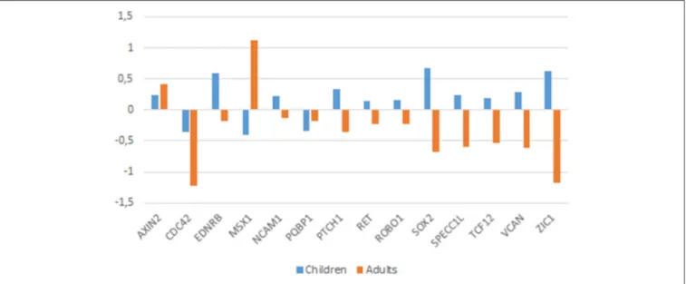
downregulated in the brain of autists (Figure 3).
Among the genes that are significantly downregulated in
the cerebellum we found AXIN2, EDNRB, SOX2, SPECC1L,
TCF12, and VCAN, whereas CDC42, and PQBP1 are found
upregulated in this region (Figure 3). Although none of them
has been associated to ASD, they stroke us as promising
candidates for the atypical presentation of the domestication
syndrome in ASD. AXIN2 is expressed in the cranial NC
and is needed for NC-derived frontal bone osteogenesis (
Yu
et al., 2005; Li et al., 2015a
). This gene is also expressed
as a specific marker for suture stem cells (
Maruyama et al.,
2016
), but also acts as a negative regulator of canonical
Wnt pathway, contributing to the stability of CTNNB1 (
Li
et al., 2015a
). Speech alterations are also observed in people
with AXIN2 mutations causing non-syndromic oligodontia (
Liu
et al., 2015
). EDNRB encodes a receptor for endothelins,
known to be potent vasoactive peptides. Mutations in this
gene are associated to increased susceptibility to Hirschsprung
disease (OMIM#600155), a neurocristopathy characterized by
congenital absence of intrinsic ganglion cells in the enteric
nervous plexa (
Amiel et al., 2008
). Waardenburg syndrome
(OMIM#277580), a genetically heterogeneous condition which
may involve developmental delay subsidiary to sensorineural
hearing loss, has also been associated with mutations in EDNRB
(
Read and Newton, 1997
). SOX2, one of core candidates for
domestication (
Wilkins et al., 2014
), encodes an interactors of the
GLI factors as part of the SHH-GLI signaling pathway involved in
NCC fate (
Oosterveen et al., 2012, 2013; Peterson et al., 2012
), but
also in the globularization of the AMH skull/brain (see Boeckx
et al., submitted for details). SOX2 interacts as well with the BMP
signaling (
Li et al., 2015b
). Interestingly, SOX2 regulates PQBP1,
highlighted above as one of POU3F2 interactors. SPECC1L is
found mutated in Opitz G/BBB syndrome (OMIM# #145410)
and in facial clefting (
Kruszka et al., 2015
). This gene functions in
NC development (
Wilson et al., 2016
) and is specifically involved
in facial morphogenesis (
Saadi et al., 2011
). TCF12 is highly
expressed in embryonic precursors of skull/brain structures,
including NC-derived head mesenchyme (
Uittenbogaard and
Chiaramello, 2002
). TCF12 directly interacts with TWIST1,
mutated in Saethre-Chotzen syndrome (OMIM#601622), which
features complex craniosynostosys with variable degrees of
intellectual disability, including ASD traits (
Maliepaard et al.,
2014
). Indeed, loss-of-function mutations of TCF12 have been
identified in patients with coronal synostosis, which sometimes
involves intellectual disability (
Sharma et al., 2013; di Rocco
et al., 2014; Paumard-Hernández et al., 2015; Piard et al., 2015
).
VCAN encodes versican-1, a protein that guides migratory NCCs
(
Dutt et al., 2006
) and which shows a fixed N3042D change
in AMHs (
Pääbo, 2014
). Finally, CDC42 controls NC stem
FIGURE 3 | Expression profiles of candidate genes in the ASD brain. Data were gathered from the following microarray expression datasets available on the Gene Expression Omnibus database (GEO datasets, http://www.ncbi.nlm.nih.gov/gds): GSE28521 (Voineagu et al., 2011) for the temporal and frontal cortices, GSE38322 (Ginsberg et al., 2012) for the cerebellum and the occipital cortex. Data are shown as log transformation of fold changes (logFC) between patients and corresponding controls. Only genes showing statistically significant (p < 0.05) differential expression were considered. Additional details may be found in the Supplemental information file.