Case report
Spinal giant cell tumor in tuberous sclerosis:
Case report and review of the literature
Mario Francesco Fraioli
1, Mario Lecce
1, Chiara Fraioli
2, Curatolo Paolo
31
Department of Neurosciences, Neurosurgery, University of Rome“Tor Vergata”, Rome, Italy, 2Department of Radiotherapy, CIRAD Villa Benedetta, Rome, Italy,3Department of Neurosciences, Pediatric Neurology Unit, University of Rome“Tor Vergata“, Rome, Italy
Background:Patients affected by tuberous sclerosis (TS) have a greater incidence of tumors than the healthy population. Spinal tumours in TS are reported very rarely and consist mainly of sacrococcygeal and cervical chordomas.
Method:Case report.
Findings: A 21-year-old man, affected by TS, presented a spinal dorsal T2 tumor that caused medullary compression. He underwent decompressive laminectomy and microsurgical excision of a giant cell tumor and an associated aneurysmal bone cyst. Postoperative hypofractionated radiotherapy was performed on the surgical field. At 2.4 years of follow-up the patient reported total recovery of neurological deficits and was free from tumor recurrence.
Conclusion:Considering this association, which is the first reported in the literature, spinal magnetic resonance imaging with gadolinium should be performed at the onset of spinal pain in patients affected by TS.
Keywords: Tuberous sclerosis, Paraparesis, Giant cell spinal tumours, Aneurysmal bone cyst, Hypofractionated radiotherapy
Introduction
Patients affected by tuberous sclerosis (TS) have a greater incidence of tumors than the healthy population because of mutations in one of two genes, TSC1 or TSC2. The normal cellular proteins encoded by these genes, harmatin and tuberin, form a heterodimer that suppresses cell growth in the central nervous system.1
The tumors associated with TS are usually located in the brain and are represented in particular by the sube-pendymal giant cell astrocytoma, which was described in only one exceptional case as multiple intra-axial ence-phalic and spinal localizations.2 Spinal tumours have been reported very rarely and they are mainly rep-resented by sacrococcygeal3 and cervical4 chordomas. We report an original case of a progressive paraparesis due to a giant cell bone tumor of the dorsal spine in a patient affected by TS.
Case report
A 21-year-old man, with prenatal diagnosis of TS and presence of tuberous lesions involving the brain and
kidney, presented to the emergency unit for the onset of rapidly worsening spastic paraparesis and initial urinary retention during the previous 3 days. The patient also reported persistent back pain that had started 3 weeks earlier. Neurological examination showed severe spastic paraparesis with inability to stand, a level of hypoesthesia at the mammillary line (T4), bilateral Achilles clonus, and urinary retention. Urgently performed cervical-dorsal computed tomography (CT) and magnetic resonance imaging (MRI) documented the presence of an expansive spinal lesion, with spinal cord compression at the level of T2–T3 (30 × 20 × 23 mm); complete replacement by tumor tissue of the laminae and the spinous processes was present at this level; T2 weighted MRI showed the compressive effect and an intramedullary hyperintense signal indicating spinal lesion (Fig. 1A,B). Decompressive laminectomy and microsurgical excision of the tumor were performed, which caused extensive bleeding. The postoperative course was characterized by rapid improve-ment of the paraparesis, with return of the ability to stand autonomously and after 2 weeks, to walk. The patient also recovered sphincter control; neurorehabilitation was started on the third postoperative day.
Correspondence to: Mario Francesco Fraioli, Department of Neurosciences, Neurosurgery, University of Rome Tor Vergata, Via Oxford 81, 00133 Rome, Italy. Email: [email protected]
© The Academy of Spinal Cord Injury Professionals, Inc. 2013
A total body positron emission tomography-CT scan performed 10 days after surgery showed the already known tuberous brain and kidney lesions, with no other areas of impaired metabolism.
Histological examination revealed a giant cell tumor with associated aneurysmal bone cyst that had arisen in the lesion. Spinal MRI with gadolinium performed 15 days after surgery, documented the excision of the lesion, showing complete re-expansion and decompres-sion of the dorsal spinal cord (Fig. 2A,B).
Hypofractionated stereotactic radiotherapy (HSRT) was then administered for 12 sessions, three times a week, for a total dose of 4000 cGy on the surgical field, using thin layer CT images (1-mm thickness) fused with 1-mm 1.5-Tesla MR images for target contour. The treatment was well tolerated with no side effects. Medical therapy with prednisone 5 mg three times a week (each day of treatment) was administered. Spinal MRI performed 6 months after surgery and 3 months after HSRT showed the absence of tumor relapses and of spinal cord compression (Fig. 2C,D).
After a follow-up period of 2.4 years, the patient is autonomous for all activities of his life with no tumor recurrence.
Discussion
TS is typically associated with subependymal giant cell astrocytoma of the brain; spinal tumours related to TS are represented mainly by cervical and sacrococcygeal chordomas, suggesting that the genes causing TS may have an aetiological role in chordomas.3,4 This case is, to our knowledge, the first giant cell tumor of the spinal bone associated with TS. Moreover, an aneurys-mal bone cyst was associated with the tumor; the
relationship between giant cell tumor and aneurysmal bone cyst has also been described also by other authors, who found a non-negligible rate of local tumor recurrence and lung metastasis, suggesting that postoperative radiotherapy should be performed on the surgical field.5 Aneurysmal bone cysts seem to arise because of hemodynamic changes in reaction to trauma or to a primary benign (giant cell tumor, chondroblastoma, etc.) or malignant (osteosarcoma) bone tumor.6
Giant cell tumors of the bone are benign primary bone tumors with great propensity for local recurrence after surgical excision.7 This tumor occurs more frequently in patients aged 20–45 years without sex preference. The most common clinical presentation reported in the literature, to our knowledge, is radiating back pain followed by rapidly progressive neurological deficits according to the localization of the tumor, which may be associated with sphincter dysfunction that can acutely worsen. Thus, early diagnosis is essen-tial to prevent spinal cord injury.
En bloc resection should be the treatment of choice to avoid local relapse which can, however, occur because of the marked tumor infiltration and replacement of the normal bone tissue.7 For this reason, adjuvant post-operative therapies, such as arterial embolization, are indicated when radical removal is not possible because of high risks of postoperative neurological deficits.8 In this patient, postoperative radiotherapy was per-formed, although its role in the treatment of giant cell spinal tumours is still controversial, according to some authors.8In contrast, other authors5suggest postopera-tive radiotherapy for aneurysmatic bone cysts. Concerning the choice of the radiotherapy treatment,
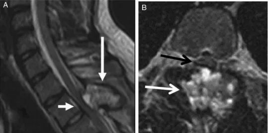
Figure 1 (A) Preoperative sagittal T2 weighted MR image of cervical and dorsal spine showing a tumoral mass (long white arrow) causing medullary compression at D2 level (short white arrow). (B) Preoperative T2 weighted MR image showing the tumor (black arrow) compressing the spinal cord (white arrow) in axial slice.
Fraioli et al. Spinal tumor in tuberous sclerosis
The Journal of Spinal Cord Medicine 2013 VOL.36 NO.2 158
HSRT was performed in this patient in order to avoid a single high radiosurgical dose that could have been potentially harmful to the adjacent spinal cord, which had already been damaged because of the previous tumoral compression.
Conclusion
In conclusion, the occurrence of a rare dorsal spine giant cell tumor associated with an aneurysmal bone cyst in a patient affected by TS is reported for the first time in the literature. Considering this association and the rapid onset and worsening of the neurological symptoms after a period of only pain, in our opinion, a spine MRI with gadolinium should be performed early in young patients with TS who develop spinal pain.
References
1 McCall T, Chin SS, Salzman KL, Fults DW. Tuberous sclerosis: a syndrome of incomplete tumor suppression. Neurosurg Focus 2006;20(1):pE3.
2 Telfeian AE, Judkins A, Younkin D, Pollock AN, Crino P. Subependymal giant cell astrocytoma with cranial and spinal metastases in a patient with tuberous sclerosis. Case report. J Neurosurg 2004;100(5 Suppl Pediatrics):498–500.
3 Lee-Jones L, Aligianis I, Davies PA,et al. Sarococcygeal chordomas in patients with tuberous sclerosis complex show somatic loss of TSC1 or TSC2. Cancer 2004;41(1):80–5.
4 Storm PB, Magge SN, Kazahaya K, Sutton LN. Cervical chordoma in a patient with tuberous sclerosis presenting with shoulder pain. Pediatr Neurosurg 2007;43(2):167–9.
5 Wu Z, Yang X, Xiao J, Feng D, Huang Q, Zheng W, et al. Aneurysmal bone cyst secondary to giant cell tumor of the mobile spine: a report of 11 cases. Spine (Philadelphia 1976). 2011;36(21): E1385–90.
6 Bello Báez A, López Pino MA, Azorín , Cuadrillero D, Sirvent , Cerdá S. Aneurysmatic bone cyst coexisting with osteosarcoma. Radiopathologic discussion. Radiologia 2010;52(3):247–50. Figure 2 (A) Postoperative sagittal T2 weighted MR image showing the surgical field after tumor removal (long white arrow) and the re-expansion of the spinal cord. (B) Postoperative T2 weighted MR image showing the surgical cavity after tumor removal (long black arrow) and spinal cord re-expansion in axial slice (short white arrows). (C) T1 weighted sagittal MRI performed 3 months after the end of hypofractionated stereotactic radiotherapy (6 months after surgery), showing absence of tumoral recurrence (long white arrow) or new medullary compression (short white arrow). (D) Axial slice of the same MR image of C, showing absence of medullary compression (white arrows).
Fraioli et al. Spinal tumor in tuberous sclerosis
7 Saikia KC, Bhattacharyya TD, Bhuyan SK, Bordoloi B, Durgia B, Ahmed F. Local recurrences after curettage and cementing in long bone giant cell tumor. J Orthop 2011;45(2):168–73.
8 Martin C, McCarthy EF. Giant cell tumor of the sacrum and spine: series of 23 cases and a review of the literature. Iowa Orthop J 2010; 30:69–75.
Fraioli et al. Spinal tumor in tuberous sclerosis
The Journal of Spinal Cord Medicine 2013 VOL.36 NO.2 160