DOI 10.1393/ncc/i2020-20005-8 Colloquia: FATA 2019
Progress reports on the MEDAMI 2019 and CTR research
at the DMIL in i3M
A. J. Gonzalez(∗), J. Barrio, E. Lamprou, V. Ilisie, F. Sanchez and J. M. Belloch
Instituto de Instrumentaci´on para Imagen Molecular, Centro Mixto CSIC-Universitat Polit`ecnica de Val`encia - 46022 Valencia, Spain
received 2 March 2020
Summary. — This contribution reports on the recently held MEDAMI 2019
work-shop in Valencia (15–17th May 2019). This workwork-shop is about advanced molecular imaging and the main topic of this last edition was Imaging in Immunotherapy. Around 70 attenders met together during three days. This meeting made it possi-ble to join medical doctors and instrumentalists. In MEDAMI 2019 it was exposed the new immunotherapies from a clinical and research point of view. It was shown the already observed improvements when using these therapies. At the same time, we heard about the difficulties and limitations of current molecular imaging in this particular field. It was clear that improvements in system sensitivity and resolution are demanded. Timing information can be utilized in different ways to improve the image quality in PET systems. Precise Coincidence Time Resolution (CTR) improves the signal-to-noise ratio and, therefore, the image contrast, allowing for instance to distinguish low uptake tumors, multicentric lesions, or tumor hetero-geneity, to name but a few. Both high time resolution and angular coverage in a PET system can improve the effective sensitivity. An example of a system bench-marking the timing resolution is the Siemens Biograph Vision with 214 ps FWHM, enhancing the detectability. The Explorer total-body PET from UC Davis improves the system sensitivity by having a 2 meters long PET scanner. Deep investigations, from different research groups, are being carried out to further push the limits of timing resolution. This work also describes some of the projects on high timing performance that are being carried out at the Detector for Molecular Imaging Lab (DMIL) at the Institute for Instrumentation in Molecular Imaging (i3M) in Valen-cia. The DMIL group has extensively worked on detectors and implementation of PET systems enabling the use of accurate timing information. In this progress re-port we describe the results obtained at the DMIL regarding timing determination in gamma-ray detectors both based on monolithic and pixelated crystals. Although
(∗) Corresponding author. E-mail: [email protected]
with 15 mm thick LYSO blocks it was tough to obtain values of CTR below 500 ps when using analog SiPMs and ASIC-based readout, this was improved down to 250 ps if small 3 mm size and 6 mm height pixels under the one-to-one coupling approach were enabled. This type of approach, the one-to-one coupling, seems to benefit from the light collection in a single photosensor element and, therefore, to improve the timing properties. Monolithic blocks offer, on the contrary, advantages such as photon depth of interaction. In order to separate Compton and photoelectric events we have thought of a detector block design with a high aspect ratio, using LYSO crystals of 51 mm size vs. 3 mm thickness, read-out by the four lateral sides. We have demonstrated the possibility to reach below 2 mm FWHM spatial resolution with an energy resolution of 12%.
1. – MEDAMI 2019
The 6th Mediterranean Thematic Workshop in Advanced Molecular Imaging (MEDAMI 2019) was held in Valencia, Spain, May 15th to 17th. MEDAMI workshops have 18 years of history, starting with meetings suggested by Franco Garibaldi on Breast (2001), Prostate (2009) or Brain imaging (2012). One could say that the meeting on Brain imaging is the first of the MEDAMI series. Jose M. Benlloch or Paul Lecoq have also organized some of the past editions with topics varying from “Low Dose Screening” to “Personalized Medicine”, among others.
The central topic of the present edition was “Imaging in Inmunotherapy”. Im-munotherapy has been one of the highlights of the recent congress of the American Society of Clinical Oncology in Chicago in June 2018. The responses seen in some pa-tients suggest that immunotherapy can drive advanced cancer “into remission, which can be durable”. This is the first time and “an extraordinary moment in human history” when the word “cure” can be seriously used in relation to cancer (from Georges Coukos). MEDAMI 2019 also addressed some key questions, such as molecular imaging techniques for high sensitivity immunotherapy guidance, follow up, evaluation of mechanisms of ac-tion, effectiveness and toxicity. During the meeting, a number of key opinion leaders from the biology, medicine and technology fields were presented. They were top-class speakers especially from the imaging in immunotherapy field, healthcare administration/funding and pharma industry associations. Among others, Israt S. Alam (Research Scientist, Sam Gambhir lab, Department of Radiology, Stanford University), Angela Krackhardt (Klinik und Poliklinik fr Innerer Medizin III, Klinikum rechts der Isar TU Mnchen) or John Prior (Lausanne University Hospital, Switzerland), gave lectures on imaging in im-munotherapy from different perspectives. The workshop had about 70 attenders, and included about 35 talks.
The meeting was divided in several sessions highlighting the imaging in immunother-apy issue from different perspectives, namely “Precision Medicine”, “Cancer”, “Preclini-cal imaging”, “Trends in Industry” and “New Frontiers”. This conference also allocated two special sessions on the “10 ps challenge”, introduced by Christian Morel and Paul Lecoq.
From a more clinical perspective, immunotherapy, as is nowadays known, presents a paradigm shift in cancer treatment. It is defined as a treatment that enhances the immune system to better recognize and kill cancer cells. There are several categories
such as agonist antibodies, vaccines and adjuvants, adoptive cell therapies or immune checkpoint blockade. There exist several limitations of current molecular imaging in immunotherapy [1]. For instance, anatomical imaging with [18F] FDG-PET can lack ability to distinguish pseudo-progression due to immune cell influx, from disease progres-sion. This is because immune cells and cancer cells can both be FDG-avid. Biopsies are invasive and fail to capture the heterogeneity within a tumor and between lesions within a patient.
There are several challenges that need to be faced in PET imaging of immune cells. There is a small number of cells in a given volume, this requires high sensitivity and res-olution systems. One way to go in order to achieve this is by implementing systems with high timing resolution [2]. In most of these studies, a high radiation dose, or long-lived isotopes, or serial imaging, is enabled. An alternative way to the high timing resolu-tion approach is to use larger axial coverage PET designs such as total-body imaging (already done in the preclinical field). These therapies account for complex spatial tem-poral dynamics that could be overcome using machine-learning approaches for system level interrogation. In few words: we are all unique, our life styles are unique and also cancer we may unfortunately acquire during life would be unique and, therefore, this problem cannot be widely attempted. In this conference we had interesting discussions about the recently launched whole-body PET system called Biograph Vision by Siemens. It is the highest, commercially available, timing resolution PET system, reaching 214 ps FWHM. From a technical, but more exciting clinical point of view, the advantages were shown in terms of better detectability of this system when compared to former versions. 2. – Progress report on developments at DMIL challenging accurate CTR
The Detectors for Molecular Imaging Lab (DMIL) is one of the research groups at the Institute for Instrumentation in Molecular Imaging (i3M), a join center between CSIC and UPV. The group has accumulated great knowledge in the development of high-performance gamma-ray detectors for medical applications [3-6]. One of the main drivers of the DMIL has been the development of transferrable technology. DMIL has carried out multiple projects and endeavors since its origins. Gamma-ray detectors, either for gamma cameras, PET or other molecular imaging modality, are mainly based on three components, namely a scintillation block, a photosensor device and dedicated readout electronics [7, 8]. The DMIL has deeply worked with the so-called monolithic (continuous) crystal geometry. First detector developments focused on gamma cameras suitable for surgery [9]. These cameras were based on bright but slow scintillation crystals such as NaI or CsI(Na), coupled to bulky Position Sensitivity PhotoMultiplier Tubes (PSPMT). The group has also developed the small animal PET named Albira first based on PSPMT [5] and nowadays on Silicon PhotoMultipliers (SiPMs) [6]. In the clinical domain, it contributed to the gamma camera Sentinella with which up to 700000 patients have been imaged since its conception [9]. A breast-dedicated PET named MAMMI was the result of an FP6 EU grant [4]. More recently, an FP7 grant has allowed us to developed the first brain PET insert for 3T Magnetic Resonance (MRI) systems also based on monolithic blocks [3, 10].
All the technology described above does not incorporate TOF in the image recon-struction, because the developments either referred to small aperture PET scanners or did not account for electronics with capabilities to reach significant CTR. However, the DMIL team is nowadays working on few projects where accurate CTR is mandatory. In the following we will describe these projects and current achievements.
2.1. 4D-PET, a PET development including Compton information. – 4D-PET is the acronym of an EU grant of the ERC Advance type. One of the main technological goals of this projects is to be able to distinguish both photoelectric and Compton events belong-ing to the same gamma-ray and, therefore, introduce the Compton kinematics into the reconstruction process. The advantages in the reconstructed images were recently demon-strated in terms of contrast improvement when this information is considered [11, 12]. In particular, this refers to making use of the Compton cones during the reconstruction process.
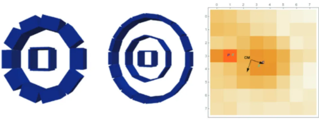
The project envisages two research lines, namely identifying the multiple interactions within a single block or using a multiple-layers detector approach (see fig. 1). On the one hand, the probability for a first Compton interaction is high in dense scintillation crystals (it can be 70% of all impacts). However, the distance between the first hit and the photoelectric interaction typically does not exceed on average the 2–3 mm and it is not always in the forward direction. On the other hand, differentiating the two events in a single block is challenging. One requires high timing resolution, but also accurate spatial performance. Therefore, we have first followed the multilayer approach.
Initial tests were carried out with Bromide-based scintillators (LaBr3 and CeBr3).
We studied their capabilities in terms of spatial and energy resolution. We used a CeBr3
block with dimensions of 51.5×51.5×3 mm3, the large faces of 51.5×51.5 mm3being the
entrance and exit faces of the gamma-ray radiation. We used 4 custom arrays of 1× 16 SiPMs with 3× 3 mm3 each coupled to the lateral sides of the crystal. We digitized all
64 SiPM signals and applied different impact reconstruction algorithms based on charge collection. The results with the CeBr3 block were limited, most likely, due to the use of
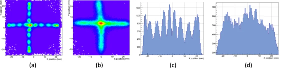
a light guide which served to protect the crystal from hygroscopicity, but simultaneously affected the transfer of scintillation photons to the SiPMs. Figure 2 shows the obtained result when moving a collimated small-size22Na source (1 mm in diameter) across one
horizontal and vertical axis of an LYSO and CeBr3block, respectively. It can be observed
how the spatial resolution obtained with the CeBr3 detector is significantly lower than
that for the LYSO block.
Afterwards, we run experiments with high statistics, but with two identical LYSO blocks in coincidence. An array of 11× 11 22Na sources with 4.6 mm pitch and 1 mm
in diameter was placed in front of the detector under characterization (see fig. 3). This
Fig. 1. – Left: PET designs based on multiple layers blocks. Right: flood map with collection of charges of a gamma-ray event having first a Compton interaction (C) and later a photoelectric one (P).
Fig. 2. – Flood maps obtained when scanning a source on a LYSO (a) and on a CeBr3 (b) block.
Projections of one of the row of sources for the LYSO (c) and CeBr3 (d) blocks.
array covers the whole scintillation volume with sources as close as 2.5 mm to the edge where the photosensors are placed. After center of gravity calculation and calibration of the data [13], a good resolvability of the system was observed, including both crystal center and edges. Average intrinsic spatial resolutions as good as 1.6 mm FWHM have been measured. The system reached an energy resolution of 12%. We are currently carrying out measurements to determine the CTR of this system, but we expect about 350–400 ps FWHM.
2.2. Coincidence time resolution with monolithic scintillators. – We have worked both using analog (SensL, KETEK, Hamamatsu Photonics) and digital SiPM (Philips) to-gether with monolithic crystals on the search of optimum CTR. The feasibility of reaching sub-200 ps CTR (FWHM) using monolithic LYSO blocks of 24× 24 × 20 mm3coupled to
digital SiPMs has been published by other groups [14]. These values are obtained by dis-abling noisy cells (only possible for digital-SiPMs) and by running the set-up at very low temperatures (−25◦C). Averaging timestamps methods as well as MLEM approaches for the timing determination were enabled. Using the same technology but different crystal sizes, we recently carried out also experiments to determine the CTR of these blocks as a function of the temperature. In particular we used two crystals with larger aspect ratio, in coincidence. The crystals had dimensions of 32× 32 × 12 mm3 and 32× 32 × 5 mm3
each, covered with white painting. We observed the effect of the temperature on the CTR, reaching values nearing 200 ps FWHM below −10◦C, see fig. 4. Trigger 1 refers to an acquisition protocol with very low threshold benefit timing capabilities, whereas in Trigger 2 a higher threshold is selected, best suitable for impact identification and dark counts reduction. Accurate timing performance is possible with monolithic blocks and
(a) (b)
Fig. 3. – (a) Set-up with two LYSO layers with lateral sides readout in coincidence. (b) Flood map of a 121 sources array before (left) and after calibration (right).
Fig. 4. – CTR using dSiPMs vs. system temperature obtained with Trigger 1 and 2. Two monolithic crystals with 12 and 5 mm thickness were used in coincidence.
digital SiPMs but it requires low-temperature environments.
In parallel, we have experienced with analog SiPMs readout using ASICs. In partic-ular, we have extensively tested the so-called TOFPET devices from PETsys. We have deeply studied monolithic LYSO blocks, although several crystal geometries, including single pixelated crystals, have also been tested. We have extensively worked with crystals of 50× 50 × 15 mm3, with all faces polished, but the lateral sides black painted and a retroreflector layer at the entrance surface [15, 16]. We made it possible to obtain a very good spatial resolution performance, as well as depth of interaction (DOI), as shown in fig. 5. Arrays of 8×8 SiPMs of 6 × 6 mm2 size each were used (J-Series, 35 microns cell size, SensL). The same photosensor array and ASIC allowed us, for instance, to resolve well 32× 32 crystal pixels of 1.5 mm size (6 mm height). However, timing information requires a major description, as follows.
When a gamma-ray impinges one of these continuous crystals and light reaches the photosensors array, the signal-to-noise ratio (SNR) collected by each SiPM is relatively poor. Notice that the scintillation light spreads among as much as 30–40 SiPMs (depend-ing on the 3D impact position) out of the 64 elements for this crystal and photosensor
Fig. 5. – Right, calibrated flood map of 11× 1122Na collimated sources on a monolithic block
with 50× 50 × 15 mm3dimensions. Center, flood map obtained with a crystal array of 32× 32 elements (1.5 mm size and 6 mm height) on an 8× 8 SiPM array (6 × 6 mm2 each). Right, time difference as a function of charge for a pair of channels before time-walk calibration.
Fig. 6. – Timing performance of a 50× 50 × 15 mm3LYSO block in coincidence with a 6× 6 × 15 mm3pixel. Left: CTR as a function of number of timestamps for different averaging methods.
Right: CTR (FWHM) vs. DOI layer, also for different timestamps averaging methods.
array. With crystals that use reflectors (ESR, white paint, Teflon, etc.) in all faces, there is a high probability that all SiPMs collect light and, moreover, an individual higher sig-nal would be observed. However, this would challenge the determination of the impact determination. For SiPM signals with limited SNR, an accurate time-walk correction is required, in addition to the skew-time correction for the whole acquisition channel path. We have developed over the years approaches to take those time corrections into account [15]. When two of those large monolithic crystals were in coincidence, it was possible to reach a CTR of 660 ps FWHM. Using the same photosensor and readout system but single pixels of 3× 3 × 5 mm3 LYSO, one could obtain a precise CTR of
180 ps FWHM. We have done an exhaustive analysis of the CTR dependency with dif-ferent parameters when using monolithic blocks. Figure 6 shows some examples of these tests. On the left-hand side, we depict the measured CTR as a function of the number of timestamps (SiPMs), when just considering the earliest timestamp, a simple average or an energy weighted average. Notice that this was measured with the monolithic block described above against a reference detector using a single 6× 6 × 15 mm3 LYSO pixel. On the right-hand side, we depict the found CTR but as a function of the depth of interaction and again for different methods to determine this. DOI3 stands for impacts occurring in the 5 mm layer closer to the photosensor and, thus, DOI1 refers to impact in the 5 mm entrance layer.
The CTR limitations on monolithic crystals strongly depend on their geometry, sur-face treatment but also on the photosensor granularity. Using the same photosensor array (8× 8 SiPMs and 6 × 6 mm2 area each), but a smaller block of 25× 25 × 10 mm3
with all faces white painted except the exit one and in contact with the photosensor, we reached a CTR of 385 ps FWHM (about 250 ps FWHM detector resolution when sub-tracting the reference detector contribution). The same block and treatment but read out using 3× 3 mm2 SiPMs exhibited a worst resolution of roughly 450 ps FWHM. This
suggested that there should be a compromise between light collection and SiPM active area (capacitance related) when using monolithic crystals for TOF PET developments.
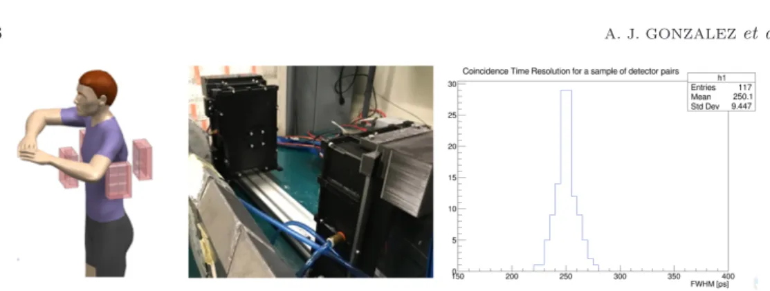
2.3. Two panels PET for cardiac studies. – We have designed a cardiac PET for patients under stress conditions, see fig. 7. Ideally, patients will be sitting in upright position in a fixed one-wheel bike, instead of running in a treadmill or being injected with a drug generating stress conditions. We initially planned to use monolithic crystals
Fig. 7. – Cardiac PET. Left: sketch of PET panels around the chest of a patient. Center: real system with two panels. Right: distribution of CTR values for 117 coincidence pairs.
of 15 mm height coupled to SiPMs with 6×6 mm2active area. However, the CTR results obtained in the previous section with similar monolithic crystals pointed out that the expected CTR with this geometry will not be sufficient to recover the missing angular information. To overcome these limitations, we are using crystal arrays and one-to-one coupling (crystal pixel size to photosensor active area). In particular, each detector panel includes 24 arrays in a 64 arrangement of miniblocks, covering about 15× 10 cm2.
These miniblocks use 8× 8 SiPM arrays with 3 × 3 mm2 active area each, and coupled
to 3× 3 × 10 mm3 LYSO crystals. As depicted in fig. 7 right, with this detector block it was possible to obtain an average CTR of 250 ps FWHM for all pairs of channels. Pilot acquisitions and reconstructions using 22Na sources already exhibited an artifact
reduction when the TOF information is considered during the reconstruction process. With this set-up it was possible to reach a stable energy resolution, after calibration, of about 11.5% at the 511 keV photopeak. A full system characterization following the NEMA protocol is expected in the coming months.
2.4. Discussion on developments performed at DMIL. – Our experience when analog SiPMs and ASIC based readouts are utilized (at temperatures above the drew point in normal conditions), they exhibited certain limitations to reach sub-200 ps. Most likely, this is related to poor signal-to-noise ratio reached at each photosensor. We observed improvements when thinner crystals and reflective material at the surfaces were used. Detector timing resolutions of 250 ps (CTR of 350 ps FWHM) was obtained but for 10 mm blocks. The ability of digital SiPMs to disable noisy cells plays an important role, in addition to running the detectors at low temperatures.
We exhaustively keep investigating how to improve current results by enabling more accurate time-walk corrections. However, we considered that the current status of 350 ps FWHM with 10 mm thick blocks, was not enough to be used in the two panels cardiac system. Therefore, we implemented detector blocks using LYSO pixels of 3× 3 × 10 mm3
dimensions directly coupled to 3× 3 mm2 photosensor. This design allowed us to reach
250 ps FWHM. We have observed in pilot tests that this resolution makes it possible to reduce the artifact effect in a two panels system. Obtaining high-quality images with this geometry would significantly improve diagnosis using molecular imaging, and therapy of a number of diseases. Not only for heart applications, but also breast or brain, among others, could benefit from this design.
We have investigated recovering Compton events during PET imaging. Compton events can amount to more than 70% in a 20 mm thick crystal. Recovering these events
would increase system sensitivity and improve image contrast. We have tried to ac-complish this in a single block, but realized that this is still complex with the current instrumentation. That means that analog SiPMs and monolithic blocks cannot easily allow to distinguish them. On the contrary, digital SiPMs technology makes use of TDC for a photosensor area of about 8×8 mm2, challenging to identify multiple impacts using charge and timing information. We have therefore proposed to use thin LYSO crystals (3 mm) in a separate multilayer configuration with lateral sides readout. This approach significantly reduces photosensor material as well as possible scatter events in the pho-tosensor material itself. Moreover, this design helps separating scatter and phopho-tosensor events in different layers. Notice that the distance from sequential layers would be re-lated to position, energy and timing resolutions. We have shown in this work that spatial resolutions as good as 1.6 mm FWHM for a very high aspect ratio block (51.5 mm size and 3 mm height) is possible.
3. – Conclusions
MEDAMI 2019 was successfully held in Valencia with a number of attenders nearing 70. The main topic of this edition was imaging in immunotherapy, motivated by the latest advances in this area, and its requirements for better imaging tools, especially related to molecular imaging. It was very quickly identified that both improved spatial resolution and higher system sensitivity are needed to better enable the new techniques in immunotherapy. Higher sensitivity is possible by increasing the systems angular coverage, but also by increasing the timing capabilities. Reaching CTR values well below 100 ps FWHM would boost the efficiency gain and, therefore, the image contrast.
Linked to MEDAMI 2019 findings, we have also summarized in this work, the achieve-ments of the DMIL group at the i3M. Accurate CTR could also be used to design PET systems with a limited angle tomography geometry, such as two panels PET or a ring with an aperture (for instance for biopsy procedures). Moreover, information on the tim-ing arrival of scintillation photons can be used to disttim-inguish scatter from photoelectric events and, therefore, to use the Compton kinematics during PET image reconstruction. Therefore, the time-of-flight information of the two annihilation 511 keV gamma-rays, can be used in different ways to improve PET instrumentation and its image.
∗ ∗ ∗
The DMIL work presented in this paper has received funding from the European Re-search Council (ERC) under the European Union’s Horizon 2020 reRe-search and innovation program (grant agreement No. 695536). It has also been supported by the Spanish Min-isterio de Econom´ıa, Industria y Competitividad under Grant TEC2016-79884-C2-1-R. The author would like to thank all current and former members of the DMIL at i3M for their continuous contributions to this work.
REFERENCES
[1] Beer L. et al., Memo, 11 (2018) 138.
[2] Lecoq P., IEEE Trans. Radiat. Plasma Med. Sci., 1 (2017) 473.
[3] Gonzalez A. J. et al., IEEE Trans. Rad. Plasma Med. Sci., 3 (2019) 343. [4] Moliner L. et al., Med. Phys., 39 (2012) 5393.
[5] Sanchez F. et al., Med. Phys., 40 (2013) 051906.
[7] Cherry S. R. and Dahlbom M., PET: Physics, Instrumentation, and Scanners (Springer) 2006.
[8] Gonzalez-Montoro Andrea, Design and implementation of PET detectors based on monolithic crystals and SiPMs, PhD dissertation, 2018.
[9] Sanchez F. et al., Med. Phys., 31 (2004) 1384. [10] Benlloch J. M. et al., Eur. Psychiatry, 50 (2018) 21. [11] Ilisie V. et al., Nucl. Instrum. Methods A, 895 (2018) 74. [12] Ilisie V. et al., J. Phys.: Conf. Ser., 931 (2017) 012012.
[13] Freire M. et al., IEEE Trans. Radiat. Plasma Med. Sci. (2019) https://doi.org/ 10.1109/TRPMS.2019.2947716.
[14] van Dam H. T. et al., Phys. Med. Biol., 58 (2013) 3242. [15] Lamprou E. et al., Nucl. Instrum. Methods A, 912 (2018) 132.