UNIVERSITÀ DEGLI STUDI DI ROMA
"TOR VERGATA"
FACOLTA' DI MEDICINA E CHIRURGIA
DOTTORATO DI RICERCA
IN MICROBIOLOGIA MEDICA E IMMUNOLOGIA
XXII CICLO
COLORECTAL CARCINOGENESIS:
MOLECULAR AND IMMUNOLOGICAL EVENTS
IN A PRECLINICAL MODEL
Dottoranda: Dott.ssa Noemi Moroni
Docente Guida: Prof.ssa Paola Sinibaldi-Vallebona
Tutor: Dr.ssa Annalucia Serafino
Coordinatore: Prof. Enrico Garaci
II
Introduction………..1
1. Colorectal Cancer……… ………..…………...2
1.1
Cytological and histological characteristics of colon………...…....3
1.2
Neoplastic manifestation in the colon………...…5
1.3
Mutations leading to colorectal cancer……….6
1.4
Histological type of lesions………...10
1.5
Wnt pathway and colorectal cancer………....11
1.6
Colorectal cancer in animal model (rodents)...16
1.7
Chemical Carcinogenesis………...18
1.8
Immune Response in Colorectal Cancer………20
Aim of the study……….…24
Materials and Methods……….26
1. Animal Model………27
2. Tissue samples preparation………28
2.1
Procedure for O.C.T. embedding samples…….………...….29
2.2
Procedure for paraffin embedding samples………...…….….29
3. Histological techniques………...30
3.1
Haematoxylin Eosin staining……….30
3.2
Immunohistochemistry………..31
4. Laser Capture Microdissection (LCM)………..33
5. Reverse Phase Protein Array……….35
III
5.2
Reverse Phase Protein Microarray Printing………..35
5.2.1
Slides pretreatment………..36
5.2.2
Total Protein amount determination: Sypro Ruby Staining……36
5.3
Microarray Immunostaining……….36
6. Western Blotting………...37
7. Statistical Analysis………....40
Results...41
1. Colorectal carcinogenetic steps in BDIX preclinical animal model...42
2. Immunohistochemical analysis of Wnt/β-catenin pathway...44
2.1 β-catenin expression and tissue distribution...44
2.2 E-cadherin expression and tissue distribution...46
2.3 APC expression and tissue distribution...46
2.4 GSK3β expression and tissue distribution...49
2.5 C-Myc expression and tissue distribution...49
2.6 Cyclin-D1 expression and tissue distribution...52
2.7 K-Ras expression and tissue distribution...52
3. Immunohistochemical analysis of immune-response markers...55
3.1 T-regulatory cells (T-reg) during tumour progression...55
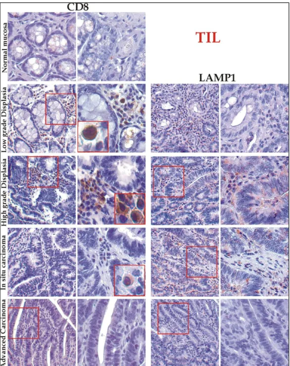
3.2 Tumour infiltrate T-lymphocytes during tumour progression...56
4. Proteomic analysis...58
Discussion...62
IV
2. Validation of pre-clinical models of colorectal cancer induced in BDIX
rats...63
3. New technologies for developing innovative preventive or therapeutic
strategies against colorectal cancer: the phospho/proteomic approach....65
4. The host immune response to cancer: intervention of Regulatory T-cells
(T-Reg) and Tumour Infiltrating T-lymphocytes (TIL) during colorectal
cancer development in the BDIX rat model...66
5. Conclusive Remarks...69
References...70
2
1. Colorectal cancer.
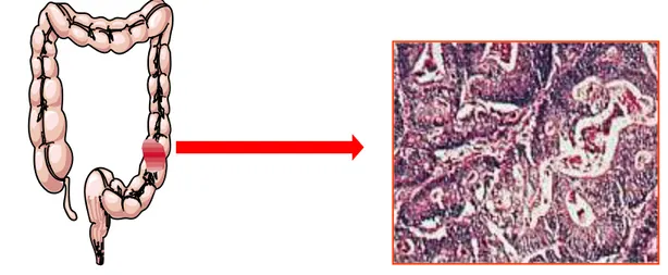
Colorectal cancer (CRC) is defined as a malignant neoplasm arising from the inner lining of the colonic epithelium, and is the third most common cancer worldwide. The incidence of colon cancer is higher in developed countries, where it is the second most common cancer (Wingo et al., 1998). It has a leading position in malignant cancer-related morbidity and mortality.
The 5-years survival rate of CRC patients after diagnosis at an early and localized stage is 90%; however, when distant metastasis (the preferential sites are liver, lung and peritoneum) has occurred, the 5-years survival rate drops to 10%. The occurrence of colon cancer is strongly related to age, with 90% of the cases arising in people who are 50 years or older; until age 50, both men and women have equal risk for colon cancer, but in later life males predominate with this malignancy (American Cancer Society, 2008).
Colon cancer most commonly occurs in the large intestine. The predominant localization is rectum (50–60%) and sigmoid colon (15–25%) (Figure 1).
.
3
1.
1. Cytological and histological characteristics of colon.
The principal functions of the large intestine are the recovery of water and salt and the propulsion of increasingly solid feces to the rectum before defecation. The colonic mucosa is folded in the non-distended state, but it doesn‟t exhibit distinct plicae circulares. The characteristic muscularis mucosae is fundamental for the rhythmic contractions showing a thick wall for peristaltic activity. The muscularis propria consists of inner circular and outer longitudinal layers but, except in the rectum, the longitudinal layer forms three separate longitudinal bands called taenia coli. Consistent with its functions, the mucosa consists of two types of cells: absorptive cells and mucus-secreting goblet cells arranged in closely packed straight tubular glands or crypts, which extend to the muscularis mucosa. Goblet cells predominate in the base of the glands, whereas the luminal surface is almost entirely lined by enterocytes, columnar absorptive cells, the most abundant cell type of intestine. Entero-endocrine cells are located throughout the crypt-villus axis and secrete intestinal hormones. Paneth cells are found at the bottom of crypts and release lysozyme and anti-microbial molecules (Crosnier et al., 2006) (Figure 2a). The lamina propria fills the space between the
glands and contains numerous blood and lymphatic vessels and also collagen as well as lymphocytes and plasma cells. These form part of the defence mechanisms against invading pathogens with intra-epithelial lymphocytes and lymphoid aggregates, which are smaller than the Peyer‟s patches, found in the lamina propria and submucosa. The large intestine is inhabited by a variety of commensal bacteria, which further degrade food residues. Differentiated cells (enterocytes, entero-endocrine cells and goblet cells) occupy the crypts. With the exception of Paneth cells, terminally differentiated cells migrate along the crypt-villi axis and are shed into lumen after 5-7 days (Reya et al., 2005) (Figure 2b).
4 Figure 2. a) Main colon cellular types; b) Scheme of enterocyte differentiation
Stem cells reside near the bottom of the crypt and give rise to progenitor cells that are capable of differentiating toward all epithelial lineages. Stem cells self-renew to regenerate the epithelium after injury while progenitor cells arrest their cell cycle and differentiate, when they reach the tip of the crypt. Epithelial renewal occurs through a coordinated series of events such as proliferation, differentiation and migration (Clevers et al., 2006). In this way, the large
number of cells produced by the crypt compartment is compensated by apoptosis at the tip of the crypt in a process that requires about 2–3 days. Recent studies suggest that a small subset of cells in tumours has stem cell like characteristics. It has been also reported the identification of a colorectal cancer initiating cell based on the surface marker CD133 (O‟Brien et al., 2007). An abnormal pattern of cell replication has been detected in several clinical
conditions associated with an increased risk for colorectal malignancies. The cells with damaged DNA do not cause apoptosis and reach the uppermost part in the crypt and, continuing proliferation process, generate a pre-cancerous change (Bird, 1995). There are two
models for the development of adenomas from stem cells. In the „top-down‟ model, mutant b
5 cells appear in the intra-cryptal zone between crypt openings (Shih et al., 2001). Therefore,
whereas the stem cell, that is the likely oncogenic precursor, must have originated in the base or depths of the crypt, the lesion originates in the top or in the space between the crypts. In the “top-down” model, there may be the establishment of a new source of stem cells in the intra-cryptal zone; a mutated stem cell may migrate toward intra-intra-cryptal area and, with a „second hit‟ conferring growth potential, can expand from this location. In the more intuitive „bottom-up‟ model, a stem cell resident in the base of the crypt, with a mutational defect in growth control, normally proliferates; the cells pushed up to the intra-cryptal area, still retain this mutation. Thus, during the migration, they can accumulate other mutation and generate a neoplastic lesion (Preston et al., 2003).
1.
2. Neoplastic manifestation in the colon.
Epidemiological studies have suggested that colon cancer can be a manifestation of a number of inherited cancer predisposition syndromes, including Familial Adenomatous Polyposis (FAP), Hereditary Non-polyposis Colorectal Cancer (HNCC), and personal or family history of colorectal cancer and/or polyps and inflammatory bowel disease (Rowley, 2004). Furthermore,
other factors such as obesity, lack of exercise, smoking, alcohol consumption, diet rich in high fat, red and processed meats and inadequate intake of dietary fibres, fruits and vegetables are also associated with increased risk of sporadic colon cancer (Cappell et al., 2007) (Figure 3).
6
1.
3. Mutations leading to colorectal cancer.
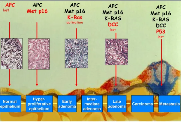
Colorectal cancer is a multi-factorial disease involving the interaction between a large number of genes and their environment. Vogelstein et al. in 1988 first described the time dependent accumulation of genetic mutations and sequential phenotypic correlation in the colonic epithelium. Genetic changes in colorectal cancer include an Adenomatous Polyposis Coli (APC, a tumour suppressor gene) mutation in 85% of all colorectal cancers (Takahashi et al., 2004).
APC is a fundamental regulator of Wnt pathway; in general, the de-regulation of this pathway leads to colon cancer development. p16-INK4a is a tumour suppressor gene that inhibits the cell cycle in response to DNA damage (Liuet al.,2009). Hypermethylation of p16-INK4a, which
silences this tumour suppressor gene, is an early event in colorectal neoplasia, which can occur in adenomas and aberrant crypt foci (Kim et al., 2010).
Oncogenic K-ras apparently contributes to tumour progression relatively early, during the transition from moderate to late adenomas (Furukawa et al., 2002). Ras-guanosine 5‟-triphosphate
(GTP) binds cytoplasmic Raf-1 and translocates it to the plasma membrane, where Raf-1 becomes activated by poorly understood mechanisms. The signal is transmitted to the extracellular signal-regulated portion of mitogen-activated protein kinase which downstream activates and promotes cell proliferation and differentiation. Activating mutations in K-ras genes have been identified in a great variety of human cancer. The mutated forms are found to stimulate cell proliferation, transformation and differentiation. CRCs contain a K-ras mutation in exon 12 in about 40–50% of the cases; the most of the mutations are Guanosine → Thymidine transversions (Pretlow et al., 1993). It is assumed that the K-ras mutation
occurs after the APC gene mutation in the CRC (Takahashi and Wakabayashi, 2004). The epigenetic
inactivation of Ras-associated factor (RASSF) 1A by hypermethylation of the promoter region is frequently detected in flat-type carcinoma. RASSF1A regulates a pro-apoptotic pathway through heterodimerization with the Ras effector NORE1 and interacts with pro-apoptotic
7 protein kinase MST1, which mediates the apoptotic effect of ras. Therefore, it is thought that the inactivation of RASSF1A causes an aberration in the ras signaling pathway without involving the K-ras gene mutation (Khokhlatchev et al., 2002).
More than 90% of the primary CRCs with Loss Of Heterozygosis (LOH) of chromosome 18q show a deletion in the deleted colorectal carcinoma (DCC) gene included in the region of allelic loss. Recent studies reported DCC functions as part of a receptor complex for netrin-1. Furthermore, in various cell lines, DCC, on netrin-1 binding, activates the ERK pathway and in the absence of netrin-1, induces apoptosis via caspase-9. The presence of netrin-1 blocks DCC induced apoptosis. A mutation of DCC that yields cell immortality is caused by continual transmission of the living signal in the absence of netrin-1 (Mehlen et al., 1998).
The most important point that determines the borderline between the adenoma and the adenocarcinoma is a mutation of the p53 gene. The p53 gene is a typical tumour suppressor gene and its mutation has been detected in a variety of cancers and about 75% of CRCs. The p53 protein acts as a cellular stress sensor and a rise in p53 levels causes arrest in the G1 phase of cell cycle, cellular senescence or apoptosis by inducing various target genes. This mechanism limits the propagation of potentially oncogenic mutations. The p53-dependent apoptotic pathway is also induced by DNA damage in certain cell types as well as in cells undergoing inappropriate proliferation. The major players of the p53-induced cell cycle arrest are p21 and growth arrest and DNA damage inducible gene 45 (GADD45)(Vousden and Lu, 2002).
The p21 gene is a cyclin-dependent kinase inhibitor that can influence cell cycle progression from G1 to S phase, by controlling the activity of CDK. GADD45 inhibits the cell progression from G0 to S phase and plays an important role in the maintenance of the stability of the chromosome. Other major players of the p53-induced apoptosis, are pro-apoptotic Bcl-2, protein Bax and BH-3-only proteins Noxa (Nakano et al., 2001).Thus, trans-activation of their
promoters through p53 might induce caspases activation. Therefore, the loss of p53 function as a transcription factor affects cellular malignant transformation (Figure 4).
8 In humans, others important mutations leading to CRC are localized in TGF-β gene. TGF-β signalling can inhibit the growth rate of epithelial cells but the response to TGF-β is often lost in cancers. TGF-β receptor type II mutations are relatively common in replication error–prone colorectal tumour cell lines (Pretlow et al., 1994)Furthermore, a small but significant fraction of
colorectal tumours show loss of the tumour suppressor DPC4, the gene encoding human Smad4 which co-transduces all TGF-β-like signals, and some harbour mutations in the TGF-β transducing Smad2 (Pretlow et al., 1997). Consistently, in humans the earliest mutations in the
TGF-β receptor are found at the late adenoma stage, apparently correlating with the transition from benign adenoma to malignant carcinoma (Paulsen et al., 2005).
In sporadic colorectal tumours, that retain wild-type APC, mutations are frequently found in the β-catenin gene (CTNNB1) or Axin2.
Colorectal cancer progression model
Colorectal cancer progression model
Normal Normal epithelium epithelium Hyper Hyper- -proliferative proliferative epithelium epithelium Early Early adenoma adenoma Inter Inter- -mediate mediate adenoma adenoma Late Late adenoma
adenoma CarcinomaCarcinoma MetastasisMetastasis APC
lost Met p16APC Met p16APC
K-Ras activation APC Met p16 K-RAS DCC lost APC Met p16 K-RAS DCC P53 lost
Figure 4. Accumulation of genetic mutations and sequential histological alteration in the colonic epithelium.
9 The relationship between a stability gene aberration and CRC is revealed by HNPCC, also termed Lynch syndrome. Instability of short tandem repeats, or microsatellites (MSI), is a characteristic of these tumours (Haydon and Jass, 2002). In most HNPCC CRCs, MSI has been
shown to result from mutations in the DNA mismatch repair, hMSH2, hMLH1, hPMS1,
hPMS2 and hMSH6 genes. Others gene may contain MSI within their coding regions such as Transforming Growth Factor-β Receptor, insulin-like growth factor receptor, regulators for
cell cycle and regulators of apoptosis. The transformation to malignancy thus occurs when these target genes are mutated (Rampino et al., 1997).
Several inflammation-related proteins, transcribed by Nuclear Factor-κB (NF-κB), such as cyclo-oxygenase (COX2), Inducible Nitric Oxide Synthase (iNOS), Interferon (IFN ), Tumour Necrosis Factor-α (TNF-α) and Interleukin-1 (IL-1), are increased in inflamed mucosa and remain elevated in colonic neoplasms. NF-κB is also a central regulator of the transcriptional activation of a number of genes involved in cell adhesion, immune and pro-inflammatory responses, apoptosis, differentiation and growth. However, chronic activation of NF-κB induces promotion of epithelial cell turnover and generation of reactive oxygen and nitrogen species (ROS) causing DNA damages that drive the carcinogenesis processes (Tanaka, 2009).
There are several studies that demonstrate a role for DNA methylation very early in colorectal tumorigenesis involving p16, MGMT, hMLH1, MINT31, MINT2, and/or MINT1 (Chan et al., 2002). A high frequency of methylation was found for the newly described SLC5A8 gene, a
sodium transporter that is implicated in colon cancer (Li et al., 2003). It was also showed
hypermethylation of the cellular retinol-binding protein 1 (CRBP1), MINT31, or H-cadherin (CDH13) (Luo et al., 2005). One of the most interesting findings is the frequent methylation of
the secreted frizzle-related protein (SFRP) genes. This epigenetic inactivation allows constitutive Wnt signalling in some precursors lesions that usually lack APC mutations (Suzuki et al., 2004).
10
1.
4. Histological type of lesions.
Carcinogenesis is a multistep process involving the clonal selection and expansion of initiated preneoplastic cells. The clonally expanding cell population is generally termed as a preneoplastic lesion (Bird and Good, 2000).
Aberrant crypts are defined by several characteristics (McLellan and Bird, 1988): larger than the
normal crypts, with increased pericryptal space, having a thicker layer of epithelial cells that often stain darker, and generally having oval rather than circular openings. The occurrence of colon cancer is mainly associated with the incidence of aberrant crypt foci (ACF), an earliest neoplastic lesion, which are clusters of mucosal cells with an enlarged and thicker layer of epithelia than the surrounding normal crypts that progress into polyps followed by adenomas and adenocarcinomas. Other early pre-neoplastic lesions are “β-catenin-accumulated crypts” (BCAC) and “mucin-depleted foci”.
Polyp is a circumscribed mass of cells that project above the surface of the surrounding normal mucosa. Colorectal polyps can be defined as well demarcated, circumscribed lumps of epithelial dysplasia with uncontrolled crypt cell division. Most adenomas remain benign. However, a small fraction of these lesions may evolve into malignancy and there are evidences indicating that a large majority of colorectal carcinomas develop from adenomatous polyps. Adenomas can be classified into three major histological types: tubular, villous and tubulo-villous adenomas. An adenoma is pedunculated when it possesses a stalk. Sessile adenomas rise above the background mucosa without any stalk. They can show different grades of dysplasia (a structural and cytological alteration in the epithelium that predispose an organ to cancer development). There are also flat adenomas, difficult to detect but with high malignant potential. They can be completely flat or show a central area of depression (depressed adenoma) (Cappell, 2007).
11 Patients with Ulcerative Colitis (UC) and Crohn’s disease (CD) have an augmented risk for colorectal malignancies, increasing with the duration of disease and the extent of colorectal involvement. In Inflammation Bowel Disease (such as UC and CD), elevated, sessile and reddish nodules, which are known as pseudo-polyps or inflammatory polyps, are often seen in the otherwise flat mucosa. These lesions are typically small and multiple and largely composed of granulation tissue, mixed with inflamed and hyperemic mucosa. Dysplasia may grow as a flat lesion or as a “dysplasia-associated lesion or mass” (DALM). Recently, several molecular alterations have been detected in long-standing UC. These include oncogene mutations, inactivation of tumour suppressor genes, LOH and chromosomal and microsatellite instability (Xie and Itzkowitz, 2008).
Adenocarcinoma is the typical tumoural lesion, able to invade the muscolaris mucosae and reach the tunica submucosa and so can generate metastases. It is characterized from different extents of peduncular infiltration, several degrees of differentiation and vascularisation.
1.
5. Wnt pathway and colorectal cancer.
Wnt proteins constitute a large family of cysteine-rich, lipid-modified signaling proteins that control development in organisms ranging from nematode worms to mammals (Wodarz and Nusse, 1998). Wnt proteins control diverse developmental processes such as gastrulation, limb, and
central nervous system development (Huelsken and Birchmeier, 2001). Recent studies have also
shown that Wnt signaling may regulate the maintenance and differentiation of stem cells (Taipale and Beachy, 2001). The intracellular signaling pathway of Wnt is also conserved
evolutionally and regulates cellular proliferation, morphology, motility, fate, axis formation, and organ development (Polakis, 2000). Wnt regulates at least three distinct pathways: the
canonical β-catenin pathway, planar cell polarity pathway, and Ca2+ pathway (Veeman et al., 2003). Among these intracellular cascades, the canonical β-catenin pathway has been most
12 extensively studied. In humans it has been shown that abnormalities of this pathway lead to several human diseases, including tumour formation in specific tissues such as the intestine, liver, skin, and mammary gland and bone abnormalities. The NH2 and COOH termini of
β-catenin are unstructured regulatory regions that largely recruit essential co-factor for adhesion and signaling (Taipale and Beachy, 2001; Van Noort et al., 2002). According to the most widely accepted
current model of the β-catenin pathway, in absence of Wnt signaling, Casein Kinase I (CKI) and Glycogen Synthase Kinase-3β (GSK-3β a serine threonine kinase) can phosphorylate β-catenin in the Axin complex for the presence of the “scaffold” protein APC (Kikuchi, 1999): APC
binds to the RGS domain of Axin. In this way, GSK-3 β, β-catenin, and CKI interact with the different sites of the central region of Axin to form the cytoplasmatic complex. Dvl binds to the following C-terminal region of Axin including the DIX domain (Kishida et al., 2001). The
amino acid sequence specifying the phosphorylation of β-catenin is 32SGXXSXXXTXXXS45. CKI–dependent phosphorylation of Ser45 proceeds and the phosphorylation induces subsequent GSK-3β-dependent phosphorylation of Thr41, Ser37, and Ser33. Asp32 and Gly34 are necessary for the interaction of phosphorylated β-catenin with
Fbw1. Therefore, Fbw1 directly links the phosphorylation machinery to the ubiquitination
apparatus. Phosphorylated β-catenin is ubiquitinated, resulting in the degradation of β-catenin by the proteasome. In general, the degradation of the proteins by the ubiquitin-proteasome pathway involves a ubiquitin-activation enzyme (E1), a ubiquitin-conjugating enzyme (E2), and a ubiquitin-ligase (E3) (Matsuzawa and Reed, 2001). The ubiquitin ligase is generally thought to
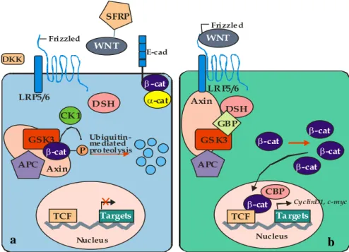
be directly involved in substrate recognition and consists of a multi-protein complex. As a result, the cytoplasmic β-catenin level is lowered. When Wnt acts on its cell-surface receptor consisting of Frizzled and Lipoprotein Receptor-related Protein 5/6 (LRP5/6), β-catenin escapes from degradation in the cytoplasmatic Axin complex. The accumulated β-catenin is translocated to the nucleus, where it binds to the transcription factor T cell factor (Tcf)/lymphoid enhancer factor (Lef) and thereby stimulates the expression of various genes
13 such as C-myc, cyclin D1, MMP-7, etc. (Figure 5). One of the strategies that a cell uses to combat the aberrant expression of β–catenin is to utilize additional regulatory pathways that can modulate its level. For instance, β–catenin induces its own negative regulators Naked and
Axin2 (Jho et al., 2002).
Figure 5. Wnt/ β-catenin pathway scheme in: a) normal cellular condition and b) in tumoural cellular condition.
APC acts as a critical component for β-catenin destruction. In colon cancers, mutations of APC correlate with high levels of β-catenin and transcriptionally active Tcf/β-catenin complexes. Expression of wild type APC in colorectal cancer cells reduces the β-catenin level, and the fragment of APC containing the 20-amino acids repeats is sufficient for this activity. However, an APC fragment with either a mutated β-catenin binding site or Axin-binding site fails to induce the degradation of β-catenin. Therefore, the interaction of APC with both Axin and β-catenin is required for the ability of APC to degrade β-catenin. In the complex, GSK-3β bound to Axin phosphorylates APC, enhancing the stability of β-catenin/APC complex, and leading to a more efficient β-catenin phosphorylation by GSK-3β (Hinoi et al., 2000). APC has
also an important function as a shuttle protein between the nucleus and cytoplasm, and its
WNT SFRP LRP5/6 LRP5/6 DKK Frizzled E-cad Frizzled Ubiquitin-mediated pro teolysis Nucleus Nucleus TCF Targets Ta rgets CyclinD1, c-myc Axin Axin APC APC GSK3 GS K3 -cat -cat -cat -cat -cat -cat -cat -cat P CK 1 DSH CBP DSH WNT GBP TCF a b
14 -CATENIN TCF -CATENIN GSK3- APC AXIN TRANSCRIPTION NUCLEUS APC SHUTTLING -CATENIN APC -CATENIN STABILIZATION
nuclear export may affect β-catenin localization and turnover. Mutant APC lacking a C-terminal NES is trapped in the nucleus. The ability of APC to exit from the nucleus may be important for its tumour suppressor function
(
Rosin-Arbesfeld et al.,2003)
(Figure 6).
Figure 6. APC shuttle protein function between nucleus and cytoplasm.
APC is also involved in polarized cell migration and cell-cell adhesion. Early studies showed that APC is localized to the tips of plasma membrane projections in migrating cells in association with bundles of microtubules. More recently, this APC distribution has been linked to the activation of a signaling complex by integrin-based adhesion that orients the cell for polarized migration. Thus, APC is a multifunctional protein that provides further links between cell-cell adhesion and β-catenin stability and is involved in processes that are not linked directly to Wnt signaling but that contribute to cellular morphogenesis.
In addition to its function in the Wnt signaling pathway, β-catenin also plays a major role in the cell-cell adhesion function, binding tightly to the cytoplasmic domain of type I cadherins and playing an essential role in the structural organization and function of cadherins by linking E-cadherins to the actin cytoskeleton. Another catenin, p120, binds to the membrane proximal domain of cadherin and regulates the structural integrity and function of the cadherin complex (Brembeck et al., 2006). Phosphorylation of p120 by Src or Fer results in loss of cadherin
15 complexes from the cell surface, perhaps as a consequence of simultaneous phosphorylation of β-catenin or because p120 is a binding site for several protein tyrosine phosphatases (PTPases) that antagonize the effects of these tyrosine kinases. In general, activation of tyrosine kinases results in a loss of cadherin mediated cell-cell adhesion and an increase in the level of cytoplasmic β-catenin, either by direct release of β-catenin into the cytoplasm or by activating cadherin endocytosis (Korinek et al., 1997).
In the carcinogenetic process, other protein are involved. E–cadherin is a cell surface protein involved in homophilic Ca2+-dependent cell–cell interactions. Specific adhesive binding is conferred by the cadherin ecto-domain, which engages an identical molecule on the surface of an adjacent cell (Boller et al., 1985;Leckband et al., 2000), whereas the cadherin cytoplasmatic domain
mediates the structural and signalling activity required for adhesion. In addition to the interaction with β-catenin, cadherin associates with two other catenin proteins, termed α and p120 catenin. Α catenin looks as an armadillo domain and is, therefore, structurally unrelated to catenin. Although α-catenin and p120 are important regulators of cell-cell adhesion, β-catenin binding to cadherin remains a prerequisite for adhesion due to its role in protecting the cadherin cytoplasmatic domain from rapid degradation (Huber et al., 2001) enhancing the
efficiency of protein transport from endoplasmic reticulum to cell surface (Chen et al., 1999) and
recruiting α-catenin at cell-cell contacts (Drees et al., 2005; Yamada et al., 2005). Therefore,
posttranslational modification, that regulate β-catenin/cadherin interaction, will have important consequence for cell–cell adhesion. In colon cancer, mutations of E–cadherin gene rarely occur but modifications of expression level and cellular localization have been reported. E-cadherin expression in colon carcinoma was found to be associated with tumour stage, lymphonodal metastases, and patient survival (Bellovin et al., 2005). In fact, loss of E-cadherin
causes a decrease of cell-cell adhesion and increased invasion and motility (Thompson et al., 1994).
Cell-cell adhesion mediated by E-cadherin is required for the maintenance of epithelial tissue architecture in adult organisms (Vleminckx et al., 1999). Down-modulation of E-cadherin is
16 observed during the later stages of tumorigenesis (Thiery, 2002). This down-regulation of
E-cadherin is accompanied by a loss of epithelial characteristics and the acquisition of mesenchymal properties, a process known as epithelial to mesenchymal transition (EMT). During EMT, carcinoma cells become more motile and invasive acquiring characteristics similar to embryonic mesenchymal cells, thereby allowing penetration of the stroma surrounding the initial neoplastic focus (Guarino et al., 2007). CRC metastasis is a multi-hit,
multistage process (Chiang and Massague, 2008). In addition to greater motility, cells must be able to
invade the extracellular matrix, to survive at low density outside the tumour microenvironment, and to develop resistance to apoptosis triggered by loss of cell-matrix interaction (Tse and Kalluri, 2007).
It was been demonstrated the important contribution of oncogenic Ras to altered regulation of E-cadherin and β-catenin. The overactive mutated Ras promotes transformation of intestinal epithelial cells associated with altered regulation of E-cadherin (Schmidt et al., 2003).
1.
6. Colorectal cancer in animal models (rodents).
There is good evidence demonstrating reduced morbidity and mortality associated with early detection of invasive lesions and precursor adenomatous polyps. However, most CRC in the world is diagnosed at an advanced stage. Therefore, most attention has focused on screening for targets for cancer chemoprevention to reduce the number of CRC patients. The identification/discovery of these biomarkers ranges from exposure assessment, risk assessment and management to clinical trials. Along with these, there is also a need to develop and validate molecular biomarkers reflective of exposure and risk from etiological factors. For these reasons it‟s very important to understand the mechanisms leading to neoplastic transformation and progression of human CRC, using an animal model mimicking the histological and molecular alterations observed in humans (Freedman, 2007).
17 Similar neoplastic human lesions could be obtained in animal models (rodents) in a short period of time with high doses of carcinogens that are not ordinary in the human environment. These animal models are chemically induced and genetically modified. More importantly, animal ACF provide the earliest identified lesions in the colon to investigate the changes that take place during the transformation of normal colonic epithelial cells to colorectal cancer. Many characteristics identified in ACF in rodents are also seen in the ACF from humans (Rosenberg et al., 2009).
One of the most frequent alterations (93%) in human ACF is the increased expression of carcinoembryonic antigen (CEA) (Pretlow et al., 1994). The expression of CEA was not associated
with the degree of dysplasia but increase as a function of size of the ACF (Augenlicht, 1994). P-cadherin is not expressed in normal colonic epithelium, but it was expressed in 65% human
ACF. The expression was independent of dysplasia. All of these ACF continued to have normal E-cadherin expression, a few had cytoplasmic expression of β-catenin (Hardy et al., 2002).
The expression of hexosaminidase and α-naphthyl butyrate esterase activities is increased in human ACF compared to adjacent normal mucosa (Pretlow et al., 1991). While only a small
proportion of ACF showed reduced expression of fragile histidine triad (FHIT) gene, its reduced expression was strongly associated with dysplasia (Hao et al., 2000) and may play a role
in the progression of lesions in human colon tumorigenesis. hTERT can be detected at a low level in some normal cells including lymphocytes and at the base of colonic crypts (Hiyama et al., 2001; Pretlow et al., 2003). The expression of iNOS is strong cytoplasmatic in the normal colonic
epithelial cells while 50% ACF and 56% carcinomas showed a marked reduction of iNOS expression (Hao et al., 2001). Sialyl Lewisx (Lex) and sialyl Tn antigens are overexpressed in
70-89% of colon cancers but are not detectable in normal colonic mucosa and are detected only rarely in hyperplastic polyps (Itzkowitz, et al., 1986). Overexpression of glutathione-S-transferase
P1-1 (GSTP1-1) was observed 89% human ACF. All of these GSTP1-1 positive ACF also stained for p21K-ras. The overexpression of GSTP1-1 appears to be induced by mutant KRAS
18 (Miyanishi et al., 2001). COX-2 was not expressed in these same ACF, and apoptosis was
decreased compared to normal mucosa. It appears that GSTP1-1 may protect ACF from apoptosis and thus contribute to the progression of ACF to cancer (Nobuoka et al., 2004). The
increased expression of p16INK4a correlates inversely with proliferation markers (Dai et al., 2000).
Increased expression of c-myc, a target of β-catenin signalling, was seen in 34.9% ACF and in 55.6% dysplastic ACF (T.P. Pretlow and T.G. Pretlow, 2002). The increased expression of c-myc in
ACF suggests an expansion of the immature colonocytes that normally express c-myc at higher levels than their mature counterparts (Mariadason et al., 2005). ACF in rats and mice express
multiple phenotypic alterations:
i) reduced expression of hexosaminidase and α-napththyl butyrate esterase activities. The alteration of these two enzymes is more marked or frequent as that observed in the human ACF, and exhibit opposite changes in expression, decreased rather than increased;
ii) increased expression of periodic acid Schiff reactive material; iii) increased expression of glutathione-S-transferase isoforms.
The expression of iNOS and 2-amino-1-methyl-6-phenylimidazo[4,5-b] pyridine (PhIP) in rat are quite different from that in human, but not relevant. The first alteration identified in rodent ACF and tumours, induced with the chemical carcinogenetic, was mutation in K-ras, varied from 7% to 32 %. Generally, induced colon tumours had a higher frequency of β-catenin (Ctnnb1) gene mutations (75%) than Adenomatous Polyposis Coli (APC) mutations (25%).
P53 mutations were not detected in tumours induced with 1,2 dimethylhydrazine (DMH)
19
1.
7. Chemical Carcinogenesis.
The ability to induce colon tumours in animals has provided the opportunity to study various aspects of the carcinogenesis process. Oncogenesis studies using these models have also elucidated the role of genetic and environmental factors and other influences on the various aspects of this complex disease. The direct-acting carcinogens are compounds that do not require biological catalysis, such as the action of enzymes to form the ultimate reactive species that alters cellular macromolecules. These agents spontaneously break down in an aqueous environment to electrophilic species that react with nucleophilic centers on the DNA molecule. Indirect-acting carcinogens require enzymatic action (e.g. intestinal commensal bacteria) to be converted into the electrophilic species.
The carcinogen most used to obtained tumours in rodent is DMH. This substance induces colon cancer providing a useful model to study early carcinogenesis and sporadic cancer development mimicking many of the clinical, pathologic, and molecular features of human colon cancer (Whiteley and Klurfeld, 2000). DMH is a specific colon carcinogen that induced large
bowel tumours in rodents. In the liver DMH is converted to its main active metabolite azoxymethane (AOM): the chemical structure changes, by oxidative steps, from CH3-NH-NH-CH3 to CH3-[O]N=N-CH3; then to methylazoxymethanol CH3-[O]NH-NH-CH2OH (MAM), which leads to CH3+ methylcarbonium ion, that seems to be the ultimate carcinogen which binds cellular DNA in the colon at the bottom of the crypts via the bloodstream or possibly via biliary secretion (Sunter et al., 1990). The exact nature of the mutations, caused by the
carcinogen DMH, is still unknown. Pro-mutagenic lesions O6-methylguanine has been detected in DNA from various rat and mouse tissues following exposure to DMH. DMH induces the formation of adducts with guanosine in the GTCCA sequences, especially in
Ctnnb1 gene, resulting in point mutation or deletions in codons 32-49 causing removal of 4
20 DMH induced tumours are often mutated in K-ras gene and show microsatellite instability. However, unlike human tumours, they are rarely mutated (15%) in Adenomatous Polyposis
Coli APC gene, never mutated in p53 gene. Nevertheless, even if APC is rarely mutated, rat
tumours accumulate β-catenin in the nucleus like human tumours but this is due to Ctnnb1 mutation. Thus, Wnt/B-catenin pathway plays a major role in carcinogen-induced rat tumours similarly to human tumours (Schwartz et al., 1995).
1.
8. Immune Response in Colorectal Cancer.
Colorectal carcinoma, like most epithelial solid tumours, has been long considered poorly immunogenic and refractory to immunotherapy; these observations are supported from several epidemiological studies based on lack of spontaneous regression of cancer also if the host immune system should be able to react against a neoplastic cell. In effect, CRC spontaneous regression is only exceptionally observed, and does not appear to be associated with an immune response (Francis et al., 1997). In vitro studies performed on tumour infiltrating
lymphocytes (TIL) cultures purified from colorectal carcinomas failed to demonstrate substantial lytic activity against autologous cancer cells (Rosenberg and Karnofsky 1992). Moreover,
classical immunotherapeutic interventions known to be active against other type of tumour (e.g. melanoma), such as systemic administration of cytokines (IL-2, INF-α) or adoptive transfer of autologous lymphocyte effectors (LAK, lymphokine-activated killer cells, TIL), have proved ineffective in colorectal tumours (Wolmark et al., 1998).
Indeed, several evidences indicate that also colorectal cancer may express tumour associated antigens (TAA) recognised by T-cells, influencing patient prognosis and determining the tumour immunological profile (Dalerba et al., 2003). In general, histopathological studies have
shown that spontaneously regressing tumours are heavily infiltrated by T-cells (Mackensen et al., 1993) and that T-cell infiltrates within the primary tumour are strong predictors of better
21 inflammatory infiltrates, but their results are contradictory, supporting a protective role of inflammatory infiltrates (Guidoboni et al., 2001), or not (Nielsen et al., 1999). In effect, the
inflammatory infiltrates are biologically heterogeneous and can originate through different mechanisms, reflecting diversities in tumour biology and tumour-host interaction. From an immunologist‟s point of view, the type of immune effectors that can more consistently be considered as a sign of a systemic anti-tumour immune response are cytotoxic T lymphocytes (CTLs), classically CD8+T-cells (Riddell and Greenberg, 1995). Distinction among different subsets
of lymphocytes is important: for example the lamina propria of normal colorectal mucosa is rich in B-cells (Lee et al., 1988). CD8+-Tumour Infiltrating Lymphocytes (TIL) in primary
colorectal carcinomas are dividing in three groups (Guidoboni et al., 2001): 1) peri-tumoural, when
distributed along the invasive margin of the tumour; 2) stromal, when infiltrating the tumour stroma; 3) intra-epithelial, when infiltrating within cancer cells and taking direct contact with tumour cells. TIL are characterized from the expression contemporary of the marker CD8 and CD25 (cell surface antigen that is expressed on T cells following activation) (Guidoboni et al., 2001).
Among the different immune cells involved in the control of human tumours, T-cells appear to have a correlation between the function of tumour-infiltrating lymphocytes (CD8+-TIL and CD4+-T regulatory, or T-Reg) and prognosis in different tumours (Alvaro et al., 2005). Type,
density, and location of immune cells within human colorectal tumours predict clinical outcome (Jarnicki et al., 2006).
CD4+ T-Reg activated express CD25 are dependent on the transcription factor Foxp3, for their development and function (Fontenot et al., 2003). CD4+CD25+Foxp3+ T-Reg cells are present in
the normal colonic lamina propria, suggesting a constitutive role in the prevention of aberrant responses to risk-free intestinal antigens (Uhlig et al., 2006). These cells have a protective role
against autoimmune and other inflammatory diseases (including colitis) in several animal models (Holmen et al., 2006). It is possible to speculate that local suppression by T-Reg
22 lymphocytes in chronic inflammation may contribute to tumour growth by preventing effective early immune-surveillance. Conversely, an increasing wealth of studies suggests a direct role for T-Reg, not in the initiation of tumour growth, but in the prevention of immunity against established tumours (Zou, 2006). T-Reg inhibit classical cytotoxic cells such as TIL and
NK cells (Ghiringhelli, et al., 2005) and contribute to tumour escape and poor survival. This would
explain the inefficiency of the immune system to adequately attack primary tumours. More significantly, however, tumours seem to have evolved mechanisms to actively submerge and suppress potential anti-tumour immune responses (Zou, 2005).
Several studies have suggested a protective role of eosinophil infiltration in colorectal carcinoma (Fernandez-Acenero et al., 2000), but this effect frequently disappears when results are
stratified and corrected for stage (Fisher et al., 1989). Moreover, eosinophils are normally well
represented in the normal colonic mucosa (Lee et al., 1988), and their number can display
significant geographical variations (Pascal et al., 1997). Indeed, most studies show that eosinophils
are more abundant in early stage and adenomas than in invasive advanced carcinomas (Moezzi et al., 2000).
Similar considerations are true also for mast cells and for γ/δ T lymphocytes (Lachter et al., 1995).
Macrophages can produce angiogenic and immune-suppressive growth factors, such as TGF-β, and can promote tissue remodelling and metastatization process via secretion of matrix metalloproteinases. In colorectal carcinoma, the number of intra-tumour macrophages is higher than in normal colorectal mucosa, and progressively increases from early stage to advanced stage tumours, without any concomitant increase in T-cells (Hakansson et al., 1997). This
suggest that infiltrating macrophages have a role in the promotion of neo-angiogenesis within the tumour tissue, either by direct production of pro-angiogenic factors like Platelet-Derived Endothelial Cell Growth Factor (PD-ECGF) (Takahashi et al., 1996), or by stimulating their
production by tumour cells, such as in the case of angiogenin (Etoh et al., 2000). Interestingly,
23 dendritic cells (DC), a different subset of myeloid cells with a unique capability in antigen presentation and primary T cell activation, progressively decreases, being lower in tumours than in normal colorectal mucosa, and lower in primary than in metastatic tumours (Schwaab et al., 2001).
24
25 The aim of this study is to validate a preclinical animal model to evaluate the carcinogenesis process. The model might reproduce all the histological steps of human colorectal cancer transformation and progression. Moreover, each step has to be correlated to associated molecular events. The study was made through immunohistological analysis of several proteins involved in carcinogenesis process, specifically in Wnt/β-catenin pathway well known to be modified in neoplastic human lesions. The preclinical animal model was also utilized to study the immunological response against the tumour and compared to the “immune-surveillance/immune-escape theory” identified in human colorectal cancer. This model could represent a useful experimental system to study differential biomarker expression and pathway activation during tumorigenesis processes. The biomarkers discovery was evaluated through a proteomic/peptidomic approach. The identified molecules could represent targets of new therapies. Thus, learning information about timing and sequencing of molecular events in our animal model is essential to control and prevent tumour growth and to identify the best therapeutic approach for the specific disease step in an individualized therapy.
26
27
1. Animal Model.
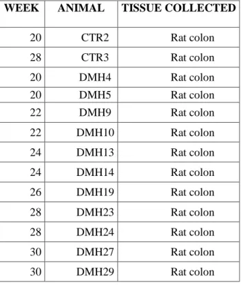
The animal model consist in the induction of colorectal carcinoma with a carcinogenic substance in the BDIX rat strain at age of 9 weeks; the carcinogen used was 1,2 Dymethylhydrazine (DMH). From literature data, DMH was administered subcutaneously once a week at dosage 18mg/Kg/rat for 5 weeks. The animals were sacrificed starting from to the 20th the 30th week after the first carcinogenic injection. At each time the rat colon was collected.
Table 1. Sample of first carcinogenesis experiment.
WEEK ANIMAL TISSUE COLLECTED
20 CTR2 Rat colon 28 CTR3 Rat colon 20 DMH4 Rat colon 20 DMH5 Rat colon 22 DMH9 Rat colon 22 DMH10 Rat colon 24 DMH13 Rat colon 24 DMH14 Rat colon 26 DMH19 Rat colon 28 DMH23 Rat colon 28 DMH24 Rat colon 30 DMH27 Rat colon 30 DMH29 Rat colon
To confirm the results obtained in the first experiment and to observe the connection between inflammatory disease and cancer, we repeated the same experiment sacrificing the animals starting from the 6th week to the 35th week after the first injection of the carcinogen.
28 Table 2. Sample of second carcinogenesis experiment.
The new weeks inserted respect to first experiment are marked in grey.
WEEK ANIMAL TISSUE COLLECTED
6 DMH2 Rat colon 6 DMH4 Rat colon 12 DMH6 Rat colon 12 DMH8 Rat colon 16 DMH10 (x2) Rat colon 18 DMH12 Rat colon 18 DMH14 (x2) Rat colon 19 DMH16 (x2) Rat colon 20 DMH20(x2) Rat colon 20 DMH22 (x2) Rat colon 21 DMH24(x2) Rat colon 22 DMH26(x2) Rat colon 24 DMH27(x2) Rat colon 24 DMH28(x2) Rat colon 26 DMH30(x2) Rat colon 26 DMH32(x2) Rat colon 28 DMH33(x2) Rat colon 28 DMH34(x2) Rat colon 32 DMH37(x2) Rat colon 35 DMH38(x2) Rat colon 35 DMH39(x2) Rat colon
29
2. Tissue Samples Preparation.
2.
1
Procedure for O.C.T. embedding samples.
The tissue sample are been frozen as soon as possible using the embedding media O.C.T. (Sakura Finetech‟s Compound); this aqueous glycerol compound provide protection of the specimen during long-term, gives support to the tissue and aids in the cryo-sectioning process. The tissue was cut to a size no greater than one half the area of the cryomold (Sakura Finetech‟s), so that it will fit into the cryomold without touching the sides of the mold. For the standard cryomold, specimen samples didn‟t exceed 1cm in height or width, or a thickness of more than 0.5 cm. The frozen tissue were frozen at -800C and then cut in a cryostate (Leica) approximately 8 microns and affixed onto the LCM slide (Arcturus). Once mounted, the slides were frozen at -20°C until LCM processesment.
2.
2
Procedure for paraffin embedding samples.
After collection, the tissue samples were washed with medium until the fixation procedure (necessary to preventing antigen elution or degradation and to preserve the position of the antigen, whether nuclear, cytoplasmic or membrane-bound). The tissues were fixed with 10% tamponate formalin (Bio-Optica) for 24h, washed in water, immersed in alcohol 70%, alcohol 95%, alcohol 100% (Fluka), histolemon (Bio-Optica) and finally embedded in liquid paraffin. After fixation, the tissue block was embedded in paraffin, then cut in a microtome (Leica) approximately 5 microns and affixed onto the positively charged slide (Carlo Erba). Once mounted, the slides were dried to remove any water and incubated at 60ºC for a few hours.
30
3. Histological techniques.
3.
1
Haematoxylin Eosin staining.
Before proceeding with the staining protocol, the slides were deparaffinized and rehydrated following these passages:
- Bioclear (Bio-Optica): 30min - 100% ethanol: 10 min - 90% ethanol: 5min - 80% ethanol: 5 min - 70% ethanol: 5 min - 50% ethanol: 5 min - H20: 10 min
- Staining with haematoxylin (Bio-Optica): 10 min - H20: 5 min
- Counterstaining with eosin 1% (Bio-Optica): 5 min The tissue were dehydrated following these passages: - 50% ethanol: 5 min
- 70% ethanol: 5 min - 90% ethanol: 5min - 100% ethanol: 10 min - Bioclear: 30min.
In the final step, the slides was mounted using Fast drying mounting medium for cover slipping (Bio-Optica).
31
3.
2
Immunohistochemistry.
Immunohistochemistry (or IHC) is a method used for demonstrating the presence and location of proteins in tissue sections. The antibody-antigen interaction is visualized using a chromogenic detection, in which an enzyme conjugated to the antibody cleaves a substrate to produce a coloured precipitate at the location of the protein. We used horseradish peroxidase (HRP) and its substrate peroxide/DAB for visible light microscopy (DAKO LSAB + System HRP).
Before proceeding with the staining protocol, the slides were deparaffinised and rehydrated following these passages:
- Bioclear: 30min - 100% ethanol: 10 min - 95% ethanol: 5min - 75 % ethanol: 5 min - 50 % ethanol: 5 min
- 1X Dulbecco‟s Phosphate Buffered Saline – PBS (Euroclone) 5 min x2
The formalin-fixed tissues required an antigen retrieval step before immunohistochemical staining to break the methylene bridges formed during fixation and expose the antigenic sites in order to allow the antibodies to bind. The method used was the “heat induced” antigen retrieval with Sodium citrate buffer (10 mM Sodium Citrate - SIGMA, 0.05% Tween 20 - Biorad, pH 6.0) or Tris-EDTA Buffer (10 mM Tris Base - SIGMA, 1 mM EDTA - SIGMA, 0.05% Tween 20, pH 9.0). After rinsing with PBS for 20‟, the activity of endogenous peroxidase was suppressed incubating the slides in 0.3% H2O2 (Fluka) added to methanol
(Carlo Erba) for 20 min. From this point, all the steps were made in a humidified chamber to avoid drying of the tissue; a-specific binding sites were blocked with 1% BSA (SIGMA) in
32 PBS for 15 min at room temperature. It was applied primary antibody diluted in 1% BSA in PBS and incubated overnight at 4°C. A biotinylated secondary antibody was then bounded to the primary antibody, incubating the slides with a biotinylated link (DAKO) for 30 min at room temperature. In a separate reaction, a complex of avidin and biotinylated enzyme was formed by mixing the two in a ratio that leaves some of the binding sites on avidin unoccupied. This complex, called streptavidin-HRP (DAKO), was then incubated with the tissue section for 30 min at room temperature. The unoccupied biotin-binding sites on the complex bind to the biotinylated secondary antibody (DAKO). Finally the slides, were incubated with the substrate of HRP, 3,3'- Diaminobenzidine (DAB) (DAKO) for 9 min at room temperature and counterstained with haematoxylin for 5 minutes. The tissue were dehydrated following these passages:
- 50% ethanol: 5 min - 75% ethanol: 5 min - 95% ethanol: 5min - 100% ethanol: 10 min - Bioclear: 30min.
In the final step, the slides were mounted using Fast drying mounting medium for cover slipping.
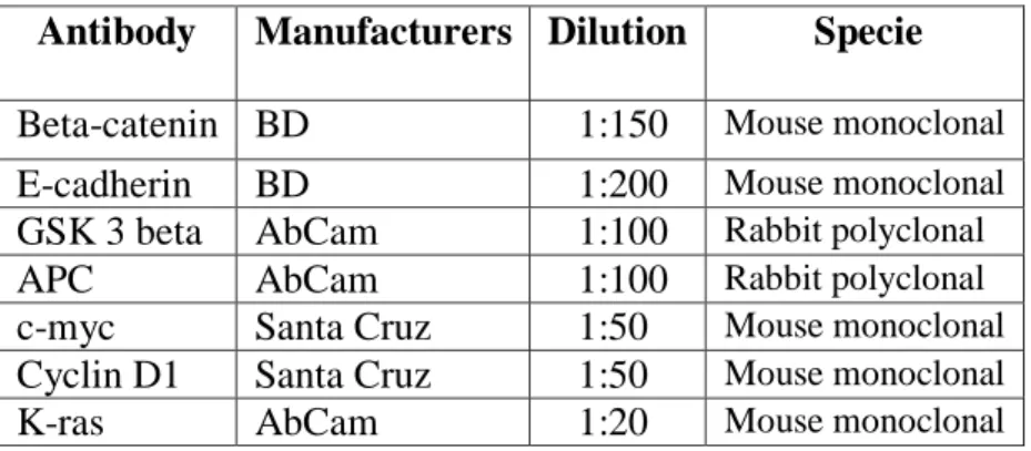
33 Table 3. Primary antibodies used for Wnt/β-catenin pathaway detection.
Antibody Manufacturers Dilution Specie Beta-catenin BD 1:150 Mouse monoclonal
E-cadherin BD 1:200 Mouse monoclonal
GSK 3 beta AbCam 1:100 Rabbit polyclonal
APC AbCam 1:100 Rabbit polyclonal
c-myc Santa Cruz 1:50 Mouse monoclonal
Cyclin D1 Santa Cruz 1:50 Mouse monoclonal
K-ras AbCam 1:20 Mouse monoclonal
Table 4. Primary antibody for immunological analysis of infiltrated lymphocytes.
4. Laser Capture Microdissection (LCM).
LCM incorporates an inverted light microscope and a near infrared laser to facilitate the procurement of desired cells. After direct visualization of the cells of interest, using a laser pulses to activate a thermoplastic polymer film that expands and impregnates the cells of interest that can then be lifted from the slide. The exact morphology as well as the DNA, RNA and proteins of the procured cells remain intact and bound to the film. Using LCM frozen tissues have been successfully dissected and recovered cells used for protein analysis. Normal epithelium, premalignant (dysplasia), in situ cancer, invasive cancer and metastatic cancer cells have been obtained from tissues. Frozen tissue has been cut at 8μm, mounted on plain, uncharged microscope slides and stained with haematoxylin protocol. Complete Mini protease inhibitor tablets (Roche Applied Science), were added to the 70% ethanol and haematoxylin staining solutions to reduce protein activation derangement.
Antibody Manufacturers Dilution Specie CD4 Millipore 1:100 Mouse monoclonal
LAMP1 AbCam 1:200 Rabbit polyclonal
CD25 Thermo Scientific 1:80 Mouse monoclonal
34 Protocol for Staining Frozen Tissue
- 70% Ethanol: 5 seconds - Deionised Water: 10 seconds
- Mayer‟s Haematoxylin: 15-30 seconds - Deionised Water: 10 seconds
- Scott‟s Tap Water (Sigma): 10 seconds - 70% Ethanol: 10 seconds - 95% Ethanol: 10 seconds - 95% Ethanol: 10 seconds - 100% Ethanol : 1 minute - 100% Ethanol: 1 minute - Xylene: 1 minute - Xylene: 1 minute
Laser capture microdissection was performed using a Pixcell II Laser Capture Microdissection system (Arcturus) to procure enriched tumour cell populations. A total of 25,000 cells, procured over several sections, were microdissected for each case and stored on microdissection caps (CapSure® Macro LCM Caps, Arcturus) at –80°C until lysed. It is possible to estimate the number of cells captured based upon the number of pulse fired during the collection of cells, using this formula:
35
5. Reverse Phase Protein Array.
Reverse Phase Protein Microarrays are a multiplexed proteomic platform used to evaluate cell signalling activity in many samples at once. Approximately 150 slides can be printed with 40µl of protein lysates and each slide is probed with a single antibody.
5.
1
Tissue Lysates Preparation.
Cell lysis volume is calculated by assuming 1µl tissue extraction buffer/1,000 cells. The tissue lysis buffer (950µl Tris-Glycine SDS Sample Buffer 2X (Invitrogen), 50µl 2 β-mercaptoethanol (PIERCE), 1ml TPER Reagent (PIERCE)) was repetitively pipetted among on the LCM caps; the protein lysate was transferred to a screw cap tube and heated at 100˚C for 7 minutes. The lysated can be stored at -80˚C.
5.
2 Reverse Phase Protein Microarray Printing.
The lysates were then spotted onto nitrocellulose-coated glass slides (Whatman) using a 2470 Arrayer (Aushon BioSystems), outfitted with 350-µm pins. Cases were printed in duplicate, in 5-point dilution curves, thus assuring that the linear detection range was encompassed for the chosen antibody concentration. As a high and low internal control for antibodies staining specificity, lysates derived from human cervical cancer HeLa cells and pervanadate treated HeLa cells, human immortalized T-cells Jurkat, calyculin treated Jurkat cells and etoposide treated Jurkat cells, were used and spotted onto every array along with the experimental samples. Slides were stored desiccated (Drierite anhydrous calcium sulfate) at –20°C until staining.
36
5.
2.1
Slides pre-treatment.
All microarray slides, with the exception of the one probed with the chemoluminescent Sypro Ruby (Invitrogen) used to determine the total protein, should be blocked prior to staining procedure.
First, incubate the slides with 1X Mild Reblot™ Mild Antigen Stripping solution (Chemicon) in deionised water for 15 minutes on a shaker.
After washing PBS calcium and magnesium free solution (GIBCO) the slides were blocked with I-Block Protein Blocking Solution (Tropix) for a minimum of 60 minutes.
5.
2.2
Total Protein amount determination: Sypro Ruby Staining.
Sypro Ruby staining is a fundamental step to quantify the amount of proteins that are present in the printed sample. Sypro Ruby procedure is based on fluorescent dye detection. For more accurate quantification of protein concentration, it was stained with Sypro Ruby 1 of every 25 slides. The slides were warmed to room temperature for 5-10 minutes, then incubated on a shaker for 15 minutes with fixative solution (3.5 ml acetic acid for a final dilution of 7% (Fisher) + 5 ml methanol for a final dilution of 10% (Fisher). After washing with deionised water the slides were incubated with Sypro Ruby solution (Molecular Probes) for 30 minutes and then rinsed with deionised water.
5.
3
Microarray Immunostaining.
This method requires a single antibody-epitope interaction on the protein of interest. The number of slides to be stained was chosen in relation to the number of endpoints of interest. The Dako autostainer (Dako Cytomation) allows simultaneous staining of 48 slides. To quantify the unspecific background signal generated from the interaction between the secondary antibody and samples, it is essential in each staining run, that one slide is probed
37 only with each secondary antibody used. The subtraction of signal produced by the negative control from the primary antibody stained slides provides a more accurate intensity value for the protein of interest. Each antibody was validated to confirm specific interaction by Western blot analysis. Antibodies producing a single band in correspondence to the molecular weight of interest were considerate validated and eligible for immunostaining. It was used the Catalyzed Signal Amplification System kit according to the manufacturer‟s recommendation (CSA; Dako Cytomation). Development was completed using diaminobenzadine/hydrogen peroxide as chromogen/substrate. All protein values were normalized to total protein to account for differences in intensity due solely to starting lysate concentration variance. Stained slides were scanned individually on a UMAX PowerLook III scanner (UMAX) at 600 dpi (dots per inch) and saved as TIFF files in Photoshop 6.0 (Adobe). The TIFF images for antibody-stained slides and Sypro-stained slide were analyzed with array analysis software designed for protein microarray analysis: version 2.X00 (Vigene). The software performed spot finding, local background subtraction, replicate averaging, and total protein normalization, producing a single value for each sample at each endpoint.
6. Western Blotting.
Western Blotting has been used for validating the antibodies used in phospho proteomic analysis. Laser capture microdissected cells were lysed directly in SDS sample buffer and run on 4%-20% SDS-PAGE gels (Invitrogen), and subjected to Western transfer onto Immobilon PVDF membrane (Sigma-Aldrich).
38
Table 5. List of used antibodies and manufacturers.
Antibodies Manufacturers
Akt Cell Signaling
APC1 Ab-1 Lab Vision
E-cadherin Cell Signaling
Catenin (beta) Cell Signaling
CD44 Cell Signaling
CD133 Milteny
Cox2 Upstate
CiclinD1 Cell Signaling
EGFR Cell Signaling
ErbB2/HER2 DAKO Cytomation
ErbB3/HER3 Cell Signaling
EGFR L858R Mutant Cell Signaling
ERK1/2 Cell Signaling
c-Myc Cell Signaling
Ras-GRF1 Cell Signaling
Smac/Diablo Cell Signaling
Phospho-4E-BP1 (S65) Cell Signaling
Phospho-4E-BP1 (T70) Cell Signaling
Phospho-Acetyl-CoA Carboxylase (s79) Cell Signaling
Phospho-Adducin (S662) Upstate
Phospho-Akt (S473) Cell Signaling
Phospho-Akt (T308) Cell Signaling
Phospho-AMPKalpha1 (S485) Cell Signaling
Phospho-AMPKbeta1 (S108) Cell Signaling
Phospho-ASK1 (S83) Cell Signaling
Phospho-BAD (S112) Cell Signaling
Phospho-BAD (S136) Cell Signaling
Phospho-Bcl-2 (S70) Cell Signaling
Phospho-c-Abl (T735) Cell Signaling
Phospho-c-Abl (Y245) Cell Signaling
Phospho-Caspase 3, cleaved (D175) Cell Signaling Phospho-Caspase 6, cleaved (D162) Cell Signaling Phospho-Caspase 7, cleaved (D198) Cell Signaling Phospho-Caspase 9, cleaved (D315) Cell Signaling Phospho-Catenin (beta) (S33/37/T41) Cell Signaling Phospho-Catenin (beta) (T41/S45) Cell Signaling
Phospho-Chk-2 (S33/35) Cell Signaling
Phospho-CREB (S133) Cell Signaling
Phospho-cofilin (S3) Cell Signaling
39
Phospho-EGFR (Y1068) Cell Signaling
Phospho-EGFR (Y992) Cell Signaling
Phospho-EGFR (Y1148) Biosource
Phospho-EGFR (Y1173) Biosource
Phospho-eIF4E (S209) Cell Signaling
Phospho-eIF4G (S1108) Cell Signaling
Phospho-eNOS (S1177) Cell Signaling
Phospho-ErbB2/HER2 (Y1248) Upstate
Phospho-ERK 1/2 (T202/Y204) Cell Signaling
Phospho-FADD (S194) Cell Signaling
Phospho-FAK (Y397) BD
Phospho-FAK (Y576/577) Cell Signaling
Phospho-FKHR (S256) Cell Signaling
Phospho-FKHR (T24)/FKHRL1 (T32) Cell Signaling Phospho-GSK-3alpha/beta (S219) Cell Signaling Phospho-GSK-3alpha/beta (Y279/216) Biosource
Phospho-GSK-3beta (S9) Cell Signaling
Phospho-Histone H3 (S10) Upstate
Phospho-IGF-1 Rec (Y1131/Insulin Rec (Y1146) Cell Signaling Phospho-IGF-1 Rec (Y1135/36IR (Y1150/51) Cell Signaling
Phospho-IkappaB-alpha (S32) Cell Signaling
Phospho-IkappaB-alpha (S32/36) (39A1431) BD
Phospho-IRS-1 (S612) Cell Signaling
Phospho-Jak1 (Y1022/1023) Cell Signaling
Phospho-c-Kit (Y703) Cell Signaling
Phospho-MARCKs (S152/156) Cell Signaling
Phospho-Met (Y1234/1235) Cell Signaling
Phospho-MSK1 (S360) Cell Signaling
Phospho-mTOR (S2481) Cell Signaling
Phospho-mTOR (S2448) Cell Signaling
Phospho-NF-kappaB p65 (S536) Cell Signaling
Phospho-p27 (T187/Y182) Zymed
Phospho-p38 MAP Kinase (T180/Y182) Cell Signaling
Phospho-p70 S6 Kinase (S371) Cell Signaling
Phospho-p70 S6 Kinase (T389) Cell Signaling
Phospho-p90RSK (S380) Cell Signaling
Phospho-PAK1 (S199/204)/PAK2 (S192/197) Cell Signaling Phospho-PDGF Receptor beta (Y716) Upstate Phospho-PDGF Receptor beta (Y751) Cell Signaling
Phospho-PKC alpha (S657) Upstate
Phospho-PKC zeta/lambda (T410/403) Cell Signaling
Phospho-PKCa/B II (T638/641) Cell Signaling
Phospho-PKCtheta (T538) Cell Signaling
Phospho-PRAS40 (T246) Biosource