President
Zhong-Ping Chen, M.D., Ph.D. (Guangzhou, China)
President Elect
Hao-Zhe Piao, M.D. (Shenyang, China)
Vice Presidents
Xiu-Wu Bian, M.D., Ph.D. (Chongqing, China)
Xian-Shu Gao, M.D., Ph.D. (Beijing, China)
Shi-Zhu Yu, M.D., Ph.D. (Tianjin, China)
Jian-Hong Zhu, M.D., Ph.D. (Shanghai, China)
Past Presidents
2004 –2008 Zhong-Ping Chen, M.D., Ph.D. (Guangzhou, China)
2008 –2012 Zhong-Ping Chen, M.D., Ph.D. (Guangzhou, China)
2012 –2016 Shi-Guang Zhao, M.D., Ph.D. (Harbin, China)
2016 –2018 Chao You, M.D., Ph.D. (Chengdu, China)
Volume 1 • Issue 6 • November-December 2018
Glioma Volume 1 Issue 6 November-December 2018 Pages ***-***
i
ntRoduCtionGlioblastoma (GBM) is one of the most frequent primary tumors of the central nervous system (CNS).[1] According to
the World Health Organization classification of CNS tumors, it can be subdivided into IDH mutant and IDH wild-type, on the basis of the mutational status of IDH1/IDH2 genes.[1] GBM IDH wild-type is the most common subtype; it mainly affects
elder patients and it carries worse prognosis, as compared to GBM IDH mutant.[1]
Regardless of IDH mutational status, the current standard of treatment of GBM includes surgery, followed by chemotherapy with temozolomide and radiotherapy.[2] However, despite
treatment, the most GBM patients undergo recurrence and die within 12–24 months, while only about 10% of them survive ≥5 years.[2,3]
A subset of GBMs have a silenced O-6-methylguanine-DNA methyltransferase (MGMT) gene, due to promoter methylation.[4]
Since MGMT encodes for an enzyme which repairs DNA
O‑6‑methylguanine‑DNA methyltransferase promoter
methylation can change in glioblastoma recurrence due to
intratumor heterogeneity
Valeria Barresi1, Maria Caffo2, Giuseppa De Luca3, Giuseppe Giuffrè4
1Department of Diagnostics and Public Health, University of Verona, Verona, Italy.
2Department of Biomedical Sciences and Morphological and Functional Images, University of Messina, Messina, Italy. 3Ircss Policlinico San Martino, Genova, Italy.
4Department of Human Pathology in Adulthood and Evolutive Age, University of Messina, Messina, Italy.
Background and Aim: The standard-of-care for patients with glioblastoma (GBM) is surgery followed by concurrent chemotherapy with
temozolomide and radiotherapy. O-6-methylguanine-DNA methyltransferase (MGMT) promoter methylation is commonly assessed in GBM as a predictive marker of response to temozolomide. Although MGMT methylation status has been shown to change between primary and recurrent GBM, no indication exists on retesting MGMT in recurrent GBM. In addition, what causes the change in MGMT methylation has yet to be identified. In this study, we aimed to investigate whether MGMT promoter methylation in recurrent GBM was influenced by
intratumor heterogeneity in the initial GBM tumor. Materials and Methods: We investigated the status of MGMT promoter methylation in
different samples taken from concentric layers of 24 GBMs and in 11-paired surgically resected recurrences. The neoplastic nature of samples submitted for methylation analysis was preliminary verified through histological examination; the fragments were accurately chosen to have
adequate cellularity and minimal amount of nontumor contaminants. Results: About 27% (3 out of 11) of the recurrences had changed MGMT
methylation status compared to the initial tumor. Initial tumor heterogeneity might play a role in this change, as all three cases had intratumor
heterogeneity (with the central part of the tumor methylated and the peripheral part unmethylated) in the primary GBM. Conclusion: This
study suggests that MGMT methylation variation in recurrent GBM may depend on intratumor heterogeneity in the initial tumor. Intratumor heterogeneity and possible changes in the recurrence should be taken into account when testing MGMT promoter methylation status as a predictive factor orienting therapeutic decisions in patients with GBM.
Keywords: Glioblastoma, heterogeneity, IDH, O-6-methylguanine-DNA methyltransferase, recurrence
Address for correspondence: Prof. Valeria Barresi,
Department of Diagnostics and Public Health, Polyclinic G.B. Rossi, P.le L.A. Scuro, 1, 37134 Verona, Italy. E‑mail: [email protected]
Access this article online Quick Response Code:
Website:
www.jglioma.com
DOI:
10.4103/glioma.glioma_38_18
This is an open access journal, and articles are distributed under the terms of the Creative Commons Attribution‑NonCommercial‑ShareAlike 4.0 License, which allows others to remix, tweak, and build upon the work non‑commercially, as long as appropriate credit is given and the new creations are licensed under the identical terms.
For reprints contact: [email protected]
How to cite this article: Barresi V, Caffo M, De Luca G, Giuffrè G.
O-6-methylguanine-DNA methyltransferase promoter methylation can change in glioblastoma recurrence due to intratumor heterogeneity. Glioma 2018;1:208-13.
Barresi, et al.: MGMT heterogeneity in glioblastoma
209
Glioma ¦ Volume 1 ¦ Issue 6 ¦ November-December 2018 damage from alkylating agents, its expression is correlated to resistance to alkylating drugs, such as temozolomide.[4] On
the other hand, MGMT promoter methylation is associated with higher sensitivity to temozolomide.[4] Thus, MGMT
promoter methylation status is commonly assessed to predict responsiveness to temozolomide in GBM patients.
At present, no indication exists for the retesting (after initial tumor MGMT testing) of recurrent tumors after treatment. Indeed, contrasting results were reported on
MGMT promoter methylation changing in GBM recurrence
after chemoradiotherapy, with variable rates of change and conversion from methylated to unmethylated status and vice versa.[5-12]
The reasons for MGMT methylation change have been hypothesized to be the result of clonal selection during therapy, technical problems, and inadequacy of sampling, or tumor heterogeneity.
MGMT intratumor heterogeneity has been evaluated in
several studies.[13-17] However, the main drawback of these
studies was that the tissue was not preselected before the methylation analysis, and thus did not exclude areas of necrosis or inflammation.
In this study, we evaluated whether the change in MGMT promoter methylation during recurrence in GBM was due to initial tumor intratumor heterogeneity. We took samples from different concentric areas, including the periphery and central aspect of the tumor in 24 formalin-fixed and paraffin-embedded GBMs, and 11-paired posttreatment recurrences. Tumor samples used for methylation analysis were preselected for adequate cellularity and without evidence of necrosis or inflammation.
m
ateRialsandm
ethodsAll procedures were performed in compliance with ethical standards and with Helsinki declaration principles. Patient’s consent was obtained before the beginning of the study. Ethical issues were discussed with local ethics committee and it was decided that no formal approval was necessary. The study included 24 patients with surgically resected GBM (13 females and 11 male patients; age range: 42–73 years; mean age at diagnosis: 60.5 years). In all cases, the gross total resection was achieved. In the operation room, each tumor was subdivided into two different portions: peripheral (tumor portion adjacent to normal brain) and central, which were placed in different jars and fixed in formalin for 24 h at room temperature. Then, all samples were paraffin embedded and submitted for histological examination with hematoxylin and eosin (H and E) stain and immunohistochemical procedures.
Immunohistochemistry was performed using an automated immunostainer (Dako Autostainer Link 48 Instruments; Glostrup, Denmark) and the following antibodies against Olig-2 (clone 211F1.1, Cell Marque, Rocklin, CA, USA; 1:100), Glial Fibrillary Acidic Protein (clone 6F2; Dako,
Glostrup, Denmark; 1:500), ATRX (Polyclonal; Life Science Sigma, St Louis, MO, USA; 1:750), IDH1 R132H (clone H09, Dianova, Gmbh, Germany; 1:200), p53 (clone DO-7, Glostrup, Denmark; 1:100), and Ki-67 (clone MIB-1, Glostrup, Denmark; 1:100).
In cases with negative IDH1 R132H stain, the mutational status of IDH1/2 genes was further evaluated by DNA sequencing.
IDH1, IDH2 genes were amplified by polymerase chain
reaction (PCR) and both strands were sequenced using the ABI PRISM 3500 Genetic Analyzer (Applied Biosystems, Foster City, CA, USA). PCR conditions for IDH1 and IDH2 were the following: (1) initial denaturation step at 95°C for 5 min, (2) 40 cycles at 95°C/30 s, 58°C/30 s, and 72°C/30 s, and (3) a final step at 72°C/5 min. We used the following primers: IDH1-F CCATCACTGCAGTTGTAGGTT;
I D H 1 - R G C A A A AT C A C AT TAT T G C C A A C ; IDH2-F TGCAGTGGGACCACTATTATC; IDH2-R
GTGCCCAGGTCAGTGGAT.
Thus, GBMs were subdivided into IDH mutant or IDH wild type. After surgery, all patients were submitted to concurrent radiotherapy and chemotherapy with temozolomide, according to the Stupp protocol.[2]
Follow-up data, including overall survival (OS) and recurrence-free survival, were available for all cases. Nineteen GBMs recurred during the follow-up time. In 11 cases, recurrent tumors were surgically resected. Surgical specimens were again split into two parts, peripheral and central, which were submitted to the same procedures described above.
O‑6‑methylguanine‑DNA methyltransferase promoter methylation analysis
In each tumor specimen (peripheral or central), the areas with the highest number of tumor cells, and minimal amount of necrosis and inflammation were identified in a control H and E slide and marked by a pathologist. Those areas were manually dissected under microscopic guidance from the corresponding 20 µm section using a sterile blade and collected in a microtube. Finally, all samples (central and peripheral of each case) had at least 100 neoplastic cells, and a proportion between neoplastic cells and nonneoplastic contaminants >80%.
MGMT promoter methylation status was assessed by
methylation-specific PCR (MS-PCR) using AlphaReal MGMT kit, which combines DNA extraction, DNA bisulfonation, and real-time PCR, according to the manufacturer instructions (Alphagenic Biotech Srl; Trieste, Italy).
Statistical analyses
The Fisher’s exact test was used to analyze the statistical correlations between the intratumor heterogeneity or changing of MGMT promoter methylation status and the gender of the patients or IDH mutational status of the tumor.
The Mantel–Cox log-rank test was applied to assess the strength of association between OS and each of the parameters (age and gender of the patient, MGMT methylation status, IDH
mutational status, and intratumor heterogeneity) as a single variable FS.
Statistical analysis was performed using MedCalc 12.1.4.0 statistical software (MedCalc Software, Mariakerke, Belgium).
P < 0.05 was considered statistically significant.
R
esultsClinicopathological variables of GBMs in our cohort are shown in Table 1. Following immunohistochemistry and molecular analyses, GBMs were subdivided into 19 IDH wild-type (9 female and 10 male patients; age range: 50–73 years; mean age: 58.8 years) and 5 IDH mutant (4 female and 1 male patients; age range: 42–51 years; mean age: 46.4 years). All IDH mutant GBMs had R132H mutation; among them, two GBMs had originated from the progression of diffuse astrocytoma (DA), grade II. Thus, MGMT promoter methylation analysis was carried out in the preceding DA as well.
MGMT promoter methylation status was homogenous
throughout the tumor (i.e., peripheral and central part of the tumor had the same methylation status) in 19 (80%) GBMs and heterogeneous (i. e., peripheral and central part of the tumor had different methylation status) in 5 (20%) [Figure 1].
Among homogeneous GBMs, 12 (63%) were unmethylated and 7 (37%) were methylated [Figure 2]. All 12 unmethylated GBMs were IDH wild-type. Among 7 methylated tumors, 4 were IDH wild-type and 3 IDH mutant.
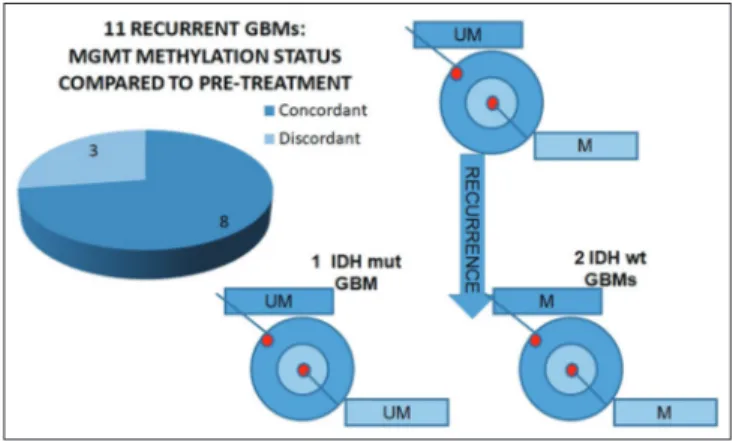
In all five of the GBMs with heterogeneous MGMT promoter methylation status, the central part of the tumor was methylated and the peripheral part was unmethylated [Figure 3]. Two of five cases were IDH mutant GBMs, which had originated from the progression of DAs with an unmethylated MGMT promoter [Figure 3]. Three of the five cases were IDH wild-type. Follow-up time ranged between 5 and 48 months. During this time, 18 cases recurred and 11 were submitted for surgical excision of the recurrent tumor. Time to progression ranged between 1 and 34 months. When we analyzed MGMT promoter methylation status in the recurrences, 8 (73%) cases were concordant with the primary tumor, while 3 (27%) were discordant [Figure 4]. In all three discordant cases, the primary tumor had heterogeneous MGMT promoter methylation status (peripheral part unmethylated and central part methylated) [Figure 5]. In one case, the initial tumor was an IDH mutant GBM, and its paired recurrent tumor was MGMT unmethylated [Figure 4], whereas, the other two cases were IDH wild-type GBMs, with recurrent MGMT methylated tumors [Figure 4].
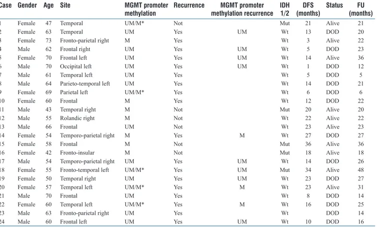
Table 1: Clinicopathological characteristics and O‑6‑methylguanine‑DNA methyltransferase promoter methylation status in 24 glioblastomas and in 11 paired recurrences
Case Gender Age Site MGMT promoter
methylation Recurrence methylation recurrenceMGMT promoter IDH 1/2 (months)DFS Status (months)FU
1 Female 47 Temporal UM/M* Not Mut 21 Alive 21
2 Female 63 Temporal UM Yes UM Wt 13 DOD 20
3 Female 73 Fronto-parietal right M Yes Wt 3 Alive 22
4 Male 62 Frontal right UM Yes UM Wt 5 DOD 23
5 Female 70 Frontal left UM Yes UM Wt 14 Alive 36
6 Male 70 Occipital left UM Yes UM Wt 1 DOD 12
7 Male 61 Temporal left UM Yes Wt 5 DOD 5
8 Male 64 Parieto-temporal left UM Yes Wt 14 DOD 21
9 Female 69 Parietal left UM/M* Yes Wt 6 DOD 6
10 Female 60 Frontal M Yes Wt 12 DOD 22
11 Male 43 Temporal right M Not Mut 20 Alive 20
12 Male 55 Rolandic right M Not Wt 22 Alive 22
13 Male 66 Frontal UM Not Wt 23 Alive 23
14 Female 54 Temporo-parietal right M Yes M Wt 27 DOD 27
15 Female 58 Frontal M Not Mut 36 Alive 36
16 Female 42 Fronto-insular M Not Mut 18 Alive 18
17 Male 54 Temporo-parietal right UM Yes UM Wt 14 DOD 26
18 Female 55 Fronto-temporal left UM/M* Yes UM Mut 34 Alive 48
19 Female 50 Temporal right UM Yes UM Wt 23 DOD 27
20 Female 57 Temporal left UM/M* Yes M Wt 23 Alive 31
21 Male 70 Frontal UM Yes Wt 8 DOD 14
22 Female 60 Temporal left UM/M* Yes M Wt 16 DOD 25
23 Male 63 Fronto-parietal right UM Yes Wt DOD 14
24 Male 60 Frontal left UM Yes UM Wt 10 DOD 16
*The central part of the tumor was methylated and the peripheral one was unmethylated. DFS: Disease-free survival, FU: Follow-up, M: Methylated, UM: Unmethylated, Wt: Wild-type, Mut: Mutant, DOD: Died of disease, MGMT: O-6-methylguanine-DNA methyltransferase, IDH: Isocitrate dehydrogenase
Barresi, et al.: MGMT heterogeneity in glioblastoma
211
Glioma ¦ Volume 1 ¦ Issue 6 ¦ November-December 2018 Intratumor heterogeneity was significantly more frequent in GBMs taken from female patients (P = 0.0411). The switching of MGMT promoter methylation status was not associated with any clinicopathological variables.
In univariate analysis, IDH mutation (P = 0.0298), MGMT promoter methylation (P = 0.0115), and female gender (P = 0.0154) were significantly associated with longer OS. Due to the small number of cases, we could not perform multivariate analyses. Among recurring cases, surgical resection of recurrent tumor was significantly associated with longer OS (P = 0.0017).
d
isCussionDynamic methylation changes of the MGMT promoter has been documented in several recurrent GBM studies but with different prevalences.[5-12] In this study, 27% of recurrent
GBMs had different MGMT promoter methylation status compared to the corresponding primary tumor. Interestingly, discordance was observed only in cases having a primary GBM with heterogeneous MGMT methylation status. Therefore, it is tempting to speculate that the changes in MGMT promoter methylation status in recurrent GBMs may descend from heterogeneity of tumor cells in the initial tumor, with subsequent subclonal expansion in the recurrence.
Several studies previously analyzed intratumor heterogeneity of MGMT promoter methylation status in GBM and with
conflicting results.[13-17] Parkinson et al.[13] reported that MGMT
promoter methylation status is homogeneous across different samples of GBM. However, all samples they analyzed had been taken from the peripheral part of the tumors, and none from their central part.[13] In two other studies, MGMT promoter
methylation heterogeneity was found in a small proportion of GBMs.[14,16] However, the authors claimed that heterogeneity
could depend on the presence of high number of nontumor contaminants in the unmethylated parts of the tumors.[14,16]
Interestingly, Della Puppa et al.[17] found intratumor
heterogeneity of MGMT promoter methylation in 33.3% of GBMs. In their study, MGMT methylation analysis was carried out in different concentric samples of GBMs.[17] Indeed,
they hypothesized that tumor stem cells, which are the most resistant to temozolomide, mainly reside in the central part of the tumor.[18] In their heterogeneous samples, MGMT promoter
methylation status was different in the intermediate part of the tumor compared to the peripheral and inner parts.[18] However,
methylation analysis had been carried out on frozen samples, with no histological verification, and eventual exclusion of nonneoplastic contaminants.[18]
In this study, we found MGMT promoter methylation at the center, but not at the periphery of the tumor, in 20% of GBMs. Heterogeneity could not be related to sample inadequacy. Indeed, methylation analysis was performed on Figure 4: O‑6‑methylguanine‑DNA methyltransferase promoter
methylation status in 11 surgically resected glioblastoma recurrences
Figure 3: Glioblastomas with heterogeneous O‑6‑methylguanine‑DNA
methyltransferase promoter methylation status
Figure 2: Glioblastomas with homogeneous O‑6‑methylguanine‑DNA
methyltransferase promoter methylation status
Figure 1: O‑6‑methylguanine‑DNA methyltransferase promoter
histologically verified neoplastic samples, with selected areas having adequate number of neoplastic cells and low amount of nontumor contaminants. Interestingly, two of the heterogeneous cases were IDH mutant GBMs, which had originated from the progression of LGAs. Since the corresponding LGAs were unmethylated, we can suppose that MGMT methylation was acquired in a subclone of tumor cells during progression, thus leading to heterogeneous methylation status in the secondary GBM. This is intriguing given that IDH mutations have been shown to induce extensive DNA methylation in gliomas.[19]
Accordingly, all the homogeneously unmethylated GBMs in our cohort were IDH wild type tumors.
To the best of our knowledge, only one study in LGAs investigated MGMT methylation status in relation to IDH mutational status.[20] In that study, the majority of IDH mutant
LGAs had MGMT promoter methylation,[20] in accordance
with the hypermethylated status of IDH mutant tumors.[19]
However, a small proportion of IDH mutant astrocytomas had unmethylated MGMT and this condition was associated with worse prognosis,[20] similarly to that observed in our cases,
which progressed to GBM.
In our cohort of patients, MGMT methylation and IDH mutation were significantly associated with longer OS, which confirm their relevant prognostic value in GBM patients.[1,21] In
addition, female gender was a favorable prognostic factor in our cohort, similarly to that reported in the study by Franceschi
et al.[22] Interestingly, among recurring tumors, surgery of
recurrences increased the OS, which is in line with the findings in other studies.[23,24]
C
onClusionThis study confirms that MGMT methylation status can vary in recurrent GBM, and that it can change from methylated to unmethylated and vice versa in comparison to the original
tumor. The fact that MGMT promoter methylation can change from initial tumor to recurrent tumor, raises the question of whether retesting of recurrent tumors is needed for therapeutic decisions. For instance, if the initial tumor is unmethylated but at recurrence becomes methylated, would a rechallenge with temozolomide be indicated? According to our results,
MGMT variation may depend on intratumor heterogeneity in
the primary GBM, which can be observed in both IDH mutant and IDH wild type tumors. Since MGMT promoter methylation status seems to vary according to a spatial criterion, further studies on larger cohorts are needed to clarify the site from which, the optimal tumor specimen for methylation analysis should be taken.
Financial support and sponsorship
Nil.
Conflicts of interest
There are no conflicts of interest.
R
efeRenCes1. Luois DN, Ohgaki H, Wisteler OD, Cavenee WK, Ellison DW, Figarella-Branger D, et al. WHO Classification of Tumors of the Central Nervous System. Lyon: IARC Press; 2016.
2. Stupp R, Mason WP, van den Bent MJ, Weller M, Fisher B, Taphoorn MJ,
et al. Radiotherapy plus concomitant and adjuvant temozolomide for
glioblastoma. N Engl J Med 2005;352:987-96.
3. Stupp R, Hegi ME, Mason WP, van den Bent MJ, Taphoorn MJ, Janzer RC, et al. Effects of radiotherapy with concomitant and adjuvant temozolomide versus radiotherapy alone on survival in glioblastoma in a randomised phase III study: 5-year analysis of the EORTC-NCIC trial. Lancet Oncol 2009;10:459-66.
4. Esteller M, Garcia-Foncillas J, Andion E, Goodman SN, Hidalgo OF, Vanaclocha V, et al. Inactivation of the DNA-repair gene MGMT and the clinical response of gliomas to alkylating agents. N Engl J Med 2000;343:1350-4.
5. Brandes AA, Franceschi E, Tosoni A, Bartolini S, Bacci A, Agati R,
et al. O (6)-methylguanine DNA-methyltransferase methylation status
can change between first surgery for newly diagnosed glioblastoma and second surgery for recurrence: Clinical implications. Neuro Oncol 2010;12:283-8.
6. Felsberg J, Thon N, Eigenbrod S, Hentschel B, Sabel MC, Westphal M,
et al. Promoter methylation and expression of MGMT and the DNA
mismatch repair genes MLH1, MSH2, MSH6 and PMS2 in paired primary and recurrent glioblastomas. Int J Cancer 2011;129:659-70. 7. Metellus P, Coulibaly B, Nanni I, Fina F, Eudes N, Giorgi R, et al.
Prognostic impact of O6-methylguanine-DNA methyltransferase silencing in patients with recurrent glioblastoma multiforme who undergo surgery and carmustine wafer implantation: A prospective patient cohort. Cancer 2009;115:4783-94.
8. Jung TY, Jung S, Moon KS, Kim IY, Kang SS, Kim YH, et al. Changes of the O6-methylguanine-DNA methyltransferase promoter methylation and MGMT protein expression after adjuvant treatment in glioblastoma. Oncol Rep 2010;23:1269-76.
9. Park CK, Kim JE, Kim JY, Song SW, Kim JW, Choi SH, et al. The changes in MGMT promoter methylation status in initial and recurrent glioblastomas. Transl Oncol 2012;5:393-7.
10. Brandes AA, Franceschi E, Paccapelo A, Tallini G, De Biase D, Ghimenton C, et al. Role of MGMT methylation status at time of diagnosis and recurrence for patients with glioblastoma: Clinical implications. Oncologist 2017;22:432-7.
11. O’Regan CJ, Kearney H, Beausang A, Farrell MA, Brett FM, Cryan JB, et al. Temporal stability of MGMT promoter methylation in glioblastoma patients undergoing STUPP protocol. J Neurooncol 2018;137:233-40.
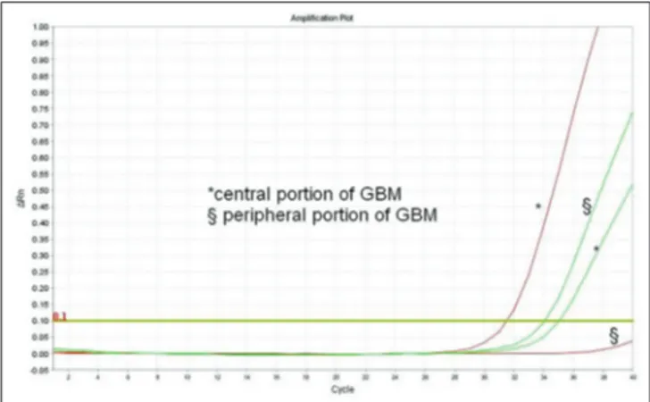
Figure 5: Polymerase chain reaction amplification plots relative to a
heterogeneous glioblastoma, with methylation changes occurring at the recurrence. The red curves refer to amplification of promoter methylated O‑6‑methylguanine‑DNA methyltransferase, while the green ones refer to the amplification of promoter unmethylated O‑6‑methylguanine‑DNA methyltransferase. In the primary tumor, the central part was methylated and peripheral one was unmethylated
Barresi, et al.: MGMT heterogeneity in glioblastoma
213
Glioma ¦ Volume 1 ¦ Issue 6 ¦ November-December 2018 12. Christmann M, Nagel G, Horn S, Krahn U, Wiewrodt D, Sommer C,
et al. MGMT activity, promoter methylation and immunohistochemistry
of pretreatment and recurrent malignant gliomas: A comparative study on astrocytoma and glioblastoma. Int J Cancer 2010;127:2106-18. 13. Parkinson JF, Wheeler HR, Clarkson A, McKenzie CA,
Biggs MT, Little NS, et al. Variation of O(6)-methylguanine-DNA methyltransferase (MGMT) promoter methylation in serial samples in glioblastoma. J Neurooncol 2008;87:71-8.
14. Grasbon-Frodl EM, Kreth FW, Ruiter M, Schnell O, Bise K, Felsberg J,
et al. Intratumoral homogeneity of MGMT promoter hypermethylation
as demonstrated in serial stereotactic specimens from anaplastic astrocytomas and glioblastomas. Int J Cancer 2007;121:2458-64. 15. Parker NR, Hudson AL, Khong P, Parkinson JF, Dwight T, Ikin RJ,
et al. Intratumoral heterogeneity identified at the epigenetic, genetic
and transcriptional level in glioblastoma. Sci Rep 2016;6:22477. 16. Hamilton MG, Roldán G, Magliocco A, McIntyre JB, Parney I,
Easaw JC, et al. Determination of the methylation status of MGMT in different regions within glioblastoma multiforme. J Neurooncol 2011;102:255-60.
17. Della Puppa A, Persano L, Masi G, Rampazzo E, Sinigaglia A, Pistollato F,
et al. MGMT expression and promoter methylation status may depend
on the site of surgical sample collection within glioblastoma: A possible pitfall in stratification of patients? J Neurooncol 2012;106:33-41. 18. Pistollato F, Abbadi S, Rampazzo E, Persano L, Della Puppa A,
Frasson C, et al. Intratumoral hypoxic gradient drives stem cells
distribution and MGMT expression in glioblastoma. Stem Cells 2010;28:851-62.
19. Bell EH, Zhang P, Fisher BJ, Macdonald DR, McElroy JP, Lesser GJ,
et al. Association of MGMT promoter methylation status with survival
outcomes in patients with high-risk glioma treated with radiotherapy and temozolomide: An analysis from the NRG oncology/RTOG 0424 trial. JAMA Oncol 2018;4:1405-9.
20. Turcan S, Rohle D, Goenka A, Walsh LA, Fang F, Yilmaz E, et al. IDH1 mutation is sufficient to establish the glioma hypermethylator phenotype. Nature 2012;483:479-83.
21. Hegi ME, Diserens AC, Gorlia T, Hamou MF, de Tribolet N, Weller M,
et al. MGMT gene silencing and benefit from temozolomide in
glioblastoma. N Engl J Med 2005;352:997-1003.
22. Franceschi E, Tosoni A, Minichillo S, Depenni R, Paccapelo A, Bartolini S,
et al. The prognostic roles of gender and O6-methylguanine-DNA
methyltransferase methylation status in glioblastoma patients: The female power. World Neurosurg 2018;112:e342-7.
23. Perrini P, Gambacciani C, Weiss A, Pasqualetti F, Delishaj D, Paiar F,
et al. Survival outcomes following repeat surgery for recurrent
glioblastoma: A single-center retrospective analysis. J Neurooncol 2017;131:585-91.
24. Pala A, Schmitz AL, Knoll A, Schneider M, Hlavac M, König R, et al. Is MGMT promoter methylation to be considered in the decision making for recurrent surgery in glioblastoma patients? Clin Neurol Neurosurg 2018;167:6-10.