Università degli Studi di Ferrara
DOTTORATO DI RICERCA IN
FARMACOLOGIA ED ONCOLOGIA MOLECOLARE
CICLO XXII
COORDINATORE Prof. Pier Andrea Borea
Coagulation-balance gene predictors influencing visual
prognosis in patients treated with photodynamic therapy for
classic choroidal neovascularization secondary to age-related
macular degeneration
Settore Scientifico Disciplinare MED/30
Dottorando
Tutore
Dott. ROMANO MARIO
Prof. SEBASTIANI ADOLFO
Abstract
Purpose: To determine whether different coagulation-balance genetic polymorphisms explain the variable clinical outcomes of photodynamic therapy with verteporfin (PDT-V) in Caucasian patients with classic or predominantly classic choroidal neovascularization (CNV) due to age-related macular degeneration (AMD).
Methods: The clinical records of consecutive AMD patients with classic or predominantly classic CNV, treated with PDT-V according to the Treatment of Age-Related Macular Degeneration with Photodynamic Therapy study criteria, were retrospectively examined. Eighty-six eligible patients were subdivided in responder and non-responder basing on the modifications of best-corrected visual acuity between baseline and 12-month checks. Six gene polymorphisms, i.e. factor V G1691A, prothrombin G20210A, factor XIII-A G185T, methylenetetrahydrofolate reductase C677T, methionine synthase A2756G, and methionine synthase reductase A66G, were genotyped in each patient. Binary logistic regression models were used to explore the predictive role of phenotypic and genotypic variables for PDT-V effectiveness.
Results: PDT-V responders were more prevalent among the combined carriers for factor V 1691A and prothrombin 20210A alleles (OR = 5.1 with a 95% CI of 1.0-24.4; P = 0.05), methylenetetrahydrofolate reductase 677 T-allele (OR = 4.3 with a 95% CI of 1.8-10.8; P = 0.002), and methionine synthase reductase 66 G-allele (OR = 2.8 with a 95% CI
of 1.1-6.8; P = 0.04). Conversely, PDT-V non-responders were over-represented in patients with factor XIII-A 185 T-allele (OR = 0.22 with a 95% CI of 0.09-0.56; P = 0.001). The other considered predictors did not significantly modify PDT-V effectiveness.
Conclusion: Our study provides evidences for the presence of pharmacogenetic relationship between peculiar coagulation-balance gene polymorphisms and different levels of visual prognosis at 12 months in AMD patients treated with standardized PDT-V protocol for classic subfoveal CNV.
Abstract
Scopo: determinare la correlazione tra differente polimorfismo dei geni della coagulazione e i diversi risultati clinici della terapia fotodinamica con verteporfina (PDT-V) in pazienti di razza caucasica affetti da degenerazione maculare legata all'età (DMLE) complicata da neovascolarizzazione coroideale (CNV) di tipo classico
Metodo: Sono state esaminate le cartelle cliniche di ottantasei pazienti consecutivi con DMLE classica trattati con PDT-V secondo le linee guida del Treatment of Age-Related Macular Degeneration with Photodynamic Therapy study. I pazienti eleggibili allo studio sono stati suddivisi in responder e non-responder basandosi sui miglioramenti della acuita’ visiva tra il basaline ed il controllo a 12 mesi. Sei polimorfismi del gene sono stati analizzati in ciascun paziente: fattore V G1691A, protrombina G20210A, Fattore XIII-A G185T, metilentetraidrofolato reduttasi C677T, metionina sintetasi A2756G, e metionina sintetasi reduttasi A66G. Il campione e’ stato analizzato con il modello binario di regressione logistica per valutare l’efficacia della PDT-V in base alle variabili fenotipiche e genotipiche.
Risultati: i pazienti PDT-V responders presentavano fattore V 1691A e alleli 20210A protrombina (OR = 5.1, con un IC 95% 1,0-24,4; P = 0,05), metilentetraidrofolato reduttasi 677 T-allele (OR = 4,3 con un IC 95% di 1,8-10,8; P = 0,002), e la metionina reduttasi sintasi 66 G-allele (OR = 2,8, con un IC 95% di 1,1 - 6,8, P = 0,04). Al contrario, i pazienti
PDT-V non-responders alla V presentavano fattore XIII-A 185 T-allele (OR = 0,22 con un CI del 95% di 0,09-0,56, P = 0.001). Gli altri fattori considerati non modificavano sostanzialmente l'efficacia PDT-V.
Conclusioni: Il nostro studio fornisce evidenze della presenza di un rapporto farmacogenetico tra il polimorfismo dei geni della coagulazione e diversi livelli di prognosi visiva a 12 mesi nei pazienti trattati con terapia fotodinamica per degenerazione maculare legata all'età complicata da neovascolarizzazione coroideale di tipo classico.
Introduction
Choroidal neovascularization (CNV) beneath the fovea represents a common cause of central blindness or low-vision in Caucasian populations affected by age-related macular degeneration (AMD) (ARMD1 [MIM 603075]). 1, 2 Although intravitreal drugs acting against
vascular endothelial growth factor (anti-VEGF) have recently shown a remarkable potential towards several ocular diseases complicated by aberrant angiogenesis, 3-6 photodynamic therapy with verteporfin (PDT-V) still remains an evidence-based treatment that maybe realizes the inactivation of AMD-related subfoveal CNV. 6-8 Experimental and
clinical evidences indicate that the combined use of both these strategies is the most promising therapeutic approach towards this harmful disease.
9-14
The therapeutic effect of PDT-V is achieved by a laser-light-induced thrombosis of CNV, which has been previously photosensitized by the administration of verteporfin (benzoporphyrin derivative monoacid A).
15-19 PDT-V for CNV is currently performed as indicated by the protocol
utilizing in the Treatment of Age-Related Macular Degeneration with Photodynamic Therapy (TAP) and the Visudyne in Photodynamic Therapy (VIP) studies. 7, 20, 21 Standardized PDT-V consists of a 6 mg/m2
dose of liposomal verteporfin formulation intravenously infused over 10 minutes, and a light dose of 50 J/cm2 delivered over a period of 83 seconds; the time for the low-intensity, non-thermal laser irradiation
(light at a wavelength of 689 nm and an irradiance of 600 mW/cm2) was 15 minutes after the start of verteporfin injection; the diameter of the irradiated area corresponds to the greatest linear dimension (GLD) of CNV added to 1000 micron to provide a 500 micron safety margin all-around the lesion. 20, 21 The PDT-V mechanism of action has been
initially considered ideal for the treatment of subfoveal CNV. 15 However, the individually variable efficacy of standardized PDT-V is clearly noticeable reviewing the outcomes of TAP, VIP, and Visudyne in Minimally Classic Choroidal Neovascularization studies. In fact, the rather proportioned percentage of cases with or without severe visual loss after PDT-V is evident for all the AMD-related forms of CNV. 6, 20-22 Moreover, differences in CNV responsiveness to standardized PDT-V between Asian and Caucasian patients have been recently pointed out. 23,
24 Despite both these evidences of individual and racial variability, and
the well-known therapeutic PDT-V effect aimed at the photo-thrombotic perturbation of hemostasis within the neovascular complex, 15-19 the predictors of visual acuity modifications in AMD patients treated with verteporfin-therapy protocol have been hitherto examined without considering the role of coagulation-balance genetic backgrounds in changing its effectiveness. 25-29 Several gene variations can affect the
balance between pro- and anti-coagulant mechanisms, accounting for the occurrence of thrombophilic or hemorrhagic diatheses. 30-40 Once thrombo-coagulative process starts, physiologically or therapeutically
triggered, both anticoagulation and fibrinolysis are involved to limit and regulate the thrombosis in the ill- or treated-area. A non-optimal restoring of the hemostatic balance is able to cause abnormalities in thrombus formation or dissolution. Recently, in patients with AMD complicated by classic and occult CNV, we have documented the presence of predictive correlations between peculiar mutated alleles modulating the thrombo-fibrinolytic equilibrium and different angiographic levels of short-term responsiveness to PDT-V. 41, 42
The aim of this study was to investigate several single nucleotide polymorphisms (SNPs), encoding enzymes involved in the coagulative process, as predictors of the difference in visual acuity after a 12-month PDT-V protocol, carried out in Caucasian AMD patients with classic subfoveal CNV. In particular, the post-operative long-term variability of this clinical parameter was evaluated considering its putative association with common or uncommon gene variants, schematically shared in two typologies: i. gain-of-function SNPs directly influencing the coagulation cascade, i.e. factor V Leiden (FVL-G1691A), prothrombin G20210A (FII-G20210A), and factor XIII-A G185T (FXIIIA-G185T); 31-35 ii. SNPs modulating homocysteine (Hcy) metabolism, which indirectly affect the hemostatic balance, i.e. methylenetetrahydrofolate reductase C677T (MTHFR-C677T), methionine synthase A2756G (MS-A2756G) and methionine synthase reductase A66G (MTRR-A66G). 36-40
Methods
We conducted a retrospective analysis of the clinical records of Caucasian patients with AMD complicated by new-diagnosed, subfoveal, classic or predominantly classic CNV, and exclusively treated with TAP/VIP-standardized verteporfin protocol during a 12-month follow-up period. The diagnosis of exudative AMD was based on the criteria of the International ARM Epidemiological Study Group. 43 The classification of CNV was based on the definitions from TAP/VIP Study Groups. 20, 21 All patients underwent, within two weeks after the onset of CNV-related visual symptoms, both fluorescein angiography (FA) and indocyanine green angiography (ICGA) to well-detail the presence of AMD-related subfoveal CNV. For the purposes of this study, AMD patients were consecutively selected to achieve a consistent study cluster of, at least, 80 cases. Each course of standardized PDT-V was performed within 7 days from diagnosis or follow-up angiographies in reference to the published guidelines for standardized PDT-V application (TAP/VIP protocol). 7 The patients were evaluated at 3-month intervals for one year; additional courses of treatment were achieved in case of persistence of fluorescein leakage from the CNV according to TAP/VIP criteria. 20, 21 During all clinical examinations, patients underwent medical and ophthalmologic histories, auto-refraction, best-correct visual acuity (BCVA), slit-lamp biomicroscopy, applanation tonometry, 60-diopter lens ophthalmoscopy, and FA. Snellen BCVA was measured using a standard logarithmic chart
at a test distance of three meters. Each BCVA datum was converted to the logarithm of the minimum angle of resolution (logMAR) scale for statistical analyses. FA and ICGA were performed employing IMAGEnet digitizing system (Topcon Corp., Japan). After angiographic examinations, the CNV area (mm2) was directly measured on angiograms
using the software available into IMAGEnet package. In the course of a masked retrospective assessment, each patient was considered responder or non-responder to PDT-V quoting the BCVA difference calculated between final and baseline values: post-PDT-V BCVA – pre-PDT-V BCVA. These data were recorded, respectively, after 12 months ± 1 week from the first PDT-V and 7 or less days before the first PDT-V. At the end of follow-up period, patients with a BCVA decrease greater than 0.1 logMAR were arbitrarily defined non-responder, whereas the cases with minor vision loss, as well as those characterized by stabilization or improvement of BCVA were labeled as responder.
Inclusion and exclusion criteria are listed in Table 1. All the enrolled patients gave their written informed consent to participate to the study, after a detailed description of the aim-work and of the procedures to be used. The local Ethic Committee reviewed and approved the clinical trial. The study followed the tenets of the Declaration of Helsinki. Since the typology of the investigated clinical parameters, the sample size calculation, accomplished for the amount of study population (86 cases),
has provided a value constantly upper than 85%. This test was performed using the PASS 97 statistical program (NCSS Inc., Kaysville, UT, USA). Blood samples were collected for the SNPs genotyping prior to the first PDT-V application. Genomic DNA was isolated from peripheral blood by using standard proteinase K treatment, followed by phenol-chloroform extraction and ethanol precipitation. Samples were polymerase chain reaction (PCR)-genotyped for FVL-G1691A, FII-G20210A, FXIIIA-G185T, MTRR-A66G, MS-A2756G, and MTHFR-C677T gene variants, according to previous reports of Gemmati and co-workers. 44-46 Genotypes were confirmed by re-genotyping a random selection of samples for each polymorphism investigated. There were no discrepancies between genotypes determined in duplicate. Genotyping was carried out in a blinded fashion relative to the clinical phenotype of patients. The expected allele frequencies in the whole group of investigated cases were checked by the Hardy-Weinberg equilibrium test for those SNPs showing a rate < 5% and compared with a cluster of normal subjects. They were matched for sex, age and ethnicity with the case group.
One-way, repeated-measures analysis of variance was used to assess the modifications of both BCVA and CNV area within follow-up times. Odds ratios (OR) and 95% confidence intervals (CI) were used to estimate the probability of belonging to responder or non-responder to PDT-V. Adjusted ORs for single or combined comparisons were
calculated with binary logistic regression models, controlled for sex and age. Thus, univariate and multivariate analyses were performed to determine which variables were predictive of PDT-V responder, using responder/non-responder as dependent binary variable. In these regression models, the putative predictors were included according to the clinical plausibility of their possible influence on the dependent variable. The following parameters were considered as PDT-V predictors: patient’s age, pre-PDT-V BCVA, pre-PDT-V CNV area, FVL-1691 GA/AA, FII-20210 GA/AA, FXIIIA-185 GT/TT, MTRR-66 AG/GG, MS-2756 AG/GG, and MTHFR-677 CT/TT. Those putative predictors that did not significantly contribute to the univariate logistic regression were ruled out from all the final multivariate selected regression models (exclusion threshold, P > 0.10). All analyses were performed by Systat V.5.0 All analyses were performed by Systat V.5.0 (Systat Inc., Evanston, IL, USA) and SPSS Statistical Package (SPSS Inc., Chicago, IL, USA). A probability of P < 0.05 was considered statistically significant.
Table 1. Inclusion and exclusion criteria.
Inclusion criteria
- patient’s age > 65 years - diagnosis of AMD
- best-correct visual acuity better than 20/200 (Snellen equivalent)
- FA/ICGA signs of classic or predominantly classic CNV secondary to AMD - CNV under the geometric center of the foveal avascular zone (subfoveal) - greatest linear dimension of entire CNV less than 5400 micron
Exclusion criteria
- retinal photocoagulative treatment of the posterior pole before PDT-V
- presence of any other possible cause of CNV, such as degenerative myopia, angioid streaks, chorioretinal inflammatory diseases, hereditary retinal disorders, presumed ocular histoplasmosis syndrome, and/or severe ocular trauma
- intraocular surgery and any ocular laser-treatment during the 6 months before or the 3 months after PDT-V
- presence of any significant side effect, condition and/or event influencing PDT-V outcome
- active or chronic systemic diseases (such as porphyria, diabetes mellitus, hepatopathies, metabolic, cardiovascular, and hematological disorders), as well as assumption of any medication, known to affect the hemostatic balance
- protein intake, during breakfast or lunch, occurred 12 hours before PDT-V
Footnotes: AMD = age-related macular degeneration; FA = fluorescein angiography; ICGA = indocyanine green angiography; CNV= choroidal neovascularization; PDT-V = photodynamic therapy with verteporfin
Results
Demographic characteristics, pre- and post-PDT-V ophthalmologic data, and PDT-V-application attributes of the enrolled sample are summarized in Table 2. Over the follow-up period, mean BCVA moderately decreased from 0.68 to 0.82 logMAR, but an evident overlap between the upper bound of the baseline 95% CI and 95% CI lower bound at the 12th month was observable. Although a reduction of mean CNV area occurred (2.32 vs 1.43 mm2), it did not arrive at a significant rank at the end of the 12-month follow-up period. In the present series of AMD patients with classic or predominantly classic CNV, the visual outcome was not as good as that reported by the TAP trial. 20 However, the 12-month mean change in BCVA of this investigated cluster (0.14 logMAR) is comparable to those observed in other clinical-setting studies, performed utilizing the same standardized PDT-V protocol in patients with analogous forms of AMD-related CNV. 47-50
Genotype frequencies in total study cluster, as well as in responders and non-responders to PDT-V are reported in Table 3. Both FVL-G1691A and FII-G20210A showed an allele frequency < 5%. In the investigated case group, no significant deviations from Hardy-Weinberg equilibrium (all P values > 0.50) and from genotype distribution of a healthy control population were observed for these SNPs. In detail: i. FVL-G1691A – total cases n = 86; GG = 80; AG = 6; AA = 0 vs control group n = 100; GG = 97; GA = 3; AA = 0); ii. FII-G20210A – total cases n = 86; GG =
81; AG = 5; AA = 0 vs control group n = 100; GG = 98; GA = 2; AA = 0). Genotype distributions of FVL-G1691A and FII-G20210A detected within our control cluster were in accordance with those previously reported in Caucasians. 30, 31, 44 Since both FVL 1691 and FII 20210 A-alleles result in a well-known pro-coagulant predisposition in Caucasian subjects 31-33 and they are not so frequent as the other examined SNPs, these genetic variants have been also collectively analyzed (FVL-1691 plus FII-20210 GA/AA) owing to the absence of concomitant-carrier case.
The ORs, adjusted by univariate logistic regression for the probability estimation of clinical efficacy (responders) or inefficacy (non-responders) of PDT-V, are shown in Table 4. When each phenotypic or genotypic factor was examined on univariate basis as putative PDT-V predictive covariate, FXIIIA 185-T carriers were more frequent within non-responders (OR = 0.22 [GT or TT vs GG], 95% CI: 0.09-0.56; P < 0.001), hence this mutated genotypic trait was related to a higher risk of severe BCVA reduction at the end of 12-month, standardized, verteporfin therapy protocol. Conversely, the predictors of PDT-V clinical efficacy were, from the less to the more common: FVL-1691 plus FII-20210 GA/AA (OR = 5.1 [AA or GA vs GG], 95% CI: 1.0-24.4; P = 0.05), MTRR-66 AG/GG (OR = 2.8 [AG or GG vs AA], 95% CI: 1.1-6.8; P = 0.04), and MTHFR-677 CT/TT (OR = 4.3 [CT or TT vs CC], 95% CI: 1.8-10.8; P = 0.002). All the other considered predictive factors did not
significantly modify the long-term BCVA modification after PDT-V application (Table 4). On selected multivariate analysis including factors with a univariate P-value ≤ 0.10, all the covariates, with the exception of pre-PDT-V CNV area and MTRR 66G-allele, still displayed influence on PDT-V outcome. In particular, both FVL-1691 plus FII-20210 GA/AA (OR = 8.5 [AA or GA vs GG], 95% CI: 1.3-54.1; P = 0.03) and MTHFR-677 CT/TT (OR = 3.9 [CT or TT vs CC], 95% CI: 1.3-11.6; P = 0.02) were significantly over-represented among the cases with acceptable PDT-V response, whereas FXIIIA-185 GT/TT had a higher prevalence in patients with final marked vision loss (OR = 0.39 [GT or TT vs GG], 95% CI: 0.1-0.9; P = 0.05) (Table 5).
Table 2. Demographic characteristics, pre- and post-PDT-V ophthalmologic data, and PDT-V-application attributes of the study group.
n of patients 86
Sex (males / females) (n) 39 / 47 Age (mean ± SD; range) (years) 73.91 ± 6.48 (66-89) Pre-PDT-V CNV area (mean ± SD; range) (mm2) 2.32 ± 1.19 (0.4-5.1) Pre-PDT-V BCVA (mean ± SD; range) (logMAR) 0.68 ± 0.27 (0.1-1.0) Number of PDT-V (mean ± SD; range) 2.99 ± 0.81 (1-4) Post-PDT-V CNV area (mean ± SD; range) (mm2) 1.43 ± 2.11 (0.3-5.3)
Post-PDT-V BCVA (mean ± SD; range) (logMAR) 0.82 ± 0.50 (0.1-2.0) Post-PDT-V BCVA – pre-PDT-V BCVA (mean ± SD; range) (logMAR) 0.14 ± 0.47 (– 0.8-1.7) PDT-V responder / non-responder 44 / 42
Footnotes: SD = standard deviation; PDT-V = photodynamic therapy with verteporfin; CNV= choroidal neovascularization; mm = millimeter; BCVA= best-correct visual acuity; MAR = minimum angle of resolution;.
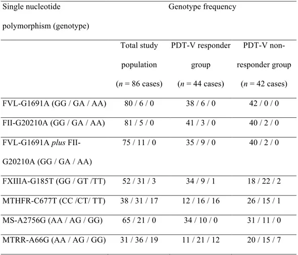
Table 3. Genotype frequencies in total study population, PDT-V responder and PDT-V non-responder groups. Single nucleotide polymorphism (genotype) Genotype frequency Total study population (n = 86 cases) PDT-V responder group (n = 44 cases) PDT-V non-responder group (n = 42 cases) FVL-G1691A (GG / GA / AA) 80 / 6 / 0 38 / 6 / 0 42 / 0 / 0 FII-G20210A (GG / GA / AA) 81 / 5 / 0 41 / 3 / 0 40 / 2 / 0 FVL-G1691A plus
FII-G20210A (GG / GA / AA) 75 / 11 / 0 35 / 9 / 0 40 / 2 / 0 FXIIIA-G185T (GG / GT /TT) 52 / 31 / 3 34 / 9 / 1 18 / 22 / 2 MTHFR-C677T (CC /CT/ TT) 38 / 31 / 17 12 / 16 / 16 26 / 15 / 1 MS-A2756G (AA / AG / GG) 65 / 21 / 0 34 / 10 / 0 31 / 11 / 0 MTRR-A66G (AA / AG / GG) 31 / 36 / 19 11 / 21 / 12 20 / 15 / 7
Footnotes: PDT-V = photodynamic therapy with verteporfin; FVL-G1691A = factor V Leiden; FII-G20210A = prothrombin G20210A; FXIIIA-G185T = factor XIII-A G185T; MTHFR-C677T = methylenetetrahydrofolate reductase MTHFR-C677T; MS-A2756G = methionine synthase A2756G; MTRR-A66G = methionine synthase reductase A66G.
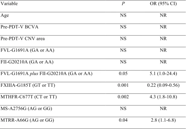
Table 4. Summary of the univariate logistic regression analyses.
Variable P OR (95% CI)
Age NS NR
Pre-PDT-V BCVA NS NR Pre-PDT-V CNV area NS NR FVL-G1691A (GA or AA) NS NR FII-G20210A (GA or AA) NS NR FVL-G1691A plus FII-G20210A (GA or AA) 0.05 5.1 (1.0-24.4) FXIIIA-G185T (GT or TT) 0.001 0.22 (0.09-0.56) MTHFR-C677T (CT or TT) 0.002 4.3 (1.8-10.8) MS-A2756G (AG or GG) NS NR MTRR-A66G (AG or GG) 0.04 2.8 (1.1-6.8)
Footnotes: PDT-V = photodynamic therapy with verteporfin; BCVA= best-correct visual acuity; CNV= choroidal neovascularization; FVL-G1691A = factor V Leiden; FII-G20210A = prothrombin G20210A; FXIIIA-G185T = factor XIII-A G185T; MTHFR-C677T = methylenetetrahydrofolate reductase C677T; MS-A2756G = methionine synthase A2756G; MTRR-A66G = methionine synthase reductase A66G; OR = Odds ratio; CI = confidence interval; NS = not significant; NR = not relevant.
19
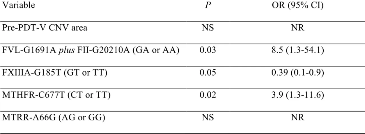
Table 5. Summary of the multivariate logistic regression analyses, including the variables with a univariate P-value ≤ 0.10.
Variable P OR (95% CI) Pre-PDT-V CNV area NS NR FVL-G1691A plus FII-G20210A (GA or AA) 0.03 8.5 (1.3-54.1) FXIIIA-G185T (GT or TT) 0.05 0.39 (0.1-0.9) MTHFR-C677T (CT or TT) 0.02 3.9 (1.3-11.6) MTRR-A66G (AG or GG) NS NR
Footnotes: PDT-V = photodynamic therapy with verteporfin; FVL-G1691A = factor V Leiden; FII-G20210A = prothrombin G20210A; FXIIIA-G185T = factor XIII-A G185T; MTHFR-C677T = methylenetetrahydrofolate reductase C677T; MTRR-A66G = methionine synthase reductase A66G; OR = Odds ratio; CI = confidence interval; NS = not significant; NR = not relevant.
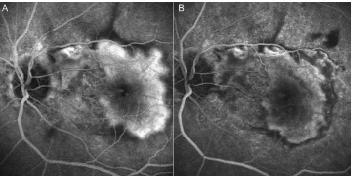
Figure 1. Two cases of classic choroidal neovascularization (CNV) lesion treated with PDT-V. The angiogram shows a regression of choroidal neovascular activity pre (A) and post PDT-V treatment (B).
21
Discussion
Several inherited or acquired clotting abnormalities are associated with a lopsided hemostasis. 30-40 The balancing of pro- and anti-coagulant processes has a key role in establishment and/or progression of thrombophilia, which can be defined as an increased tendency towards thrombosis induced by a multi-causal disorder wherein genetic and environmental factors interact dynamically. 30-31 The photothrombotic effect, obtained within the neovascular lesion following PDT-V application, is essentially due to a prevalent photosensitizer binding to the endothelium of CNV in comparison with that of normal macular vasculature. In blood circulation, the liposomally delivered verteporfin couples with plasma low density lipoproteins (LDL) to form a complex, which is then preferentially up-taken into neovascular endothelial cells via endocytosis for an overexpression of LDL receptors at CNV level. Intensive damage of neovascular endothelium induced by PDT-V results from the photo-oxidative action of several types of reactive oxygen species (ROS), representing the trigger that leads to therapeutic blood flow stasis and tissue hypoxia within CNV. These conditions can elicit a cascade of events (i.e. amplification of platelet activation, thrombosis, vasoconstriction, and increased vascular permeability), aimed at the final shutdown of CNV vasculature. 15 In accordance with the hypothesized work postulations, the present predictive findings indicate that the
mechanism of action of PDT-V is characterized by several phases which are strongly modifiable by peculiar coagulation-balance SNPs. Significant correlations were identified between satisfactory or unsatisfactory visual outcomes at the end of 12-month, TAP/VIP-standardized verteporfin protocol and, respectively, thrombophilic or anti-thrombophilic genotypic predictors. In this pharmacogenetic investigative approach, retrospectively applied in Caucasian patients with AMD-related classic CNV, three crucial steps are involved in determining the clinical success of PDT-V, 15, 17, 19 and each of them appears to be dependent on particular genetic backgrounds: i. the triggering of photo-chemical damages at the level of neovascular endothelium is positively influenced by two common hyperhomocysteinemic gene variants, i.e. MTHFR-C677T and MTRR-A66G; ii. the progressive photothrombotic closure of neovascular complex is enhanced by the FVL-1691 or by FII-20210 A-alleles, both predisposing to thrombotic diathesis and higher thrombin generation; iii. the persistence and the extensiveness of the hemodynamic occlusion within CNV are negatively affected by the hyperfibrinolytic FXIII-185 T-allele.
23
i. Post-PDT-V damages of CNV endothelium and
hyperhomocysteinemic gene polymorphisms
At the level of CNV endothelium, the modalities by which PDT-V photo-chemical action triggers the therapeutic pro-thrombotic process 15, 41, 51 are largely overlapped with those occurring in the course of hyperhomocysteinemia (HHcy) owing to the presence of folate-related gene variants. 37, 39 These SNPs are able to cause appreciable enzymatic defects in MTHFR, MTRR and other enzymes, inducing endovascular damage via hyper-activation of endothelial cells and platelets. 38, 52 HHcy leads to thrombophilia as a result of ROS-related triggering, 53-55 which
initiates lipid peroxidation in endothelial cell membranes and in circulating LDL, 36 overexpresses lectin-like oxidized LDL receptor-1, 56,
57 and enhances platelet activation. 58 All these hyperhomocysteinemic
effects, occurring in both MTHFR 677T-carriers and MTRR 66G-carriers, 37, 39, 52 could play a key role in photodynamic therapeutic effect,
strongly supporting the concept of a gene-environment interaction between PDT-V efficacy and thrombophilic SNPs affecting Hcy metabolism. 38, 40, 41 Therefore, the lower odds of post-PDT-V severe BCVA reduction at one year, recorded in carriers of hyperhomocysteinemic SNPs with AMD-related classic CNV, is attributable to a more effectual photo-oxidative activation during the first phases of CNV photothrombosis. 15, 38-41 Moreover, oxidation of
LDL-vesicles in the sub-endothelial spaces may affect the uncoagulable properties of endothelium giving back basically a more activable surface responsible for local hyper-reactivity. 59 In the course of HHcy, an increased vascular oxidant stress has been shown to activate inflammatory signaling pathways in endothelial cells, like the transcriptional nuclear factor kappa B. 54 These items should be considered for a further possible explanation of the present results. In fact, inflammatory and oxidative damages are implicated during the nuclear factor kappa B activation in human endothelial cells after photodynamic therapy, 15, 60 suggesting a linear correlation between the
efficacy of this treatment and these changes within the ill-treated area.
ii. Post-PDT-V magnitude of CNV occlusion and gene
polymorphisms affecting thrombin generation
In patients with neovascular AMD, the sequence of early vascular events after PDT-V has been investigated using scanning laser system to achieve confocal FA and ICGA images for the evaluation of CNV size and leakage. These exams show that the photo-thrombotic occlusion of the entire CNV does not immediately take place, but it happens over a prolonged period of about 24 hours. 17 During this time course,
pro-coagulant, anti-coagulant and fibrinolytic factors may influence the extent of thrombo-coagulative process inside CNV. Thus, thrombophilic
25
gain-of-function SNPs can modify this photodynamic phase of CNV thrombosis. Both the uncommon SNPs G1691A of FV gene and G20210A of FII gene induce thrombophilia increasing the level of thrombin bio-availability in plasma. 31-33 FV is a cofactor enzyme with pivotal roles in hemostatic equilibrium especially stimulating the inactivation of factor VIIIa by activated protein C; in FV-1691A carriers, this anticoagulant function is altered with a consequent increased thrombin generation causing a pro-thrombotic state. 31, 32 Prothrombin is the central component of coagulation cascade which in its active form, thrombin, regulates both pro- and anti-coagulant processes; in FII-20210A carriers the elevation of prothrombin expression is due to functional abnormalities of its mRNA metabolism, able to predispose to thrombosis affecting a tightly balanced architecture of non-canonical 3’ end formation signals. 31, 33 Consistently, as a consequence of their individual thrombophilic predisposition, the heterozygous A-allele carriers of FV 1691 or FII 20210 gene appear to be characterized by an higher possibility to exhibit clinical benefit after PDT-V owing to a greater magnitude of CNV photo-thrombosis. This correlation, also present on selected multivariate analysis, supports the pharmacogenetic suitability of the work postulations, even if it has not been observed considering FVL-G1691A and FII-G20210A polymorphisms alone,
probably because of their low prevalence in the Caucasian general population. 30, 31, 44
iii. Post-PDT-V hemodynamic recanalization of CNV and
hyperfibrinolytic gene polymorphisms
In all AMD patients affected by subfoveal CNV and treated with verteporfin therapy, the three-dimensional analyses of retinochoroidal angiograms after PDT-V disclose several distinctive features of the “dark spot”, i.e. the typical circular hypofluorescence corresponding to the laser-exposed area. During the first hours following PDT-V, this angiographic pattern is only partially due to photo-thromboses of CNV and collateral choroid, which are also associated with a massive fluid extravasation. One week later, the slowly regression of exudation results in displaying the whole extent of photodynamic occlusive effects. 19 However, this phase of blood non-perfusion is individually variable and, at 7-day check after PDT-V, the angiographic findings show that CNV vascular net is still apparent in about half of the treated patients. 18 This changeability in the post-PDT-V retinochoroidal appearance may be related to differences in fibrin structure, able to influence the fibrinolysis rate of each individual. 34, 35 Plasma coagulation factor XIII (FXIII) is
directly involved in the final steps of coagulation cascade and indirectly in fibrinolytic process. It is the precursor of a transglutaminase that
cross-27
links fibrin and, altering its network and properties, regulates fibrinolysis. The FXIII rate of activation is modulated by FXIIIA-G185T polymorphism, 35 a relatively common genetic variation among Caucasian population. 34 Both GT and TT genotypes are associated with remarkable modification in FXIII transglutaminase activity, which appears to be highly increased in homozygotes and exhibits an intermediate function in heterozygous carriers. 34 In the present study cluster, a reduced fibrin-clot stability and persistence, together with an increased fibrin-network permeability, could explain the correlation between this fibrinolytic-related polymorphism and the long-term inefficacy of the verteporfin protocol, owing to the early CNV recanalization after each PDT-V application. In this view, the very different frequency of FXIIIA 185T-allele among Caucasian and Asian races (respectively, 25-30% and close to 0%), 61, 62 could notionally explain the better outcomes obtained in Asians after verteporfin-therapy protocol. 23, 24 Otherwise, also the neo-angiogenic properties ascribed to FXIII may in part account for our predictive findings. In fact, a more active FXIII molecule at the site of injury (i.e. in the FXIII T-carriers) could downgrade PDT-V benefit by contrasting the laser-induced CNV deactivation. Our speculation is supported by the findings of neovascularization development after FXIIIA injection in experimental cornea model. 63
In Caucasian patients affected by AMD-related CNV and treated with standardized TAP/VIP protocol during a 12-month period, the present findings document the presence of predictive, gene-environment interactions between different levels of final visual outcome and several SNPs encoding enzymes involved in coagulation and/or fibrinolysis. Particularly, a satisfactory, post-PDT-V differential value of BCVA appears to be directly dependent on common folate-pathway gene variants (i.e. MTHFR-C677T and MTRR-A66G), which convert vascular endothelium and natural anticoagulant pathway to a more pro-thrombotic phenotype increasing plasma Hcy level, 36-40 consequently enhancing the post-PDT-V oxidative triggering of CNV endothelium. Likewise, an adequate visual result is also associated with the presence of rare thrombophilic SNPs (i.e. FV-1691A plus FII-20210A), able to increase the thrombin concentration and activation in plasma, 31-33 intensifying the
magnitude of thrombosis within the neovascular complex. On the other hand, in carriers of common hyperfibrinolytic genotypes (i.e. FXIIIA-185 GT or TT), which modify the polymerized fibrin structure affecting thrombin ability to effectively hydrolyze the factor XIII activation peptide, 34, 35 the most frequent occurrence of severe visual loss is
reliably related to a minor persistence of hemodynamic CNV shutdown after each application of verteporfin therapy. These long-term results
29
resemble those previously reported by Parmeggiani and co-workers in another separate cluster of Caucasians with AMD-related classic CNV investigated for a 3-month follow-up period, 41 and definitely support the prospect to utilize, during the routine ophthalmologic practice, some pharmacogenetic predictors for the optimization of PDT-V application in either single or combined treatment modality. In fact, the clinical applicability of the present findings mainly concerns the opportunity to improve the eligibility criteria of TAP/VIP standardized PDT-V, currently centered just on the morphological composition of CNV. 6, 7,
25-27 A pre-operative assessment of individual genetic backgrounds for
designated coagulation-balance SNPs should make possible a more rational selection of candidates to PDT-V which, at present, is especially employed in combined treatment modalities. 9-14 This pharmacogenetic approach towards PDT-V predictors could rationally guide the interventional planning for exudative AMD: i. associating this CNV photo-thrombosis with anti-angiogenic therapy only in patients with thrombophilic diathesis that facilitates CNV photothrombosis; 15, 31-33,
36-41, 59, 60 ii. preferentially choosing anti-VEGF drugs in carriers of the
hyperfibrinolytic FXIIIA 185 T-allele as a consequence of its both anti-thrombophilic and pro-angiogenic effects. 3-5, 34, 35, 63, 64 Moreover, the
present predictive findings prospectively indicate the possibility to re-evaluate the suitability of the previous notions regarding the optimized
laser-light dose of PDT-V. 7, 65-67 Currently, the socio-economic burden related to the treatment of neovascular AMD represents a very important puzzle, leading to increased health resource utilization and high societal costs. 68 Incongruously, as a consequence of both individual and racial PDT-V-efficacy variability, 6, 7, 20-24 there is a great interest in proposing
the intravitreal anti-angiogenic treatments of CNV 4, 69 without considering a crucial bias that could be occurred during the definition of the parameters for the application of verteporfin therapy. In fact, this standardized procedure has been delineated after phase I/II clinical trials, conducted on a rather low number of patients who underwent PDT-V with different treatment regimens, which overlooked that the different coagulation-balance genetic backgrounds of each treated patient can significantly influence the final PDT-V effectiveness. 65-67 Hypothetically, the ideal PDT-V may be accomplished differentiating the laser-light dose on the basis of the individual pro- and anti-thrombotic diatheses. In view of this innovative exploratory attitude, verteporfin therapy, anticipated by the intravitreal injection of an anti-VEGF drug, might be performed in a customized manner, aimed to induce the definitive shutdown of subfoveal CNV minimizing the required number of therapeutic combined approaches, which should be characterized by an higher (i.e. 100-125 J/cm2) or lower (i.e. 50-75 J/cm2) PDT-V light
31
dose, respectively, in patients with hyperfibrinolytic or thrombophilic predisposition.
The results of this retrospective study open a new scenario for the realization of prospective, randomized and controlled, pharmacogenetic trials, which are warranted to: i. improve the international guidelines for PDT-V application; ii. develop appropriate therapeutic strategies combining customized PDT-V and anti-VEGF compounds; iii. downgrade the socio-economic burden of the interventive planning for subfoveal CNV secondary to AMD.
Acknowledgements
The Author is indebted to Dr Francesco Parmeggiani and Prof Ciro Costagliola to for the conception and design of the study, to Mr. Giuseppe Gilli for assistance in the statistical analyses, and to Ms. Graziella Ferraresi for the logistic support.
References
1. Ferris FL 3rd, Fine SL, Hyman L. Age-related macular degeneration and blindness due to neovascular maculopathy. Arch Ophthalmol 1984; 102: 1640-1642.
2. Klein R, Klein BE, Knudtson MD, et al. Prevalence of age-related macular degeneration in 4 racial/ethnic groups in the multi-ethnic study of atherosclerosis. Ophthalmology 2006; 113: 373-380.
3. Gragoudas ES, Adamis AP, Cunningham ET Jr, et al.; VEGF Inhibition Study in Ocular Neovascularization Clinical Trial Group. Pegaptanib for neovascular age-related macular degeneration. N Engl J Med 2004; 351: 2805-2816.
4. Rosenfeld PJ, Brown DM, Heier JS, et al.; MARINA Study Group. Ranibizumab for neovascular age-related macular degeneration. N Engl J Med 2006; 355: 1419-1431.
5. Kaiser PK. Antivascular endothelial growth factor agents and their development: therapeutic implications in ocular diseases. Am J Ophthalmol 2006; 142: 660-668.
6. Chakravarthy U, Soubrane G, Bandello F, et al. Evolving European guidance on the medical management of neovascular age related macular degeneration. Br J Ophthalmol 2006; 90: 1188-1196.
7. Verteporfin Roundtable Participants. Guidelines for using verteporfin (Visudyne) in photodynamic therapy for choroidal
33
neovascularization due to age-related macular degeneration and other causes: update. Retina 2005; 25: 119-134.
8. Wickens J, Blinder KJ. A preliminary benefit-risk assessment of verteporfin in age-related macular degeneration. Drug Saf 2006; 29: 189-199.
9. Heier JS, Boyer DS, Ciulla TA, et al.; FOCUS Study Group. Ranibizumab combined with verteporfin photodynamic therapy in neovascular age-related macular degeneration: year 1 results of the FOCUS Study. Arch Ophthalmol 2006; 124: 1532-1542.
10. Lazic R, Gabric N. Verteporfin therapy and intravitreal bevacizumab combined and alone in choroidal neovascularization due to age-related macular degeneration. Ophthalmology 2007; 114: 1179-1185. 11. Spaide RF. Rationale for combination therapies for choroidal neovascularization. Am J Ophthalmol 2006; 141: 149-156.
12. Zuluaga MF, Mailhos C, Robinson G, et al. Synergies of VEGF inhibition and photodynamic therapy in the treatment of age-related macular degeneration. Invest Ophthalmol Vis Sci 2007; 48: 1767-1772. 13. Bradley J, Ju M, Robinson GS. Combination therapy for the treatment of ocular neovascularization. Angiogenesis 2007; 10: 141-148. 14. Schmidt-Erfurth UM, Pruente C. Management of neovascular age-related macular degeneration. Prog Retin Eye Res 2007; 26: 437-451.
15. Schmidt-Erfurth U, Hasan T. Mechanisms of action of photodynamic therapy with verteporfin for the treatment of age-related macular degeneration. Surv Ophthalmol 2000; 45: 195-214.
16. Schlotzer-Schrehardt U, Viestenz A, Naumann GO, et al. Dose-related structural effects of photodynamic therapy on choroidal and retinal structures of human eyes. Graefes Arch Clin Exp Ophthalmol 2002; 240: 748-757.
17. Schmidt-Erfurth U, Michels S, Barbazetto I, et al. Photodynamic effects on choroidal neovascularization and physiological choroid. Invest Ophthalmol Vis Sci 2002; 43: 830-841.
18. Michels S, Schmidt-Erfurth U. Sequence of early vascular events after photodynamic therapy. Invest Ophthalmol Vis Sci 2003; 44: 2147-2154.
19. Schmidt-Erfurth U, Niemeyer M, Geitzenauer W, et al. Time course and morphology of vascular effects associated with photodynamic therapy. Ophthalmology 2005; 112: 2061-2069.
20. Treatment of Age-Related Macular Degeneration with Photodynamic Therapy (TAP) study group. Photodynamic therapy of subfoveal choroidal neovascularization in age-related macular degeneration with verteporfin: one-year results of 2 randomized clinical trials – TAP Report No. 1. Arch Ophthalmol 1999; 117: 1329-1345.
35
21. Verteporfin in Photodynamic Therapy (VIP) Study Group. Verteporfin therapy of subfoveal choroidal neovascularization in age related macular degeneration: 2 year results of a randomized clinical trial including lesions with occult with no classic choroidal neovascularization – VIP Report No. 2. Am J Ophthalmol 2001; 131: 541-560.
22. Azab M, Boyer DS, Bressler NM, et al.; Visudyne in Minimally Classic Choroidal Neovascularization Study Group. Verteporfin therapy of subfoveal minimally classic choroidal neovascularization in age-related macular degeneration: 2-year results of a randomized clinical trial. Arch Ophthalmol 2005; 123: 448-457.
23. Chan WM, Lai TY, Tano Y, et al. Photodynamic therapy in macular diseases of asian populations: when East meets West. Jpn J Ophthalmol 2006; 50: 161-169.
24. Yang N, Fan CM, Ho CK. Review of first year result of photodynamic therapy on age-related macular degeneration in Chinese population. Eye 2006; 20: 523-526.
25. Blinder KJ, Bradley S, Bressler NM, et al.; TAP study group; VIP study group. Effect of lesion size, visual acuity, and lesion composition on visual acuity change with and without verteporfin therapy for choroidal neovascularization secondary to age-related macular degeneration: TAP and VIP report 1. Am J Ophthalmol 2003; 136: 407-418.
26. Axer-Siegel R, Ehrlich R, Yassur Y, et al. Photodynamic therapy for age-related macular degeneration in a clinical setting: visual results and angiographic patterns. Am J Ophthalmol 2004; 137: 258-264.
27. Arias L, Pujol O, Berniell J, et al. Impact of lesion size on photodynamic therapy with verteporfin of predominantly classic lesions in age related macular degeneration. Br J Ophthalmol 2005; 89: 312-315. 28. Sivaprasad S, Saleh GM, Jackson H. Does lesion size determine the success rate of photodynamic therapy for age-related macular degeneration? Eye 2006; 20: 43-45.
29. Manku KK, Rotchford A, Whitaker J, et al. Factors influencing poor visual outcome in patients treated with photodynamic therapy for choroidal neovascularization secondary to age-related macular degeneration. Clin Experiment Ophthalmol 2007; 35: 330-334.
30. Bernardi F, Marchetti G. Modulation of thrombophilia genes by environmental factors. Pathophysiol Haemost Thromb 2002; 32: 335-337.
31. Lane DA, Grant PJ. Role of hemostatic gene polymorphisms in venous and arterial thrombotic disease. Blood 2000; 95: 1517-30.
32. Castoldi E, Rosing J. Factor V Leiden: a disorder of factor V anticoagulant function. Curr Opin Hematol 2004; 11: 176-81.
37
33. Danckwardt S, Hartmann K, Gehring NH, et al. 3’ end processing of the prothrombin mRNA in thrombophilia. Acta Haematol 2006; 115: 192-7.
34. Ariens RA, Lai TS, Weisel JW, et al. Role of factor XIII in fibrin clot formation and effects of genetic polymorphisms. Blood 2002; 100: 743-54.
35. Shemirani AH, Haramura G, Bagoly Z, et al. The combined effect of fibrin formation and factor XIII A subunit Val34Leu polymorphism on the activation of factor XIII in whole plasma. Biochim Biophys Acta 2006; 1764: 1420-3.
36. Coppola A, Davi G, De Stefano V, et al. Homocysteine, coagulation, platelet function, and thrombosis. Semin Thromb Hemost 2000; 26: 243-54.
37. Schwahn B, Rozen R. Polymorphisms in the methylenetetrahydrofolate reductase gene: clinical consequences. Am J Pharmacogenomics 2001; 1: 189-201.
38. D’Angelo A, Mazzola G, Fermo I. Gene-gene and gene-environment interactions in mild hyperhomocysteinemia. Pathophysiol Haemost Thromb 2003-2004; 33: 337-41.
39. Castro R, Rivera I, Blom HJ, et al. Homocysteine metabolism, hyperhomocysteinaemia and vascular disease: an overview. J Inherit Metab Dis 2006; 29: 3-20.
40. Matthews RG, Elmore CL. Defects in homocysteine metabolism: diversity among hyperhomocyst(e)inemias. Clin Chem Lab Med 2007; 45: 1700-3.
41. Parmeggiani F, Costagliola C, Gemmati D, et al. Predictive role of coagulation-balance gene polymorphisms in the efficacy of photodynamic therapy with verteporfin for classic choroidal neovascularization secondary to age-related macular degeneration. Pharmacogenet Genomics 2007; 17: 1039-1046.
42. Parmeggiani F, Costagliola C, Gemmati D, et al. Coagulation-balance genetic predictors for efficacy of photodynamic therapy in occult choroidal neovascularization secondary to age-related macular degeneration. Invest Ophthalmol Vis Sci; In press.
43. Bird AC, Bressler NM, Bressler SB, et al. An international classification and grading system for related maculopathy and age-related macular degeneration. The International ARM Epidemiological Study Group. Surv Ophthalmol 1995; 39: 367-374.
44. Gemmati D, Serino ML, Moratelli S, et al. Coexistence of factor V G1691A and factor II G20210A gene mutations in a thrombotic family is associated with recurrence and early onset of venous thrombosis. Haemostasis 2001; 31: 99-105.
45. Gemmati D, Serino ML, Ongaro A, et al. A common mutation in the gene for coagulation factor XIII-A (VAL34Leu): a risk factor for
39
primary intracerebral hemorrhage is protective against atherothrombotic diseases. Am J Hematol 2001; 67: 183-188.
46. Gemmati D, Ongaro A, Scapoli GL, et al. Common gene polymorphisms in the metabolic folate and methylation pathway and the risk of acute lymphoblastic leukemia and non-Hodgkin’s lymphoma in adults. Cancer Epidemiol Biomarkers Prev 2004; 13: 787-794.
47. Armbrecht AM, Aspinall PA, Dhillon B. A prospective study of visual function and quality of life following PDT in patients with wet age related macular degeneration. Br J Ophthalmol 2004; 88: 1270-1273. 48. Frennesson CI, Nilsson SE. Encouraging results of photodynamic therapy with Visudyne in a clinical patient material of age-related macular degeneration. Acta Ophthalmol Scand 2004; 82: 645-650.
49. Wachtlin J, Stroux A, Wehner A, et al. Photodynamic therapy with verteporfin for choroidal neovascularisations in clinical routine outside the TAP study. One and two-year results including juxtafoveal and extrafoveal CNV. Graefes Arch Clin Exp Ophthalmol 2005; 243: 438-445.
50. Incorvaia C, Campa C, Parmeggiani F, et al. 12-month retrospective study and review of photodynamic therapy with verteporfin for subfoveal choroidal neovascularization in age-related macular degeneration. Retina 2008; 28: 289-297.
51. Parmeggiani F, Costagliola C, Incorvaia C, et al. Vision loss after PDT. Ophthalmology 2006; 113: 157.
52. Rongioletti M, Baldassini M, Papa F, et al. Homocysteinemia is inversely correlated with platelet count and directly correlated with sE- and sP-selectin levels in females homozygous for C677T methylenetetrahydrofolate reductase. Platelets 2005; 16: 185-190.
53. Austin RC, Lentz SR, Werstuck GH. Role of hyperhomocysteinemia in endothelial dysfunction and atherothrombotic disease. Cell Death Differ 2004; 11 (Suppl 1): S56-S64.
54. Weiss N. Mechanisms of increased vascular oxidant stress in hyperhomocysteinemia and its impact on endothelial function. Curr Drug Metab 2005; 6: 27-36.
55. Kanani PM, Sinkey CA, Browning RL, et al. Role of oxidant stress in endothelial dysfunction produced by experimental hyperhomocyst(e)inemia in humans. Circulation 1999; 100: 1161-1168. 56. Holven KB, Scholz H, Halvorsen B, et al. Hyperhomocysteinemic subjects have enhanced expression of lectin-like oxidized LDL receptor-1 in mononuclear cells. J Nutr 2003; receptor-133: 3588-359receptor-1.
57. Sakurai K, Sawamura T. Stress and vascular responses: endothelial dysfunction via lectin-like oxidized low-density lipoprotein receptor-1: close relationships with oxidative stress. J Pharmacol Sci 2003; 91: 182-186.
41
58. Holven KB, Aukrust P, Pedersen TM, et al. Enhanced platelet activation in hyperhomocysteinemic individuals. J Thromb Haemost 2007; 5: 193-195.
59. Postea O, Krotz F, Henger A, et al. Stereospecific and redox-sensitive increase in monocyte adhesion to endothelial cells by homocysteine. Arterioscler Thromb Vasc Biol 2006; 26: 508-513.
60. Volanti C, Matroule JY, Piette J. Involvement of oxidative stress in NF-kappaB activation in endothelial cells treated by photodynamic therapy. Photochem Photobiol 2002; 75: 36-45.
61. Attié-Castro FA, Zago MA, Lavinha J, et al. Ethnic heterogeneity of the factor XIII Val34Leu polymorphism. Thromb Haemost 2000; 84: 601-603.
62. Okumura T, Yamada T, Park SC, et al. No Val34Leu polymorphism of the gene for factor XIIIA subunit was detected by ARMS-RACE method in three Asian populations. J Thromb Haemost 2003; 1: 1856-1857.
63. Dardik R, Solomon A, Loscalzo J, et al. Novel proangiogenic effect of factor XIII associated with suppression of thrombospondin 1 expression. Arterioscler Thromb Vasc Biol 2003; 23: 1472-1477.
64. Dardik R, Loscalzo J, Inbal A. Factor XIII (FXIII) and angiogenesis. J Thromb Haemost 2006; 4: 19-25.
65. Schmidt-Erfurth U, Miller J, Sickenberg M, et al. Photodynamic therapy of subfoveal choroidal neovascularization: clinical and angiographic examples. Graefes Arch Clin Exp Ophthalmol 1998; 236: 365-374.
66. Miller JW, Schmidt-Erfurth U, Sickenberg M, et al. Photodynamic therapy with verteporfin for choroidal neovascularization caused by age-related macular degeneration: results of a single treatment in a phase 1 and 2 study. Arch Ophthalmol 1999; 117: 1161-1173.
67. Schmidt-Erfurth U, Miller JW, Sickenberg M, et al. Photodynamic therapy with verteporfin for choroidal neovascularization caused by age-related macular degeneration: results of retreatments in a phase 1 and 2 study. Arch Ophthalmol 1999; 117: 1177-1187.
68. Cruess AF, Zlateva G, Xu X, et al. Economic burden of bilateral neovascular age-related macular degeneration: multi-country observational study. Pharmacoeconomics 2008; 26: 57-73.
69. Brown DM, Kaiser PK, Michels M, et al.; ANCHOR Study Group. Ranibizumab versus verteporfin for neovascular age-related macular degeneration. N Engl J Med 2006; 355: 1432-1444.
43
Dichiarazione di conformità
Io sottoscritto Dott. ROMANO MARIOnato a NAPOLI Provincia NAPOLI il giorno 03/07/1978
E-Mail Address [email protected]
avendo frequentato il corso di Dottorato di Ricerca in: FARMACOLOGIA ED ONCOLOGIA MOLECOLARE Ciclo di Dottorato XXII
Titolo della tesi in Italiano
Influenza dei geni della coagulazione nella prognosi visiva dei pazienti trattati con terapia fotodinamica per degenerazione maculare legata all'età complicata da neovascolarizzazione coroideale di tipo classico
Titolo della tesi in Inglese
Coagulation-balance gene predictors influencing visual prognosis in patients treated with photodynamic therapy for classic choroidal neovascularization secondary to age-related macular degeneration
Tutore - Prof: ADOLFO SEBASTIANI Settore Scientifico Disciplinare (SSD) MED/30
Parole chiave (max 10) Coagulation-balance gene, photodynamic therapy, choroidal neovascularization, age-related macular degeneration
Consapevole - Dichiara
CONSAPEVOLE --- 1) del fatto che in caso di dichiarazioni mendaci, oltre alle sanzioni previste dal codice penale e dalle Leggi speciali per l’ipotesi di falsità in atti ed uso di atti falsi, decade fin dall’inizio e senza necessità di alcuna formalità dai benefici conseguenti al provvedimento emanato sulla base di tali dichiarazioni; -- 2) dell’obbligo per l’Università di provvedere al deposito di legge delle tesi di dottorato al fine di assicurarne la conservazione e la consultabilità da parte di terzi; -- 3) della procedura adottata dall’Università di Ferrara ove si richiede che la tesi sia consegnata dal dottorando in 4 copie di cui una in formato cartaceo e tre in formato .pdf, non modificabile su idonei supporti (CD-ROM, DVD) secondo le istruzioni pubblicate sul sito : http://www.unife.it/dottorati/dottorati.htm alla voce ESAME FINALE – disposizioni e modulistica; -- 4) del fatto che l’Università sulla base dei dati forniti, archivierà e renderà consultabile in rete il testo completo della tesi di dottorato di cui
alla presente dichiarazione attraverso l’Archivio istituzionale ad accesso aperto “EPRINTS.unife.it” oltre che attraverso i Cataloghi delle Biblioteche Nazionali Centrali di Roma e Firenze. - DICHIARO SOTTO LA MIA RESPONSABILITA' --- 1) che la copia della tesi depositata presso l’Università di Ferrara in formato cartaceo, è del tutto identica a quelle presentate in formato elettronico (CD-ROM, DVD), a quelle da inviare ai Commissari di esame finale e alla copia che produrrò in seduta d’esame finale. Di conseguenza va esclusa qualsiasi responsabilità dell’Ateneo stesso per quanto riguarda eventuali errori, imprecisioni o omissioni nei contenuti della tesi; -- 2) di prendere atto che la tesi in formato cartaceo è l’unica alla quale farà riferimento l’Università per rilasciare, a mia richiesta, la dichiarazione di conformità di eventuali copie; -- 3) che il contenuto e l’organizzazione della tesi è opera originale da me realizzata e non compromette in alcun modo i diritti di terzi, ivi compresi quelli relativi alla sicurezza dei dati personali; che pertanto l’Università è in ogni caso esente da responsabilità di qualsivoglia natura civile, amministrativa o penale e sarà da me tenuta indenne da qualsiasi richiesta o rivendicazione da parte di terzi; -- 4) che la tesi di dottorato non è il risultato di attività rientranti nella normativa sulla proprietà industriale, non è stata prodotta nell’ambito di progetti finanziati da soggetti pubblici o privati con vincoli alla divulgazione dei risultati, non è oggetto di eventuali registrazioni di tipo brevettale o di tutela. --- PER ACCETAZIONE DI QUANTO SOPRA RIPORTATO
Firma Dottorando Ferrara, lì _31.01.2010__
Firma del Dottorando _________________________________ Firma Tutore
Visto: Il Tutore Si approva
Firma del Tutore _______________________________________
FORMAZIONE POSTLAUREA