_______________________________________________________________________________________________________________________________ ID Design Press, Skopje, Republic of Macedonia
Open Access Macedonian Journal of Medical Sciences. 2018 May 20; 6(5):843-847. https://doi.org/10.3889/oamjms.2018.186
eISSN: 1857-9655
Case Report
Unusual Signs and Symptoms in HIV-Positive Patients
Coinfected with Leishmania spp: The Importance of Neglected
Tropical Disease in Differential Diagnosis
Manuela Ceccarelli1*, Emmanuele Venanzi Rullo1, Fabrizio Condorelli2, Fabrizio Vitale3, Vincenzo Di Marco4, Giuseppe
Nunnari1, Giovanni Francesco Pellicanò5
1
Department of Clinical and Experimental Medicine, Unit of Infectious Diseases, University of Messina, Italy; 2Department of Pharmacological Sciences, Università del Piemonte Orientale "A. Avogadro", Novara, Italy; 3
Zoo-prophylactic Experimental Institute of Sicily, Palermo (PA), Italy; 4Zoo-prophylactic Experimental Institute of Sicily, Barcellona Pozzo di Gotto (ME), Italy; 5Department of Human Pathology of the Adult and the Developmental Age “G. Barresi”, Unit of Infectious Diseases, University of Messina, Italy
Citation: Ceccarelli M, Venanzi Rullo E, Condorelli F, Vitale F, Di Marco V, Nunnari G, Pellicanò GF.Unusual Signs and Symptoms in HIV-Positive Patients Coinfected with Leishmania spp: The Importance of Neglected Tropical Disease in Differential Diagnosis. Open Access Maced J Med Sci. 2018 May 20; 6(5):843-847. https://doi.org/10.3889/oamjms.2018.186
Keywords: Leishmania; HIV; Visceral Leishmania; CD4: CD8 ratio; Coinfection
*Correspondence: Manuela Ceccarelli. Department of Clinical and Experimental Medicine, Unit of Infectious Diseases, University of Messina, Italy. E-mail: [email protected]
Received: 28-Jan-2018; Revised: 25-Feb-2018; Accepted: 27-Mar-2018; Online first: 25-Apr-2018 Copyright: © 2018 Manuela Ceccarelli, Emmanuele Venanzi Rullo, Fabrizio Condorelli, Fabrizio Vitale, Vincenzo Di Marco, Giuseppe Nunnari, Giovanni Francesco Pellicanò. This is an open-access article distributed under the terms of the Creative Commons Attribution-NonCommercial 4.0 International License (CC BY-NC 4.0)
Funding: This research did not receive any financial support
Competing Interests: The authors have declared that no competing interests exist
Abstract
BACKGROUND: Leishmaniasis is a parasitic disease affecting both animals and humans, acquired with the bite
of sand flies or, in Injection Drug Users (IDUs), with contaminated needles, still hypoendemic in Sicily and the Mediterranean basin. Even though it is responsible for 20,000 to 40,000 deaths per year, this parasitic infection is still considered a neglected tropical disease. People Living with HIV (PLWH) are considered at high-risk of developing Leishmaniasis and, despite the introduction of Highly Active Anti-Retroviral Therapy (HAART), mortality rate and relapses prevalence are still high in coinfected people.
CASE REPORT: We present a case of HIV-Leishmania coinfection, posing the attention on the atypical signs and
symptoms and the importance of thinking about other causes than the HIV infection progression when the patient presents with a worsening of his immune status during HAART.
CONCLUSION: This parasitic disease has a high mortality rate, so it is mandatory to think about it in all the
patients having a low CD4+ T-cell count and an inverted CD4/CD8 ratio under HAART.
Introduction
After the introduction of Highly Active
Anti-Retroviral
Therapy
(HAART)
mean
age
and
comorbidities related to ageing, immune suppression,
coinfections and persistent inflammation increased in
People Living with HIV (PLWH) [1] [2] [3] [4] [5] [6] [7]
[8] [9] [10] [11] [12] [13] [14] [15] [16] [17] [18] [19] [20]
[21] [22] [23] [24] [25] [26] [27].
Among the coinfections commonly observed
in PLWH, there is Leishmaniasis, which is still
considered a neglected tropical disease, affecting
immunocompromised people, and especially PLWH,
more than the immunocompetent population, being
responsible for 20,000 to 40,000 deaths per year [28].
PLWH are considered at high-risk of
developing Leishmaniasis because of the two
infections geographical distribution: areas, where the
HIV infection have a high prevalence, are usually also
areas where the Leishmania infection is widespread
[29].
Despite
the
introduction
of
HAART
significantly reduced the coinfection prevalence,
mortality rate and relapses prevalence are still high in
immunocompromised people [29][31]. Prognosis is
also affected by the nutritional status of the patient
[29].
_______________________________________________________________________________________________________________________________
Treatment is difficult in immunocompromised
people, because of the role of both constitutive
immunodepression
and
Leishmania-related
immunodepression on the response to the infection
[28] [31] [32] [33] [34].
Here we present a case of HIV-Leishmania
coinfection during which the patient developed some
atypical signs before the appearance of a more typical
Leishmaniasis clinical presentation allowed the
diagnosis.
Case Report
A 52-years-old HIV-positive man, followed in
our outpatient clinic since 1999 for his infection, came
to the Emergency Ward of our University Hospital in
August 2017, complaining of diarrhoea (defined as
defecation of yellow liquid faeces at least five times
per day) for two months. He lived in a poor health
condition setting, and he was a smoker. He always
lived in Sicily and never left the island.
Until November 2015 he was on combined
anti-retroviral
therapy
(cART)
with
emtricitabine/tenofovir
disoproxil
(FTC/TDF)
and
raltegravir (RAL), with successful virologic (HIV-RNA
not detectable) and partially successful immunological
(CD4+ 491/μl, 24%) control, but he was lost to
follow-up for a year. In January 2017 he was admitted to the
Thoracic Surgery Unit of our hospital for a massive left
pleural effusion, drained through a chest tube. An
incomplete immunological control was highlighted
(CD4+ T-cells 62/
μl, 7%; CD8+ T-cells 630/ μl, 71%,
CD4/CD8 0.09) at that time. Therefore, he started a
cART with darunavir/cobicistat (DRV/COBI) and RAL,
with a successful virologic control (HIV-RNA not
detectable), and a slight improvement of his
immunological control (CD4+
82/μl, 11 %) at the last
blood testing, which took place in May.
At the admission, he complained of asthenia.
He reported that he had autonomously suspended
cART, thinking that the diarrhoea was an adverse
effect of the therapy, with no improvement. The
physical examination revealed extreme dehydration
and moderate hepatosplenomegaly. Moreover, a
hyperemic hyperthermic painful lesion was highlighted
on his left side, around the area where the chest tube
was positioned in January. He was feverish (37.5 °C),
while blood pressure and heart rate were normal.
Blood tests showed anemia (Hb 9.9 g/dl), leukopenia
(WBC
3,260/μl) and thrombocytopenia (PLTS
109,000/μl); hypoalbuminemia (1.7 g/dl); monoclonal
hypergammaglobulinemia (65.58%, normal 10.5 -
19.5%). He began an intravenous (IV) therapy with
albumin and IV hydration, and a cART with DRV/COBI
and RAL was started again.
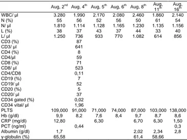
During
the
admission
his
conditions
worsened, making it necessary to perform a blood
transfusion on August, 5
th. Table 1 resumes altered
results of the blood tests performed on our patient
during the admission.
Table 1: Blood test results during the admission
Aug, 2nd Aug, 4th Aug, 5th Aug, 6th Aug, 8th Aug, 11th Aug, 16th WBC/ μl 3.280 1.990 2.170 2.080 2.460 1.860 2.140 N (%) 55 56 52 56 50 61 54 N/ μl 1.810 1.114 1.128 1.165 1.230 1.135 1.156 L (%) 38 37 43 37 44 33 40 L/ μl 1.250 736 933 770 1.082 614 856 CD3 (%) 87 CD3/ μl 641 CD4 (%) 8 CD4/μl 59 CD8 (%) 71 CD8/ μl 523 CD4/CD8 0,11 CD19 (%) 7 CD19/ μl 52 CD20 (%) 5 CD20/ μl 37 CD34 gated (%) 0,02 CD34 vital/ μl 1,96 PLTS 109,000 91,000 71,000 74,000 87,000 103,000 138,000 Hb (g/dl) 9,9 8,2 7,6 8,4 9,7 8,7 8,6 CRP (mg/dl) 12,80 6,30 6,70 6,30 1,50 PCT (ng/ml) 0,44 0,3 Albumin (g/dl) 1,7 2,02 2,34 2,8 γ-globulin (%) 65,58 61,4 58,66
As it can be seen, his immunological control
had worsened, revealing a CD4+ T-cell count of 59/μl
(8%), with a CD8+ T-cell count of 523/μl (71%) and a
resulting CD4+/CD8+ ratio of 0.11.
Stool
examinations
(research
of
the
Clostridium difficile toxin, parasitic infections and stool
cultures, faecal occult blood) were performed,
resulting in negative. Suspecting multiple myeloma,
the patient underwent a bone marrow biopsy to
determine the cause of the pancytopenia. No biopsy
was performed on the flank skin lesion.
Indirect immunofluorescence assay (IFAT)
and Polymerase Chain Reaction (PCR) for the
research of a Leishmania infection were performed on
the 4
thday after the admission, resulting in positive
(PCR 11,500 Leishmania/ml; IFAT 1:5120) on the 8
thday after the admission.
He then began a treatment with Liposomal
Amphotericin B (L-AMB) 4 mg/kg/day on days 1 to 5,
according to the Italian guidelines for the diagnosis
and management of HIV infection (2016 edition) of the
Italian Society of Infectious and Tropical Diseases
(ISITD), and he repeated the treatment on days 10,
17, 24, 31 and 38, completing the cycle [35]. He was
discharged on the 14
thday after the admission and
completed the treatment as an outpatient. Diarrhoea
and the hyperemic lesion on his flank completely
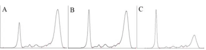
disappeared after the fifth day of therapy with L-AMB.
Figure 1 shows the Serum Protein Electrophoresis
(S-PEP) trend before, during and at the end of the
therapy. Secondary prophylaxis was not started, and
the patient is still in follow up for the possibility of VL
relapses.
_______________________________________________________________________________________________________________________________
_______________________________________________________________________________________________________________________________
Figure 1: S-PEP before (A); during (B) and after completing (C) the therapy with L-AMB
Discussion
Leishmaniasis is a parasitic disease affecting
both animals and humans, acquired with the bite of
sand flies or, in Injection Drug Users (IDUs), with
contaminated needles, still hypoendemic in Sicily and
the Mediterranean basin [34] [36].
It is a known opportunistic disease in People
Living with HIV (PLWH), whose immunodeficiency
promotes visceral localisation, even though the
coinfection prevalence reduced after the introduction
of HAART [33] [36] [37]. Moreover, Leishmania spp
can promote viral replication and enhance progression
to Acquired Immuno-Deficiency Syndrome (AIDS)
[34]. As a result, despite the introduction of HAART,
relapses are still common, and mortality is three times
higher in Leishmania coinfection than in
HIV-negative people affected by Leishmania infection [30]
[31].
Cota and al [33] reported in 2017 that CD4
T-cell count at the moment of the diagnosis is not able
to foresee the patient’s prognosis. However,
resolution of the infection depends on an efficient
CD4+ T-cell response, and it was observed that a
higher incidence of symptoms is related to a lower
CD4+ T-cell count [31] [32].
The most frequent signs and symptoms of this
infection are fever, asthenia, weight loss and
splenomegaly; unspecific symptoms that could often
lead to a delay in the diagnosis [31] [34]. Also, PLWH
can complain of misleading non-classical symptoms
[29].
In our case, the patient came to our attention
in January, after more than a year of likely therapeutic
vacation, with a seriously impaired immunological
control. At the time he did not have any classical sign
of the infection (normal S-PEP, no signs of
pancytopenia, no hepatosplenomegaly), but it has
been reported that Leishmaniasis can be related to
pleural effusion, especially in PLWH with a very low
CD4+ T-cell count [38]. Moreover, Infectious Diseases
Society of America (IDSA) 2016 guidelines report that
in PLWH the number of asymptomatic carriers of
Leishmania seems to be higher than in the
immunocompetent host [39]. Therefore, it can be
supposed that our patient’s disease began some time
before the classical signs appeared.
An active response to Leishmania, leading to
the infection control, is associated to host adaptive
immunity, but, at the same time, to natural immunity
[40]. The most influential factor in the immune
response in Leishmaniasis seems to be the early
interaction of the parasite with macrophages and
dendritic cells [41].
Moreover, it has been observed an enhanced
secretion of Th2 cytokines, and in particular IL-10, in
Visceral Leishmaniasis associated to HIV-infection,
which can promote the dissemination of both the virus
and the parasite [28] [32] [40] [41] [42] [43].
Our patient presented to our attention, both in
January and August, with a profoundly impaired
immunological control, defined as a very low CD4+
T-cell count and an inverted CD4/CD8 ratio, despite
having a suppressed VL, a common sign during
visceral Leishmaniasis [42]. However, the fact the
patient came from a period of therapeutic vacation
made difficult to think to other causes of severe
immunodepression than his HIV infection, leading to
the possibility of a diagnostic delay.
The CD4/CD8 ratio is a marker of immune
dysfunction leading to persistent inflammation in
PLWH, and a low ratio can predict an impaired CD4+
T-cell count recovery before the start of the HAART
[44]. During visceral leishmaniasis, CD8+ T-cells, and
especially those expressing CD38, or activated CD8+
T-cells, increase, leading to a status of chronic
inflammation which results in a T cell depletion,
establishing a vicious circle that worsens the
immune-depression [42].
Although both IDSA and ISITD guidelines
recommend secondary prophylaxis with L-AMB in
patients with a CD4+ T-cell count lower than 200/μl,
our patient refused it [35] [39]. He is still in follow up
for the possibility of relapses and recently completed
his 5
thmonth from the end of the therapy with L-AMB.
In conclusion, visceral Leishmaniasis is an
important opportunistic disease in PLWH, with a
complicated differential diagnosis because of its
unspecific symptoms and signs. It is even more
difficult because of the possibility of atypical
manifestations.
However, this parasitic disease has a high
mortality rate, so it is mandatory to think about it in all
the patients having a low CD4+ T-cell count and an
inverted CD4/CD8 ratio under HAART.
Further studies are needed to clear the
pathogenesis of the infection and to establish the
duration of the secondary prophylaxis.
_______________________________________________________________________________________________________________________________
References
1. Lai V, Zizi B, Calia GM, Bagella P, Fiore V, Peruzzu F, Caruana G, Babudieri S, Mura MS, Madeddu G. Fib-4 values and
neurocognitive function in hiv-infected patients without hepatic coinfections. Infect Dis Trop Med. 2016; 2:e293.
2. La Ferla L, Lo Presti Costantino MR, Mondello P. Kaposi's sarcoma in HIV-infected patients: a review of the literature. Infect Dis Trop Med. 2016; 2:e239.
3. Bruno R, Scuderi D, Locatelli ME, Pampaloni A, Pinzone MR. Prevalence of micronutrients deficiencies in a cohort of HIV-positive individuals on ART. Infect Dis Trop Med. 2017; 3:e431. 4. Pinzone MR, Di Rosa M, Celesia BM, Condorelli F,
Malaguarnera M, Madeddu G, Martellotta F, Castronuovo D, Gussio M, Coco C, Palermo F, Cosentino S, Cacopardo B, Nunnari G. LPS and HIV gp120 modulate monocyte/macrophage CYP27B1 and CYP24A1 expression leading to vitamin D consumption and hypovitaminosis D in HIV-infected individuals. Eur Rev Med Pharmacol Sci. 2013; 17:1938–1950. PMid:23877860
5. Celesia BM, Nigro L, Pinzone MR, Coco C, La Rosa R, Bisicchia F, Mavilla S, Gussio M, Pellicanò G, Milioni V, Palermo F, Russo R, Mughini MT, Martellotta F, Taibi R, Cacopardo B, Nunnari G. High prevalence of undiagnosed anxiety symptoms among HIV-positive individuals on cART: A cross-sectional study. Eur Rev Med Pharmacol Sci. 2013; 17:2040–2046. PMid:23884824
6. Scarpino M, Santoro M, Pellicanò G. HIV infection and kidney disease: literature review. Infectious Diseases and Tropical Medicine. 2015; 1:e195.
7. Facciolà A, Venanzi Rullo E, Ceccarelli M, D'Aleo F, Di Rosa M, Pinzone MR, Condorelli F, Visalli G, Picerno I, Fisichella R, Nunnari G, Pellicanò GF. Kaposi's sarcoma in HIV-infected patients in the era of new antiretrovirals. Eur Rev Med Pharmacol Sci. 2017; 21:5868–5879. PMid:29272026
8. Montrucchio C, Biagini R, Alcantarini C, Calcagno A, Barco A, Ferrara M, Milesi M, Costa C, Trentalange A, Trunfio M, Tettoni MC, Grosso Marra W, D'Ascenzo F, Ballocca F, Lonni E, Gili S, Vai D, Imperiale D, Gaita F, Bonora S, Di Perri G. Cardiovascular risk and neurocognitive deficits in HIV-positive individuals. Infect Dis Trop Med. 2017; 3:e370.
9. Castronuovo D, Cacopardo B, Pinzone MR, Di Rosa M, Martellotta F, Schioppa O, Moreno S, Nunnari G. Bone disease in the setting of HIV infection: update and review of the literature. Eur Rev Med Pharmacol Sci. 2013; 17:2413–2419. PMid:24089217 10. Di Rosa M, Tibullo D, Vecchio M, Nunnari G, Saccone S, Di Raimondo F, Malaguarnera L. Determination of chitinases family during osteoclastogenesis. Bone. 2014; 61:55–63.
https://doi.org/10.1016/j.bone.2014.01.005 PMid:24440516 11. Visalli G, Bertuccio MP, Currò M, Pellicanò G, Sturniolo G, Carnevali A, Spataro P, Ientile R, Picerno I, Cavallari V, Piedimonte G. Bioenergetics of T cell activation and death in HIV type 1 infection. AIDS Res Hum Retroviruses. 2012; 28:1110–
1118. https://doi.org/10.1089/aid.2011.0197
12. Pinzone MR, Nunnari G. Prevalence of comorbidities in a cohort of women living with HIV. Infect Dis Trop Med. 2015; 1:e165.
13. Kulkosky J, Bouhamdan M, Geist A, Nunnari G, Phinney DG, Pomerantz RJ. Pathogenesis of HIV-1 infection within bone marrow cells. Leukemia and Lymphoma. 2000; 37:497–515.
https://doi.org/10.3109/10428190009058502 PMid:11042510 14. Bellissimo F, Pinzone MR, Cacopardo B, Nunnari G. Diagnostic and therapeutic management of hepatocellular carcinoma. World J Gastroenterol. 2015; 21:12003–12021.
https://doi.org/10.3748/wjg.v21.i42.12003 PMid:26576088 PMCid:PMC4641121
15. Nunnari G, Otero M, Dornadula G, Vanella M, Zhang H, Frank I, Pomerantz RJ. Residual HIV-1 disease in seminal cells of HIV-1-infected men on suppressive HAART: Latency without on-going
https://doi.org/10.1097/00002030-200201040-00006
PMid:11741161
16. D'Aleo F, Ceccarelli M, Venanzi Rullo E, Facciolà A, Di Rosa M, Pinzone MR, Condorelli F, Visalli G, Picerno I, Berretta M, Pellicanò GF, Nunnari G. Hepatitis C-related hepatocellular carcinoma: diagnostic and therapeutic management in HIV-patients. Eur Rev Med Pharmacol Sci. 2017; 21:5859–5867. PMid:29272025
17. Squillace N, Pellicanò GF, Ricci E, Quirino T, Bonfanti P, Gori A, Bandera A, Carenzi L, De Socio GV, Orofino G, Martinelli C, Madeddu G, Rusconi S, Maggi P, Celesia BM, Cordier L, Vichi F, Calza L, Falasca K, Di Biagio A, Pellicanò GF, Bonfanti P. Safety and tolerability of Elvitegravir/Cobicistat/Emtricitabine/Tenofovir Disoproxil fumarate in a real life setting: Data from surveillance cohort long-term toxicity antiretrovirals/antivirals (SCOLTA) project. PLoS ONE. 2017; 12:e0179254.
https://doi.org/10.1371/journal.pone.0179254 PMid:28632758 PMCid:PMC5478131
18. Pomerantz RJ, Nunnari G. HIV and GB Virus C - Can Two Viruses be Better Than One? N Engl J Med. 2004; 350:963–965.
https://doi.org/10.1056/NEJMp048004 PMid:14999105
19. Colafigli M, Bonadies A, Ferraresi V, Tonachella R, Cristaudo A, Latini A. Kaposi Sarcoma in HIV-infected patients: an infectious-dermatological outpatient service experience. Infect Dis Trop Med. 2017; 3:e410.
20. Ceccarelli G, Vassalini P, Corano Scheri G, Cavallari EN, Bianchi L, Di Girolamo G, Fratino M, Vullo V, D'Ettorre G.
Improvement of neuropsychological performances and reduction of immune-activation markers after probiotic supplementation and change of life-style in an HIV positive male: targeting the microbiota to act on gut-brain axis. Infect Dis Trop Med. 2017; 3:e404.
21. Scarpino M, Pinzone MR, Di Rosa M, Madeddu G, Focà E, Martellotta F, Schioppa O, Ceccarelli G, Celesia BM, D'Ettorre G, Vullo V, Berretta S, Cacopardo B, Nunnari G. Kidney disease in HIV-infected patients. Eur Rev Med Pharmacol Sci. 2013; 17:2660–2667. PMid:24142615
22. Visalli G, Paiardini, Chirico C, Cervasi B, Currò M, Ferlazzo N, Bertuccio MP, Favaloro A, Pellicanò G, Sturniolo G, Spataro P, Ientile R, Picerno I, Piedimonte G. Intracellular accumulation of cell cycle regulatory proteins and nucleolin re-localization are
associated with pre-lethal ultrastructural lesions in circulating T lymphocytes: The HIV-induced cell cycle dysregulation revisited. Cell Cycle. 2010; 9:2130–2140.
https://doi.org/10.4161/cc.9.11.11754 PMid:20505329
23. Trovato M, Ruggeri RM, Sciacchitano S, Vicchio TM, Picerno I, Pellicanò G, Valenti A, Visalli G. Serum interleukin-6 levels are increased in HIV-infected patients that develop autoimmune disease during long-term follow-up. Immunobiology. 2017. 24. Nunnari G, Coco C, Pinzone MR, Pavone P, Berretta M, Di Rosa M, Schnell M, Calabrese G, Cacopardo B. The role of micronutrients in the diet of HIV-1 infected individuals. Front Biosci. 2012; e4:2442–2456.
25. Castronuovo D, Pinzone MR, Moreno S, Cacopardo B, Nunnari G. HIV infection and bone disease: a review of the literature. Infect Dis Trop Med. 2015; 1:e116.
26. Malaguarnera M, Vacante M, Motta M, Giordano M, Malaguarnera G, Bella R, Nunnari G, Rampello L, Pennisi G. Acetyl-L-carnitine improves cognitive functions in severe hepatic encephalopathy: a randomized and controlled clinical trial. Metabolic Brain Disease. 2011; 26:281–289.
https://doi.org/10.1007/s11011-011-9260-z PMid:21870121 27. Calcagno A, Simiele M, Alberione MC, Bracchi M, Marinaro L, Ecclesia S, Di Perri G, D'Avolio A, Bonora S. Cerebrospinal Fluid Inhibitory Quotients of Antiretroviral Drugs in HIV-Infected Patients Are Associated With Compartmental Viral Control. Clin Infect Dis.
2015; 60:311–317. https://doi.org/10.1093/cid/ciu773
PMid:25281609
28. Pagliano P, Esposito S. Visceral leishmaniosis in immunocompromised host: an update and literature review. J
_______________________________________________________________________________________________________________________________
_______________________________________________________________________________________________________________________________
Chemother. 2017; 29:261–266.
https://doi.org/10.1080/1120009X.2017.1323150 PMid:28490252 29. Pagliano P, Ascione T, Di Flumeri G, Boccia G, De Caro F. Visceral leishmaniasis in immunocompromised: Diagnostic and therapeutic approach and evaluation of the recently released IDSA guidelines. Infez Med. 2016; 24:265. PMid:28011960
30. Madeddu G, Fiori ML, Ena P, Riu F, Lovigu C, Nunnari G, Bagella P, Maida I, Babudieri S, Mura MS. Mucocutaneous leishmaniasis as presentation of HIV infection in Sardinia, insular Italy. Parasitol Int. 2013; 63:35–36.
https://doi.org/10.1016/j.parint.2013.10.002 PMid:24126182 31. de Sousa-Gomes ML, Romero GAS, Werneck GL. Visceral leishmaniasis and HIV/AIDS in Brazil: Are we aware enough? PLoS Negl Trop Dis. 2017; 11:e0005772.
https://doi.org/10.1371/journal.pntd.0005772 PMid:28945816 PMCid:PMC5612457
32. Ezra N, Ochoa MT, Craft N. Human immunodeficiency virus and leishmaniasis. J Glob Infect Dis. 2010; 2:248–257.
https://doi.org/10.4103/0974-777X.68528 PMid:20927287 PMCid:PMC2946682
33. Cota GF, de Sousa MR, de Assis TSM, Pinto BF, Rabello A. Exploring prognosis in chronic relapsing visceral leishmaniasis among HIV-infected patients: Circulating Leishmania DNA. Acta Trop. 2017; 172:186–191.
https://doi.org/10.1016/j.actatropica.2017.05.011 PMid:28501450 34. Guarneri C, Tchernev G, Bevelacqua V, Lotti T, Nunnari G. The unwelcome trio: HIV plus cutaneous and visceral leishmaniasis.
Dermatol Ther. 2016; 29:88–91. https://doi.org/10.1111/dth.12303
PMid:26555699
35. Linee Guida Italiane sull'utilizzo dei farmaci antiretrovirali e sulla gestione diagnostico-clinica delle persone con infezione da HIV-1.
http://www.simit.org/medias/1047-lg-hiv-2016-c17pubblicazioni2545allegato.pdf Accessed 14 January 2018. 36. Lindoso JAL, Cunha MA, Queiroz IT, Moreira CHV. Leishmaniasis-HIV coinfection: current challenges. HIV AIDS
(Auckl). 2016; 8:147–156. https://doi.org/10.2147/HIV.S93789
PMid:27785103 PMCid:PMC5063600
37. Cocuzza S, Strazzulla A, Pinzone MR, Cosentino S, Serra A, Caltabiano R, Lanzafame S, Cacopardo B, Nunnari G. Isolated laryngeal leishmaniasis in immunocompetent patients: an
underdiagnosed disease. Case Rep Infect Dis. 2013; 2013:165409.
https://doi.org/10.1155/2013/165409
38. Chenoweth CE, Singal S, Pearson RD, Betts RF, Markovitz DM. Acquired Immunodeficiency Syndrome-Related Visceral Leishmaniasis Presenting in a Pleural Effusion. Chest. 1993;
103:648-649. https://doi.org/10.1378/chest.103.2.648
PMid:8432183
39. Aronson N, Herwaldt BL, Libman M, Pearson R, Lopez-Velez R, Weina P, Carvalho EM, Ephros M, Jeronimo S, Magill A. Diagnosis and Treatment of Leishmaniasis: Clinical Practice Guidelines by the Infectious Diseases Society of America (IDSA) and the American Society of Tropical Medicine and Hygiene (ASTMH). Clin Infect Dis. 2016; 63:e202–e264.
https://doi.org/10.1093/cid/ciw670 PMid:27941151 40. Strazzulla A, Cocuzza S, Pinzone MR, Postorino MC, Cosentino S, Serra A, Cacopardo B, Nunnari G. Mucosal leishmaniasis: an underestimated presentation of a neglected disease. Biomed Res Int. 2013; 2013:805108.
https://doi.org/10.1155/2013/805108 PMid:23853773 PMCid:PMC3703408
41. Khadem F, Uzonna JE. Immunity to visceral leishmaniasis: implications for immunotherapy. Future Microbiol. 2014; 9:901–
915. https://doi.org/10.2217/fmb.14.43 PMid:25156379
42. Santos-Oliveira JR, Giacoia-Gripp CBW, de Oliveira PA, Amato VS, Lindoso JÂL, Goto H, Oliveira-Neto MP, Mattos MS, Grinsztejn B, Morgado MG, Da-Cruz AM. High levels of T lymphocyte activation in Leishmania-HIV-1 co-infected individuals despite low HIV viral load. BMC Infect Dis. 2010; 10:358.
https://doi.org/10.1186/1471-2334-10-358 PMid:21171992 PMCid:PMC3022832
43. Celesia BM, Cacopardo B, Massimino D, Gussio M, Tosto S, Nunnari G, Pinzone MR. Atypical Presentation of PKDL due to Leishmania infantum in an HIV-Infected Patient with Relapsing Visceral Leishmaniasis. Case Rep Infect Dis. 2014; 2014:370286. 44. Lu W, Mehraj V, Vyboh K, Cao W, Li T, Routy J-P. CD4:CD8 ratio as a frontier marker for clinical outcome, immune dysfunction and viral reservoir size in virologically suppressed HIV-positive patients. J Int AIDS Soc. 2015; 18:20052.
https://doi.org/10.7448/IAS.18.1.20052 PMid:26130226 PMCid:PMC4486418