Letter | Dermatol Pract Concept 2020;10(3):e2020053
1
Dermatology
Practical & Conceptual
Introduction
Delayed tattoo reactions include a wide range of clinical presentations and overlapping forms; thus diagnosis can be challenging. We present a case of a delayed tattoo reaction from red dye.
Case Presentation
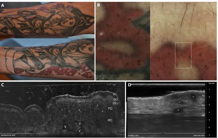
An otherwise healthy 40-year-old man presented with firm elevated plaques that arose 4 months previously, within a multicolored tattoo on the right forearm, 8 months after the execution of the tattoo (Figure 1A). The patient claimed minimal and only occasional itching. Polarized dermoscopy (Luminis Visiomed, Caliber) highlighted multiple keratotic plugs within the lesion, limited to the red-dyed areas (Figure 1). Examination with line-field confocal optical coherence tomography (LC-OCT) [1] of both the transitional
lesional-to-healthy part (Figure 1C) and the central part of the lesion (Figure 2A) showed a hyperkeratotic flattened epidermis and a blurred basal membrane due to the presence of inflam-matory infiltrate, suggesting a lichenoid/interface dermatitis pattern. Comparative examination with high-frequency ultrasound 70 MHz (HFUS) (VEVO MD, VisualSonics) up to a depth of 5.5 mm performed at the same location revealed multiple anechoic holes and posterior shadowing due to dilated vessels, and inflammation in the superficial and deep dermis (Figure 1D). Punch biopsy taken at the same location for histopathological examination revealed hyperkeratosis, flattened epidermis with hyperkeratosis and blurring of the basal membrane; lymphocytic lichenoid inflammatory infiltrate in the superficial dermis (Figure 2B); and clustered infiltrate around red pigment deposits in reticular dermis, focally periadnexal and perivascular (Figure 2C). Immuno-histochemical study revealed a mixed inflammatory chronic infiltrate composed of B and T lymphocytes. Red dye
depos-Delayed Tattoo Reaction From Red Dye With
Overlapping Clinicopathological Features:
Examination With High-Frequency Ultrasound and
Line-Field Optical Coherence Tomography
Linda Tognetti,
1Sean Ekinde,
1Cyril Habougit,
2Elisa Cinotti,
2Pietro Rubegni,
1Jean Luc Perrot
31 Department of Dermatology, Division of Medical, Surgical and NeuroSciences, University of Siena, Italy 2 Anatomopathology Service, University Hospital of Saint-Etienne, Saint-Etienne, France
3 Department of Dermatology, University Hospital of Saint-Etienne, Saint-Etienne, France
Keywords: tattoo reaction, high-frequency ultrasound, line-field optical coherence tomography
Citation: Tognetti L, Ekinde S, Habougit C, Cinotti E, Rubegni P, Perrot JL. Delayed tattoo reaction from red dye with overlapping clinicopathological features: examination with high-frequency ultrasound and line-field optical coherence tomography. Dermatol Pract
Concept. 2020;10(3):e2020053. DOI: https://doi.org/10.5826/dpc.1003a53
Accepted: February 21, 2020; Published: June 29, 2020
Copyright: ©2020 Tognetti et al. This is an open-access article distributed under the terms of the Creative Commons Attribution License, which permits unrestricted use, distribution, and reproduction in any medium, provided the original author and source are credited.
Funding: None.
Competing interests: The authors have no conflicts of interest to disclose.
Authorship: All authors have contributed significantly to this publication.
Corresponding author: Linda Tognetti, MD, Dermatology Unit, Division of Medical, Surgical and NeuroSciences, University Hospital of Siena, Viale Bracci, 53100 Siena, Italy. Email: [email protected]
2
Letter | Dermatol Pract Concept 2020;10(3):e2020053Figure 1. (A) Severe persistent tattoo reaction from red dye. (B) Dermoscopic examination (×20) of a swelling elevated area (white circle in A) highlights the presence of yellowish circles within the stained skin corresponding to perifollicular openings filled with keratin, dyed by exogenous pigment. Examination of transition healthy-to-lesional zone (white square) with (C) line-field confocal optical coherence tomog-raphy and (D) high-frequency ultrasound 70 MHz: inflammatory infiltrates (asterisks) and dilated vessels are visible in papillary and reticular dermis of lesional skin, generating diffuse anechoic holes in the papillary and reticular dermis, respectively. DEJ = dermoepidermal junction; E = epidermis; MF = muscularis fascia; PD = papillary dermis; RD = reticular dermis; SC = stratum corneum; SG = stratum granulosum; V = vessels.
Figure 2. (A) Line-field confocal optical coherence tomography image (0.5 mm in depth, axial and lateral resolution of 1 µm) of lesional skin. (B) Histological correlation: PAS OM200× shows thickened and hyperreflective stratum corneum (SC) due to hyperorthokeratosis; narrowed stratum granulosum (SG) and stratum spinosum (SS); blurred dermoepidermal junction (DEJ) due to infiltrating lymphocytes; dilated vessels (V) and chronic inflammatory infiltrate (asterisks) in the papillary (PD) and reticular dermis (RD) appearing as hyporeflective areas sur-rounding red pigment deposits. (figure continues next page)
Letter | Dermatol Pract Concept 2020;10(3):e2020053
3
egorized into a rigid classification, the present case should be interpreted as a severe delayed tattoo reaction from red dye with overlapping features of the pseudolymphomatous, lichenoid, and hyperkeratotic pattern.Getting closer to an in vivo histology and providing higher resolution than conventional OCT, the new LC-OCT [1] can be proposed to better characterize the superficial part of a tattoo reaction of uncertain pattern and/or with clinical over-lapping features, in combination with HFUS, which is able to reveal the inflammation in the deep dermis [1]. Moreover, the 2 techniques can be used to monitor treatment response to laser or intralesional therapy.
References
1. Dubois A, Levecq O, Azimani H, et al. Line-field confocal optical coherence tomography for high-resolution noninvasive imaging of skin tumors. J Biomed Opt. 2018;10(23):1-9. https://doi. org/10.1117/1.JBO.23.10.106007.
2. Forbat E, Al-Niaimi F. Patterns of reactions to red pigment tattoo and treatment methods. Dermatol Ther (Heidelb). 2016;6(1):13-23. https://doi.org/10.1007/s13555-016-0104-y.
its were distributed in the deep dermis only and spared the superficial dermis (Figure 2D).
Conclusions
In the last decade, delayed tattoo reactions have been on the increase due to the popularity of tattoos [2]. Metal compo-nents of the red dye (eg, nickel, mercury, and cadmium) have been addressed as the chronic antigenic stimulating agent causing a polyclonal proliferation of lymphoid cells. Tradi-tionally, 4 histological patterns were described for delayed tattoo reactions, ie, hyperkeratotic pattern, lichenoid der-matitis, pseudolymphomatous pattern, and granulomatous pattern. However, this classification often fails to correlate significantly with clinical appearance of the lesions and has limited utility in daily practice, especially when multiple or overlapping clinicopathological features can be detected inside the same lesion [2]. Indeed, in this case we can observe keratotic cysts on dermoscopy, in favor of the hyperkeratotic pattern; histological features of lichenoid dermatitis; and evolution and clustered deep dermal infiltrate typical of the pseudolymphomatous pattern. Thus, rather than being
cat-Figure 2. (continued) (C) H&E staining (OM25×) demonstrates a lymphocytic infiltrate exhibiting both a lichenoid and a cluster distribu-tion, focally periadnexal and perivascular. Red pigment deposits are distributed in the whole thickness of RD and reach the upper part of the hypodermis (HD). (D) No staining (OM25×).