Università degli studi della Tuscia
Dipartimento di Scienze Ecologiche e Biologiche (DEB)
DOTTORATO DI RICERCA
IN GENETICA E BIOLOGIA CELLULARE
XXVII Ciclo
BIO/11
Aging-related anatomical and biochemical changes in lymphatic
collectors impair lymph transport, fluid homeostasis and pathogen
clearance
Coordinatore: Giorgio Prantera
Firma: ………..………
Tutor: Sara Rinalducci Dottorando: Valerio Zolla
INDEX
1. INTRODUCTION ...10
1.1 General introduction ...11
1.2 Aim of the thesis ...12
1.3 The lymph ...13
1.4 Lymph Formation ...13
1.5 Lymph composition ...16
1.6 LYMPH TRANSPORT ...16
1.6.1 Lymphatic Capillaries and Collectors ...16
1.6.2 Structure of lymphatic capillaries and collectors ...18
1.6.3 Lymphatic capillaries (both initial and pre collecting) ...18
1.6.4 Lymphatic collectors (or collecting lymphatics) ...18
1.6.5 Differences between blood and lymphatic vessels ...19
1.7 Lymphatic Extracellular Matrix ...21
1.9 Lymphatic smooth muscles cells ...23
1.10 Extrinsic and intrinsic forces ...25
1.11 Lymph Circulation under pathological condition. ...25
1.12 FREE- RADICALS AND AGING ...26
1.13 IMMUNOSENESCENCE ...28
2. ABSTRACT ...30
3. PREFACE ...32
4. RESULTS ...35
4.1 Decreased extracellular matrix and contractile proteins in
aged lymphatic collectors...36
4.2 Decreased contractility and pumping efficiency in aged
lymphatic collectors ...47
4.3 Decreased glycocalyx and junctional proteins in aging
lymphatic collectors ...49
4.5 Impaired pathogen clearance by aged lymphatic collectors.
...55
4.6 Impaired permeability in aged lymphatic collectors. ...60
5. DISCUSSION ...63
6. MATERIAL AND METHODS ...67
6.1 Rats and procedures for in vivo measurements of lymphatic
contractility ...68
6.2 In vivo analysis of lymphatic contractility. ...69
6.3 Mice and in vivo procedure ...71
6.4 Growth conditions and CFU determination for Cryptococcus
Neoformans H99 (Serotype A) ...72
6.5 Growth conditions and CFU determination for
Mycobacterium smegmatis (mc
2155) ...72
6.6 Transmission electron microscopy and Electron
Tomography ...73
6.7 Scanning electron microscopy ...73
6.8 Western blot analysis of oxidized proteins ...74
6.9 Preparation, imaging and analysis of bacterial transport in
the lymphatic collectors ...74
6.10 Evaluations of bacterial traffic through isolated mesenteric
lymphatic vessels. ...76
6.11 Sample preparation for FACS analysis and CFU ...77
6.12 Rat mesenteric lymphatic vessels proteomics analysis ...78
6.13 Generation of Heat Maps ...80
6.14 Permeability Assay: ...81
6.15 Evans Blue Injections and Measurement of Collector’s
Leakage ...82
7. BIBLIOGRAPHY ...84
PARTICIPATION TO MEETINGS WITH POSTER PRESENTATION
...97
GLOSSARY
ROS: reactive oxygen species
SEM: scanning electron microscopy
TEM: transmission electron microscopy
MS/MS: tandem mass spectrometry
AFP: amplitude frequency product
FPT: fractional pump flow
PTM: post translation modification
CFU: colony forming unit
GFP: green fluorescent protein
TRITC: tetramethylrhodamine isothiocyanate
VEGF: vascular endothelial growth factor
EDD: end-diastolic diameter
ESD: end-systolic diameters
DMSO: dimethylsulfoxide
MLV: mesenteric lymphatic vessel
FACS: fluorescence-activated cell sorting
LEC: lymphatic endothelial cell
ECM: Extracellullar matrix
1.1 General introduction
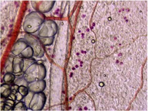
Figure 1: Imaging of parallel running blood and lymphatic vessels
Figure 2: Actin immunostaining of a collector’s valve
Lymphatic collectors and capillaries are present in every parenchymal organ, except the brain; they run parallel to the blood vessel and collect products of cellular metabolism/catabolism, products of cellular apoptosis/necrosis, extracellular matrix processing and immune cell trafficking to the lymph nodes. Pathogens and cancer cells are also drained by the lymphatic network to the lymph nodes. Thus, any functional defects in the draining lymphatics will affect lymph transport and consequently body fluid and
parenchymal organs homeostasis, immune cell trafficking and pathogen clearance. The blood flows through by thin walled capillaries to each organ, delivering nutrients and
oxygen. Nutrients and oxygen diffuse into the interstitial space and through each layer of parenchymal cells. The majority of blood filtrate collected in the interstitial space is not
reabsorbed into the blood vessels but enters instead into the lymphatic capillaries as lymph. In recent years, lymphatic dysfunction and its correlation to increased inflammation and cancer has become a topic of extreme interest. Mice with decreased lymphangiogenesis have immune system imbalances and enhanced edema. Virtually nothing is known about how lymphatic functionality, in particular lymphatic drainage, is affected by the process of aging. In the current literature, a paucity of papers reported a negative correlation between
aging and intrinsic lymphatic pump (1).
1.2 Aim of the thesis
Over the last 20 years a deeper understanding of the lymphatic circulatory system, lymph formation, and composition has emerged. Recent studies yield to new insights on the organization of the lymphatic vascular tree, the formation of lymph from the extracellular fluid, lymph circulation and lymph proteomic composition during physiological and pathological conditions. Formation of the lymph fluid is dependent on capillary bed pressure gradients and the composition of the endothelial cell glycocalyx, which acts as a molecular sieve. Lymph “omics” composition is dependent on the ultrafiltration of plasma proteins as well as proteins and molecules derived from the metabolic and catabolic activities of each parenchymal organ from which the lymph drains. Altogether, these new discoveries have brought to light the importance of the lymphatic system in human physiology and pathology.
However, how the aging process influence lymph transport and pathogen clearance still
needs to be clarified. Aged individuals have a compromised immune response to pathogens and increased tissue
edema. Remarkably, a paucity of studies have investigated age-related changes in lymphatic collectors and how these changes could lead to impairment in lymphatic function. Evidence suggests a possible correlation between oxidative stress, a typical hallmark of aging, endothelial cell dysfunction and decreased lymphatic vessel contractility (1-3). Decreased function of lymphatic muscle cells has also been observed in aged animals
but the causes are still unknown. Because the lymphatics are pivotal in maintaining fluid homeostasis and immune responses, we propose to perform an anatomical, biochemical and functional comparative analysis of lymphatics between young and old rodents. The ultimate goal is to provide a comprehensive analysis and determine the pathogenesis of aging-associated decline in lymph transport.
1.3 The lymph
Lymph is the fluid that circulates throughout the lymphatic system and it is formed from the interstitial fluid collected through the lymph capillaries. Lymphatic collectors transport the lymph to the draining lymph nodes before emptying ultimately into the right or the left subclavian vein, where it mixes back with blood. The lymph has a pivotal role in every immunological processes including maintenance of immunological tolerance, autoimmunity, inflammation, cancer metastasis, and cardiovascular and metabolic disorders. Until recently the detailed composition of the lymphatic fluid was virtually unknown. This was primarily due to the technical difficulties of cannulating lymphatic collectors, the low sensitivity of mass spectrometry instruments for proteome mapping, and the scarcity of the collected lymph, which precluded many biological assays. Many of these issues have been resolved, and lymph biology has progressively received more attention. During the last ten years important progress has been made towards understanding the mechanisms of lymph formation, circulation and composition.
1.4 Lymph Formation
The Interstitial fluid, which is the precursor of the pre-nodal lymph, is formed as an ultra-filtrate of the capillary microcirculation (Figure 3); as such many of the proteins found in the blood are present in the lymph as well. According to the Starling principle, the microvascular ultrafiltration process is determined by the net balance between hydrostatic and osmotic pressures across the microvascular endothelium (4). The principle states that
the pressure gradient between the arterial and venous halves of the capillary beds drives fluid filtering from the arterial end into the interstitial space and reabsorption into the venous system. This concept, however, which has been accepted for over 100 years, has recently been revised. Indeed, more sophisticated measurements of fluid exchange in the capillary beds indicate that in most tissues the steady state pressures (hydrostatic and osmotic) provide a low level of fluid filtration from the microcirculation, at both arterial and venous ends, towards the interstitial fluid (5) (Figure 4).
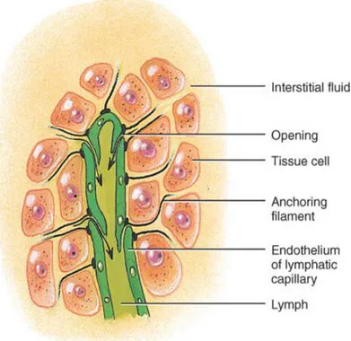
Figure 3: The monolayer of endothelial cells is fundamental for allowing interstitial liquid
to enter into the lumen of the lymphatic capillaries. The gaps and spaces between these cells are larger; and by moving, the endothelial cells allow liquid to flow in.
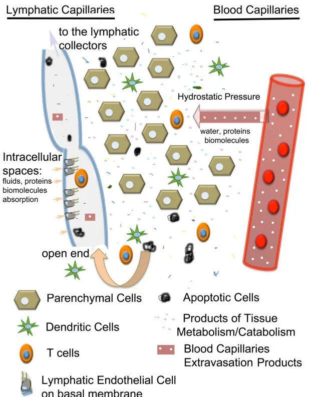
Figure 4: Representation of the interstitial space between lymphatic and blood capillaries;
here are present parenchymal cells, immune cells, products of tissue metabolism/catabolism, apoptotic cells and other products extravasated from the blood capillaries. The hydrostatic pressure drives the fluid exchange between the plasma and the lymphatic lumen; once cells and biomolecules are filtered through the lymphatic
endothelium, they are rapidly transported along the lymphatic capillaries toward the lymphatic vessels.
1.5 Lymph composition
As recently as ten years ago it was thought that the qualitative protein composition of the lymph overlapped with that of the plasma. However, this notion has been challenged by several proteomic analyses performed on lymph fluid (6), collected in physiological and pathological conditions, which have indicated how the metabolic/catabolic activities of parenchymal organs contribute to the lymph proteome. Analyses performed on human(6), rodents (7), ovine (8) and bovine lymph samples have revealed qualitative and quantitative differences between plasma and lymph. Two major differences between plasma and lymph can be elucidated. Lymph, unlike plasma, is rich with protein derived from extracellular matrix processing, tissue growth and remodeling, cellular metabolic/catabolic activities and cell death. In addition, tissue specific proteins have been mapped draining from different organs, indicating a regional-specific “omic” signature. Tissue-derived proteins are
eventually phagocytosed by tissue-resident and nodal antigen presenting cells, leaving only a small fraction to reach the venous circulation.
1.6 LYMPH TRANSPORT
1.6.1 Lymphatic Capillaries and Collectors
The lymph transport system is comprised of capillaries, vessels and ducts. Lymph capillaries are involved in the collection of the interstitial fluid. They have a diameter of 54.8 +/- 8.2 microns and are formed by a monolayer of endothelial cells supported by an incomplete basal membrane. Lymph capillaries are slightly larger than their counterpart in the vascular system and more importantly have one open end, which facilitates entry of fluids and immune cells. Lymphatic vessels are characterized by a
monolayer of endothelial cells supported by a complete basal membrane as well as by lymphatic muscle cells. The pumping activity of the muscle cells and the presence of several valves in the vessels facilitate lymph transport. There are different categories of lymphatics, which are distinguished by their diameter, anatomical structure and function (Figure 5). Lymph vessels are lined by endothelial cells, a thin layer of smooth muscle, and an organized extracellular matrix that is essential for support and communication with the surrounding tissues. The afferent lymph vessels carry the lymph to a draining lymph node, while the efferent lymph vessels exit the lymph node. Efferent vessels join together to form larger vessels, which transport the lymph into the thoracic duct or the right lymphatic duct. These two ducts (thoracic duct or right lymphatic duct) then drain into the subclavian veins, returning the post-filtration lymph to the vascular circulation.
Figura 5: There are three kinds of lymphatics. Initial capillaries have blunt-ends and are
implicated in the absorption of interstitial fluid. The liquid is then transported to the bigger pre-collecting lymphatic capillaries; both initial and pre-collecting lymphatics are surrounded only by endothelium. At the end, the lymph reaches the lymphatic vessel, called the collecting lymphatic, which is bigger in size. Since they have few smooth muscle cells on their wall, they can propel the lymph forward by intrinsic forces.
1.6.2 Structure of lymphatic capillaries and collectors
Both lymph capillaries and collectors are lined with a monolayer of endothelial cells. These cells are connected by specialized intercellular junctions, containing PECAM1 and VE cadherin, which function as one-way valves that facilitate the entry of immune cells, proteins, fluids, macro- and small molecules. Mechanical pressure from the surrounding tissue can enlarge the gaps between the endothelial cell monolayer.
1.6.3 Lymphatic capillaries (both initial and pre collecting)
The capillaries are classified into two groups: initial capillaries and general capillaries. The initial capillaries collect fluid, molecules and immune cells from the interstitial space, and thus have larger gaps between the endothelial cells that comprise the monolayer. The general capillaries serve to transport the initial lymph into the collectors.
1.6.4 Lymphatic vessels (or collectors or collecting lymphatics)
Unique to the lymphatic collectors is a layer of muscle cells that surrounds the endothelial cell monolayer. These muscle cells, which share characteristics with both smooth and striated muscle, are important for the maintenance of basal vessel tone and for spontaneous contractions that propel the lymph towards the draining lymph node(9). The directional flow of lymph is also maintained through a series of unidirectional valves positioned along the collectors, which open and close in synchrony with the vessel contraction. The valves are bicuspid and are formed by connective tissue overlaid by lymphatic endothelial cells
(10). The segment of lymphatic collectors positioned between the two sets of valves is called lymphangion (Figure 6). Contractions from the more distal lymphangion towards the one closer to the lymph node, in synchrony with directional valve closure, favors unidirectional lymph transport and prevents backflow, enabling the collectors to work as pumps (11). The outermost layer of the lymphatic collectors is the extracellular matrix that consists of a network of collagens, laminin and additional extracellular matrix proteins.
Figure 6: Structure of a lymphagion wall. The adventitia forms the most external layer of
the lymphatic collector wall. Here many proteins, including collagens, make a connective tissue. Below, and mixed with the extracellular matrix, are smooth muscle cells which are implicated in contraction. A monolayer of endothelial cells surrounds the lumen of the
lymphatic collectors. The extracellular matrix and the few smooth muscle cells are present only in lymphatic vessels and not in capillaries.
1.6.5 Differences between blood and lymphatic vessels.
Although lymph vessels are anatomically similar to blood vessels, there are important differences to be considered. Blood capillaries have an arterial and a venous end whereas the former is absent in the lymphatic counterpart, which originates as an open-end tube. Furthermore, lymph vessels have a larger and more irregular lumen (the inner space) than blood vessels. Compared to blood vessels, lymph vessels are more permeable, and many of the endothelial cells in the lumen of the lymphatic collector overlap with each other. When fluid outside the capillary pushes against these overlapping cells, they swing open and allow the entrance of the parenchymal fluid. In addition, lymphatic capillaries have a relatively low hydrostatic pressure, thus they can easily absorb tissue fluid from intracellular spaces. Blood capillaries, in comparison, are connected to arterioles at one side and to venules at the other side and the hydrostatic pressure pushes liquid and molecules from the capillary beds into the extracellular spaces.
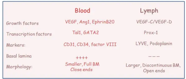
Figure 7: The growth factors VEGF-C and VEGF-D are the two known VEGF
homologues, which bind VEGF receptor-3 (VEGFR-3) on lymphatic endothelial cells (LECs) and promote endothelial cell proliferation and differentiation. An additional marker of lymphatic endothelial cells is the hyaluronan receptor LYVE-1. The transcription factor Prox1 has been identified as the master regulator of lymphatic endothelial cell (LEC)
differentiation and is uniquely express on LEC. After Prox1 induction, the early LECs upregulate expression of several lymphatic markers, as compared with surrounding blood vascular endothelium. The best-characterized lymphatic markers include LYVE-1, vascular endothelial growth factor receptor-3 and the cell surface glycoprotein podoplanin
1.7 Lymphatic Extracellular Matrix
The extracellular matrix forms the external layer of the lymphatic collector wall (Figure 8). Generally the adventitia, the other name for the extracellular matrix, plays several roles: it is implicated in the control of proliferation, differentiation, migration and cell-cell communication. The extracellular matrix comprises matrix proteins, such as collagens laminins, elastin, and glycosaminoglycans, as well as cell adhesion proteins including fibronectin, vitronectin and thrombospondins. The interactions at the matrix level occur through recognition sites present in the proteins and oligosaccharide sequences. The cells present on the outside of the lymphatic collectors interact with hyaluronan, discoidin domain receptors present in the collagen, and cell surface proteoglycans (12). In addition to the roles listed above, it appears that the interaction of the extracellular matrix component among nearby lymphatic vessels is important in the modulation of lymphangiogenesis.
Figure 8: The extracellular matrix is essential for support and communication. Many of the
proteins present can interact with solutes in the surrounding environment
1.8 Lymphatic endothelial cells glycocalyx
Both lymphatic and vascular endothelial cells secrete a complex mixture of glycoproteins called the glycocalyx (Figure 9 and 10). The glycocalyx serves several different functions, including cell-cell recognition, communication, and adhesion. It is also critical to the maintenance of tissue fluid homeostasis and fluid exchanges between the intra and extra-capillary milieu. Fluid, proteins and small molecules filter across the walls of the microvessels into the interstitium. These exchanges occur through the glycocalyx, a matrix of glycoproteins and glycosaminoglycans with a thickness of 50 to 500 nm present on the luminal surface of the endothelial cells extending over the intercellular clefts (13). The glycocalyx structure and organization has been shown to be similar in both fenestrated and continuous endothelial cells (13). Fluids, small molecules and proteins, with the exception of large macromolecules, are filtered through the glycocalyx, which acts as a molecular sieve of various porosity, influencing the filtration rate from the capillary lumen to the interstitium (14). Through this mechanism, plasma proteins contribute to the lymph proteome.
Much more is known about the blood endothelium. For instance, it has been demonstrated that the glycocalyx is involved in the regulation of osmotic pressure. Solutes and molecules leave or enter the blood vessels by filtering through the vascular endothelium. The monolayer of endothelial cells thus works as a selective permeable membrane for the passage of specific molecules. This role is essential for the regulation of blood composition and osmotic equilibrium. Recently, some studies have suggested a similar role for lymphatic endothelial cell-associated glycocalyx (14). It is likely that lymphatic endothelial cells, as previously shown for vascular endothelial cells, are able to selectively filter the lymph and the solutes through the glycocalyx, which act as a molecular sieve. However, very little is known about the LEC-associated glycocalyx.
For this reason, we decided to perform an ultrastructural and proteomic analysis of the LEC-associated glycocalyx in young and old lymphatic collectors to identify both the molecular composition of the glycocalyx and possible age-related differences.
Figure 9: Transmission electron microscopy of vascular endothelium glycocalyx (modified
from Daniel Chappell, 2009).
Figure 10: Interaction and communication of glycoproteins with the lymph flow inside the
lymphatic lumen (modified from http://matrixrepatterning.com/thescientist).
1.9 Lymphatic smooth muscles cells
The lymph flows and progresses through the lymphatic system by rhythmic and phasic contractions of the vessels while constrictions of lymphatic smooth muscles modulate lymph flow resistance (15). Lymphatic smooth muscle is regulated by physical and chemical stimuli (Figure 11). Lymphatic muscles cells are present only in lymphatic collectors and vessels; the amount and the organization of the muscle cells changes as they progress proximally (10). The distribution of the muscle cells along the lymphangions is interrupted at the level of the valves, where only endothelium and adventitia are present.
One peculiarity of these cells is their ability to spontaneously contract; similar to the heart pacemaker, these cells can spontaneously depolarize and contract.
The lymphatic muscle performs its functions under precise modulation by physical factors such as transmural pressure, luminal flow and shear stress. It is also modulated by chemical agents like neurotransmitters, circulating hormones or substances released by surroundings cells into the interstitial space.
The myosin heavy chain and the actin present in these cells are the same found in vascular, cardiac and visceral myocytes (9).
It is very likely that each action potential/phasic contraction is preceded by a membrane depolarization event. Indeed several ion channels, such as Na, Ca and K channels, which are important for the generation of the action potentials and smooth muscle contraction have been found on LEC (your paper). These include nonselective cation channels that allow Na+ and Ca2+ and K+ to permeate into smooth muscle cells.
Ion channels are the targets of many intracellular signaling pathways, which act through protein phosphorylation and dephosphorylation. Nearly every type of voltage-gated K+, Ca2+, and Na+ channels is regulated to some extent by phosphorylation of serine/threonine residues on intracellular domains of the channel proteins.
Growth factors, which act through receptor protein tyrosine kinases, regulate the long-term expression of ion channels but also have acute actions on channel activity. Several studies have already provided strong evidence that extracellular matrix proteins and other integrin ligands can modulate ion channels through nonreceptor protein tyrosine kinases.
Figure 11: Representation of a smooth muscle cell with the elements involved in the
generation of a contraction.
1.10 Extrinsic and intrinsic forces
The lymph flow progresses from one point to another by the action of two kind of forces: extrinsic and intrinsic. Surrounding tissues apply a pressure on the surface of the lymphatic collector wall by expanding and compressing. The compression causes a constriction toward the lymphatic lumen, which pushes the lymph forward. The contraction of muscle cells works in parallel with the action of external forces. When the lymph pressure increases the intrinsic lymph pump becomes subsequently activated. The working together, the action of extrinsic and intrinsic forces will propel the lymph forward.
1.11 Lymph Circulation under pathological condition.
Original studies by Smith et al. indicate a flow rate of 1-5 mL/h in both pre and post-nodal lymph under physiological conditions(16). However, lymph flux to the draining lymph nodes can change notably under pathological conditions, which can be associated with increased pro-inflammatory mediators that affect lymphatic contractility, increased lymphangiogenesis, increased cellular trafficking, as well as increased lymph volume and flux. Increased lymphangiogenesis has been observed in several pathological conditions including primary and secondary lymphedema, acute and chronic inflammation, and cancer. In all these conditions the increased lymphangiogenesis relates to an increased production of different vascular endothelial growth factors (VEGF-A, VEGF-C, VEGF-D, VEGFR-3) released by immune and stromal cells (17). Additionally, NFκB also up-regulates the transcription factor Prox1 that promotes lymphatic endothelial cell proliferation. At the moment it is still unclear whether the increased lymphangiogenesis observed under inflammatory conditions serves a beneficial role. While it decreases tissue edema and clears inflammatory cells and cytokines/chemokines, it also expands the
dissemination of pro-inflammatory cells and mediators. Less controversial is the role of lymphangiogenesis in cancer where the expansion of lymphatic vessels facilitates metastasis and is a strong negative prognostic factor (18).
1.12 FREE- RADICALS AND AGING
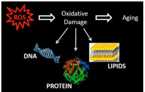
Aging is a multifactorial process driven by genetic, environmental and metabolic conditions. Metabolically, senescence is often associated with increased oxidative stress, protein and DNA carbonylation, glycation and lipoxidation and consequent alteration of immune cells; this leads to a chronic inflammatory process often referred as inflamm-aging (19). Over the last several decades many studies have brought to attention the role of metabolism in aging; in particular, the free-radicals theory of aging. The free-radical theory asserts that during aging there is an imbalance in the production and clearance of of reactive oxygen species. Free radicals are normally produced in cells as byproducts of cellular metabolism, however cellular antioxidant enzymes and molecules are able to reduce and control the ROS and RSN-mediated damage. Examples of the most common free radicals are: hydrogen (H•), nitric oxide (NO•), hydroxyl radical (•OH), peroxyl radicals (RO2), alkoxyl radicals (RO•), transition metals (copper and iron) or two unpaired electrons (the diatomic oxygen molecule O2, and its superoxide (O•−2) .
Electrons might escape from the mitochondrial electron transport chain. Complexes I and III are large multisubunit complexes in the mitochondrial chain that are
coupled with the extrusion of protons from the mitochondrial matrix into the intermembrane space. As a consequence, reactive oxygen species can be produced and in particular superoxide. Other enzymes independent of the respiratory chain may also contribute to the mitochondrial and cytosolic pool of ROS (α-ketoglutarate dehydrogenase, mitochondrial flavoenzyme pyruvate dehydrogenase, palmitoyl-carnitine and cytosolic glycerol-3-phosphate dehydrogenase).
At the end of the mitochondrial electron transport chain O•−2 is produced and then converted to hydrogen peroxide by the mitochondrial superoxide dismutases in a two-step chemical reaction. In the last step, H2O2 generates the highly reactive hydroxyl radical •OH
in the presence of metals such as Fe by means of a Fenton reaction. Three major scavenging enzymes, mitochondrial glutathione peroxidase, mitochondrial peroxiredoxin, and catalase, regulate the amount of free radicals produced to protect cells from oxidative damage (20).
Another source of ROS is the oxidative burst. Upon microbe invasion, neutrophils, macrophages, dendritic cells and monocytes release ROS to produce strong oxidizing agents that function as highly reactive microbicidal components (21-23). The glucose monophosphate catabolic pathway is activated when a pathogen binds to the plasma membrane. This host-pathogen interaction leads to conversion of O2, into its superoxide (O•−2), by the NADPH oxidase complex, which is then converted into H2O2 by the superoxide dismutase. Once superoxide dismutase has been converted to its reduced status, it binds superoxide anions and release H2O2. A Fenton reaction then converts H2O2 into reactive free radicals (•OH and H•). All of these molecules possess a strong microbicidal activity. During aging, an increase in the oxidative burst has been reported due to an
increased incidence of pathologies that present with chronic inflammation. With aging there is a reduced ability of antioxidants to lessen cellular damage and this leads
to cross-linking of molecular structures, such as proteins, carbohydrates, lipids and nucleic acids, with subsequent damage to macromolecular structures, such as DNA, lipids and larger proteins. Cross-linking between proteins results in the formation of multi-complex called aggregates which are known to lead to several disorders and diseases, including Alzheimer's and Parkinson's diseases. The majority of the protein carbonyls reported in aging are on the lysine, arginine, threonine and proline residues (24). Nitration occurs at tyrosine residues(25), while Cystein-s-thiolation has also been implicated as an aging – related post-translational protein modification (26, 27). ROS and RSN-associated protein oxidation can result in protein fragmentation, unfolding, aggregation and loss of function, altogether altering the cellular proteostasis.
Figure 12: Generation of reactive oxygen species can trigger a chain reaction with
consequent damage of cellular components; this phenomenon particularly occurs during aging (http://www.vai.org/en/vari/research-groups/lab-of-aging-and-neurodegenerative-disease/projects.aspx).
1.13 IMMUNOSENESCENCE
With the term Immunosenescence we refer to a gradual deterioration of the immune response that occurs during aging and leads to increased mortality among the elderly. An increased amount of free-radicals has been observed in cells of the immune system (28). Decreased cellular function and increased cellular apoptosis, upon accumulation of oxidized products, are two of the causes of immunosenescence. Oxidative post-translational modifications occurring on different biomolecules can compromise the function of several cellular components. For instance, post-translational modifications of lipids on the plasma membrane can decrease endosomal organelle formation, which is important for the assembly of signaling complexes. In addition, lipid modification affects fluid phase and receptor-mediated phagocytosis. Protein and lipid carbonylation then leads to the formation of indigestible carbonylated protein aggregates in endosomal compartments, which affect pathogen processing ability of antigen presenting cells (29). The proteasome degradative processes are also affected by aging causing further accumulation of oxidized proteins and decreased ability to correctly process cytosolic proteins (30). Oxidative stress also reduces cellular ability to process and present MHC class II restricted antigens. Once antigens are modified, there is an increased proteolysis of the immunogenic peptides, or
aggregation-prone modification that sequesters some of the epitopes, rendering them less available for MHC class II presentation (31). Another way oxidative stress affects the immune response is the glutathionylation occurring in T lymphocytes--several enzymes and ribosomal proteins undergo this modification. Furthermore, many signaling receptors present on the membrane of both T and B cells are carbonylated by aging-related oxidative stress (32). Ultimately, an oxidative stress consequence is self-antigen modification; oxidative stress can modify the structure of self antigens and is an important factor in the pathogenesis of autoimmunity. A second correlation between oxidative stress and immunosenescence is the induction of cellular apoptosis caused by accumulation of protein aggregates. Misfolded protein aggregates are found next to the proteasome, in tubular microelements, and in endosomal compartments. The accumulation of protein aggregates is due to phagocytosis of extracellular proteins and the accumulation of oxidatively damaged molecules through macroautophagy. Finally, the accumulation of modified proteins has been correlated to apoptotic cell death.
2. ABSTRACT
Lymphatic collectors are present in every parenchymal organ, except the brain. They carry the lymph and collect extracellular fluid from interstitial spaces. Interstitial spaces contain proteins extravasated from the blood capillaries and products of tissue metabolism, cellular secretion, cellular apoptosis, receptor editing and extracellular matrix degradation. By collecting extracellular fluid, lymphatic collectors control body fluid homeostasis. Importantly, pathogens and cancer cells are also drained by the lymphatic network to lymph nodes where they are destroyed. Functional defects in draining lymphatics may affect lymph transport and consequent pathogen clearance. Aged individuals have a compromised immune response to pathogens and increased tissue edema. Remarkably, only few studies have investigated age-related changes in lymphatic collectors and how these changes could lead to impairment in lymphatic function. Evidence suggests a possible correlation between oxidative stress, a typical hallmark of aging, and endothelial cell dysfunction and a decreased lymphatic vessel contraction. And although loss of smooth muscle has been observed in aged animals, it’s unclear how this might affect lymphatic function and an immune system response. Because the lymphatics are so important for fluid homeostasis and immune response, and because lymphatic function is compromised in aged individuals, herein we analyze how the aging process affects the dynamic and structural elements of lymph flow. Ultrastructural, biochemical and proteomic analysis indicated loss of matrix protein in aged lymph vessels, as well as an increase in protein oxidative modifications. This resulted in decrease of amplitude and frequency of lymphatic contractions with reduction of total productivity of lymph pump, as measured in vivo on lymph collectors. Functionally, this impairment translated into a reduced ability to transport bacteria to the draining lymph nodes, as determined by time-lapse microscopy. Ultrastructural analysis also indicated a decrease in the thickness of the endothelial cell glycocalyx. Redox proteomics determined an increase in glycation and carbonylation of endothelial cell glycocalyx structural proteins. The glycocalyx changes in structure resulted in increased vessel permeability. Altogether, our data provide a mechanistic analysis of how the anatomical and biochemical changes, as occurring in aged lymphatic vessels, compromise lymphatic transport.
The major function of the lymphatic vessels is to transport lymph. The role of lymph transport makes lymphatics pivotal to the movement of fluid, soluble molecules and immune cells from the interstitium of parenchymal organs to the draining lymph nodes. Networks of lymphatic vessels dispersed in interstitial spaces collect proteins and molecule ultrafiltrates from the blood capillaries, products of tissue metabolism/catabolism, tissue remodeling and injury, apoptotic cells, pathogens and serve as a conduit for immune cell homing to the lymph nodes. Thus, altogether the lymphatics play a major role in body fluid homeostasis, tissue proteostasis and innate and adaptive immune responses (6, 11, 14, 33-37).
The driving force that propels the lymph forward is generated by the active spontaneous contractions of the lymphatic muscle cells that surround the basal membrane and the endothelial cells of the lymphatic vessels (15, 38). The activity of the vessel’s intrinsic pump is crucial to lymph transport against an opposing net hydrostatic gradient (1, 15, 33, 38, 39). Additionally, a set of intraluminal valves positioned along the lymphatic vessels, which close in coordination with the lymphatic muscle contractile activity, prevent lymphatic backflow at the end of each contraction (39, 40, 41). The coordination between the pumping activity of the lymphangions, the sections of lymphatic vessels between adjacent valves (42), and the closing of the valves promote the unidirectional flow of the lymph from the parenchymal tissue to the draining lymph nodes (11, 15, 43).
An important component of lymphatic transport is the presence of a thick glycocalyx on the lumen side of the endothelial cells generated by a backbone of proteoglycans and glycoproteins (13). The proteoglycans are formed by a core protein bound to glycosaminoglycan side chains (13). The core protein can be anchored to the cell membrane (syndecans, glypicans) or secreted into the glycocalyx structure (mimecan, perlecan, and biglycan). The glycosaminoglycans are bound, as side chains, to the core protein and comprise heparan, chondroitin, dermatan and keratan sulfates and hyaluronic acid. Several more glycoproteins constitute the backbone of the glycocalyx including adhesion molecules; selectins, integrins and Ig superfamilies, and components of the coagulation and fibrinolysis cascade. Soluble proteins, such as albumin and orosomucoid, are also retained in the glycocalyx and are pivotal, together with the proteoglycans, in
maintaining its negative charge and selective permeability. The function of the glycocalyx is to aid lymphocyte rolling, prevent immune cells from adhering to the endothelium, generating a cytokine/chemokine gradient and balancing the exchanges between the lymphatic and the surrounding interstitium (13, 14).
The magnitude and the complexity of the many tasks performed by the lymphatics is highlighted by the extensive tissue edema and impaired immunity observed in mice and humans with decreased lymphangiogenesis (44) or surgical removal of draining lymph nodes (45). Many aspects of the lymphatic transport in physiological and pathological conditions have been elucidated but a number of important aspects of lymphatic functions have yet to be investigated. Several recent reports demonstrate that a consequence of the aging process is the loss of lymphatic muscle cells in the valvular zones of the lymphatic vessel wall(3) with signs of diminished lymphatic contractility and flow (46, 33, 1), which in part may be linked to increased oxidative stress (2). Indeed an increase in superoxide dismutase activity, lipid peroxidation and cellular superoxide and mitochondrial ROS was reported in aged lymphatic collectors (2) and oxygen-derived free radicals have been shown to inhibit lymphatic contractile function (47, 48).
However, the effect of aging-related changes on the movement of fluids, molecules and cells through the lymphatic vessels, or on the lymphatic permeability remains unknown. Knowledge of how the aging process affects the dynamics of lymph flow and the function of lymphatics is important to further our understanding of the pathogenesis of altered fluid homeostasis and microvascular hyper-permeability in aging and to elucidate some of the aspects of immunosenescence and altered response to pathogens.
In this study we provide an ultrastructural, biochemical and functional comparative analysis between adult and aged lymphatic vessels. The observed changes provide a mechanistic explanation for the development of tissue edema and the compromised response to pathogens often observed in aging (49, 50).
4.1 Decreased extracellular matrix and contractile proteins in aged
lymphatic collectors
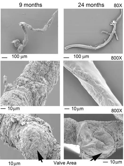
As a first step to determine how the aging process affects lymphatic vessels we performed ultrastructural analysis using scanning and transmission electron microscopy (SEM and TEM respectively). Mesenteric lymphatic vessels were isolated from 9- and 24-months old Fisher-344 rats. Scanning electron microscopic analysis of these tissues demonstrated substantial structural differences in the vessels isolated from adult and old rats (Figure 13). Examination of the adventitia, or external surface of the 9-months old collectors, revealed tight connective tissue surrounding the lymphatic (Figure 13). However, lymphatic collectors isolated from 24-months old rats exhibited tissue degeneration with a substantial loss of extracellular matrix (Figure 13). Additionally, visual analysis of vessels from 9-months old rats established that the collector’s valve area was surrounded by extracellular matrix, whereas a loss of extracellular matrix was observed in valves of collectors isolated from aged rats (Figure 13).
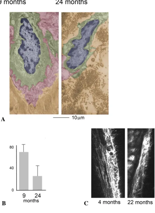
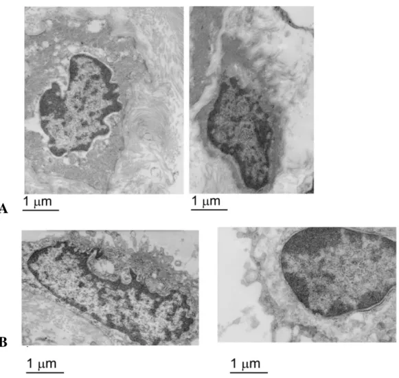
Ultrastructural examination of lymphatic vessel thin sections by TEM also revealed a marked decrease of the extracellular matrix associated with the lymphatic collectors, with more dispersed areas of collagen bundles, surrounding endothelial cells in aged samples as compared to the adult ones (Figure 14a,b). Decreased collagen surrounding the lymphatic collectors was also observed in 22 month old mice as compared to 4 month old mice, by intravital two-photon laser scanning microscopy (Figure 14c).
To confirm the observed structural differences among the two age groups, we performed a global proteomic analysis on the isolated rat mesenteric lymphatic vessels. The protein expression profiling was performed on three different sample preparations each consisting of two/three isolated vessels for each age group using one-dimensional SDS PAGE coupled with NanoLC-ESI-MS/MS analysis of Lys-C/tryptic/Glu-C/Asp-N peptides. The three independent biological replicates for each age group were analyzed by Scaffold analysis (version Scaffold 4.0.7, Proteome Software Inc., Portland, OR) and the label-free quantification of the protein expression in each biological sample was used to generate an un-clustered heat map (Figure 15).
Global proteomic profiling of the lymphatic vessels revealed statistically significant differences (p<0.05 and a False Discovery Rate of <0.4) in protein expression in the two age groups (Figure 13 and Table 1). Many muscle contractile and regulatory proteins, such as troponins, myosin, and their cytoskeleton associated proteins, such as actin, gelsolin, dynein and myosin binding proteins were at least two-fold lower in the proteome of 24-months old lymph collectors (Figure 13 and Table 1). Other proteins, including Na+, K+ and Ca++ channels, known to be involved in the generation of the muscle cell action potential, induction of depolarization and Ca++ release were also decreased in the aged lymphatic collectors (Figure 13 and Table 1). In addition, extracellular matrix proteins, including fibronectins, collagens, cartilage oligomeric protein, were also substantially down-regulated in the 24-month old lymph collectors (Figure 13 and Table 1). Finally, adrenergic receptors, and associated kinase, involved in the regulation of the lymphatic vessel contractility during stress-associated events were also affected by the aging process (Figure 13 and Table 1). Altogether, the down-regulation of several proteins associated with muscle contraction, generation of the action potential, Ca++ release and formation of basal membrane/extracellular matrix, in large degree parallel the differences observed in matrix and ultrastructural proteins between adult and aged lymphatic vessels (Figures 13-15 and Table 1).
Figure 13: Scanning electron microscopy of mesenteric lymphatic vessels isolated from 9
and 24 month old rats. Different magnifications show the reduced amount of extracellular matrix in old sample. The decrease was also observed in the valves area.
A
B
C
Figure 14: (A) Transmission electron microscopy of mesenteric lymphatic vessels isolated
from 9 and 24 month old rats. Disrupted collagens around endothelial cells were observed in old samples. Nucleus is colored in blue, cytoplasm in green, glycocalyx in red and basal membrane in brown. (B) Quantification of basal membrane thickness in adult versus old
lymphatics. Average and standard deviation from 15 separate measurements. ** p < 0.005. (C) Intravital multi-photon microscopy of collagen fibers surrounding lymphatic collector of 4 and 23 months old mice.
Figure 15: Heat map representation of the proteomic analysis performed on 9 and 24
month old rat mesenteric lymphatic vessels. The color-coded heat map shows proteins with the highest level of expression in red and the lowest abundance proteins in green, with intermediates shades for the rest of the expression levels. Data compile three independent biological replicates.
Table 1: List of proteins identified from samples harvested from both 9 and 24 month old
rats. We found proteins belonging to the following functional groups: lymphatic muscle contraction, extracellular matrix proteins, adrenergic receptors, membrane potential-related ion channel proteins, glycocalyx -associated proteins, ion channel related proteins and adherence molecules.
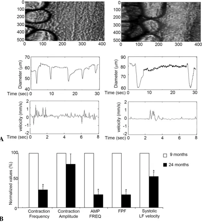
4.2 Decreased contractility and pumping efficiency in aged lymphatic
collectors
To analyze whether aging-associated changes in lymphatic ultrastructure and proteome composition could affect the vessel functionality, we analyzed the lymphatic contractile activity of mesenteric collectors in adult and aging rats. Using intravital microscopy we determined that, when compared to adult collectors, aged lymphatic vessels can still contract comparatively strong with around 20% decrease in contraction amplitude (Figure 16 a, b, Supplement movie 1a and 1b). However, the contraction frequency is drastically diminished; up to a 70% decrease is observed in aged collectors as compared to adult ones (Figure 16 a, b, Supplement movie 1a and 1b). These aging-associated changes in mesenteric lymphatic contractility led to a significant reduction in their pumping indices. Both the amplitude frequency product [AFP and the fractional pump flow (FPF)] were reduced to 24% of what measured in the adult mesenteric collectors. Consequently, systolic lymph flow velocity was also significantly diminished in aged collectors to a value corresponding to almost half of what observed in the adult collectors. Altogether, the in
vivo functional data support the notion that the aging process decreases the lymphatic
A
B
Figure 16: (A) In vivo analysis of mesenteric lymphatic vessels contractility. Microscopic
view and representative tracings of contractile activity of adult (9 months) and aged (24 months) rat mesenteric lymphatic vessels. (B) Aging-associated changes in vesselss contractile activity. AMP*FREQ Product - amplitude-frequency product, FPF – fractional pump flow, LF- lymph flow. Significant differences (P < 0.05) between active lymph pump parameters indicated by* - 9-mo versus 24-mo specimens.
4.3 Decreased glycocalyx and junctional proteins in aging lymphatic
collectors
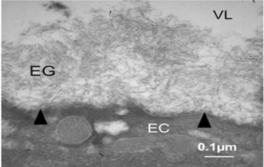
The glycocalyx is an important component of the endothelial barrier in lymphatic collectors. To examine possible age-related ultrastructural changes to the endothelial glycocalyx, lymphatic collectors were isolated from 9- and 24-months old rats, stained with Alcian blue and examined by transmission electron microscopy. The glycocalyx of endothelial cells from 9-months old rats was observed as an intact, continuous, electron-dense material covering the cell membrane (Figures 17a and 18a). On the other hand a significant loss of the glycocalyx, with reduction in size and continuity, was observed in lymphatic endothelial cells from 24-months old rats (Figures 17b,18b and c). Electron tomography and 3D modeling were employed to examine the glycocalyx of Alcian blue stained endothelial cells from 9-months old rats. The tomographic analysis confirmed a well demarcated glycocalyx of varying height spanning the luminal side of an endothelial cell in collectors from 9-months old rats (Figure 19).
Possible proteomic differences among glycocalyx proteins from 9 and 24 old lymphatic collectors were also explored. Glycoproteins were separated from the collector’s total proteome and run on a SDS-PAGE followed by in gel digestion and MS/MS analysis as described above.
Global proteomic profiling of the lymphatic vessels revealed statistically significant differences (p<0.05 and a False Discovery Rate of <0.4) in proteins expression between the two age groups (Figure 20 and Table 1). Cadherins, protocadherins, integrins and GAP junction proteins all showed a significant decrease (at least two fold, p<0.05) in collectors harvested from aging as compared to adult rats (Figures 3g, 3h). Additionally, complementing the ultrastructural analysis on the glycocalyx, proteomic analysis determined that several glycocalyx-associated proteins, including versicans, aggrecans, proteoglycans, brevican, galectins, mucins, agrin and clusterins were at least two-fold down-regulated in aged collectors as compared to adult ones (Figure 20 and Table 1).
Altogether, the down-regulation of several proteins associated with glycocalyx and GAP-junction support the aging-related changes observed by ultrastructural microscopy.
A
B
Figure 17: (A) Ultrastructural analysis of the lymphatic endothelial cell glycocalyx from 9
month old mesenteric lymphatic vessels. (B) Ultrastructural analysis of the lymphatic endothelial cell glycocalyx from 24 month old mesenteric lymphatic vessels
A
B
C
Figure 18: (A) Transmission electron micrograph and pseudocolored micrograph of the
lymphatic endothelial cell glycocalyx from a 9 month old rat 9; cytoplasm is colored in red and glycocalyx in green. (B) Transmission electron micrograph and pseudocolored micrograph of the lymphatic endothelial cell glycocalyx from a 24 month old rat; cytoplasm is colored in red and glycocalyx in green. Bar corresponds to 200 nm. (C) Quantification of glycocalyx thickness in adult versus old lymphatics. Average and standard deviation from 15 separate measurements. * p < 0.001
Figure 19: Tomographic reconstruction and 3D model of the lymphatic endothelial cell
glycocalyx in a 9 month old rat. 3D model view presents the glycocalyx coat in red, the cytosol in yellow and the nucleus in blue. Bar corresponds to 1 µm.
Figure 20: Heat map representation of the proteomic analysis performed on 9 and 24
month old rat mesenteric lymphatic vessels. The color-coded heat map shows proteins with the highest level of expression in red and the lowest abundance proteins in green, with intermediates shades for the rest of the expression levels. Data compile three independent biological replicates.
4.4 Increased oxidative stress in aged lymphatic collectors
Aging-related increases in oxidative stress have been associated with endothelial dysfunction and a diminution of mesenteric lymphatic vessel contractility(1, 2). To examine whether an aging-associated increase in oxidative stress would result in an elevation of the oxidatively modified proteins comprising the mesenteric lymphatic vessel, proteins were extracted from 9- and 24-months old rat lymphatic collectors and incubated with 2,4-dinitrophenylhydrazine (DNPH). DNPH specifically reacts with carbonylated proline, lysine, histidine, and arginine residues. Derivatized samples were separated by gradient SDS-PAGE and analyzed by western blotting using an anti-DNPH primary antibody. Protein carbonyls were detected in mesenteric lymphatic vessels isolated from 9- and 24-months old rats (Figure 21a). However, the accumulation of carbonylated proteins was significantly elevated in the lymphatic collectors from 24-month old rats (Figure 21a). To further probe into lymphatic endothelial protein oxidative damage, we isolated mesenteric lymphatic vessels from 9- and 24-months old rats and their extracted proteins were run on an SDS-PAGE and analyzed by one-dimensional liquid chromatography coupled with tandem mass spectrometry on a nanoLC/Orbitrap system. In 24-months old collectors the percentage of chemically modified proteins was significantly higher as compared to the adult samples (Figure 21b). The most frequent post-translational modifications (PTM) we identified were: monoxidation, pyrrolidinone, 4-hydroxynonenal (HNE), carboxymethyl, carboxethyl, glutamic semialdehyde (Glu SA) and 3-deoxyglucosone. Mono-oxidation on lysine, proline, methionine and arginine was found to be about 55% while the pyrrolidone and pyrrolidinone derivatives of proline were present in higher abundance (56.2% and 49.2% respectively). Arginine conversion to GluSa (57.3%) and 3-deoxyglucosone (26.3%) was another post-translational oxidative modification significantly increased. In addition, the lysine residues were observed to be significantly modified as 4 Hydroxynonenal (37.3%), carboxymethyl (29%) and carboxethyl (23.6%). In the 9-months old samples the percentages of previously stated modifications were generally lower (Figures 21b, 21c). Altogether the data support the presence of increased oxidative stress in aged lymphatic collectors.
Figure 21: (A) Western blot analysis of oxidatively modified (carbonylated) proteins
detected in MLV isolated form 9 and 24 month old rats. Lanes marked as ‘‘-’’ indicate non-derivatized proteins (control) and ‘‘+’’ indicate non-derivatized proteins. (B) Number (expressed in %) of proteins with post-translation oxidative modifications. (C) Examples of MS/MS mapping of oxidative modifications of amino acid side chains across the peptide sequences in MLV proteomes isolated from 24month-old mice shows Pyro-Glu,
Carboxymethyl, Pyrrolidone and HNE modifications in ATPase WRNIP1, Receptor-type tyrosine-protein phosphatase, Collagen Vα1 and Cubilin, respectively. Data compile three independent biological replicates.
4.5 Impaired pathogen clearance by aged lymphatic collectors.
Reduced innate and adaptive immune responses are a hallmarks of aging. Several reports have indicated how aging antigen presenting cells have a decreased ability to phagocytose and process pathogens and aging T cells have decreased proliferative responses (49, 50). However, so far, little is known about how the aging process affects bacterial clearance associated with lymphatic transport. To study whether aging-related anatomical, biochemical and functional differences would interfere with pathogens clearance we set up different functional assays using three different pathogens, S. aureus, C. neoformans and M. smegmatis. Pathogens (1x10⁸-1x10⁹ CFU) were injected in the footpad of 4- and 22-months old mice and draining lymphatics from the lower limbs, below the popliteal node, were collected at different time points (5 minutes, 20 minutes, 1 hour and 2 hour) (Figure 22a). Following tissue digestion with collagenase, free fluorescently-labeled pathogens transported by the lymphatic collectors were analyzed by flow cytometry and quantified (Figure 22b). Over the analyzed time-course more than 85% of the pathogens were present as free bacteria or fungi (Figure 22b). The total number of free pathogens was consistently and significantly higher in samples collected from aged mice, at each of the examined time points (Figure 23). C. neoformans and M. smegmatis were also quantified by colony forming units; again, the number of CFU was significantly higher in aging samples (Figure 24). To further analyze pathogen transport in the lymphatic collectors we performed intravital two-photon microscopy of lymph-carried pathogens (Supplement movies 2a, 2b, 2c). GFP-expressing S. aureus (1x10⁸ CFU) and a fluorescent tracer, (Tetramethylrhodamine isothiocyanate (TRITC)–Dextran or PEG-D680)(51), were injected in the lower limb footpads of 4 and 22-months old mice to visualize the transit of the bacteria through the
collectors in the legs. In both groups, we could visualize transit of the bacteria through the collectors in the leg (Supplement movies 2a, 2b, 2). In separate experiments, mice were injected with bacteria one hour prior to surgery, to allow transport through the collectors, and subsequently imaged. In both groups of mice, bacteria could be sporadically found within the vessel space (Figure 25a). In young mice, there were occasionally bacteria found in the fat pad surrounding the collectors, however, in aged mice, we observed 100 times more bacteria accumulated in the fat pad and along the outside of the vessel (Figure 25b). These bacteria-rich regions were unevenly distributed within the fat, suggesting that the lymphatic leakage from the vessels into the surrounding interstitium, was more predominant in selective areas of the lymphangions.
A
B
Figure 22: (A) Mice lymphatic collector labeled with Evans Blue. (B) FACS analysis of C.
neoformans present in lymphatic collectors, as free bacteria or phagocytosed by CD11c+/CD11b+ cells.
Figure 23: Quantification of C. neoformans and S. aureus, present at different time points,
in the lymphatic collectors following injection in the hind limb food-pad of 4 and 22 months old mice. **p < 0.01; ***p < 0.001; **** p < 0.0001.
Figure 24:Amount of C. neoformans (above) and M. smegmatis( below), as measured by colony forming unit, present in the same sample as in figure 23.
A
B
C
Figure 25:
(
A)
Representative (xy and yz) images of lymphatic collectors in the hind leg taken 1 hour after footpad injection with a TRITC-dextran sinus marker (red) and S. Aureus (cyan) bacteria in the footpad from 4 month and 22 month-old mice. (B) Density of bacteria in the surrounding footpad was quantified in both groups, and compared by Mann-Whitney test in 4 or more mice per condition, taken from 3 experiments. (C) Bacteria highlighted with yellow arrows.To confirm these data, 4 and 22 months old mice were injected in the lower limb footpad one hour prior to surgery, after which popliteal lymph nodes were isolated and imaged to
analyze bacteria distribution. We observed 3 times more bacteria accumulated in the lymph nodes of 4 month old mice as compared to those of the 22 month old mice. This further suggests a reduced ability of lymphatic collectors to drain bacteria to lymph nodes in old mice (Figure 26).
Figure 26: Imaging of bacteria distribution in isolated popliteal lymph nodes from 4 and 22
months old mice. Analysis of the ratio between bacteria and lymph nodes volume was performed; 3 times more bacteria were found in young mice. Representative images of 3 different experiments.
4.6 Impaired permeability in aged lymphatic collectors.
Since time-lapse microscopy clearly indicated that bacteria could easily exit from aged lymphatic collectors and since structural and proteomic differences were observed in the aging glycocalyx, we set out to further analyze the lymphatic collector’s permeability. To this end, Evans Blue dye was injected in the footpad of hind limb of 4- and 23-months old mice. After 20 minutes, mice were perfused via the left ventricle with PBS, and the collecting lymphatic vessels of mice were imaged under intravital microscopy. In young mice, the dye was mostly confined to the collectors (Figure 27), while in the aged mice, the Evans Blue was present both in the collectors and in the surrounding tissue (Figure 27). Similar results were obtained on isolated, cannulated and pressurized rat mesenteric lymphatic vessels under transmural pressure of 5 cm H2O. Bacteria were injected to the tubing connected to the input end of the collector and their transport visualized by epi-fluorescent microscopy (Figure 28). In adult collectors the bacteria were observed moving along the vessel and often can be visible close to the lymphatic valve, before its opening which propelled them downstream (Figure 28). On the other hand in aged collectors the bacteria were mostly seen lingering close to the vessel wall (Figure 28) and often visually observed exiting the lumen through the lymphatic wall.
To recapitulate the experiment on aging collectors a Transwell plate assay was performed to assess permeability of human lymphatic endothelial cells, after treatment with Paraquat, which is known to induce aging-like oxidative stress. Lymphatic endothelial cells were grown to confluence on Transwell filters, and FITC-dextran was added to the upper Transwell chambers. The integrity of the endothelial cells monolayer was subsequently determined spectrophotometrically by measuring the concentration of FITC-dextran that had diffused into the bottom well over 15 minutes. In this setup, overnight treatment with paraquat (20 nM ) significantly increased lymphatic endothelial cells permeability (Figure 29). Notably, this effect was comparable to the one induced by 20 minutes of treatment with vascular endothelial growth factor (VEGF)-A (20 nM), a well-known inducer of vascular permeability (Figure 29) (52).
Figure 27: Evans Blue distribution in the calf lymphatic collectors after lower limb
foot-pad injection in 4 month old mice and 22 month old mice. Representative image from 4 independent experiments.
Figure 28: Representative images of 9-month (k) and 24-months (l) isolated and
cannulated segments of mesenteric lymphatic vessels under conditions of perfusion of bacteria-containing solution
Figure 29: Quantification of FITC-dextran molecules passing through a monolayer of
lymphatic endothelial grown to confluence on transwell filters. A FITC-dextran (70 kDa) solution was added to the upper chambers, and the concentration of fluorescence (i.e. absorbance) in the lower chamber was measured 15 minutes later using a spectrophotometer. VEGF-A (20nM) as well as paraquat (20 nM) significantly enhanced LEC permeability over control levels. Data from one representative out of three similar experiments are shown. **p < 0.01; ***p < 0.001; **** p < 0.0001.