I
Scuola Superiore Sant’Anna
di Studi Universitari e di Perfezionamento
Dissertation for the Degree of Doctor of Philosophy in Biorobotics
Smart nanomaterials
to overcome highly invasive tumor resistance
Tutor
Prof. Gianni Ciofani
Supervisor
Prof. Paolo Dario
Ph.D. Candidate
Agostina Francesca Grillone
1
Index
Abstract 4
Chapter 1
6Introduction 6
Outline of the thesis 9
References 10
Chapter 2
112.1 Introduction 11
2.2 Magnetic fluid hyperthermia 12
2.3 Magnetic drug delivery 13
2.4 Control of cell functions 15
2.5 Conclusion 16
References 17
Chapter 3
193.1 Introduction 19
3.2 Experimental section 22
3.2.1 Solid lipid nanoparticles preparation and characterization 22
3.2.2 Magnetic and relaxometric characterizations 25
3.2.3 Cell cultures and in vitro cytotoxicity study 25
3.2.4 Western blotting analysis 26
3.2.5 Cellular uptake investigation 27
3.2.6 Dynamic cell culture and magnetic targeting experiment 27
3.2.7 Statistical analysis 29
3.3 Results 29
3.3.1 Sorafenib-loaded magnetic solid lipid nanoparticles 29
2
3.3.3 Cellular uptake studies and magnetic drug targeting 39
3.4 Discussion 42
References 44
Chapter 4
474.1 Introduction 47
4.2 Experimental section 49
4.2.1 Solid lipid nanoparticle preparation and characterization 49 4.2.2 Cell cultures and in vitro cytotoxicity study 50
4.2.3 Cellular uptake investigation 50
4.2.4 In vitro bbb model 51
4.2.5 Transendothelial electrical resistance and permeability assays 51
4.2.6 Immunostaining of bbb model 52
4.2.7 Statistical analysis 52
4.3 Results 52
4.3.1 Nutlin-loaded magnetic solid lipid nanoparticles 52 4.3.2 In vitro cytotoxicity study and cellular uptake studies 54
4.3.3 Characterization of bbb 56 4.4 Discussion 58 References 60
Chapter 5
61 5.1 Introduction 61 5.2 Experimental section 635.2.1 Preparation of lipid mixture and liposomes 63
5.2.2 Liposome characterization 63
3
5.2.4 Cellular uptake and investigation 64
5.2.5 Intracellular antioxidant activity evaluation 65
5.2.6 Statistical analyses 65
5.3 Results 66
5.3.1 Nanoceria-loaded liposomes 66
5.3.2 In vitro study and cellular uptake studies 67
5.4 Discussion 70
References 72
Conclusions 74
4
Abstract
The aim of this PhD Thesis has been the investigation of nanotechnological solutions for cancer therapy. In particular, hybrid nanomaterials composed by both inorganic and organic components have been synthesized, characterized, and in vitro studied, demonstrating their potentialities for biomedical application.
In the first part of the thesis magnetic solid lipid nanoparticles (Mag-SLNs) have been proposed as nanocarriers for the targeted delivery of sorafenib, a chemotherapeutic agent exploited for the treatment of hepatocarcinoma, and nutlin-3a, a potent non-genotoxic drug candidate for the cure of the glioblastoma multiforme.
Concerning Mag-SLNs as nanovectors for sorafenib some promising results have been obtained. Developed nanoparticles showed to inhibit the proliferation of cancer cells due to cytotoxic action of the encapsulated drug, and to localize this effect in a desired area thanks to possibility of controlling their movement/targeting through ax external magnetic field. Therefore, such characteristic makes Mag-SLNs extremely attractive, because it would allow to overcome the problem of lack of selectivity of traditional chemotherapy. A preliminary investigation about relaxivity properties, moreover, demonstrated that Mag-SLNs present superior features with respect to other commercially-available system, and therefore can be exploited as negative contrast agents in diagnostic imaging.
About Mag-SLNs for encapsulation of nutlin-3a, in vitro studies clearly showed superior antitumor activity of drug-loaded nanoparticles respect to free drug. Obtained results demonstrated that such nanovectors represent an ideal system to optimize the delivery of the drug into tumor cells, thus enhancing therapeutic index of nutlin-3a. A blood-brain-barrier in vitro model was also developed to allow future studies of nanoparticle crossing of the barrier and of tumor cells targeting, at level of which the action of the drug will be potentiated by magnetic nanoparticles-mediated hyperthermia.
In the final part, we will propose the use of a liposomal drug delivery system for the encapsulation of cerium oxide nanoparticles (nanoceria), a powerful nanotechnological tool for the regulation of the free radical level in the biological environment, and thus extremely useful in all those cases where oxidative stress play a key role, including
5
cancer. Nanoceria-loaded liposomes resulted stable, biocompatible, and they retain strong antioxidant activities, resulting a promising platform for the targeted delivery of the nanoparticles.
6
Chapter 1
Introduction
According to the World Health Organization, cancer is the second leading cause of death globally, and was responsible for 8.8 million deaths in 2015 [1]. Cancer is a general term used to describe a group of diseases characterized by abnormal mass of tissue, which grows in excess and in uncoordinated way with respect to other tissues [2].
Cancer development, or tumorigenesis, is a multistep process which starts with the accumulation of mutations in specific genes involved in the cellular growth, in the differentiation and in the programmed cell death [2]. After such genetic alterations, cells gradually become malignant, they grow and divide in an uncontrolled manner [3]. Moreover, through the secretion of a small subset of growth factors cells induce the development of new blood vessels from pre-existing ones, a process known as angiogenesis [3]. Due to the rapid tumor growth, the final result of angiogenetic process is an abnormal vascular network with functional defects, large fenestrations among the endothelial cells, hyperpermeability, and with a density vascular that tends to decrease in the central regions, thus leading to necrotic areas lacking of oxygen [3]. Because of insufficient blood perfusion, hypoxia, inflammation, and excessive production of acidic metabolites produced by cancer cells [4], the tumor microenvironment becomes acidic influencing the malignancy and the development of the tumor itself [5]. Exposure to chronic acidosis in fact induces chromosomal instability, clastogenicity, gene mutation, and accelerates the tumor invasion into the surrounding tissue through the secretion of metalloproteinases, enzymes involved in the degradation of the extracellular matrix [6]. In addition, secretion of various factors such as cytokines and inflammatory mediators by tumor cells forces the inactivation of tumor-infiltrating immune cells, such as antitumor lymphocytes and natural killer cells, towards an immunosuppressive state thus allowing tumor cells to elude immune response [7].
7
Tumor microenvironment is also characterized by the presence of cancer stem cells (CSCs), a small group of cancer cells different from the other ones, with higher aggressiveness and tumorigenic potential, capable to induce new tumors when injected into animal models [8]. Because of their genetic instability, CSCs are more likely to mutate and acquire many properties closely associated to tumor drug resistance, such as drug efflux transporter, detoxifing enzymes, quiescence, and DNA repair ability [9].
Definitely, tumor cells, in order to satisfy their needs of nutrition and survival, affect the organization and the functions of many distinct cell type present within stroma and of the extracellular matrix [10]. Such process induces the formation of a dynamic and heterogeneous tissue capable to support the tumor development and progression until the insurgence of the metastasis, the spread of the cancerous cell from the point of origin to other body parts through the vascular and lymphatic systems [10].
Although current radio and chemotherapies remain the first choice treatments for many tumors, a better understanding of the tumorigenesis and of the pathophysiological mechanisms has driven scientific community to design alternative anti-tumor strategies. The latter should be capable to exploit the characteristics of the cancer cells and of their microenvironment to obtain more targeted and effective therapies. One discipline using such approach is nanomedicine, which exploits devices on nanometer scales for diagnosis, prevention, and treatment of many different diseases [11]. More specifically, the basic rationale is based on the exploitation of nanoparticles as carriers to carry and direct drugs towards specific tissues, by designing the characteristics of the vectors on the basis of molecular, biochemical, and metabolic differences between tumor and healthy tissues [12]. Such approach allows encapsulated drug to avoid normal tissue, and to be selectively delivered to tumors, achieving a cytotoxic concentration higher in the tumor and a reduced toxicity for the rest of the body [13].
In order to achieve an efficient drug delivery system the optimization of the loading and release of the drug is needed, by choosing appropriate carriers on the base of the chemical characteristics of chemotherapeutic agent. Nanoparticles can be made from a huge variety of materials and can be classified into liposomes, polymeric
8
nanoparticles, micelles, dendrimers, and solid lipid nanoparticles [14], among others. Through the control of the nanoparticle size is instead possible to obtain drug delivery systems incapable to leave the vasculature in healthy tissue but able to passively across the large endothelial fenestrations of the tumor capillaries, where the poor drainage induces their accumulation. Such process, denominated enhanced permeability and retention effect, enhances extravasation and prolongs the retention of the encapsulated drug at tumor site, leading to a passive targeting of the tumor cells and of the cells of the stromal compartment [15].
Nanovectors can be also developed to increase intracellular drug release in response to physical, chemical, or biological stimuli. Through the incorporation into nanoparticles of sensitive moieties that respond to variations of pH, redox agents, ionic strength, oxygen levels and of shear stress in the tumor tissue, it is possible trigger physical/chemical modifications of the nanovectors, thus tuning the drug release [16]. Stimuli can be also applied externally of the body, by loading into nanovectors inorganic nanoparticles able to responde to physical stimuli, such as temperature, light, ultrasound, magnetic forces and electric fields [17].
Inorganic nanoparticles represent an important class of nanostructures in the field of nanomedicine. Generally made of metal, metal oxide, semiconductor, rare earth minerals and silica, such nanoparticles present interesting electric, magnetic, optical and plasmonic properties at the nanometer scale [14]. Thanks to their intrinsic quantum mechanical properties, several types of inorganic nanoparticles can be used as contrast agents for different imaging modalities, for the targeted delivery of drugs, and for thermal therapies simultaneously [14]. Their multifunctional nature allows to develop theranostic platforms capable to combine diagnosis and therapeutic treatment, and therefore to monitor the response to therapy in an unique single nanodevice [18].
9
Outline of the Thesis
This thesis presents the results obtained during the Ph.D. program in BioRobotics at Scuola Superiore Sant’Anna, Pisa (November 3th
, 2013 - April 30th, 2017) carried out at the Center for Micro-BioRobotics of the Istituto Italiano di Tecnologia. The present research was aimed at developing nanoparticle systems for tumor therapy. After this short introduction, the second chapter briefly presents the fundamental concepts of the nanomagnetism, illustrating the most important biomedical applications of the magnetic nanoparticles. In the third and fourth chapter the synthesis, the characterization, and in vitro experiments of magnetic solid lipid nanoparticles for encapsulation of sorafenib and nutlin-3a is presented, respectively, that are two chemotherapeutic agents used against hepatocarcinoma and glioblastoma multiforme. Finally, the fifth chapter illustrates the development of a liposomal drug delivery system for cerium oxide nanoparticles, an important nanomaterial with a strong antitumor potential.
10
References
[1] Y. Igarashi, H. Utsumi, H. Chiba, Y. Yamada-Sasamori, H. Tobioka, Y. Kamimura, K. Furuuchi, Y. Kokai, T. Nakagawa, M. Mori, N. Sawada, Biochem. Biophys. Res. Commun. 1999, 261, 108.
[2] P. C. Nowell, Science 1976, 194, 23.
[3] G. A. Grant, N. J. Abbott, D. Janigro, News Physiol. Sci. 1998, 13, 287.
[4] M. G. Vander Heiden, L. C. Cantley, C. B. Thompson, Science 2009, 324, 1029. [5] M. Wang, J. Zhao, L. Zhang, F. Wei, Y. Lian, Y. Wu, Z. Gong, S. Zhang, J. Zhou,
K. Cao, X. Li, W. Xiong, G. Li, Z. Zeng, C. Guo, J. Cancer 2017, 8, 761. [6] C. R. Justus, L. Dong, L. V. Yang, Front. Physiol. 2013, 4 DEC, 1.
[7] H. Dewitte, R. Verbeke, K. Breckpot, S. C. De Smedt, I. Lentacker, Nano Today 2014, 9, 743.
[8] L. Persano, E. Rampazzo, G. Basso, G. Viola, Biochem. Pharmacol. 2013, 85, 612. [9] S. Vinogradov, X. Wei, 2012, 7, 597.
[10] A. Manuscript, E. Paradigms, 2012, 147, 275.
[11] L. Zuo, W. Wei, M. Morris, J. Wei, M. Gorbounov, C. Wei, Med. Clin. North Am. 2007, 91, 845.
[12] A. Wicki, D. Witzigmann, V. Balasubramanian, J. Huwyler, J. Control. Release 2015, 200, 138.
[13] E. Pérez-Herrero, A. Fernández-Medarde, Eur. J. Pharm. Biopharm. 2015, 93, 52. [14] G. Bao, S. Mitragotri, S. Tong, Annu. Rev. Biomed. Eng. 2013, 15, 253.
[15] A. K. Iyer, G. Khaled, J. Fang, H. Maeda, Drug Discov. Today 2006, 11, 812.
[16] S. Ganta, H. Devalapally, A. Shahiwala, M. Amiji, J. Control. Release 2008, 126, 187.
[17] G. G. Genchi, A. Marino, A. Grillone, I. Pezzini, G. Ciofani, Adv. Healthc. Mater. 2017, 1700002.
11
Chapter 2
Magnetic Nanoparticles
2.1 Introduction
Since many years, magnetic nanoparticles have nurtured and largely met the expectations of revolutionizing current diagnostic and therapeutic techniques by featuring the ability of responding to and of being manipulated by external magnetic fields. This characteristic has made them very useful for a wide variety of applications both in vitro and in vivo, including magnetic resonance imaging, tissue repair, immunoassay, drug delivery, detoxification of biological fluids, cell separation, and hyperthermia [1].
Super-paramagnetism is the property at the base of many in vivo applications of magnetic nanoparticles. It is an important size-dependent phenomenon, according to which particles with size around 10 nm become magnetized up to their saturation level only under the influence of an external magnetic field, and lose their magnetization once the magnetic field is removed [2]. Nanoparticles of enough small size in fact act like a single magnetic domain, and in the presence of an external magnetic field they tend to align along its direction, thus generating a strong attractive interaction. Once the external magnetic field is removed, the domains cease to be aligned with the field, thermal agitation cancels residual magnetization, and the nanoparticles return to be inactive: as a result, nanoparticles do not interact magnetically among each other, and do not undergo aggregation [3].
The behavior of magnetic materials upon the application of a magnetic field is generally described by the hysteresis loop on the B (magnetization) against H (applied field) graph, through two principal parameters: the remanence (the value of the magnetization remaining when the applied magnetic field returns to zero) and the coercivity (the magnitude of the field that must be applied to cancel the magnetization of the material). In the case of super-paramagnetic nanoparticles, both parameters are zero and thus hysteresis is absent [3].
12
In a biomedical context, the super-paramagnetic behavior offers the double advantage of magnetically influencing nanoparticle movement in a flow (as the blood stream) in order to accumulate them to a target tissue, while preventing, at the same time, the formation of dangerous aggregates due to magnetic interactions once the magnetic field is removed [4]. Due to their demonstrated optimal biocompatibility (key requirement for nanomedicine applications), nanoparticles of iron oxides are widely used, especially magnetite (Fe3O4) and maghemite (Fe2O3) [5].
2.2 Magnetic fluid hyperthermia
A promising example of in vivo applications of magnetic nanoparticles is represented by magnetic fluid hyperthermia (MFH), a form of localized heating up to 42–46 °C generated by magnetic nanoparticles during their exposure to an alternating magnetic field of sufficient intensity and adequate frequency [6]. Such technique exploits the property of magnetic nanomaterials to absorb the applied electromagnetic energy and to convert it into heat through Néel and Brownian relaxation, as well as through frictional and hysteresis losses. In the clinical practice, hyperthermia is a promising therapeutic approach, in particular for the treatment of tumors, as they have an aberrant microenvironment which hinders heat dissipation, thus rendering cancer cells more sensitive to temperature increments [7]. When the temperature reaches values above 42 °C, morphological and functional alterations of the cytoplasm, of the plasma membrane, and of the nucleus are triggered, leading to cell apoptosis or necrosis [8].
Traditional techniques for local hyperthermia treatments are for instance based on radio frequency/microwave radiations, and on infrared light, but they mostly suffer from poor spatial resolution. By exploiting magnetic nanoparticles, MFH instead offers a number of advantages which make it a possible elective technique for tumor treatment. First, MFH enables tissue-specificity of the treatment and a more precise control of the heat dose, since heat is directly generated within the tumor [9]. Magnetic nanoparticles are in fact exposed to an alternating magnetic field for heat generation after their delivery to the desired site, either systemically (through a suitable targeting strategy), or by direct in situ injection, thus limiting the increment of temperature just to the desired area of action [9]. Selective accumulation of nanoparticles in the tumor mass is facilitated by the “enhanced permeability and retention” (EPR) effect, a typical phenomenon of tumor neovascularization. The presence of blood vessels presenting poor endothelial organization
13
allows the extravasation of small nanoparticles toward tumor interstitium; on the other side, their retention is favored by a poor lymphatic drainage typical of cancer tissues [10]. Moreover, nanoparticle cellular uptake can be increased by surface modifications with specific antibodies, able to recognize receptors typically overexpressed by tumor cells, thus reducing a specific internalization in healthy tissues [11]. Finally, the remote control of magnetic nanoparticles by an externally applied magnetic field and the possibility to track magnetic nanomaterials with noninvasive techniques such as magnetic resonance imaging (MRI) render MFH a noninvasive theranostic approach [12].
Many in vitro and in vivo studies have been carried out since Gilchrist et al. [13] presented their first experimental works on MFH, and recently magnetic nanoparticles have also successfully been tested in the first clinical trials aiming at demonstrating safety and efficacy of prostate cancer and glioma treatment [14]. Nevertheless, many limits still prevent such method to become a standard medical procedure. First of all, significant limitations are inherent to the parameters of the applied magnetic fields (i.e., frequency and intensity). It is very important to work in the right range of values in order to prevent any adverse physiological reactions, including undesired muscular and cardiac stimulations, while ensuring enough heating [15]. Other significant challenges are related to the synthesis of magnetic nanoparticles with high specific absorption rate (a physical parameter that describes the conversion efficiency of electromagnetic energy into thermal energy under an alternating magnetic field) in order to reach suitable temperatures at a low dosage of magnetic nanoparticles [16]. Even if MFH offers the advantage of a direct contact between the heat source and tumor cells, the identification of a safe and efficient method for nanoparticle delivery to the tumor area and for the control of their intratumoral distribution remains another big challenge. Tumor morphology and functioning in fact cause heterogeneous intratumoral distribution of the nanoparticles, thus preventing a uniform exposition to high temperatures [14].
2.3 Magnetic drug delivery
Magnetic nanoparticles also represent an excellent platform for drug delivery purposes [17]. Generally, therapeutic agents (such as drugs, radionuclides, nucleic acids, etc.) are linked to the surface of magnetic nanoparticles, or they are encapsulated within magnetic nanocarriers, and undergo an attractive force exerted by an external magnetic field, specifically acting on the disease area. Due to a preferential accumulation of the magnetic
14
nanoparticles in the area exposed to the static magnetic field, the cytotoxic effects of the cargo are mostly limited to the diseased tissues, with a consequent reduction of side effects [18]. Magnetic targeting is thus a promising approach that offers the possibility to actively control drug distribution in vivo (a goal that can hardly be achieved with traditional strategies), and to modulate the pharmacokinetics and pharmacodynamics of a drug by enhancing its therapeutic index [19]. Many aspects and parameters have to be carefully evaluated in the design of magnetic drug delivery systems. Among these, there are physical/chemical features such as size, surface charge, and surface chemistry. The latter often needs modification through organic ligands for nanoparticles to escape from the action of the reticuloendothelial system and of the immune system, and thus to exhibit a prolonged circulation lifespan in vivo [20]. Drug delivery optimization can additionally be provided by tuning of nanoparticle magnetization during synthesis process, and by controlling strength and penetration depth of the applied magnetic gradients. Despite the fact that magnetic targeting is a highly promising strategy, currently, the magnetic field strength of commercially available devices drops rapidly from the source as distance increases, and just a penetration depth of a few millimeters is allowed, rendering difficult the targeting of deep sites in the body [21].
After delivery at the correct site of action, drug release can be achieved by many mechanisms, and it usually occurs by enzymatic activity or physiological changes of pH, osmolarity, and temperature [22]. Very interestingly, drug release can also be remotely triggered from drug-containing magnetic particles, by exploiting the ability of magnetic nanovectors of acting as transducers, and, more specifically, of capturing external electromagnetic energy and converting it into thermal energy. This mechanism is defined magnetothermally triggered drug release, and it combines magnetic nanostructures with thermoresponsive materials [23]. After the application of an external magnetic field, the heat produced by the magnetic nanoparticles triggers crevices or cracks within a thermally responsive polymeric matrix, thereby inducing the release of the encapsulated drugs. This mechanism has been proposed for drugs embedded in magnetic nanovectors. In the case of drugs directly linked to the surface of magnetic nanoparticles, heating can be used for bond breaking between drug and nanoparticles [24]. Although a significant progress in the field of magnetic targeting and release occurred in the past decade (especially in the design of magnetic nanovectors), most of the studies were conducted on cell cultures and/or small animals, and human clinical trials remain very few to date. In vivo real-time imaging of magnetic nanocarriers, rigorous safety and toxicology testing in more accurate in vivo
15
models, and magnetic field steering optimization by external magnets are some of the main limitations to be overcome before obtaining realistic applications of magnetically targeted drug therapy [21].
2.4 Control of cell functions
Delivery of magnetic nanoparticles to precise subcellular structures also represents an important tool for studying and modulating cell functions and molecular pathways. Thanks to advanced synthesis and characterization techniques, magnetic nanoparticles with suitable size were obtained, and their capability to be internalized by cells and to interact with small molecules such as enzymes and membrane receptors was demonstrated [25]. By binding these nanovectors to the surface of specific cells or to inner cell compartments, the investigation and modulation of the molecular mechanisms of certain biological activities, and even the control of specific cell functions by means of magnetically induced stimuli, are enabled [26].
Recently, Tay et al. used this new actuation mechanism for the manipulation of Ca2+ levels in cortical neuronal networks in vitro, [27] demonstrating that magnetic forces can increase the release of Ca2+ from mechanosensitive ion channels in neurons previously treated with magnetic nanoparticles. Furthermore, their approach allowed a more accurate time-space control of the stimulus with respect to other stimulation methods, for instance based on ultrasounds or optogenetics. The exploitation of magnetic nanoparticles for achieving remote manipulation of specific ion channels in neurons was also investigated by Huang et al. [28]. More in detail, authors demonstrated that the heating triggered by magnetic nanoparticles through a radio-frequency magnetic field allows the opening of TRPV1, a temperature-sensitive ion channel, and determines an influx of calcium ions. Thermal activation of these channels triggers action potentials in cultured neurons without observable toxic effects [28].
Although magnetic actuation still presents several difficulties and needs of further refinements for the study of cell mechanics and function, it paved the way to a number of important nanomedicine applications, including gene therapy, tissue engineering, and stem cell differentiation [26].
16
2.5 Conclusion
Concluding this overview on the biomedical applications of magnetic nanoparticles, we would like to stress the extraordinary potential of these nanomaterials, thanks to the possibility of their remote and noninvasive control, jointly to satisfactory biocompatibility, colloidal stability, and surface engineering capability, which render them exploitable for a wide range of in vivo biomedical applications [29]. Compared to other nanomaterials, magnetic nanoparticles exhibit the highly attractive feature of multifunctionality (Figure 2.1), that renders them actual “theranostic” platforms [30].
Figure 2.1 Biomedical multifunctionality of magnetic nanoparticles. Reproduced with permission [23]. Copyright 2011, Elsevier.
17
References
[1] L. Hajba, A. Guttman, Biotechnol. Adv. 2016, 34, 354. [2] W. Wu, C. Z. Jiang, V. A. Roy, Nanoscale 2016, 8, 1942.
[3] A. Akbarzadeh, M. Samiei, S. Davaran, Nanoscale Res. Lett. 2012, 7, 144.
[4] P. Tartaj, M. del Puerto Morales, S. Veintemillas-Verdaguer, T. Gonzalez-Carreno, C. J. Serna, J. Phys. D: Appl. Phys. 2003, 36, 182.
[5] J. Beik, Z. Abed, F. S. Ghoreishi, S. Hosseini-Nami, S. Mehrzadi, A. Shakeri-Zadeh, S. K. Kamrava, J. Controlled Release 2016, 235, 205.
[6] C. L. Dennis, R. Ivkov, Int. J. Hyperthermia 2013, 29, 715.
[7] M. Furuya, Y. Yonemitsu, Curr. Cancer Drug Targets 2008, 8, 253. [8] J. Overgaard, Cancer 1977, 39, 2637.
[9] B. Thiesen, A. Jordan, Int. J. Hyperthermia 2008, 24, 467. [10] H. Maeda, Adv. Enzyme Regul. 2001, 41, 207.
[11] R. Bazak, M. Houri, S. El Achy, S. Kamel, T. Refaat, J. Cancer Res. Clin. Oncol. 2015, 141, 769.
[12] J. Estelrich, M. J. Sánchez-Martín, M. A. Busquets, Int. J. Nanomed. 2015, 10, 1727.
[13] R. K. Gilchrist, R. Medal, W. D. Shorey, R. C. Hanselman, J. C. Parrott, C. B. Taylo, Ann. Surg. 1957, 46, 596.
[14] I. Hilger, Int. J. Hyperthermia 2013, 29, 828.
[15] S. Dutz, R. Hergt, Nanotechnology 2014, 25, 452001.
[16] P. Guardia, R. Di Corato, L. Lartigue, C. Wilhelm, A. Espinosa, M. Garcia Hernandez, F. Gazeau, L. Manna, T. Pellegrino, ACS Nano 2012, 6, 3080.
[17] V. V. Mody, A. Cox, S. Shah, A. Singh, W. Bevins, H. Parihar, Appl. Nanosci. 2014, 4, 385.
[18] A. Grillone, E. Redolfi Riva, A. Mondini, C. Forte, L. Calucci, C. Innocenti, C. de Julian Fernandez, V. Cappello, M. Gemmi, S. Moscato, F. Ronca, R. Sacco, V. Mattoli, G. Ciofani, Adv. Healthcare Mater. 2015, 4, 1681.
[19] E. Pérez-Herrero, A. Fernández-Medarde, Eur. J. Pharm. Biopharm. 2015, 93, 52. [20] B. Polyak, G. Friedman, Expert Opin. Drug Delivery 2009, 6, 53.
[21] B. Shapiro, S. Kulkarni, A. Nacev, S. Muro, P. Y. Stepanov, I. N. Weinberg, Wiley Interdiscip. Rev.: Nanomed. Nanobiotechnol. 2015, 7, 446.
[22] M. van Elk, B. P. Murphy, T. Eufrásio-da-Silva, D. P. O’Reilly, T. Vermonden, W. E. Hennink, G. P. Duffy, E. Ruiz-Hernández, Int. J. Pharm. 2016, 515, 132.
18
[23] C. S. Kumar, F. Mohammad, Adv. Drug. Delivery Rev. 2011, 63, 789. [24] C. S. Brazel, Pharm. Res. 2009, 26, 644.
[25] R. J. Mannix, S. Kumar, F. Cassiola, M. Montoya-Zavala, E. Feinstein, M. Prentiss, D.E.Ingber, Nat. Nanotechnol. 2008, 3, 36.
[26] J. Dobson, Nat. Nanotechnol. 2008, 3, 139.
[27] A. Tay, A. Kunze, C. Murray, D. Di Carlo, ACS Nano 2016, 10, 2331.
[28] H. Huang, S. Delikanli, H. Zeng, D. M. Ferkey, A. Pralle, Nat. Nanotechnol. 2010, 5, 602.
[29] A. Ali, H. Zafar, M. Zia, I. Ul Haq, A. R. Phull, J. S. Ali, A. Hussain, Nanotechnol., Sci. Appl. 2016, 9, 49.
19
Chapter 3
Sorafenib-loaded Magnetic Solid Lipid Nanoparticles
3.1 Introduction
Hepatocellular carcinoma (HCC) is the most common type of primary liver cancer, and it represents the third leading cause of cancer-related death worldwide [1]. The highest incidence rates are found in China (55% of the world total), Japan, Southeast Asia, and sub-Saharan Africa [2]. The risk factors increasing the incidence of HCC are hepatitis B and hepatitis C virus infections, alfa-toxin exposure, alcoholic cirrhosis, and cigarette smoking, [3, 4] and the therapeutic strategy may change on the basis of the liver conditions and of the tumor stadiation [5, 6].
Curative treatments, such as surgical resection, liver transplantation, and local ablation, can improve the survival of HCC patients at an early stage diagnosis; on the contrary, no effective treatments are available for patients with advanced HCC. As HCC is highly resistant to chemotherapy, target therapies have been evaluated as first-line treatment or combinational therapies [7, 8].
Interesting perspectives seem to come from sorafenib (Bayer’s proprietary compound BAY 43–9006, Figure 3.1), the first clinically approved drug for HCC and for advanced renal cell carcinoma (RCC) [9]. Indeed, in vivo and in vitro studies have demonstrated that sorafenib is able to block the tumor growth by inhibiting important components of signaling pathways involved in the proliferation and in the angiogenesis of the tumor [10]. Sorafenib is a small molecule able to block cell surface tyrosine kinase receptors (e.g., vascular endothelial growth factor receptors VEGFR-2 and VEGRF-3, and platelet-derived growth factor receptor-β PDGFR-β, c-kit, and Flt-31) and, down-stream, intracellular serine/threonine kinases (e.g., Raf-1, wild-type B-Raf, and mutant B-Raf), the action of which, when not precisely regulated, may induce oncogenesis [11]. Although sorafenib is the only systemic therapy capable of increasing the overall patient survival, as well as of delaying the progression of the pathology in patients with advanced HCC, its severe side effects (as handfoot skin reaction, decreased heart blood fl ow, heart attack, perforation of
20
the bowel, change in thyroid hormone levels, loss of appetite, tiredness, diarrhea, rash, etc.) may eventually require the interruption of the treatment. Furthermore, the poor solubility of sorafenib in aqueous environments strongly limits its application for local treatment [9]. All the mentioned drawbacks of sorafenib may be overcome by the use of nanovectors capable of selectively targeting and delivering the drug into the target tissue, for example, by exploiting magnetic transport granted by loading with superparamagnetic nanoparticles. [12] The aim of the proposed study is the development of a drug-loaded magnetic nanovector able to efficiently and selectively deliver sorafenib to the tumor site with the help of an external magnetic field. This system, targeting the drug to a specific area, would concentrate it in the intended site, thus increasing its efficacy and avoiding the administration to healthy tissues, with the consequence of reducing the aforementioned dangerous side effects.
The class of nanovectors used in this study is represented by solid lipid nanoparticles (SLNs), a drug delivery system composed of a solid lipid matrix stabilized by a surfactant in aqueous solution. [13] Thanks to the solid lipid matrix, SLNs present several benefits with respect to other types of carriers. In fact, they ensure a controlled release of the drug, protect the drug from degradation and metabolic inactivation, show a high drug loading efficiency, and finally are non-toxic at both cellular and systemic level. The lipid used in this study is cetyl palmitate, an ester manufactured from raw materials exploited in several cosmetic formulations. [14] The drug magnetic targeting is ensured by the presence of superparamagnetic iron oxide nanoparticles (SPIONs) within the lipid matrix, which render the SLNs magnetically responsive. [15, 17] Moreover, based on the contrast properties shown by SPIONs, [18, 21] our SLNs could be noninvasively tracked by means of magnetic resonance imaging (MRI) scanners.
Sorafenib and SPIONs were encapsulated in cetyl palmitate SLNs by a hot homogenization technique, and the obtained nanovectors (Sor-Mag-SLNs) were thoroughly characterized by determining size distribution, zeta potential, morphology, drug and SPION loading efficiency, and magnetic properties. Drug targeting and biological effects were tested in vitro on the human hepatocellular carcinoma cell line HepG2, taking advantage of a home-made dynamic culturing system. Moreover, longitudinal and transverse water proton relaxivities of aqueous suspensions of Sor-Mag-SLNs were investigated at two magnetic
21
fields in order to preliminarily assess the potential negative contrast properties of the proposed nanovectors, in view of their possible application as theranostic nanoplatforms.
Figure 3.1 Chemical structure of sorafenib.
22
3.2 Experimental Section
3.2.1 Solid Lipid Nanoparticles Preparation and Characterization
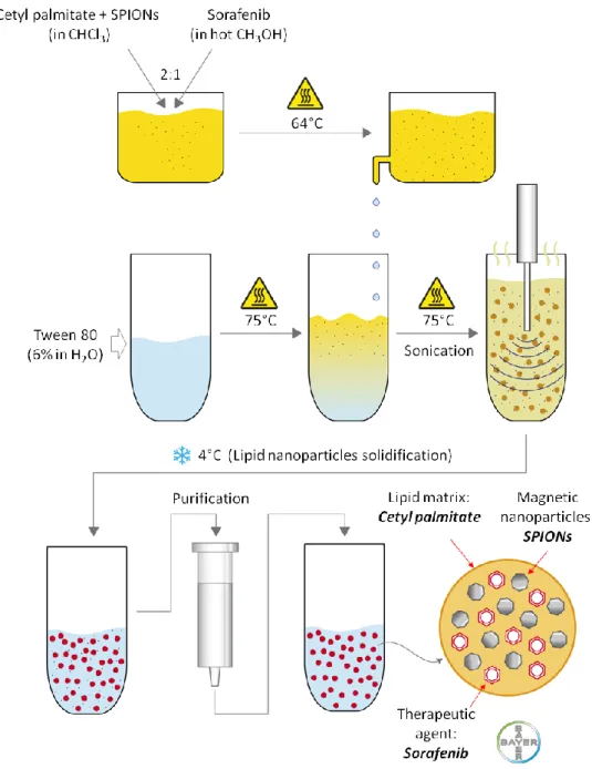
Solid lipid nanoparticles loaded with sorafenib and SPIONs (Sor-MagSLNs) were prepared by using an oil-in-water homogenization process at high temperature (Figure 3.2).
Figure 3.2 Schematization of the procedure followed for the preparation of sorafenib loaded magnetic solid lipid nanoparticles (Sor-Mag-SLNs).
23
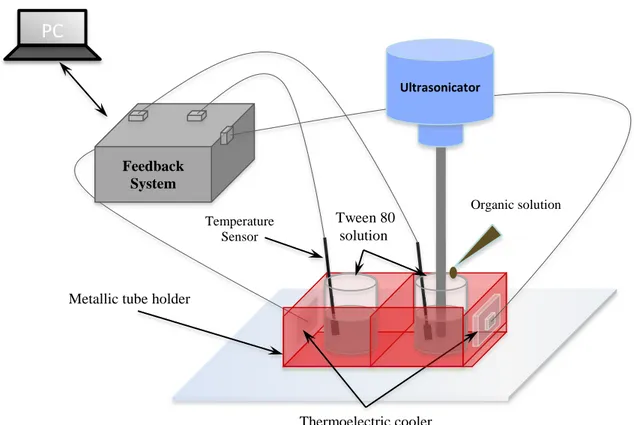
In detail, 5 mg of sorafenib tosylate (kindly provided by Bayer International) were dissolved in 500 µL of hot methanol (65 °C). Meanwhile, 210 mg of cetyl palmitate (kindly provided by Gattefossé) and 15 mg of SPIONs (average diameter of 10 nm, EMG1300 from Ferrotec) were dissolved in 1 mL of chloroform. The methanol and chloroform solutions were added and gently stirred. The obtained mixture was then transferred into a vial containing 3 mL of a Tween 80 (Sigma-Aldrich) water solution (6%), and sonicated for 5 min with a probe-tip ultrasonicator (SONOPLUS mini20, Bandelin). To allow organic solvents evaporation, the temperature of the emulsion was kept constant at 75 °C using a home-made heating system with a negative feedback control. Briefly, two thermoelectric coolers were assembled onto a precut metallic tube holder and connected to a software programmable feedback system, which ensures accurate temperature control (Figure 3.3). At the end of the procedure, the obtained dispersion was maintained at 4 °C for 15 min to allow the formation of the SLNs. In order to remove the surfactant, solvent residuals, and non-encapsulated drug and SPIONs, the final product was purified by gel chromatography, using Sephadex G-25 prepacked columns (GE Healthcare Life Sciences) with deionized water as eluent. Plain magnetic SLNs (Mag-SLNs), i.e., not loaded with sorafenib, were prepared following the same procedure, but omitting the drug, and were used as controls in biological experiments.
Feedback System
Metallic tube holder
PC
Software Interface Temperature Sensor Tween 80 solution Organic solution Thermoelectric cooler Ultrasonicator24
Sor-Mag-SLN characterization in terms of size and Z-potential was performed through dynamic light scattering with a Malvern Zetasizer Nano S90. The morphology of Sor-Mag-SLNs was investigated by AFM with an Innova SPM (Bruker), after drying a drop of appropriately diluted nanoparticle dispersion onto a silicon wafer. AFM scans were performed in tapping mode, by using NT-MDT NSG01 antimony-doped n-type silicon probes with a resonance frequency of 87–230 kHz and a force constant of 1.45–15.1 N m−1. All data scans were elaborated and analyzed with the Gwyddion SPM analysis tool (http://gwyddion.net). TEM was moreover performed. Samples underwent negative staining, the simplest and fastest method for the surface characterization of particulate samples. We used a two-step protocol for the specimens preparation, which consists in a 30 s adsorption of a properly diluted Sor-Mag-SLN suspension over carbon-coated 300 mesh grids (EMS) followed by MilliQ water washing (three times), and by a 30 s staining with 3% uranyl acetate solution in ethanol. Grids were dried before observation, carried out with a Zeiss Libra 120 Plus transmission electron microscope operating at 120 kV, equipped with an in-column omega filter, and mounted with a 16-bit 2k × 2k CCD camera. For drug loading determination, freeze-dried Sor-Mag-SLNs were dissolved in hexane, in order to solubilize the lipid matrix, and sorafenib was extracted with DMSO. The sorafenib concentration was then estimated by UV–vis spectroscopy (NanoDrop, Thermo Scientific) exploiting the absorbance peak at 254 nm. [51] Drug release investigation was performed placing a Sor-Mag-SLN suspension (600 µL, concentration 10 mg mL−1, corresponding to 320 × 10−6 M of drug) into dialysis bags (molecular weight cut-off 3.5 kDa, Sigma), immersed in a release bulk constituted by 3 mL of PBS at different pH conditions (3, 7, 10). The drug release study was carried out at 37 °C in a thermostatic shaker at a rate of 150 rpm. At predetermined time points, release bulk was replaced with fresh buffer solution, and aliquots freeze-dried; sorafenib was thereafter extracted in DMSO and its concentration assessed as previously described. TGAs were performed under air at a heating rate of 10 °C min−1 from 30 to 900 °C, by using a TG/DTA 7200 Extar instrument (SII Nanotechnology Inc). The iron content was measured by elemental analysis performed through ICP-MS using a Perkin Elmer NexION 300 spectrometer. Sample digestion was performed with 65% HNO3 and 30% H2O2 in a Milestone Start D microwave apparatus.
25
3.2.2 Magnetic and Relaxometric Characterizations
Magnetic characterization was carried out using a superconducting quantum interference device (SQUID) from Quantum Design. Magnetization curves were measured from −50 to 50 kOe at 5 K and 300 K upon zero field cooling. The temperature dependence of the ZFC and FC magnetizations were recorded applying a probe field of 25 Oe to measure the thermal dependence of the magnetization and evaluate the superparamagnetic behavior of the nanoparticles. Measurements of water proton relaxation times (T1 and T2) were carried out at 37 °C on Sor-Mag-SLNs aqueous suspensions at different concentrations obtained by dilution of a 30 mg mL−1 mother suspension with ultrapure water. Measurements at 20 MHz (0.47 T) were performed with a Spinmaster FFC-2000 Fast Field-Cycling NMR relaxometer (Stelar). The temperature of the samples was controlled within ±0.2 °C with a Stelar VTC90 variable temperature controller. In particular, T1 measurements were performed using the nonpolarized (NP) pulse sequence, while T2 measurements were performed using the nonpolarized Carr–Purcell–Meiboom–Gill (NPCPMG) pulse sequence with a τ delay of 30 µs [52]. The 90° pulse duration was 9.7 µs. All the 1H magnetization curves versus time were monoexponential within experimental error and the errors in fitting T1 and T2 were always less than 1%. Measurements of water proton T1 and
T2 at 300.13 MHz (7.05 T) were performed with a Bruker AMX300 spectrometer using the
Inversion Recovery pulse sequence and the CPMG pulse sequence [53, 54] ( τ = 100 µs) for T1 and T2 measurements, respectively. The 90° pulse duration was 6.7 µs. Relaxation
rates (R1 and R2) were calculated as the inverse of the relaxation times (R1 = 1/T1 and R2 =
1/T2) and relaxivity values (r1 and r2) were determined on the basis of the iron
concentration [Fe] of the samples, by fitting data acquired on Sor-Mag-SLN aqueous suspensions at different concentrations to the equation
Ri = R i0 + ri ⋅ [Fe] (1)
where i = 1,2 and R i0 is the Ri value of water alone.
3.2.3 Cell Cultures and In Vitro Cytotoxicity Study
Human hepatocellular carcinoma cell line (HepG2) was purchased by Sigma-Aldrich (85011430). HepG2 cells were cultured in low glucose Dulbecco’s modified Eagle medium (DMEM) containing 10% of fetal bovine serum (FBS), 1% of non-essential amino acid solution (NEAAS), and 1% of penicillin-streptomycin, at 37 °C and in 5% CO2 humidified
26
atmosphere. In order to assess the biocompatibility of Mag-SLNs and the cytotoxic effects of free sorafenib and of Sor-MagSLNs against HepG2 cells, the WST-1 assay was performed (2-(4-iodophenyl)-3-(4-nitophenyl)-5-(2,4disulfophenyl)-2H-tetrazoilium monosodium salt, provided in a pre-mix electrocoupling solution, BioVision). The cells were seeded at a density of 3 × 10⁴ cells/well in 24-well plates and incubated for 48 h at 37 °C and 5% CO2 in order to reach confluence. The medium was thereafter replaced with
fresh medium containing increasing concentrations of Mag-SLNs (0, 10, 20, 50,100, and 200 µg mL−1), Sor-Mag-SLNs (0, 10, 20, 50, 100, and 200 µg mL−1), or sorafenib at concentrations equal to those present inside Sor-Mag-SLNs (0, 0.32, 0.64, 1.60, 3.15, 6.35 × 10⁻⁶ M). Cell viability was assessed after 24 and 72 h following the WST-1 protocol: culture medium was replaced with 300 µL of medium + 30 µL of the premix solution, and incubated for 1.5 h. Finally, the absorbance was read at 450 nm with a microplate reader (Victor3, Perkin Elmer). Cytotoxic effects of Sor-Mag-SLNs (200 µg mL−1) were also qualitatively investigated at 72 h with the Live/Dead viability/cytotoxicity kit (Molecular Probes), that allows live cells (stained in green by calcein) to be discriminated from dead cells (stained in red by ethidium homodimer-1, EthD-1). The cells were rinsed with PBS, treated for 5 min at 37 °C with calcein AM, EthD-1, and Hoechst 33342 (Invitrogen, for nucleus counterstaining) in growth medium following manufacturer’s instruction, and finally observed with an inverted fluorescence microscope (TE2000U, Nikon), equipped with a cooled CCD camera (DS-5MC USB2, Nikon) and with NIS Elements imaging software for image acquisition.
3.2.4 Western Blotting Analysis
In order to investigate the molecular mechanism of action of free sorafenib and Sor-Mag-SLNs, HepG2 whole cellular lysates were examined through Western blotting analysis for the detection of phosphorylated Erk1/2 (pErk1/2). Analysis were performed after 24 and 72 h of treatment using the concentration that showed the highest cytotoxic effect observed with the WST-1 test, i.e., 200 µg mL−1 of Sor-Mag-SLNs, corresponding to 6.35 × 10⁻⁶ M of drug. Mag-SLNs (200 µg mL−1) were considered as well, as negative control. Briefly, HepG2 were cultured on Petri dishes at a density of 15 × 103 cells cm−2 for 24 h and then treated as described above. After 24 or 72 h treatment, cells were washed with D-PBS containing 1 × 10⁻³ M Na3VO4 and resuspended in a cell lysis ice-cold buffer (50 × 10−3 M
Tris-HCl, 154 × 10−3 M NaCl, 20 × 10 −3 M NaF, 1 × 10⁻³ M Na3VO4 , 1 × 10⁻³ M
27
then disrupted using two 30 s pulses applied with the Micro-Ultrasonic Cell Disrupter (Vineland) set at 50% output in an ice bath. Sample lysates were centrifuged at 15000 rpm for 20 min at 4 °C, and protein concentration in the supernatants was determined by the bicinchoninic acid microplate method, following manufacturer’s instructions (Pierce). Proteins (20 µg lane−1) were separated on a 10% polyacrylamide gel (Bio-Rad) under reducing conditions, and then transferred to a nitrocellulose membrane (Trans Turbo Blot system, Bio-Rad). The membrane was blocked with 3% bovine serum albumin in 0.1% Tween/Tris Buffer Saline (T-TBS) and incubated overnight at 4 °C with anti-pErk1/2 mouse monoclonal antibody (Santa Cruz) diluted 1:1000 in T-TBS. Anti β-actin mouse monoclonal antibody (Sigma-Aldrich) was used as protein loading control. Anti-mouse Horse Radish Peroxidase (HRP) conjugated antibodies (KPL), diluted 1:2000 in 4% dry nonfat milk in T-TBS for 1 h at room temperature, was used as secondary antibody, and the immunocomplexes were detected by chemiluminescence (ECL clarity, Bio-Rad) by using a Chemi-Doc XRS+ system (Bio-Rad). The data were analyzed by using Image Lab software (Bio-Rad).
3.2.5 Cellular Uptake Investigation
Internalization of SLNs by HepG2 cells was investigated through confocal analyses. Cells were seeded at a density of 12 × 103 cm 2 on Ibidi µ-Dish (35 mm, Ibidi) and incubated at 37 °C and 5% CO2 in order to reach confluence. Sor-Mag-SLNs were red-fluorescently
stained with DiI (Life Technologies), a lipophilic fluorescent stain for labeling cell membranes and other hydrophobic structures, and thereafter added to the cell culture medium at a concentration of 100 µg mL−1. After 24 h of incubation, the cells were rinsed with PBS and treated for 45 min with LysoTracker (Invitrogen), a fluorescent acidotropic probe for labeling and tracking of acidic organelles (lysosomes) in live cells. Finally, cells were rinsed with PBS, incubated for 10 min with Hoechst 33342 (5 µg mL−1, Invitrogen) for nucleus counterstaining, and observed with a confocal microscope (C2s, Nikon).
3.2.6 Dynamic Cell Culture and Magnetic Targeting Experiment
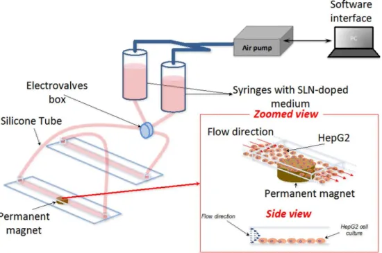
Sor-Mag-SLN magnetic targeting experiments in dynamic cell culture conditions were performed using a two syringes perfusion set-up connected to a fluidic unit with programmable switching valves and plastic channel µ-Slides (µ-Slide VI 0.4, Ibidi) for cell culturing. The home-made air pump system, built with a peristaltic pump linked to a feedback system, allows the pressure input to the syringes and the electrovalves periodic
28
switching to be controlled via software interface. Valve action and silicone tubing ensure continuous and directional cell medium flow inside the microfluidic plastic slide avoiding medium depletion. The whole perfusion set-up was placed in an incubator to ensure physiological temperature condition for the entire duration of the experiment. All the experiments where performed for 4 h at the constant flow rate of 2.3 mL min−1 inside the microfluidic channel, with a wall shear stress of about 4 dyn cm−2 set after the calibration of the perfusion set-up. These values were chosen considering wall shear stress in small diameter blood vessels (2–3 mm inner diameter) ranging between 1–10 dyn cm−2, as reported in the literature [55, 57]. No occlusions of silicon tubing and microfluidic channel due to particle aggregation was observed during the experiments, confirming the excellent stability of the SLNs in the cell culture medium. A scheme of the fluidic set-up is given in Figure 3.4. HepG2 cells were seeded at the bottom of two of the six rectangular-shaped microfluidic channels of the µ-Slide, with size 0.4 × 17 × 3.8 mm. All the seeded channels were previously incubated with gelatin (4% in PBS) for 20 min at 37 °C and then washed three times with PBS to improve cell adhesion. Cells (7 × 10⁴ per each channel) were incubated for three days at 37 °C and 5% CO2 in order to reach confluence before
performing experiments. In order to assess targeting efficiency, the syringes were loaded with medium containing red-fluorescently labeled Sor-Mag-SLNs (50 µg mL−1). A permanent neodymium magnet ( Br = 1.32 T) was positioned at the bottom of one of the
seeded channels to magnetically target Sor-Mag-SLNs near the area of action of the magnetic field, while the other channel was used as negative control. After 4 h, the experiment was stopped and cells were rinsed with PBS, fixed with paraformaldehyde (4% in PBS), treated with Triton 0.1% X-100 to allow membrane permeabilization, and blocked for 1 h with a goat serum solution (10% in PBS). Thereafter, cultures were incubated with a staining solution containing phalloidin (1:100, Millipore) and DAPI (1:1000, Millipore) to label cytoskeletal f-actin and nuclei, respectively. After extensive PBS rinsing, images were acquired by confocal microscope. An analogous procedure was followed in order to demonstrate selective magnetic-targeted cytotoxic effects of Sor-Mag-SLNs in dynamic conditions. Also in this case, medium containing 50 µg mL−1 of Sor-Mag-SLNs was circulated in the system for 4 h. Thereafter, cells were incubated in static conditions for 72 h before performing Live/Dead viability/cytotoxicity assay. Quantitative evaluation of cell mortality was performed by counting EthD-1 positive cells over the total number of cells.
29
Figure 3.4 Schematization of the set-up exploited for dynamic magnetic targeting experiments.
3.2.7 Statistical Analysis
Data were analyzed with one-way ANOVA followed by Bonferroni’s post hoc test or two-tailed unpaired t-test through KaleidaGraph (Sinergy Software). In all experiments, performed in triplicate, data with p value < 0.05 were considered statistically significant.
3.3 Results
3.3.1 Sorafenib-Loaded Magnetic Solid Lipid Nanoparticles
Sorafenib-loaded magnetic solid lipid nanoparticles (Sor-MagSLNs), prepared by a hot homogenization technique using cetyl palmitate as lipid matrix, as depicted in the schema of Figure 3.2, presented an average diameter of 248 ± 113 nm (dynamic light scattering measurement, DLS) and a Z-potential value of −23.0 ± 5.3 mV. These data indicate a good colloidal stability of the nanoparticles in aqueous environment, which prevents aggregation during all the experiments. Moreover, a satisfactory polydispersity index was determined (0.2 ± 0.1), thus suggesting monodisperse nanoparticle size distribution. Atomic force microscopy (AFM) scans confirm a regular spherical shape of Sor-Mag-SLNs,
30
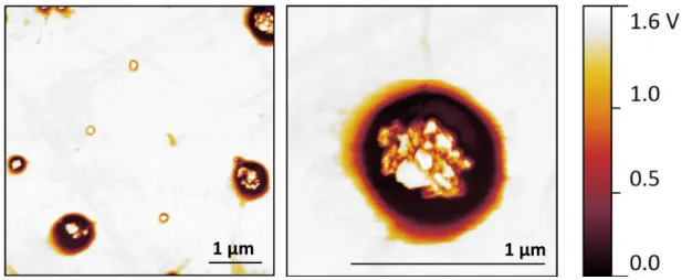
characterized by an average diameter of 420 ± 181 nm, calculated via software analysis (Figure 3.5a, b). This value is over-estimated with respect to DLS measurements, most probably because of the collapse and spreading of cetyl palmitate onto the substrate after water evaporation, as well as of the interaction of the cantilever with the nanoparticle edges.
In the topographical scans of single nanoparticles it is possible to observe an irregularly shaped area at the center of the circular edge, better evidenced by the 3D rendering of the scan (Figure 3.5c), which is probably due to drug and magnetic nanoparticle entrapment
Figure 3.5 AFM and TEM imaging of Sor-Mag-SLNs. AFM topographical scans at a) low and b) high magnification of nanoparticles deposited onto a silicon substrate;
c) 3D rendering of an AFM scan; d) TEM images of nanoparticles.
a)
b)
500 nm 1 µm
31
inside the SLNs. Moreover, a displacement in the oscillation phase of the cantilever is evidenced in AFM tapping phase scans (Figure 3.6) when the tip interacts with the two aforementioned portions of the nanoparticles evidenced by topographical scans. These results collectively indicate that the nanoparticles are composed of materials characterized by different mechanical properties, thus suggesting the presence of SPIONs inside the lipid core.
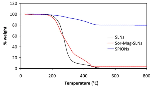
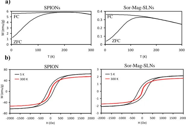
The presence of magnetic nanoparticles, characterized by different electron density and encapsulated in the lipid matrix, is in fact confirmed by transmission electron microscopy (TEM) image (Figure 3.5d), where it is clearly possible to see internalized SPIONs. The loading of SPIONs on SLNs was investigated by thermogravimetric analysis (TGA) and elemental analysis performed by inductively coupled plasma-mass spectrometry (ICP-MS). From the comparison of the thermal degradation behaviors under air flow (Figure 3.7) a SPION loading of about 3 wt% could be estimated. In agreement, elemental analysis gave an iron content of 1.4 wt%. Concerning magnetic properties, Sor-Mag-SLNs present a superparamagnetic behavior at room temperature, as the coercive field of about 200 Oe at 5 K becomes negligible at 300 K.
1 µm 1 µm
Figure 3.6 Tapping phase AFM scans of Sor-Mag-SLNs show cantilever oscillation phase contrast when the tip interacts with the edge and
32
Figure 3.7 TGA evaluation of the amount of loaded magnetic nanoparticles.
This observation was confirmed by the zero field cool (ZFC)/ field cool (FC) curve at 25 Oe, as the blocking temperature is around 190 K (Figure 3.8a). The lower blocking temperature of Sor-Mag-SLNs with respect to SPIONs and the broadening of the ZFC curve are hints of less inter-domains interactions because of the presence of the lipid matrix. The hysteresis curves of the two materials are similar (Figure 3.8b), thus indicating analogous magnetic properties. Sor-Mag-SLNs present a higher coercive field, again because of the encapsulation of SPIONs inside the lipid matrix. The magnetic saturation value of Sor-Mag-SLNs is about 1.95 emu g−1 at 300 K (60 emu g−1 for SPIONs), while 2.3 emu g−1 at 5 K (70 emu g−1 for SPIONs). Considering that the magnetic moment per particle should not be different between SPIONs and Sor-Mag-SLNs, we can conclude that the percentage in mass of iron oxide nanoparticles in the solid lipid particles is about 3%, in agreement with thermogravimetric and elemental analyses. On the other hand, the magnetization of the Sor-MagSLNs is almost saturated with a low magnetic field, i.e., 1000 Oe. 0 20 40 60 80 100 120 0 200 400 600 800 Temperature (°C) % w e ig h t SLNs Sor-Mag-SLNs SPIONs
33
This assures the easy manipulation of the magnetic nanovectors by an external magnetic field. Quantitative information on longitudinal (r1) and transverse (r2) relaxivities of
Sor-Mag-SLNs at 0.47 and 7.05 T at 37 °C was acquired from measurements of water proton T1 and T2 relaxation times on aqueous suspensions at different concentrations. In all cases,
a linear dependence was found between relaxation rates (R1 = 1/ T1 and R2 = 1/T2) and
nanoparticle concentration. The relaxivity values, determined by fitting these data and considering the Fe concentration in Sor-Mag-SLNs determined by elemental analysis, and their ratio (r2/r1) are reported in Table 3.1 together with relaxivities of commercial contrast
agents based on SPIONs. These results indicate that Sor-Mag-SLNs have good negative contrast properties at the investigated fields, in particular showing r2 (38 ± 2 s−1 m M−1 at
7.05 T, 215 ± 5 s−1 m M−1 at 0.47 T) and r2/r1 (633 at 7.05 T, 77 at 0.47 T) values much
higher than commercial agents at magnetic fields of clinical interest. [22–26] Successful sorafenib loading inside magnetic SLNs was assessed by UV–vis quantification of the drug, the estimated loading efficiency being about 90%, corresponding to a sorafenib concentration of 6.35 × 10⁻⁶ M in 200 µg mL−1 samples of Sor-Mag-SLNs. Interestingly,
-3 -2 -1 0 1 2 3 -2000 -1500 -1000 -500 0 500 1000 1500 2000 H (Oe) M (e m u/ g) 5 K 300 K . -80 -40 0 40 80 -2000 -1500 -1000 -500 0 500 1000 1500 2000 H (Oe) M (e m u/ g) 5 K 300 K . 0 0.1 0.2 0.3 0.4 0 100 200 300 T (K) M (e m u/ g) 0 1 2 3 4 5 6 0 100 200 300 T (K) M (e m u/ g) SPIONs Sor-Mag-SLNs
a)
b)
SPION s Sor-Mag-SLNs FC FC ZFC ZFCFigure 3.8 Magnetic characterization. a) Temperature dependence of theZFC and FC magnetizations measured with a magnetic field of 25 Oe and
34
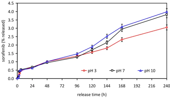
drug release investigations performed at different pH values (3, 7, and 10) revealed no significant, extremely slow, release of sorafenib (less than 5% after 10 d, Figure 3.9). These data suggest as biological effects, described in Sections 3.3.2 and 3.3.3, are ascribable to the degradation of cetyl palmitate by intracellular enzymes upon cellular internalization, and consequent release of the active drug from the dissolving lipid matrix.
Sample Field (T) T (°C) r1 (s-1 mM-1) r2 (s-1 mM-1) r2/r1 Sor-Mag-SLNs 7.05 37 0.06±0.02 38±2 633 Feridex[22] 7 - - 166.71 - Molday ION[23] 7 - - 106±4 - Feratrack[22] 7 - - 247±14 - Sor-Mag-SLNs 0.47 37 2.80±0.05 215±5 77 Endorem (AMI-25)[24] 0.47 37 27±1 152±8 5.6 Endorem (AMI-25)[24] 0.47 39 23.7±1.2 107±11 4.5 Ferumoxsil[25] 0.47 39 3.2±0.9 72±12 22.5 Resovist (SHU-555A)[24] 0.47 37 20.6±1.0 86±4 4.2 Resovist (SHU-555A)[26] 0.47 37 24.9 177 7.1 Sinerem (AMI 227)[25] 0.47 39 22.7±0.2 53.1±3.3 2.34 Table 3.1 Longitudinal and transverse relaxivity values of Sor-Mag-SLNs and some
35
Figure 3.9 Drug release from Sor‐Mag‐SLNs in PBS at different pH values.
3.3.2 In Vitro Cytotoxicity Study
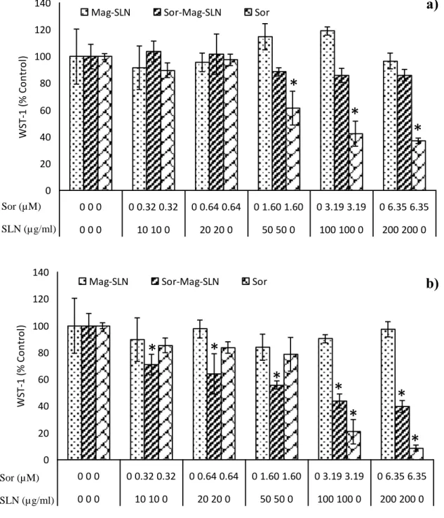
Antitumor activity of Mag-SLNs and Sor-Mag-SLNs was investigated against HepG2 cells with the WST-1 viability assay and compared to that of the free-administered drug. As shown in Figure 3.10, Mag-SLNs did not significantly inhibit cell viability after both 24 h (Figure 3.10a) and 72 h (Figure 3.10b) of incubation (at all the investigated concentrations p > 0.05 with respect to the controls). On the other hand, Sor-Mag-SLNs showed a moderate cytotoxic effect after 24 h of incubation. In fact, for Sor-Mag-SLNs concentrations of 50, 100, and 200 µg mL−1, corresponding to 1.60 × 10⁻⁶, 3.19 × 10⁻⁶, and 6.35 × 10⁻⁶ M of sorafenib, a viability reduction of about 15% was observed, which, however, is not statistically significant (Figure 3.10a). A strong dose-dependent antiproliferative effect was instead observed after 72 h of incubation (Figure 3.10b): at all the tested concentrations we have a significant reduction of viability (p < 0.05), that reaches about 40% of the control for the 200 µg mL−1 Sor-Mag-SLN concentration (6.35 × 10⁻⁶ M of sorafenib). The free drug confirmed its antitumor action both at 24 h (for concentrations of 1.60, 3.19, and 6.35 × 10⁻⁶ M, p < 0.05) and at 72 h (for concentrations of 3.19 and 6.35 × 10⁻⁶ M, p < 0.05). Overall, these results indicate good cytocompatibility of the plain Mag-SLNs; conversely, drug-loaded Sor-Mag-SLNs show an antiproliferative effect against tumor cells similar to that induced by free sorafenib, thus demonstrating their ability to deliver active drug into the cells. These quantitative results were confirmed by the Live/Dead viability/cytotoxicity assay (Figure 3.11). Also in this case, the cytocompatibility of Mag-SLNs was optimal at the tested concentration (200 µg
0.0 0.5 1.0 1.5 2.0 2.5 3.0 3.5 4.0 4.5 0 24 48 72 96 120 144 168 192 216 240 release time (h) so ra fen ib ( % rel ea sed ) pH 3 pH 7 pH 10
36
mL−1). No evidence of cell membrane damage was observed after the treatment, demonstrating the high viability of cells after 72 h of incubation with the plain nanoparticles, in line with non-treated control cultures. On the contrary, Sor-Mag-SLNs exercised a significant cytotoxic effect at the same concentration (200 µg mL−1, corresponding to 6.35 × 10⁻⁶ M of drug), as demonstrated by the great amount of red-stained cells observed at the end of the 72 h treatment. Once again, similar results were achieved with the same concentration of free drug (6.35 × 10⁻⁶ M).
0 20 40 60 80 100 120 140 0 0 0 0 0.32 0.32 0 0.64 0.64 0 1.60 1.60 0 3.19 3.19 0 6.35 6.35 0 0 0 10 10 0 20 20 0 50 50 0 100 100 0 200 200 0 W ST -1 ( % C o n tro l)
Mag-SLN Sor-Mag-SLN Sor
a)
Sor (µM) SLN (µg/ml) 0 20 40 60 80 100 120 140 0 0 0 0 0.32 0.32 0 0.64 0.64 0 1.60 1.60 0 3.19 3.19 0 6.35 6.35 0 0 0 10 10 0 20 20 0 50 50 0 100 100 0 200 200 0 W ST -1 ( % C o n tro l)
Mag-SLN Sor-Mag-SLN Sor
b)
Sor (µM) SLN (µg/ml)
*
*
*
*
*
*
*
*
*
*
Figure 3.10 Metabolic WST-1 assay on HepG2 cells after a) 24 h and
b) 72 h of incubation with increasing concentrations of Mag-SLNs, Sor-Mag-SLNs, and free sorafenib. * p < 0.05.
37
Finally, based on observations from the literature, that reported as treatment with sorafenib increases Erk1 and Erk2 phosphorylation, [27] we assessed phosphorylated Erk1/2 (pErk1/2) expression in HepG2 treated with sorafenib (6.35 × 10⁻⁶ M ) or Sor-Mag-SLNs (200 µg mL−1, corresponding to 6.35 × 10⁻⁶ M of drug). Coherently with those findings, Figure 3.12 shows increased pErk1/2 expression in cells treated with sorafenib or Sor-Mag-SLNs, which is more evident at 72 h of incubation compared to the controls. In agreement with data on cell viability, we observed that the cytotoxic effect of sorafenib and Sor-Mag-SLNs correlates with an increase of pErk1/2 in HepG2 cells rather than with
Live Cells
Dead Cells
Nuclei
M
ag
-SLN
So
r-M
ag
-SLN
500 µmSo
r
C
ontr
ol
Figure3.11 Results of Live/Dead® viability/cytotoxicity assay after incubation of HepG2 cells with plain MagSLNs (200 µg/ml), Sor‐Mag‐SLNs
(200 µg/ml, corresponding to 6.35 µM of sorafenib), and free sorafenib (6.35 µM); not‐treated cultures, as well.
38
an inhibition as reported in other studies: further investigations will be thus needed to verify whether the activation of the MAP kinase pathways is independent from the cytotoxic effect of sorafenib. [28] Taken together, all these data demonstrate that Sor-Mag-SLNs represent an excellent vector for sorafenib delivery, thanks to the nanoparticle biocompatibility and the retention of the drug activity following encapsulation.
pErk1/2
β-actin
1
2
3
4
pErk1/2
β-actin
24h
24h
72h
72h
Figure 3.12 Western blotting analysis for pErk1/2 (and β -actin as control) expression on HepG2 cellular lysates after 24 and 72 h of incubation in control
cultures (lane 1) and in cultures treated with free sorafenib 6.35 × 10⁻⁶ M (lane 2), Mag-SLNs 200 µg mL -1 (lane 3) and Sor-Mag-SLNs 200 µg mL-1.
39
3.3.3 Cellular Uptake Studies and Magnetic Drug Targeting
Cellular internalization studies revealed strong uptake of Sor-Mag-SLNs by HepG2 cells. Moreover, staining of lysosomes allowed a significant colocalization to be demonstrated, thus corroborating the hypothesis of drug release following the degradation of the lipid matrix in these digestive organelles. In Figure 3.13, Sor-Mag-SLNs are stained in red, Lysotracker probe is green-fluorescent, and overlapping is easily evidenced by the yellow signal arising from the merging of the two channels (Pearson’s index R = 0.67).
Magnetic targeting experiments were performed in dynamic conditions thanks to the set-up depicted in Figure 3.4, as widely described in the Experimental Section. Results show an enhanced Sor-Mag-SLNs uptake by cells cultured in correspondence of the region of the
Sor-Mag-SLNs
Lysosome
s
Nuclei
Merging
20 µm
Figure 3.13Investigation of cellular uptake. Confocal fluorescence images of HepG2 cells showing Sor-Mag-SLN uptake (in red) and colocalization with lysosomes (in green) after LysoTracker assay;
![Figure 2.1 Biomedical multifunctionality of magnetic nanoparticles. Reproduced with permission [23]](https://thumb-eu.123doks.com/thumbv2/123dokorg/2927732.18827/17.892.212.746.424.848/figure-biomedical-multifunctionality-magnetic-nanoparticles-reproduced-permission.webp)