Factors affecting successful mobilization with plerixafor:
an Italian prospective survey in 215 patients with multiple
myeloma and lymphoma
Francesco Lanza, Roberto M. Lemoli, Attilio Olivieri, Daniele Laszlo, Massimo Martino,
Giorgina Specchia, Vincenzo Pavone, Manuela Imola, Annalisa Pasini, Giuseppe Milone,
Ilaria Scortechini, Elisabetta Todisco, Elena Guggiari, Nicola Cascavilla, Giovanni Martinelli,
Alessandro Rambaldi, and Alberto Bosi*
BACKGROUND: Although the efficacy of plerixafor inperipheral blood stem cell (PBSC) mobilization has been explored in several studies, factors associated with successful plerixafor mobilization after administra-tion of granulocyte–colony-stimulating factor (G-CSF), with or without chemotherapy, have not been investi-gated. We analyzed data on PBSC mobilization from a large Italian database of lymphoma and myeloma plerixafor-treated patients.
STUDY DESIGN AND METHODS: Two endpoints were
established to define successful mobilization: patients with at least 2¥ 106CD34+ cells/kg collected by three leukapheresis procedures and patients achieving a peak count of at least 20¥ 106CD34+ cells/L during mobilization.
RESULTS: Plerixafor achieved successful mobilization
in both predicted (n= 64) and proven poor mobilizers (PMs; n= 143), classified according to the Gruppo Ital-iano Trapianto di Midollo Osseo (GITMO) criteria. Suc-cessful mobilization was independent of type of mobilization (steady state or chemotherapy); age; sex; disease; number or type of chemotherapy regimens preceding plerixafor; radiation therapy; prior treatment with melphalan, carmustine, lenalidomide, and radioim-mune conjugates; and laboratory variables. Multivariate analysis identified previous fludarabine treatment and premobilization platelet count as predictors of success-ful mobilization.
CONCLUSION: This large, prospective, nationwide
study confirmed plerixafor efficacy for mobilizing PBSCs when added to G-CSF with or without chemotherapy. Plerixafor can overcome negative effects of most pre-dictors of poor mobilization to achieve satisfactory harvest both in predicted and proven PM.
H
igh-dose chemotherapy followed byautolo-gous stem cell transplantation (ASCT) is a standard therapeutic approach for patients
with non-Hodgkin’s lymphoma (NHL),
ABBREVIATIONS:ASCT= autologous stem cell
transplantation; GITMO= Gruppo Italiano Trapianto di Midollo Osseo; HL= Hodgkin’s lymphoma; MM = multiple myeloma; NHL= non-Hodgkin’s lymphoma; PB = peripheral blood; PBSC= peripheral blood stem cell; PM(s) = poor mobilizer(s); ROC= receiver operating characteristic.
From the Section of Hematology and BMT Unit, Cremona Hospital, Cremona, Italy; the Department of Hematology and Oncological Sciences, Institute of Hematology “L. & A. Seràgnoli,” University of Bologna, Bologna, Italy; the Section of Hematology, University Hospital of Ancona, “Ospedali Riuniti,” Ancona, Italy; the Hemato-Oncology Unit, Istituto Europeo Oncologico, Milan, Italy; the Section of Hematology, Reggio Calabria Hospital, Reggio Calabria, Italy; the Section of Hematology, University Hospital of Bari, Bari, Italy; the Section of Hematology, Tricase Hospital, Tricase, Italy; Section the of Hematology, Rimini Hospital, Rimini, Italy; the Section of Hematology, University Hospital Ferrarotto, Policlinico Vittorio Emanuele, Catania, Italy; the Section of Hematology,
Humanitas Hospital, and the BMT Unit, S. Raffaele Hospital, Milan, Italy; the Section of Hematology, Hospital Casa Sollievo Sofferenza, S. Giovanni Rotondo, Italy; the Section of
Hematology, Bergamo Hospital, Bergamo, Italy; and the Section of Hematology and BMT Unit, University Hospital “Careggi,” Florence, Italy.
Address correspondence to: Francesco Lanza, Section of Hematology and BMT Unit, Hospital of Cremona, Via Concordia, 1, 26100 Cremona, Italy; e-mail:
*See Appendix S1, available as supporting information in the online version of this paper.
Received for publication January 16, 2013; revision received February 26, 2013, and accepted March 4, 2013.
doi: 10.1111/trf.12265
Hodgkin’s lymphoma (HL), or multiple myeloma (MM).1
The preferred source of hematopoietic stem cells for ASCT is represented by peripheral blood stem cells (PBSCs), since they are easier to collect and engraft faster than marrow-derived cells.2,3
Historically, granulocyte–colony-stimulating factor (G-CSF) or granulocyte macrophage-CSF, with or without chemotherapy, have been used to mobilize stem cells. However, these strategies do not always result in success-ful mobilization.3-6A recent survey involving 1040 patients
with NHL, HL, or MM, shows that a relevant proportion of patients (6%-27%) failed to mobilize a sufficient number of CD34+ progenitor cells for ASCT regardless of whether
G-CSF was used alone or with chemotherapy.5
Notewor-thy, factors predicting successful PBSC mobilization in patients with hematologic malignancies are still poorly
investigated,7-10 although their identification would
improve collection efficiency and prevent unnecessary apheresis procedures, thus allowing a patient-tailored mobilization and collection strategy.11
Plerixafor (Mozobil, Genzyme BV, Naarden, Nether-lands, Sanofi licencing distribution in Italy) is a novel CXCR4 chemokine receptor antagonist used in PBSC mobilization.12The interaction of the CXCR4 receptor with
the chemokine (C-X-C motif ) ligand 12 (CXCL12) plays a pivotal role in retaining CD34+ cells in the marrow niches.12Therefore, the blockade of this interaction leads
to the mobilization of stem cells into peripheral blood (PB).12The efficacy of plerixafor in the mobilization of
PBSCs is supported by the results of two randomized, double-blind, multicenter trials in adult patients with NHL or MM.13,14Moreover, the results of
compassionate-use studies conducted in patients with lymphoma or MM demonstrated that plerixafor plus G-CSF successfully and safely induces stem cell mobilization in the majority of patients who had previously failed a mobilization attempt (i.e., collected CD34+ cells did not reach the minimum number to proceed to ASCT or patients did not undergo apheresis because of low PB CD34+ cell count) or patients who were predicted to be poor mobilizers (PMs; e.g., heavily treated patients).15-20 Although the efficacy of
plerixafor in PBSC mobilization has been explored in several studies, to our knowledge the factors potentially associated with successful mobilization after the admin-istration of this agent have not been specifically investi-gated. To this end, this study analyzes a large Italian database of patients treated with plerixafor.
PATIENTS AND METHODS
Study setting and designThis multicenter, prospective, observational cohort study was conducted in 23 Italian centers with experience in the treatment of hematologic malignancies and performance of stem cell mobilization and ASCT between January 2010
and December 2011. The study was conducted in accor-dance with the Declaration of Helsinki, and the ethical committee of each center approved the study protocol. All patients signed an informed and educated consent before their inclusion in the study.
Patients and interventions
Patients with NHL, HL, or MM requiring PBSC mobiliza-tion were screened for inclusion in this study. Mobiliza-tion was performed using G-CSF, with or without chemotherapy, depending on individual center policy.
G-CSF was given at a dosage of 10mg/kg when used alone
and 5mg/kg when administered after chemotherapy.
Plerixafor was given at a dosage of 240 mcg/kg body weight. (Sanofi licencing distribution in Italy)
Patients were categorized as proven or predicted PMs, according to the Gruppo Italiano Trapianto di Midollo Osseo (GITMO) criteria.21Patients were considered to be
“proven PMs” when the peak concentration of PB CD34+ cells, after adequate mobilization, was fewer than 20¥ 106
cells/L or if the concentration of CD34+ cells collected with up to three apheresis procedures was not more than 2.0¥ 106CD34+ cells/kg and considered to be “predicted
PMs” if they had failed a previous collection attempt (not otherwise specified), previously received extensive radio-therapy or full courses of radio-therapy affecting stem cell mobi-lization, and met two of the following criteria: advanced disease (at least two lines of chemotherapy), refractory disease, extensive marrow involvement or cellularity less than 30% at the time of mobilization, or age at least 65 years.21
Data storage
Data were collected in a centralized secured database and were analyzed at the end of the study. The database con-sisted of three different parts: 1) demographic character-istics and previous history of mobilization; 2) type of mobilization; and 3) posttransplantation follow-up. Endpoints and data analysis
Two endpoints were established to evaluate mobilization success: 1) at least 2¥ 106 CD34+ cells/kg body weight
collected with up to three leukapheresis procedures and 2) a peak count of at least 20¥ 106/L CD34+ cells ¥ 106/L PB
during mobilization.21
The fold increase of CD34+ cells ¥ 106/L after the
administration of plerixafor was also calculated, to deter-mine the increase of PB CD34+ progenitor cells before and after the administration of plerixafor. The correlation between the platelet (PLT) concentration and the end-points were investigated using receiver operating charac-teristic (ROC) curves. A ROC curve permits a comparison of two operating characteristics or variables as the values change to enable thresholds to be established—in this case comparing endpoints with other variables, to better
differentiate patients likely to reach the endpoints (i.e., achieve successful mobilization) from those not reaching the endpoints.22
Patients were stratified according to several variables: age; sex; “predicted” versus “proven” PM category; chemo-therapy versus steady-state mobilization strategy; diagno-sis (NHL, HL, MM); the number of chemotherapy regimens preceding plerixafor administration; use of flu-darabine, radiation therapy, melphalan, carmustine, lena-lidomide, and radioimmune conjugates; and laboratory variables (hemoglobin, PLT count, white blood cells [WBCs], and neutrophils). Each variable was tested using univariate statistical analysis, to establish whether it could discriminate patients reaching the endpoint from those not reaching the endpoint, thus becoming a possible sig-nificant predictor of mobilization after plerixafor therapy. The chi-square test or Fisher’s exact test was used for cat-egorical variables, according to the number of samples. For continuous normally distributed variables, the t test was applied, whereas the Wilcoxon test was used for non-normally distributed variables. The two endpoints were analyzed separately and p values of less than 0.05 were considered significant.
To identify the factors able to predict the outcome of plerixafor treatment, a multivariate logistic regression model was calculated using only the variables that were significant in the univariate analysis as covariates. Covari-ates were considered to significantly affect the prediction if the p value was less than 0.05.
Finally, Kaplan-Meier curves were drawn to evaluate the relationship between the number of plerixafor doses and the endpoints. All the analyses were conducted on the population as a whole and on the subpopulations of pre-dicted PMs and proven PMs.
RESULTS
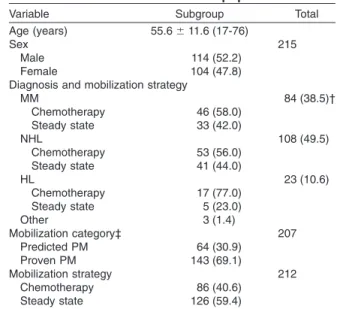
Baseline characteristicsThe baseline characteristics of the study patients are shown in Table 1. In total, 218 patients were enrolled (114
males; 52%). The mean age was 55.6⫾ 11.6 years (range,
17-76 years). Approximately half of the patients (n= 108; 49.5%) were affected by NHL; 23 patients (10.6%) had HL and 84 (38.5%) had MM. Three patients were diagnosed with other hematologic diseases (one subject with plasma cell leukemia, one with Richter syndrome, and one with immune thrombocytopenia purpura) and were not included in the subsequent analysis; therefore, 215 patients with NHL (n= 108), HL (n = 23), or MM (n = 84) requiring mobilization of PBSCs were included in the study. Concerning the mobilization status (predicted PM vs. proven PM) before plerixafor administration, data were available for 207 patients: among these, 64 (30.9%) were predicted PMs and 143 (69.1%) were proven PMs.
In total, 86 patients received G-CSF after chemo-therapy, according to different schedules (for six patients data unknown): 40 patients (50%) received high-dose cyclophosphamide (ⱖ 3 g/m2); 14 patients (17.5%) were
treated with DHAP (dexamethasone, ARA-C, cisplatin)
regimen; five patients (6.3%) with etoposide (1 g/m2,
Vepesid, Bristol-Myers Squibb, Rome, Italy); seven
patients (8.8%) with cytarabine (3 g/m2, Aracytin,
Janssen-Cilag, Berchem, Belgium); three patients (3.8%) with a mitoxantrone, cytarabine, and dexamethasone (MAD) regimen and 10 patients (12.5%) with other regi-mens. In the 126 remaining patients, PBSC mobilization
was based on G-CSF (10 mg/kg) in combination with
plerixafor (240 mg/kg body weight) without
chemo-therapy (steady state). Efficacy of plerixafor
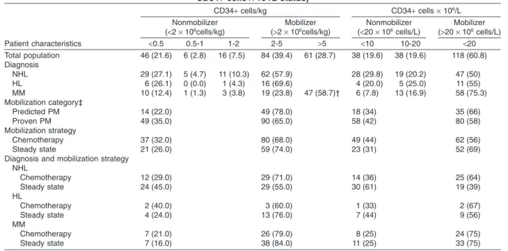
Overall, more than the 60% of patients enrolled in the study reached the endpoints, measured either as the number of CD34+ cells/kg collected (145 patients, 68.1%) or as the peak value of circulating CD34+ cells ¥ 106/L (118
patients, 60.8%; Table 2). In total, 61 patients (35.5%)
reached the endpoint of at least 2¥ 106 CD34+/kg with
one apheresis procedure, and 80 (46.5%) with two apher-esis procedures, so that more than 80% of patients
(n= 141) reached the target with no more than two
aph-eresis procedures.
The high response rate was observed after both steady state (G-CSF) and chemomobilization (Table 2). However, in NHL patients, chemomobilization was
TABLE 1. Baseline demographics and clinical characteristics in the total population
Variable Subgroup Total
Age (years) 55.6⫾ 11.6 (17-76)
Sex 215
Male 114 (52.2)
Female 104 (47.8)
Diagnosis and mobilization strategy
MM 84 (38.5)† Chemotherapy 46 (58.0) Steady state 33 (42.0) NHL 108 (49.5) Chemotherapy 53 (56.0) Steady state 41 (44.0) HL 23 (10.6) Chemotherapy 17 (77.0) Steady state 5 (23.0) Other 3 (1.4) Mobilization category‡ 207 Predicted PM 64 (30.9) Proven PM 143 (69.1) Mobilization strategy 212 Chemotherapy 86 (40.6) Steady state 126 (59.4)
* Data are reported as mean⫾ SD (range) or number (%). † Note that not all details were available for all patients.
Per-centages are given accordingly. ‡ According to GITMO criteria.21
associated with a higher proportion of patients reaching the end points compared with patients mobilized with plerixafor plus G-CSF alone (64% vs. 39% for the propor-tion of patients with peak CD34+ during mobilizapropor-tion of at least 20¥ 106/L CD34+ cells ¥ 106/L and 71% vs. 55% for
the number of CD34+ cells/kg collectedⱖ2 ¥ 106CD34+
cells/kg with up to three leukapheresis procedures), although these differences did not reach significance.
Patients with MM showed the highest response rate
with 82.5% of patients with more than 2¥ 106 CD34+
cells/kg collected in up to three apheresis procedures and
75.3% patients with a peak value of more than 20¥ 106
CD34+ cells/L, compared to other patients (p = 0.0016, Table 3).
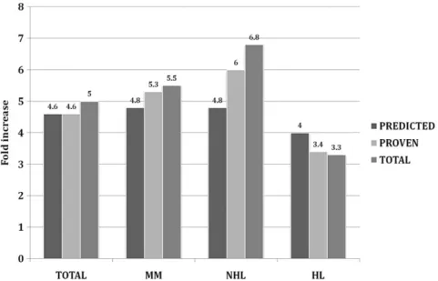
Overall, the data showed a fivefold increase of PB CD34+ cells ¥ 106/L after plerixafor administration
(Fig. 1). The fold increase was higher for NHL patients (6.8 times) and lower for HL patients (3.3 times) and there was no difference in fold increase between predicted PM and proven PM (Fig. 1).
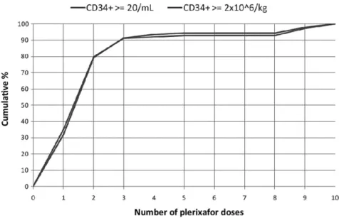
At the time of data analysis, of 126 (57.0%) patients receiving ASCT, 118 cases (95%) engrafted. Six patients did not engraft due to early death after auto-ASCT. A transplant-related mortality of 5% (six patients) was observed. Analysis of the cumulative percentage of patients reaching the endpoints (Fig. 2) showed that
almost 90% reached the defined study endpoints after three doses of plerixafor.
Predictive factors for mobilization success
Statistical analysis showed that baseline PLT concentra-tion was the most powerful predicting factor for successful stem cell mobilization (Table 3). The ROC curve estima-tion provided significant discriminaestima-tion thresholds of 140¥ 109/L to reach more than 2¥ 106CD34+ cells/kg and
143¥ 109/L to reach more than 20¥ 106 CD34+ cells/L.
Lack of radiotherapy was also a significant predictor of successful stem cell mobilization (Table 3). The only other factor predicting poor mobilization in the overall popula-tion was the previous use of fludarabine. In particular, patients previously treated with fludarabine were signifi-cantly less capable of reaching the endpoints (20% vs. 60%; p= 0.0009 in the univariate analysis). In the multi-variate analysis, these three variables, but not type of disease, still kept a significant predictive capacity for suc-cessful mobilization.
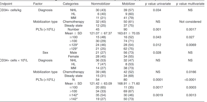
Overall similar observations came from the analysis of predicted PM (Table 4) and proven PM (Table 5): PLT concentration was the only factor with a discriminant capability. As in the total population, the estimation of the ROC curve for PLT concentration provided a threshold for
TABLE 2. Diagnosis and mobilization characteristics by endpoint (CD34+ cells/kg or peak
CD34+ cells ¥ 106/L status) Patient characteristics CD34+ cells/kg CD34+ cells ¥ 106/L Nonmobilizer (<2 ¥ 106cells/kg) Mobilizer (>2 ¥ 106cells/kg) Nonmobilizer (<20 ¥ 106cells/L) Mobilizer (>20 ¥ 106cells/L) <0.5 0.5-1 1-2 2-5 >5 <10 10-20 <20 Total population 46 (21.6) 6 (2.8) 16 (7.5) 84 (39.4) 61 (28.7) 38 (19.6) 38 (19.6) 118 (60.8) Diagnosis NHL 29 (27.1) 5 (4.7) 11 (10.3) 62 (57.9) 28 (29.8) 19 (20.2) 47 (50) HL 6 (26.1) 0 (0.0) 1 (4.3) 16 (69.6) 4 (20.0) 5 (25.0) 11 (55) MM 10 (12.4) 1 (1.3) 3 (3.8) 19 (23.8) 47 (58.7)† 6 (7.8) 13 (16.9) 58 (75.3) Mobilization category‡ Predicted PM 14 (22.0) 49 (78.0) 18 (34) 35 (66) Proven PM 49 (35.0) 90 (65.0) 58 (42) 80 (58) Mobilization strategy Chemotherapy 37 (32.0) 80 (68.0) 49 (44) 62 (56) Steady state 21 (26.0) 59 (74.0) 23 (31) 52 (69)
Diagnosis and mobilization strategy NHL Chemotherapy 12 (29.0) 29 (71.0) 14 (36) 25 (64) Steady state 24 (45.0) 29 (55.0) 30 (61) 19 (39) HL Chemotherapy 2 (40.0) 3 (60.0) 1 (33) 2 (67) Steady state 4 (24.0) 13 (76.0) 7 (44) 9 (56) MM Chemotherapy 7 (21.0) 26 (79.0) 8 (25) 24 (75) Steady state 7 (16.0) 38 (84.0) 11 (25) 33 (75)
* Data are reported as number (%).
† At least 4¥ 106CD34+ cells/kg (cut off for MM patients).
discriminating patients likely to reach the endpoints from those not likely to reach the endpoints. Notably, when examining the univariate analysis of the predicted PMs, patients who did not receive fludarabine had a higher
probability of reaching the endpoints (80% vs. 20% for>2 ¥ 106CD34+ cells/kg
and 75% vs. 0% for >20 ¥ 106 CD34+
cells/L; p< 0.05 [Table 4]), whereas this factor was not significant in the proven population in the univariate analysis (Table 5).
For CD34+ cells ¥ 106/L, the
predic-tive multivariate model was calculated only for the proven PM population, because the low number of cases in the predicted poor population did not allow a reliable estimation.
When we analyzed the number of CD34+ cells collected/kg in different patient subpopulations, the only factor with a predictive capability, both in the univariate and in the multivariate model, was the PLT concentration. This finding was present in all patient categories.
The statistical analysis also showed a correlation between the peak of CD34 cells¥ 106/L and the harvest of
CD34 cells/kg in patients treated with plerixafor (Spear-man coefficient, 0.78704; p< 0.0001). ROC curve analysis
TABLE 3. Significant predictive factors in the total population
Endpoint Factor Categories Nonmobilizer Mobilizer
p value univariate p value multivariate CD34+ cells/kg Diagnosis NHL 45 (42.1) 62 (57.9) 0.0016 NS HL 7 (30.4) 16 (69.6) MM 14 (17.5) 66 (82.5)
Mobilization status Predicted 14 (22.0) 49 (78.0) NS Not considered
Proven 49 (35.0) 90 (65.0)
Mobilization strategy Chemotherapy 37 (32.0) 80 (68.0) NS Not considered Steady state 21 (26.0) 59 (74.0) PLTs (¥109/L) Number 59 140 <0.0001 <0.0001 Mean⫾ SD 116.48⫾ 69.83 176.52⫾ 89.33 ⱕ100 22 (49) 23 (51) 0.0013 <0.0001 >100 37 (24) 117 (76) ⱕ140* 38 (44) 49 (56) 0.0001 <0.0001 >140* 21 (19) 91 (81) Radiotherapy No 46 (27) 122 (73) 0.028 0.044 Yes 15 (47) 17 (53) CD34+ cells ¥ 106/L Diagnosis NHL 47 (50) 47 (50) NS NS HL 9 (45) 11 (55) MM 19 (25) 58 (75)
Mobilization category† Predicted 18 (34) 35 (66) NS Not considered
Proven 58 (42) 80 (58)
Mobilization strategy Chemotherapy 49 (44) 62 (56) NS Not considered
Steady state 23 (31) 52 (69) PLTs (¥109/L) Number 71 117 <0.0001 <0.0001 Mean⫾ SD 122.56⫾ 67.54 178.32⫾ 89.50 ⱕ100 26 (60) 17 (40) 0.0005 0.0004 >100 45 (31) 100 (69) ⱕ143* 46 (54) 39 (46) <0.0001 0.0002 >143* 25 (24) 78 (76) Fludarabine No 63 (36) 111 (64) 0.0009 0.0175 Yes 12 (80) 3 (20)
* Cutoff obtained by ROC curve analysis. † According to GITMO criteria.21
Fig. 1. Fold increase in CD34+ cells ¥ 106/L after plerixafor treatment in predicted
further permitted the estimation of a significant discrimi-nation threshold of 4mL of CD34+ cells for a successful mobilization with plerixafor.
DISCUSSION
The results presented here confirmed, on a large database including more than 200 patients from several Italian centers, the efficacy of plerixafor in inducing stem cell mobilization and, for the first time, identified factors predictive of successful mobilization after the administra-tion of this novel agent. Our results are in agreement with those obtained in randomized, Phase III and compassionate-use program trials,8-10,13-20,23in which the
administration of plerixafor produced a successful mobi-lization with a median increase of approximately fivefold in patients with MM and NHL. We found no significant difference between the response of patients who were pre-dicted PMs or proven PMs. Moreover, we did not observe any difference between steady state and chemotherapy mobilization, thus suggesting that plerixafor can be incor-porated successfully into both regimens.
Preliminary studies in patients previously failing the first mobilization attempt reported a successful rate ranging from 37% to 90%.15,17,18Our results on a larger
Italian database were similar, with more than 60% of the total population reaching the endpoints. Our results also confirm that rates of successful mobilization tend to be lower in patients with NHL than in patients with
MM or HL.9 In addition, in our study, approximately
90% of patients reaching the endpoint needed no more than three doses of plerixafor and two apheresis procedures.
Moreover, a high correlation
between increased likelihood of mobili-zation success and increased number of CD34+ cells present in the mobilized blood was documented; a cutoff point of
4¥ 106 CD34+/L cells best predicted
success in a plerixafor-containing
mobilization regimen. Based on this, it can be postulated that the presence of at least 4¥ 106CD34+/L cells in the
mobi-lized blood, prior plerixafor administra-tion, identified patients who most benefited from plerixafor usage in all patient categories.
In our population the analysis of factors potentially able to influence the treatment outcome was different from those reported in previous studies.8,18
We failed to demonstrate that patients who underwent previous ASCT had a lower median number of CD34+ cells collected than those who did not undergo ASCT. Moreover, previous chemotherapy was not a significant factor discriminating patients reaching the endpoint from those not reaching the endpoint, neither in the total population nor in the two subgroups (predicted vs. proven), except for the use of fludarabine. In fact, in our population only 20% of the patients who were treated with fludarabine achieved the endpoint of at least 20¥ 106CD34+/L, compared to the 64% of those
who did not receive fludarabine. This difference was greater than reported in the literature (60% vs. 76%).10
The subanalysis by proven or predicted PMs showed that this effect was only seen in patients who were predicted PMs (using both endpoints).
Our analysis also allowed the definition of a baseline PLT concentration threshold that may be used as a predic-tive factor for successful mobilization with plerixafor. In fact, baseline thrombocytopenia was a significant predic-tor of reduced mobilization success, although plerixafor treatment was able to overcome this effect partially. Overall, a PLT count of fewer than 150¥ 109/L was
signifi-cantly associated with a lower probability of successful mobilization regardless of the endpoint considered or in all subgroups of patients.
In conclusion, our study shows that the admini-stration of plerixafor produced a successful mobilization in lymphoma and myeloma patients, both in predicted and in proven PMs, independent of the type of mobilization (steady state or chemotherapy). We also identified some factors, including baseline PLT concen-tration, previous fludarabine treatment, and previous radiotherapy, which, in a multivariate model, may be able to predict the successful mobilization with plerixafor.
Fig. 2. Cumulative percentage of patients reaching the efficacy endpoints in relation to the number of plerixafor doses. ( ) At least 20¥ 106CD34+/L; ( ) at least
TABLE 4. Significant predictive factors in the predicted PM population (n= 64)
Endpoint Factor Categories Nonmobilizer Mobilizer
p value Univariate Multivariate
CD34+ cells/kg Diagnosis NHL 10 (33) 20 (67) NS NS
HL 1 (13) 7 (87)
MM 3 (12) 22 (88)
Mobilization strategy Chemotherapy 5 (14) 30 (86) NS Not considered
Steady state 9 (32) 19 (68) PLTs (¥109/L) N 14 47 0.0052 0.043 Mean⫾ SD 101.71⫾ 77.98 193.38⫾ 109.88 ⱕ100 7 (50) 7 (50) 0.011 NS >100 7 (15) 40 (85) ⱕ103* 9 (53) 8 (47) 0.0005 0.027 >103* 5 (11) 39 (89) Fludarabine No 10 (18) 47 (82) 0.0014 NS Yes 4 (80) 1 (20) WBCs N 14 47 0.0199 NS Median 6.14 15.36
CD34+ cells ¥ 106/L Diagnosis NHL 11 (44) 14 (56) NS Not estimated
HL 2 (40) 3 (60)
MM 5 (22) 18 (78)
Mobilization strategy Chemotherapy 10 (33) 20 (67) NS
Steady state 8 (35) 15 (65) PLTs (¥109/L) Number 17 34 0.046 Mean⫾ SD 126.18⫾ 82.17 189.76⫾ 113.53 ⱕ100 6 (50) 6 (50) NS >100 11 (28) 28 (72) ⱕ143* 11 (55) 9 (45) 0.0084 >143* 6 (19) 25 (81) Fludarabine No 13 (28) 34 (72) 0.0033 Yes 5 (100) 0 (0) WBCs Number 17 34 0.0021 Median 4.70 22.68
Neutrophil count (¥109/L) Number 17 29 0.012
Median 3.24 8.5
* Cutoff obtained by ROC curve analysis.
TABLE 5. Significant predictive factors in the proven PM population (n= 144)
Endpoint Factor Categories Nonmobilizer Mobilizer p value univariate p value multivariate
CD34+ cells/kg Diagnosis NHL 30 (43) 39 (57) 0.034 NS
HL 6 (40) 9 (60)
MM 11 (21) 41 (79)
Mobilization type Chemotherapy 32 (40) 50 (61) NS Not considered
Steady state 12 (25) 37 (75) PLTs (¥109/L) Number 45 90 0.001 0.0017 Mean⫾ SD 121.07⫾ 67.37 163.61⫾ 70.05 ⱕ100 15 (48) 16 (52) 0.043 0.027 >100 30 (29) 74 (71) ⱕ129* 24 (46) 28 (54) 0.012 0.0069 >129* 21 (25) 62 (75) Sex Male 21 (27) 56 (73) 0.028 NS Female 28 (45) 34 (55) CD34+ cells ¥ 106/L Diagnosis NHL 36 (53) 32 (47) NS NS HL 7 (47) 8 (53) MM 14 (27) 38 (73)
Mobilization type Chemotherapy 39 (48) 42 (52) NS 0.0166
Steady state 15 (31) 34 (69) PLTs (¥109/L) N 54 80 0.0001 <0.0001 Mean⫾ SD 121.42⫾ 63.09 168.91⫾ 71.60 ⱕ100 20 (65) 11 (35) 0.0017 0.0003 >100 34 (33) 69 (67) ⱕ142* 35 (54) 30 (46) 0.0019 0.0013 >142* 19 (27) 50 (73)
ACKNOWLEDGMENTS
This study was approved by GITMO (Italian Society for Stem Cell Transplantation). Editorial assistance and statistical analysis of data were provided by Luca Giacomelli, PhD, Mary Hines, and Chiara Favero, on behalf of inScience Communications, Springer Healthcare.
CONFLICT OF INTEREST
FL, AB, RML, and AR were members of an advisory board for Genzyme s.r.l. The rest of the authors declare that they have no conflict of interest.
REFERENCES
1. Rosenbeck LL, Srivastava S, Kiel PJ. Peripheral blood stem cell mobilization tactics. Ann Pharmacother 2010;44:107-16.
2. Jantunen E, Fruehauf S. Importance of blood graft charac-teristics in auto-SCT: implications for optimizing mobiliza-tion regimens. Bone Marrow Transplant 2011;46:627-35. 3. Vose JM, Ho AD, Coiffier B, Corradini P, Khouri I, Sureda A,
Van Besien K, Dipersio J. Advances in mobilization for the optimization of autologous stem cell transplantation. Leuk Lymphoma 2009;50:1412-21.
4. Bensinger W, DiPersio JF, McCarty JM. Improving stem cell mobilization strategies: future directions. Bone Marrow Transplant 2009;43:181-95.
5. Pusic I, Jiang SY, Landua S, Uy GL, Rettig MP, Cashen AF, Westervelt P, Vij R, Abboud CN, Stockerl-Goldstein KE, Sempek DS, Smith AL, DiPersio JF. Impact of mobilization and remobilization strategies on achieving sufficient stem cell yields for autologous transplantation. Biol Blood Marrow Transplant 2008;14:1045-56.
6. Giralt S, Stadtmauer EA, Harousseau JL, Palumbo A, Bens-inger W, Comenzo RL, Kumar S, Munshi NC, Dispenzieri A, Kyle R, Merlini G, San Miguel J, Ludwig H, Hajek R, Jagan-nath S, Blade J, Lonial S, Dimopoulos MA, Einsele H, Bar-logie B, Anderson KC, Gertz M, Attal M, Tosi P, Durie BG et al. International myeloma working group (IMWG) con-sensus statement and guidelines regarding the current status of stem cell collection and high-dose therapy for multiple myeloma and the role of plerixafor (AMD 3100). Leukemia 2009;23:1904-12.
7. Han X, Ma L, Zhao L, He X, Liu P, Zhou S, Yang J, Qin Y, Yang S, Yao J, Shi Y. Predictive factors for inadequate stem cell mobilization in Chinese patients with NHL and HL: 14-year experience of a single-center study. J Clin Apher 2012;27:64-74.
8. Duarte RF, Shaw BE, Marin P, Kottaridis P, Ortiz M, Morante C, Delgado J, Gayoso J, Goterriz R, Martinez-Chamorro C, Mateos-Mazon JJ, Ramirez C, de la Rubia J, Achtereekte H, Gandhi PJ, Douglas KW, Russell NH. Plerix-afor plus granulocyte CSF can mobilize hematopoietic
stem cells from multiple myeloma and lymphoma patients failing previous mobilization attempts: EU compassionate use data. Bone Marrow Transplant 2011;46:52-8.
9. Hubel K, Fresen MM, Apperley JF, Basak GW, Douglas KW, Gabriel IH, Geraldes C, Jaksic O, Koristek Z, Kroger N, Lanza F, Lemoli RM, Mikala G, Selleslag D, Worel N, Mohty M, Duarte RF. European data on stem cell mobilization with plerixafor in non-Hodgkin’s lymphoma, Hodgkin’s lymphoma and multiple myeloma patients. A subgroup analysis of the European Consortium of stem cell mobili-zation. Bone Marrow Transplant 2012;47:1046-50. 10. Malard F, Kroger N, Gabriel IH, Hubel K, Apperley JF,
Basak GW, Douglas KW, Geraldes C, Jaksic O, Koristek Z, Lanza F, Lemoli R, Mikala G, Selleslag D, Worel N, Mohty M, Duarte RF. Plerixafor for autologous peripheral blood stem cell mobilization in patients previously treated with fludarabine or lenalidomide. Biol Blood Marrow Transplant 2012;18:314-7.
11. Duong HK, Bolwell BJ, Rybicki L, Koo A, Hsi ED, Figueroa P, Dean R, Pohlman B, Kalaycio M, Andresen S, Sobecks R, Copelan E. Predicting hematopoietic stem cell mobiliza-tion failure in patients with multiple myeloma: a simple method using day 1 CD34+ cell yield. J Clin Apher 2011;26: 111-5.
12. Keating GM. Plerixafor: a review of its use in stem-cell mobilization in patients with lymphoma or multiple myeloma. Drugs 2011;71:1623-47.
13. DiPersio JF, Micallef IN, Stiff PJ, Bolwell BJ, Maziarz RT, Jacobsen E, Nademanee A, McCarty J, Bridger G, Calandra G. Phase III prospective randomized double-blind placebo-controlled trial of plerixafor plus granulocyte colony-stimulating factor compared with placebo plus granulocyte colony-stimulating factor for autologous stem-cell mobilization and transplantation for patients with non-Hodgkin’s lymphoma. J Clin Oncol 2009;27:4767-73. 14. DiPersio JF, Stadtmauer EA, Nademanee A, Micallef IN,
Stiff PJ, Kaufman JL, Maziarz RT, Hosing C, Fruehauf S, Horwitz M, Cooper D, Bridger G, Calandra G. Plerixafor and G-CSF versus placebo and G-CSF to mobilize hemato-poietic stem cells for autologous stem cell transplantation in patients with multiple myeloma. Blood 2009;113:5720-6. 15. Arcaini L, Laszlo D, Rizzi S, Balzarotti M, Antoniazzi F,
Zilioli VR, Guggiari E, Farina L, Todisco E, Bonfichi M, Alamos SM, Rossi G, Martinelli G, Morra E. Plerixafor and G-CSF for PBSC mobilization in patients with lymphoma who failed previous attempts with G-CSF and
chemotherapy: a REL (Rete Ematologica Lombarda) expe-rience. Leuk Res 2011;35:712-4.
16. Calandra G, McCarty J, McGuirk J, Tricot G, Crocker SA, Badel K, Grove B, Dye A, Bridger G. AMD3100 plus G-CSF can successfully mobilize CD34+ cells from non-Hodgkin’s lymphoma, Hodgkin’s disease and multiple myeloma patients previously failing mobilization with chemotherapy and/or cytokine treatment: compassionate use data. Bone Marrow Transplant 2008;41:331-8.
17. Worel N, Rosskopf K, Neumeister P, Kasparu H, Nachbaur D, Russ G, Namberger K, Witt V, Schloegl E, Zojer N, Linke-sch W, Kalhs P, Greinix HT. Plerixafor and granulocyte-colony-stimulating factor (G-CSF) in patients with lymphoma and multiple myeloma previously failing mobi-lization with G-CSF with or without chemotherapy for autologous hematopoietic stem cell mobilization: the Aus-trian experience on a named patient program. Transfusion 2011;51:968-75.
18. Basak GW, Jaksic O, Koristek Z, Mikala G, Basic-Kinda S, Mayer J, Masszi T, Giebel S, Labar B, Wiktor-Jedrzejczak W. Haematopoietic stem cell mobilization with plerixafor and G-CSF in patients with multiple myeloma transplanted with autologous stem cells. Eur J Haematol 2011;86:488-95. 19. D’Addio A, Curti A, Worel N, Douglas K, Motta MR, Rizzi S,
Dan E, Taioli S, Giudice V, Agis H, Kopetzky G, Soutar R, Casadei B, Baccarani M, Lemoli RM. The addition of plerixafor is safe and allows adequate PBSC collection in multiple myeloma and lymphoma patients poor mobiliz-ers after chemotherapy and G-CSF. Bone Marrow Trans-plant 2011;46:356-63.
20. Attolico I, Pavone V, Ostuni A, Rossini B, Musso M, Cresci-manno A, Martino M, Iacopino P, Milone G, Tedeschi P, Coluzzi S, Nuccorini R, Pascale S, Di Nardo E, Olivieri A. Plerixafor added to chemotherapy plus G-CSF is safe and allows adequate PBSC collection in predicted poor
mobilizer patients with multiple myeloma or lymphoma. Biol Blood Marrow Transplant 2012;18:241-9.
21. Olivieri A, Marchetti M, Lemoli R, Tarella C, Iacone A, Lanza F, Rambaldi A, Bosi A. Proposed definition of “poor mobilizer” in lymphoma and multiple myeloma: an ana-lytic hierarchy process by ad hoc working group Gruppo Italiano Trapianto di Midollo Osseo. Bone Marrow Trans-plant 2012;47:342-51.
22. Hanley JA, McNeil BJ. The meaning and use of the area under a receiver operating characteristic (ROC) curve. Radiology 1982;143:29-36.
23. Micallef IN, Stiff PJ, DiPersio JF, Maziarz RT, McCarty JM, Bridger G, Calandra G. Successful stem cell remobilization using plerixafor (mozobil) plus granulocyte colony-stimulating factor in patients with non-Hodgkin
lymphoma: results from the plerixafor NHL phase 3 study rescue protocol. Biol Blood Marrow Transplant 2009;15: 1578-86.
SUPPORTING INFORMATION
Additional Supporting Information may be found in the online version of this article at the publisher’s web-site: