Synthesis, characterization
and cytotoxicity studies of
novel organo-metallic
compounds
UNIVERSITÀ DEGLI STUDI DI SALERNO
Dipartimento di Farmacia
Dottorato di ricerca
in Scienze Farmaceutiche
Ciclo XII NS — Anno di discussione 2014 Coordinatore: Chiar.mo Prof. Gianluca SbardellaSYNTHESIS, CHARACTERIZATION AND
CYTOTOXICITY STUDIES OF NOVEL
ORGANO-METALLIC COMPOUNDS
settore scientifico disciplinare di afferenza: CHIM/08
Dottorando Tutore
Dott. Chiar.mo Prof.
Chapter One 1
1 Introduction 2
1.1 MRI Contrast Agents 3
1.2 Radiopharmaceuticals 5
1.3 Antiinfective Agents 6
1.4 Superoxide Dismutase Mimics 8
1.5 Cardiovascular System 9
1.6 Insulin Mimetics 10
1.7 Gold Antiarthritic Drugs 11
1.8 Bismute antiulcer Drugs 13
1.9 Anticancer Agents: Platinum 15 1.9.1 Clinical Platinum Complexes 15
1.9.2 Platination of DNA 18
1.9.3 Protein Recognition 22
1.9.4 Active trans Complexes 23
1.9.5 Biotransformation 24
1.9.6 Photoactivation 25
1.9.7 Octahedral Platinum(IV) Complexes 26 1.9.8 Polynuclear Platinum Complexes 27 1.10 Palladium Complexes as Alternative Potential Antitumor Agents 28 1.10.1 Trans-Palladium(II) Complexes Containing Bulky Monodentate Ligands 29
1.10.3 Palladium complexes with Biphosphine P∩P Ligands 34 1.10.4 Palladium(II) complexes with N∩S Mixed Donor Ligand 34 1.10.5 Dinuclear Palladium(II) Complexes 35 1.11 Ruthenium (II & III) Complexes as Antitumor Agents 36 1.12 Copper Complexes as Antitumor Agents 39 1.12.1 Other Metal Anticancer Agents 41 1.13 Anticancer Agents: Titanium Complexes 42
1.13.1 Structure–Activity Relationships of Titanocene Dichloride and Related
Compounds 44
1.13.2 Structure–Activity Relationships of Budotitane and Related
Compounds 48
1.13.3 Comparison of the Biological Profiles of Titanocene Dichloride and
Budotitane 49
1.13.4 Mechanistic Aspects of Biological Activity 50 1.13.5 Phase II trial of titanocene dichloride in advanced renal-cell carcinoma
53
1.13.5.1 Patients and methods 53
1.13.5.2 Response 54
1.13.5.3 Toxicity 55
1.13.6 Methoxy alkyl substituted titanocenes 55
Chapter Two 59
2 Aim of the project 60
2.1 Ligand and Complex Design 60
2.2 Synthesis and characterization of aryl-substituted half-titanocene 65 2.2.1 Hydrolysis test of complexes 1b-11b 68
2.2.2.1 Cytotoxicity results 71 2.3 Synthesis and characterization of titanocene derivatives complexes 74 2.3.1 Hydrolysis test of titanocene compounds 77
2.3.2 Cytotoxic activity 79
2.4 Synthesis and characterization of alkyl-substituted titanocene 82
2.4.1 Hydrolysis test 83
2.4.2 Cytotoxic activity 84
2.5 Synthesis and characterization of ethoxybenzyl-cyclopentadienyl
titanocene 85
2.5.1 Hydrolysis test 87
2.5.2 Cytotoxic activity 88
2.6 Synthesis and characterization of titanocene and half-titanocene derivatives having a methyl group on the linker between Cp ring and phenyl group 91
2.6.1 Hydrolysis test 93
2.6.2 Cytotoxic activity 93
2.7 Synthesis and characterization of naphtyl-substituted complexes 94
2.7.1 Hydrolysis test 96
2.7.2 Cytotoxic activity 96
2.8 Cytotoxic activity of fulvene 97
Chapter Three 101
3 Conclusions 102
Chapter Four 105
4 Experimental section 106
4.2 Synthesis of ligands 107 4.2.1 Synthesis of 6-p-(methoxyphenyl) fulvene (1a ) 107 4.2.2 Synthesis of 6-(3’,4’- dimethoxyphenyl)fulvene (2a) 107 4.2.3 Synthsis of 6-(p-N, N-Dimethylanilinyl) fulvene (3a) 108 4.2.4 Synthesis of 6-phenylfulvene (4a) 108 4.2.5 Synthesis of 6-(2’,4’- dimethoxyphenyl)fulvene (5a) 109 4.2.6 Synthesis of 6 (2’,4’,6’-trimethoxyphenyl)fulvene (6a) 109 4.2.7 Synthesis of (tert-butyl) cyclopentadiene (7a ) 110 4.2.8 Synthesis of 1-[(2-(cyclopentadienyl)ethoxy]benzene (8a) 110 4.2.9 Synthesis of 6-methyl-6-phenyl-fulvene (9a) 111 4.2.10 Synthesis of 6-methyl-6-(4’-methoxyphenyl)fulvene (10a) 111 4.2.11 Synthesis of 6-methyl-6-(2’,4’-dimethoxyphenyl)fulvene (11a) 112 4.2.12 Syhthesis of 6-(4’-methoxynaphtalyl)fulvene (12a) 112 4.3 Synthesis of half-titanocene complexes 113 4.3.1 Synthesis of [(4-methoxybenzyl)cyclopentadienyl]-titanium (IV)trichloride (1b) 113 4.3.2 Synthesis of [(3,4-dimethoxybenzyl)-cyclopentadienyl]-titanium-trichloride (2b) 114 4.3.3 Synthesis of [(4-(N,N-dimethylbenzanilido)-cyclopentadienyl]-titanium-trichloride (3b) 115 4.3.4 Synthesis of [(benzyl)-cyclopentadienyl]-titanium-trichloride (4b) 116 4.3.5 Synthesis of [(2,4-di-methoxybenzyl)-cyclopentadienyl]-titanium-trichloride (5b) 117 4.3.6 Synthesis of [(2,4,6-tri-methoxybenzyl)-cyclopentadienyl]-titanium-trichloride (6b) 118
4.3.7 Synthesis of (tert-butyl-cyclopentadienyl)-titanium (IV) trichloride (7b) 119
4.3.8 Synthesis of (2-cyclopentadienyl-ethoxy-benzene)-titanium-(IV)
trichloride (8b) 119
4.3.9 Synthesis of 3-(phenylethyl)cyclopentadienyl-titanium(IV) trichloride (9b) 120
4.3.10 Synthesis of [3-(4-methoxyphenylethyl)cyclopentadienyl]titanium(IV)
trichloride (10b) 121
4.3.11 Synthesis of
[3-(2,4-dimethoxyphenylethyl)cyclopentadienyl]titanium(IV) trichloride (11b) 122 4.3.12 Synthesis of [(4-methoxynaphtalyl)cyclopentadienyl] titanium (IV)
trichloride (12b) 123
4.4 Synthesis of titanocene derivatives 124 4.4.1 Synthesis of bis-[(benzyl)-cyclopentadienyl]-titanium-dichloride (4c)124
4.4.2 Synthesis of 125
[1,2-Di(cyclopentadienyl)-1,2-bis1,2-di(phenyl)ethanediyl]titanium-dichloride)
(4d) 125
4.4.3 Synthesis of bis-[(2,4 dimethoxybenzyl)-cyclopentadienyl] titanium (IV)
dichloride (5c) 126
4.4.4 Synthesis of bis(tert-butyl-cyclopentadienyl) titanium (IV) dichloride (7c) 127 4.4.5 Synthesis of (bis-2-cyclopentadienyl-ethoxy-benzene)-titanium (IV)dichloride (8c) 127 4.4.6 Synthesis of (bis-2-cyclopentadienyl-ethoxy-benzene)-titanium-bis-glycine (8d) 128 4.4.7 Synthesis of (bis-2-cyclopentadienyl-ethoxy-benzene)-titanium-oxalate (8e) 128 4.4.8 Synthesis of bis-[3-(phenylethyl)cyclopentadienyl]-titanium(IV) dichloride (9c) 129
4.4.9 Synthesis of
bis-[3-(4-methoxyphenylethyl)cyclopentadienyl]titanium(IV) dichloride (10c) 130 4.4.10 Synthesis of
bis-[3-(2,4-dimethoxyphenylethyl)cyclopentadienyl]titanium(IV) dichloride (11c) 131 4.4.11 Synthesis of bis-[(4-methoxynaphtalyl)cyclopentadienyl] titanium (IV)
Despite the discovery of cis-platin in the treatment of cancer there has been a considerable exploration on the antitumoral activity of other transition metal complexes. One of the main problems about the application of transition metal complexes for chemotherapy is their potential toxicity. For instance, recently the attention has been focused on titanium based complexes, which could have significant potential effect against solid tumor. The advantage of Ti(IV) complexes is their relative biological compatibility, which mostly leads to mild and revisable side effects. However, the hydrolytic instability of known Ti(IV) complexes and formation of various different species upon water addition makes their therapeutic application problematic, and raises a strong interest in the development of relatively stable Ti(IV) complexes with well defined hydrolytic behavior that demonstrate appreciable cytotoxic activity. Strong ligand binding is also of interest to avoid complete ligand stripping by transferrin, so that the ligand may be used as a target for structure–activity relationship investigations.
Titanocene dichloride (Cp2TiCl2) shows an average antiproliferative activity in vitro but promising results in vivo. Considerable work has been performed in developing therapeutic analogues of Cp2TiCl2 by varying the central metal, the labile ligands (Cl) and the bis-cyclopentadienyl moiety. In particular, small changes to the Cp ligand can strongly affect
the hydrolytic stability and water solubility properties of the metallocenes and have an impact on the cytotoxic activity.
For a better exploration of the parameters affecting activity and its mechanistic aspects, the synthesis and investigation of particularly designed complexes based on different strongly coordinating ligands has been our main purpose
We synthesized novel titanocene and half-titanocene derivatives, having substituted cyclopentadienyl ligands; all the complexes have been fully characterized by NMR, elemental analysis and MS. Additionally we studied the rate of hydrolysis of these complexes. Starting from the reflection that the different activities of the complexes could be related to their different stabilities, the hydrolysis stability represents a first possible indication on the achievable cytotoxic effects of synthesized compounds.
The synthesized compounds have been evaluated for their cytotoxic potential against cancer cell lines. Most of these compounds showed significant anti-proliferative effects compared to cisplatin
Chapter One
This chapter contains an overview on all the medical fields in which organo-metallic compounds are involved, with a special focus on organo-metallic antcancer drugs.
1 Introduction
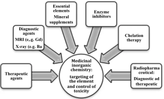
Biomedical inorganic chemistry, known as elemental medicine, is an important new area of chemistry. It really offers the potential for the design of novel therapeutic and diagnostic agents and hence for the treatment and understanding of diseases which are now intractable[1-2] (Figure 1). Inorganic elements has a central roles in biological and biomedical processes, and it is evident that many organic compounds used in medicine do not have a merely organic mode of action; some of them are activated or biotransformed by metal ions including metallo-enzymes[4],others have a direct or indirect effect on metal ion metabolism. Elemental medicine offers real possibilities to pharmaceutical industries, which have traditionally been dominated by organic chemistry alone, for the discovery of novel drugs with new mechanisms of action. This field has been encouraged by the success of cisplatin, the worlds best selling anticancer drug, and platinum complexes with oral activity, reduced toxicity, and activity against resistant tumors are now on clinical trial. [3] The organo-metallic complex titanocene dichloride has also been tested on patients, and RuIII complexes have a promising metastatic activity.
The toxicity of GdIII complexes can be controlled so that they can be
injected in gram quantities as magnetic resonance imaging (MRI) contrast agents with no risk and ligand design allows paramagnetic ions to be targeted to specific organs.
Designed ligands also allow the targeting of radiodiagnostic (e.g. 99mTc)
and radio- therapeutic isotopes (e.g. 186Re).
There has been recent impreovment in understanding the coordination chemistry and biochemistry of older metallo-drugs such as gold antiarthritic and bismuth antiulcer drugs, and further work might lead to their effective use.
Current areas with exciting clinical potential include vanadium insulin mimics, manganese superoxide dismutase mimics, lanthanide-based photo-ensitizers, ruthenium nitric oxide scavengers, and metal-targeted organic agents.
The increasing knowledge of organo-metallic chemistry will provide an opportunity for the design of new drugs (both inorganic and organic) in many other areas too, for example neuropharmaceutical and antiinfective
agents. Progress in medicinal coordination chemistry is closely dependent on understanding not only the thermodynamics (equilibria and structures) but also the kinetics (and mechanisms) of reactions of metal complexes, especially under biologically relevant conditions.
Figure 1 Some of the key areas of medicinal inorganic chemistry
1.1 MRI Contrast Agents
Magnetic resonance imaging is now a strong instrument in clinical diagnosis.[5] Diseases can be detected from differences in 1H NMR
resonances (mainly of H2O) between normal and abnormal tissues by
injections of external paramagnetic agents, known as contrast agents. Most contrast agents contain FeIII GdIII, or MnII ions which have a large
number of unpaired electrons (7, 5, and 5, respectively, high spin) and long electron spin relaxation times.[6, 7]
Four GdIII complexes have been approved for clinical use, and are
nowadays widely used, for example, for the detection of abnormalities of the blood-brain barrier.[14] Complexes containing DTPA (1, Magnevist)
and DOTA ligands (2, Dotarem) are ionic, whereas those of BMA-DTPA (3, Omniscan) and HP-DOTA (4, Prohance) are neutral; their low osmolarity decreases the pain of the injections. All these agents are extracellular, and they diffuse rapidly into the interstitial space. The GdIII
center is nine-coordinate in each complex and contains one bound H2O
Medicinal inorganic chemistry: targeting of the element and control of
toxicity Therapeutic agents Diagnostic agents MRI (e,.g. Gd) X-ray (e.g. Ba Essential elements Mineral supplements Enzyme inhibitors Chelation therapy Radiopharma ceutical: Diagnostic ad therapeutic
ligand.Water exchange on GdIII is dissociative and steric hyndrance at
the H2O site increases the exchange rate.
Complex 1 does not enter cells and is excreted almost exclusively by the kidney.
The insertion of a benzyloxymethyl substituent on the a-C atom of a terminal acetate of DTPA as in BOPTA gives a GdIII complex (5,
Gadobenate), which enters hepatocytes and is excreted in bile.[8] The
coordination sphere of GdIII in complex 5 is almost identical to that in
complex1 (distorted tricapped trigonal prism, nine-coordinate), and both complexes show similar stabilities and relaxivities (see Figure 3).[8]
Figure 2 Complexes used as MRI agents
The distorted octahedral complex 6 (Teslascan is the mangafodipir trisodium salt),[9] is currently in clinical use for increasing contrast in the
liver (detection of hepatocellular carcinomas).[10] The relaxivity of 6 is
about 35 % greater than that of Mn complexes of DTPA and DOTA, which also do not contain directly coordinated water.[11]
Figure 3 Crystal structure of 5 (on the left) and Teslacan (on the right).
1.2 Radiopharmaceuticals
Clinical interest in radionuclides centers not only on highintensity γ-ray emitters, especially 111In, 67Ga, 51Co 99mTc and 201Tl, , 51Cr, and 169Yb for diagnostic imaging, but also on β- emitters, for example 186Re, 89Sr, and
153Sm for therapy.[1]Some 99mTc-based radiopharmaceuticals and several
other radionuclides are currently utilized in clinical diagnosis. Complex 7 ([99mTcV(dl-hm-pao)], Ceretec) is an approved perfusion imaging agent
for diagnosis and evaluation of cerebral stroke. It is absorbed by the brain and is transformed into a more hydrophilic drug which is retained in the brain. Complex 8 ([99mTcI(sestamibi)], Cardiolite), instead, is used for myocardial perfusion imaging. It was projected on the idea that cationic lipophilic complexes behave as potassium mimics and are taken up by the heart.[13] The sequential metabolism of the six equivalent methoxy groups of 8 to hydroxyl groups in the liver leads to formation of 99mTc complexes with stronger hydrophilicity which are not retained in myocardial tissues.[14] Monoclonal antibodies (mAbs) combined with radionuclides such as 111Insatumomab pendetide (which contains the murine mAb B72.3, which is directed to TAG-72, an antigen expressed by many adenocarcinomas) are currently used for diagnosis of ovarian and colorectal cancer.[15] Several other murine mAbs linked to 111In and 99mTc are now in clinical trials.[16] Substantial progress has been performed recently in the development of 99mTc-based receptor-specific
radiopharmaceuticals.[17] Encapsulation in fullerenes may also give a novel method for the delivery of radionuclides to target sites.[18, 19]
Figure 4 Structure of Cardiolite.
1.3 Antiinfective Agents
Historically silver compounds have widely been used as antimicrobial agents in medicine. Silver has a low toxicity and is active at low concentrations. The practice of instilling the eyes of babies with 1% AgNO3 solution immediately after delivery is still common in some
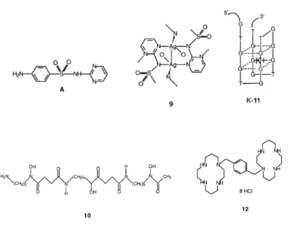
countries for prevention of ophthalmia neonatorum.[20] Silver sulfadiazine 9, made from AgI and sulfadiazine A, is currently used as an antifungal and antimicrobial agent. It is a polymeric insoluble drug which releases AgI ions slowly, and is applied topically as a cream to prevent bacterial
infections in cases of acute burns. The slow release of antimicrobial AgI ions from inorganic or organic polymer matrices[21] is important for pharmaceutical industries..
The mode of action of AgI is unknown. Cell-wall damage may be irelevant, and it has been shown that Cys150 in phosphomannose isomerase, an essential enzyme for the biosynthesis of cell walls of Candida albicans, is a AgI target in this organism.[22] Silver-resistant bacteria are well known,[20] and improvement in understanding the mechanism of resistance is now being made.
Antimony has been used for medical purposes for many centuries. Complexes of SbIII are conventionally more toxic than those of SbV. Two SbV drugs, sodium stibogluconate (Pentostam) and N-methylglucamine
antimonate (Glucantime), are currently used for the treatment of leishmaniasis, an infection caused by intracellular parasites.[23] The carbohydrates contained in the drugs may sbe useful for delivering SbV to macrophages. The SbV complexes may be considered prodrugs because the more toxic SbIII forms at or near the site of action. There is current
interest in improving the solubility, stability, and efficacy of Sb drugs, and SbIII and SbV complexes with yeast mannan are reported to be promising.[24, 25]
The iron chelating complex, named desferrioxamine 10 is clinically approved for the treatment of malaria disease. Its activity may results from the interfernce of FeIII metabolism inside the digestive vacuole of malaria parasites.[26]
Several antisense oligonucleotides are strong inhibitors of HIV-1 integrase,[27] and are currently on clinical trial. These have key sequences such as 5'-d(GTGGTGGGTGGGTGGGT) (11, T30175), and are only formed of thymidine and deoxyguanosine. They fold up in the presence of K+ ions to give four-stranded structures dominated by two stacked guanine-quartet motifs.[28]
This structure is very stable under physiological conditions (e.g., K-11 is resistant to serum nucleases with a half-life of 5 h) and is probably critical for biological properties. Macrocyclic bicyclam ligands such as 12 (JM3100) are certainly amongst the strongest inhibitors of HIV ever described, being active at nanomolar concentration.[29] Since they are safe at these levels, they have a high selectivity index (ca. 105).
The zinc complex of ligand 12 is also active.[30] The bicyclams appear to
obstacle the virus entry and membrane fusion through interaction with the CXCR4 coreceptor during the early stages of the retrovirus replicative cycle.[31]
Figure 5 Structure of complexes used as antinfective agents
1.4 Superoxide Dismutase Mimics
The free radical superoxide, O2-, is a product of endothelial cells and
activated leukocytes, and has been postulated to mediate the ischemia-reperfusion injury as well as inflammatory and vascular diseases. It can react with NO forming damaging peroxynitrite, ONO2-. The metalloenzyme superoxide dismutase, known as SOD can destroy O2-:
Cu,Zn-SOD in the cytoplasm of eukaryotic cells, Mn-SOD in mitochondria [see Eqs. (1) and (2) below].
Mn+1 + O2- Mn+ + O2 (1)
Mn+ + O2- + 2H+ Mn+1 + H2O2 (2)
However, the use of SOD in therapy is restricted by itsbrief plasma half-life (clearance by the kidney) and inability to penetrate cell membranes giving only an extracellular activity. Low molecular mass mimics of SOD
are, for this reason,of much potential medical interest.[32] For example, a variety of Fe-based and Mn- porphyrins and macrocyclic complexes show SOD mimic activity.[33-36] Notably, MnII and MnIII macrocycles appear to be promising.[37, 38] For example, complex 13 (SC-52608) is able to scavenge superoxide and therefore effectively protect the ischemic and reperfused myocardium from injury.[39] Another example is manganese(III) 5,10,15,20-tetrakis(4-benzoic acid)- porphyrin (MnTBAP), which can protect against neurodegeneration and is for this reason of potential interest for the treatment of brain diseases such as Alzheimer’s and Parkinson’s diseases.[40]
Figure 6 Structure of SC-52608
1.5 Cardiovascular System
The FeII complex sodium nitroprusside 14 is the only clinically used among the metal-nitrosyl complex.[41] It is often used to rapidly decrease the blood pressure in humans. Its hypotensive effect is evident within seconds after infjection, and the required blood pressure is usually obtained within one to two minutes. It is also useful in cases of heart attacks, emergency hypertension, or surgery.[42] The therapeutic effects of 14 is based on the quick release of nitric oxide, which relaxes vascular smooth muscle. Activation in vivo may involve reduction to [Fe(CN)5(NO)]3-, which then releases cyanide to give [Fe(CN)4(NO)]
2-and then nitric oxide.[43, 44]
Ruthenium complexes such as K[Ru(Hedta)Cl] K-15 (JM1226) have been proposed as nitric oxide scavengers for the control of NO levels under conditions of clinical interest.
In water complex 15 is in equilibrium with the aqua species 16 (JM6245; the pKa of the dangling arm carboxyl group is 2.4).[45] Both complexes bind to NO very quickly (rate constant>108m-1 s-1 at body temperature 310 K) and tightly (K>108m-1), giving the linear nitrosonium RuII -NO+ adduct 17.[45] Complex 16 has been demonstrated to reverse the poor response of
the artery to vasoconstrictor drugs,[46] which is one of the major clinical problem in the treatment of patients with septic shock (caused by very high levels of circulating bacteria in the body). The excessive production of NO not only appears to be a main contributory factor in septic shock, but also in arthritis, inflammation, diabetes and epilepsy.
Figure 7 Structures of complexes used in the cardiovascular system
1.6 Insulin Mimetics
Nearly 20 years ago It was founded that VV as vanadate and VIV as vanadyl can mimic some of the effects of insulin (stimulate glucose uptake and oxidation as well as glycoge synthesis).[47, 48] Vanadium complexes having organic ligands are often less toxic and can show improved aqueous solubility and lipophilicity. The orally active complex bis-(maltolato) oxovanadium(IV) (18,BMOV)[49] is about three times more effective in vivo as an insulin-mimetic agent than VOSO4[50]
Complex 18 has, in the solid-state, a five-coordinate square pyramidal geometry with the oxo ligand in the axial position and trans maltolato ligands.[51] In aerobic aqueous solutions, the complex is quickly oxidized to the dioxovanadium(V) species.[52]
Low molecular weight chromium-binding substance (LMWCr), a naturally occuring oligopeptide (ca. 1500 Da, consisting of CrIII, Asp, Glu, Gly, and Cys in a 4:2:4:2:2 ratio), has been demostrated to activate
the insulin-dependent tyrosine kinase activity of the insulin receptor protein, with the activity being proportional to the Cr content of the oligopeptide (maximium activity with four CrIII per oligopeptide) .[53] The trinuclear cation 19 can stimulate the tyrosine kinase activity of the insulin receptor in a manner quite identical to that of LMWCr.[54]
Interestingly when acetate groups replaces propionate in 19, the complex does not activate but rather inhibits both the kinase and membrane phosphatase activity. Its potential for diabetes treatment has yet to be discovered.
Figure 8 Structures of complexes used as insulin mimics.
1.7 Gold Antiarthritic Drugs
Some injectable 1:1 AuI thiolato complexes are used clinically for the treatment of severe cases of rheumatoid arthritis, including sodium aurothiomalate (20, Myocrisin), aurothioglucose (21, Solganol), and sodium aurothiopropanol sulfonate (22, Allochrysine).[55] Most of the gold thiolates are polymeric complexes (e.g. chain or ring forms) with thiolate S-bridging and linear AuI ions and have a gold-to-ligand ratio close to 1:1, as shown by EXAFS[56] and WAXS data.[57] Crystal structures of the gold thiolate drugs themselves have been ambiguous, the only example is given by the crystallization of 20 by Bau[58] using techniques for macromolecules crystallization. The linear S-Au-S units are disposed into polymeric double-helical chains. Hexameric Au6S6 rings of the type previously predicted [59] have also been demostrated to
exist in crystals with 2,4,6-tri(isopropyl)thiophenol as the thiolato ligand.[60] The formulated gold thiolate drugs often contain thiols in small
molar excess (e.g. 10%) over AuI, and readily undergo thiolate exchange and thiolate addition reactions.[61]
Figure 9 Structures of gold antiarthritic drugs
The only oral gold antiarthritic compound currently in use is the Auranofin, a phosphane complex. It is also highly cytotoxic to cancer cells in vitro, and is also reported to be active against psoriasis if used topically.[62]
A common metabolite found in the plasma and urineof patients treated with gold drugs is [Au(CN)2]-,[63] an ion which promptly enters cells and
is capable of inhibiting the oxidative burst of white blood cells. Therfore it could therefore be an active metabolite of gold drugs. It also shows anti- HIV and anticancer activity. The elevated Au content in the red blood cells of smokers receiving gold therapy has been realted to the inhalation of HCN through smoke.[64] Simulating the red blood cell concentrations, Elder et al.[65] have observed that [Au(CN)
2]- reacts with
GSH to form [Au(SG)2]-, which is very stable. In all probability [Au(SG)2]- is to be formed inside the cells both from auranofin and gold
thiolate drugs, and may be responsible for the inhibition of many enzymes such as the Se-enzyme glutathione peroxidase.[66] Compound 20 is a strong inhibitor of neutrophil collagenase, a zinc enzyme which includes Cys ligands in the metal-binding site.[67, 68] These interactions may be useful in joint tissue. GoldI shows a srtrong affinity for thiolate S, but
binds only weakly to N and O ligands. Hence the proteins containing cysteine thiolate groups (mainly those with low pKa values) are targets for AuI, but binding of AuI to DNA is poor. Moreover, the thiolate exchange reactions are usually very rapid. During therapy, Au levels in the blood typically reach 20mm, and Au is transported by albumin bound to Cys34.
The rate of gold binding is regulated by the rate of opening of the cleft containing Cys34,[69, 70] and the Au binding seems to induce a “flip-out” of this residue.[71] GoldI drugs can strongly bind to thiol groups in DNA-binding proteins such as the transcription factors Jun-Fos and Jun-Jun, allowing the possibility that gold can regulate the activity of transcription factor.[72] The oxidation of AuI to AuIII in vivo could be responsible for some of the side effects of gold drugs.[73,74] In inflammatory conditions, strong oxidants such as H2O2 and ClO- are potentially available in vivo
and can oxidize the metal centers in auranofin, 20 and 21 to AuIII.[75] GoldIII has the unusual capability to deprotonate peptide amide groups even at low pH values (e.g. pH 2)[76] and may modify peptide recognition by T cells. GoldIII is able tooxidize disulfide bridges in insulin and albumin[77] and methionine residues of ribonuclease.[78]
1.8 Bismute antiulcer Drugs
Some bismuth complexes have been used for treating gastrointestinal disease for at least two centuries.[79] These include alicylate salts, nitrate, sand colloidal bismuth subcitrate, all BiIII compounds. BismuthV is usually a powerful oxidant agent. The structures of Bi drugs are widely unknown. The coordination number of BiIII is highly variable (3-10), and often the coordination geometry is irregular.[80] BiIII is strongly acidic in water (first pKa ca. 1.5) and has an high tendency to form stable oxo-bridged and hydroxo clusters.
The [Bi6O4(OH)4](NO3)6 4H2O complex, known as “magisterium
bismuti” since the 17th century was used as a beauty care product, and crystallizes at pH values under 1.2.[81] The main understood in structural terms are the citrate complexes, for which several X-ray structures have been performed,[82] although none has absolutely the same composition as the drugs themselves. The dominant aspect is the dimeric [(cit)BiBi(cit)] 2-unit (H4cit:citric acid), which contains bridging citrate anions. The
Bi-O(alkoxide) bond is reallystrong and short (2.2 Å), and a stereochemical role for the 6s2 lone pair is evident in the structure. These dimers
aggregate into chains and sheets in the crystal through a network of hydrogen bonds involving citrate, counter ions, and water to give polymers. Such polymerscould be deposited on the surface of ulcers.
BismuthIII citrate complexes seem to be stable in solution in a pH range of about 3.5 to 7.5. [BiIII(Hcit)] itself is solubilized by a variety of amines,[83] and the adduct with the organic histamine antagonist ranitidine, ranitidine bismuth citrate,[84, 85] has recently been approved as a new drug. The antibacterical activity of BiIII against Helicobacter pylori
seems to be important for its antiulcer activity.[86] This organism is also implicated in other diseases such as even cancer.
The biological activities of BiIII are probably mainly due to binding to
enzymes and proteins. The binding of BiIII to DNA is unknown, so far. There is a poweful correlation between the strength of binding of BiIII to a variety of ligands and that of FeIII. Although BiIII (ionic radius 1.03 Å) is bigger than FeIII (ionic radius 0.64 Å high spin) it also strongly binds to transferrin, the serum FeIII transport protein.[87] The strength of binding of transferrin to BiIII can be related with the high acidity of BiIII. Correlated proteins (periplasmic iron-binding proteins) are implicated in Fe uptake by some virulent bacteria.
BismuthIII can promptly displace ZnII from the metallothionein, Cys-rich protein, and bismuth metallothionein is stable even under deeply acidic conditions (pH 2).[88] Cysteine and glutathione may have a crucial role in the transport of BiIII in cells and biofluids. These thiols can avoid the precipitation of colloidal bismuth subcitrate (CBS) at pH 2.0, and animal studies have shown that simultaneous oral administration of thiols and bismuth salts gives a significant increase in the bismuth concentration in blood plasma.[89, 90] The complex [Bi(SG)3] is very stable (lgK.29.6) in a
wide pH range (2-10) with bindingonly through the S atom.[91] Exchange
of GSH between bound and free forms is quite rapid at biological pH (ca. 1500 s-1).
1.9 Anticancer Agents: Platinum
1.9.1 Clinical Platinum Complexes
Platinum complexes are currently amongst the most world-widely used drugs for the treatment of cancer. Four injectable PtII complexes have
been approved for clinical use and many other cis-diam(m)ine complexes are on clinical trials, including an oral PtIV complex. Today cisplatin (23) is one of the most largely used anticancer drugs, along with the second generation drug carboplatin [Pt(NH3)2(CBDCA-O,O')] (24). The
glycolato complex 25 (nedaplatin, 254-S) and oxalato complex 26 (oxaliplatin, l-OHP, which contains R,R-1,2- diaminocyclohexane, DACH) have also been approved for clinical use in Japan and in all the countries, respectively. These drugs are particularly effective in “cocktail” or combination chemotherapy for treatment of advanced colorectal, lung, and ovarian cancers.[92, 93]
Figure 10 Stuctures of Platinum complexes
The sterically-hindered complex 27 (ZD0473) is active (by oral administration and injection) against an acquired cisplatin-resistant cell line of a human ovarian carcinoma xenograph,[94] and entered clinical trials in 1997. It is certainly less reactive than cisplatin, for example binding to plasma proteins and inducing DNA interstrand crosslinks in cells less quickly. The hydrolysis rate of 27 are at least two to three times slower than those of cisplatin (table 1).
Table. 1 Rate constants k (Scheme 1) for hydrolysis of the platinum- picoline anticancer
complex 27 and the 3-picoline analogue at 310 K (0.1m NaClO4), in comparison with
cisplatin (308 K, 0.32m KNO3)
Scheme. 1 Reaction step in the hydrolysis of a(m)mineplatinum complexes
The orally active complex 28 (JM216) is demonstrated to have stronger in
vitro and in vivo activity compared to cisplatin against small cell lung,
human cervical, and ovarian carcinoma cell lines.[96] Incubating 28 with human plasma, it is transformed into at least six biotransformation products, which include the dichloroplatinum(II) complex cis-[PtCl2(NH3)(cyclohexylamine)] and the monoand dihydroxo PtIV
complexesas the major metabolite.[97,98]
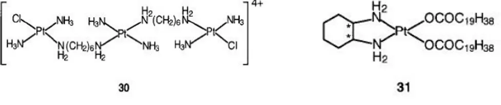
Complex 29 (Lobaplatin, D-19466) was approved into clinical trials in 1992.
Figure 11 Structure of Platinum complexes
The central Pt unit in the trinuclear complex 30 (BBR3464, counteranion = NO3-), is capable only of hydrogen-bonding interactions with DNA and
is reported to be up to 100-fold more potent than cisplatin against human tumor cell lines resistant to cisplatin .[99-101] The general charge of 4 greatly enhances DNA affinity, characterized by long-range interstrand cross-linking up to six base pairs apart with significant efficient, unwinding and irreversible conversion of B- to Z-DNA. The adducts are capable of terminating the DNA synthesis in vitro. The cytotoxic effect of the complex is insensitive to the p53 status of cisplatin-resistant cells (effective against tumors showing a mutant p53).
cis-Bis(nonadecanoato)(trans-R,R-1,2-diaminocyclohexane)- platinumII 31 (N-DDP) is a ipophilic liposome-incorporated cisplatin analogue that has demonstrated promising in vivo activity against tumors resistant to cisplatin and liver metastases.[102]
1.9.2 Platination of DNA
The guanine N7 is the most electron-rich site on DNA (most easily oxidized), and the major adducts of platinum drugs with DNA are 1,2-GpG and 1,2-ApG intrastrand crosslinks. The features of these adducts have been widely characterized and reviewed.[ 103] The NMR solution
structure of cis-[{Pt(NH3)2}2+{d(CCTG*G*TCC)⋅d(GGACCAGG)}]
(where * denotes Ptbound G) indicates that the B-DNA backbone conformation is mainly altered to accommodate the platinated lesion (Figure 13).[104]
Figure 13 NMR solution structure of cis-[{Pt(NH3)2}2+{d(CCTG*G*TCC)
⋅d(GGACCAGG)}]
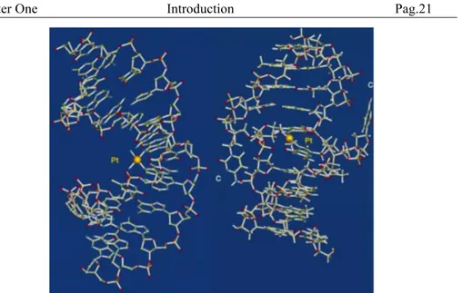
A novel X-ray crystal structure of the cisplatin adduct of the duplex d(CCTCTG*G*TCTCC)⋅ d(GGAGACCAGAGG) (Figure 4) shows that
cisplatin bends DNA by 35-40° in the direction of the major groove with a dihedral angle of 268 between the two guanine rings.[105,106] The duplex acquires a juxtaposition of A-like and B-like helical DNA segments. It is notable that the conformation surrounding the GG platination site in the solid-state X-ray crystal structure [106] is strongly similar to that in solution.[104,107] The Pt ion is removed from the planes of the coordinated
G-bases by about 0.8 Å, suggesting the presence of critical strain in this lesion. The properties and nature of minor cisplatin -DNA lesions which account for about 10%of the adducts, including 1,3-d(GpNpG) interstrand and intrastrand cross-links and monofunctional adducts, are less well know. Interstrand cross-linking of DNA occurs mainly between two guanine residues on opposite strands, and requires a distance of approximately 3 Å between the two N7 atoms. The most conventional interstrand cross-linking sequences are 5'-CG and 3'-CG.[108] In these
sequences the two guanine residues are separated by at least 7-9 Å; therefore a large distortion of double-helical B-DNA is need to achieve the cross-linking.
The solution NMR structure (see Figure 14 on the left) of cis-[{Pt(NH3)2}2+{d(CATAGCTATG)2}] shows that the duplex endures a
strong rearrangement at the lesion site, locating the platinum atom in the minor groove.[109] The deoxyribose of the platinated G residue is inverted
so that O4' is pointing in the opposite direction respect to the remaining nucleotides. Moreover, the C residue which was initially base-paired to the platinated G is extruded and is transformed in extra-helical
(see Figure 14 on the right). In the solution NMR structure of the interstrand cross-linked [{Pt(NH3)2}2+}-{d(CCTCG*CTCTC)
´d(GAGAG*CGAGG)}], the cis-{Pt- (NH3)2}2+ moiety is also placed in
the minor groove.[110] The stacking of the two cross-linked guanine residues with the surrounding bases forms a bend of 40° towards the minor groove. A very similar bend occurs in the X-ray crystal structure of this interstrand cross-link. The two cytosin residues are extruded from the double helix, and the platinum residue is embedded in a cage of nine water molecules. The general mechanism of formation of GG cross-links on DNA by cisplatin is evident in Scheme 2. Cisplatin is hydrolyzed to give monoaqua and diaqua species with a half-life of 1.7 h at 310 K. The aqua complexes are considered to be the active species towards DNA. The finding that the 5'-G monofunctional adduct formed during the reaction of cisplatin with the 14-mer DNA duplex d(ATACATGGTACATA) d(TATGTACCATGTAT) is very long lived
with a half-life of 80 h at 298 K was unexpected.[111,112] The half-life of the two monofunctional G adducts of the GG single strand are similar, proposing that the three-dimensional structure of DNA has a role in stabilizing the 5'-G adduct either by constraining the position of the
incoming 3'-G N7 ligand or by shielding the Cl ligand from hydrolysis. Molecular modeling studies prove that hydrogen-bonding between the carbonyl groups on DNA and the NH3 ligands plays a main role in
determining the orientation of the Pt-Cl bonds and their accessibility. Molecular mechanics calculations demonstrate that although the chloride ligand in the monofunctional adduct faces outward, away from the helix, the aqua ligand which replaces it after hydrolysis faces inwards on account of its strong hydrogenbonding properties.[112] Modeling of
transition states is now necessary.
Scheme. 2 Mechanism of reaction of cisplatin with DNA
Kinetic analyses based on HPLC results provide the accurate determination of the rates of both platination and chelation steps for reactions of cisplatin diaqua with oligonucleotides.[ 113,114] For the double-stranded oligonucleotide d(TTGGCCAA)2 the formation of the 5'-G
monoaqua adduct is faster than that of the 3'-G monoadduct, and macrochelate ring closure of the 5'-G monoaqua to give the bifunctional adduct (half-life of 3.2 h at 293 K) is much slower than that of the 3'-G monoadduct. The biological importance of long-lived monofunctional adducts on DNA remains to be detected, but these alone may be sufficient to kill cells if they are not repaired, which seems to be the case for complex 33, the active trans iminoether (see Section 1.9.3).[115] Long-lived monofunctional adducts may also contribute to the formation of DNA- protein cross-links.
Figure 14 Crystal structure of [{Pt(NH3)2}2+}-{d(CCTCG*CTCTC) ´
d(GAGAG*CGAGG)}] (on the left) and of cis-[{Pt(NH3)2}2+{d(CATAGCTATG)2}] (on
the right)
The stability of the Pt-N7G bond is demonstrated to be very high, and it can be broken only by a very strong nucleophiles such as thiourea or cyanide. However, recent studies have found that this bond can be labile in certain DNA adducts.
The adduct trans-{Pt(NH3)2}2+{d(TCTACG*CG*TTCT)}] (1,3-GG
cross-link) is at neutral pH unstable and rearranges to form the linkage isomer trans-[{Pt(NH3)2}2+{d(TCTAC*GCG*TTCT)}] (1,4-CG
cross-link).[116] It was indicated subsequently that intra- and interstrand transplatin -DNA adducts undergo isomerization in both double- and single-stranded and DNA.[117-119] For instance interstrand cross-links between a platinated 5'-G and the complementary C residue can be formed.[117]
Scheme. 3 Rearrangement of a trans-[{Pt(NH3)2}-DNA] intrastrand cross-link (left) to
The isomerization of the intrastrand d(CCTG*G*TCC) ⋅ d(GGACCAGG)
cross-link to a d(CCTG*GTCC) ⋅ d(GGACCAGG*) interstrand
cross-link proves that even Pt-N7 bonds in Pt - 1,2-GpG cross-cross-links can be destabilized.[ 104] This process seems to be assisted by Cl-.
1.9.3 Protein Recognition
In tumor cells the repair of platinated DNA lesions may be protected by a class of proteins named HMG high mobility group proteins.[120, 121]
The protein HMG1 sows three structural domains, two of them, known as domains A and B, are positively charged, and the third domain comprises a 30-amino-acid stretch of acidic residues at the C terminus site. Several NMR structures of HMG domains have been performed.[122,123] Domain B
of HMG1 consists of three α-helical regions joined by small loops, and folded into an L-shaped structure. The two domains, the positively charged domain A and the central domain B bind to DNA, while the acidic C-terminal domain interacts with histones only.
The binding affinity of HMG1 towards a series of cisplatin modified 15-mer DNA duplexes d(CCTCTCN1G*G*- N2TCTTC)
⋅ d(GAAGAN3CCN4GAGAGG) is strongly dependent on the bases
adjacent to the Pt lesion.[124] The affinity for HMG1 domain A decreases by at least two orders of magnitude in the order N2=dA>T>dC. When N1=N2=dA, Pt-DNA binds 100-fold stronger to HMG1 domain A (Kd.1.6nM) than to HMG1 domain B (Kd.134nM). The HMG binding
succed ind increasing the bending of platinated DNA to as much as 90°.[125] NMR studies of a GG 14-mer platinated with cisplatin show that the kinked duplex binds in the elbow region of HMG1 domain A,[126] and recent X-ray studies of HMG1 domain A bound to a 16-base-pair DNA fragment containing a cisplatin-DNA 1,2-d(GpG) intrastrand crosslink suggest that the protein inserts a Phe side chain into the platinum site. The linker histone H1, a nuclear protein, also binds much more strongly to DNA modified by cisplatin than to transplatin or unmodified DNA.[127] Competitive binding of a cisplatin modified 123-base-pair DNA fragment suggest that histone H1 binding is stronger than that of HMG1. Also the TATA box-binding protein (TBP)/TFIID, a promoter recognition factor binds selectively to and is sequestered by cisplatin-damaged DNA. This may lead TBP/TFIID away from its normal promoter sequence explaining
the inhibition of RNA synthesis.[128] Therefore these proteins may also have an important roles in the mechanism of action of platinum complexes drugs.
1.9.4 Active trans Complexes
Most of the work on the design of platinum anticancer complexes has focused on cis-diam(m)ine complexes, since it was demonstrated, years ago, that trans-[PtCl2(NH3)2] is inactive. However, the discovery that
trans-[PtCl2(py)2] (py = pyridine) is strongly cytotoxic has stimulated
renewed interest in trans complexes.[129] The trans PtIV complex 32 is active against both human and murine subcutaneous tumor models,[130] and efficiently promotes single-strand breaks and DNA interstrand cross-links.[131] These properties may explain its unusual cytotoxicity property against cisplatin-resistant tumors.[132] Strikingly the corresponding trans PtII complexes,without the axial hydroxo groups, are inactive. Direct reactions of the PtIV complexes with DNA may be crucial, but in vivo reduction could give reactive intermediates.
Trans-Imino ether platinum complexes such as 33 are more cytotoxic than the cis analogues (Ome and Pt are cis with respect to C=N in the Z isomer, and trans in the E isomer).[133,134] The mechanism of action of these agents seems to be different from that of cisplatin and may be correlated to the properties of the imino ether ligands.[135] Although these
trans complexes react with DNA more slowly than cisplatin, they give the same level of DNA binding after 24 hrs. The trans E,E complex is the m strongest complex in inhibiting DNA synthesis and cell proliferation, but does not induce large local DNA conformational changes.[136] It especially forms monofunctional adducts at guanine residues in double-helical DNA even after long incubation times (48 hrs at 310 K).[137a] The reactivity of the second chloride ligand in the monofunctional adduct is strongly reduced. The trans E,E monofunctional adducts are not recognized by HMG proteins.[137b,c]
Figure 15 Structures of trans Pt complexes
1.9.5 Biotransformation
l-Methionine (L-MetH) may have an important role in the metabolism of
platinum anticancer drugs. The PtII- L-Met complexes form quickly in
plasma after injection of cisplatin into rats.[138] The complex [Pt(Met-S,N)2] has been detected in the urine of patients[139] and exists mainly as
the cis isomer (cis:trans=87:13) in solution (Figure 16).[140]
Figure 16 Isomerization of R,R-trans- and S,R-cis-[Pt(Met)2], a metabolit of cisplatin
HAS, the human serum albumin is a single-chain 66-kDa protein which has 17 disulfide bridges and one free thiol group at Cys34, as well as six Met residues: M87, M123, M298, M329, M446, and M548. Reaction between HAS and cisplatin is believed to be the main route for platinum binding in human blood plasma.[141] Recent data suggest that cisplatin reacts strongly with methionine residues of albumin, forming a Met- S,N macrochelate together with minor adducts with Cys34 and monodentate Met-S residues.[142]
The reaction of carboplatin 24 with l-Met gives way to to a surprisingly stable ring-opened intermediate with a half-life of 28 hrs at 310 K.[143] Similar ring-opened complexes seems to be present in urine after
carboplatin administration.[144] Therefore it can be possible that the reaction of carboplatin with Met or its derivatives could give an activation pathway for the drug. The binding of the methionine sulfur atom is reversible, and it can be displaced by guanine N7.
Intracellular thiols such as glutathione (the tripeptide g- Glu-Cys-Gly, GSH), often present at concentrations of 3-10mM, are capable of
inactivating platinum drugs. The main product from reaction between GSH and cisplatin is a high molecular mass polymer with a Pt:GSH ratio of 1:2.[145] The formation of these polymers may be responsible for depletion of platinum from the circulation. The active export of cisplatin from cells is one of the main mechanisms of resistance, and a GS-X pump, an ATP-dependent export pump for GS-conjugates, is able to pump out GS-Pt complexes from cancer cells.[146] Reaction between cisplatin and the cysteine-rich protein metallothionein (MT) leads to displacement of the ammine ligands and gives rise to Pt7-10MT containing
PtS4 clusters.[147] This gives way to another pathway for the inactivation
of Pt drugs.
1.9.6 Photoactivation
Both PtII and PtIV complexes have long been considered to be photoreactive, and there are many field for the application of photodynamic reactions in pharmacology and biochemistry. A new approach to DNA platination involves the use of iodoplatinum(IV) complexes which are activated by visible light.[148] The toxicity of trans,cis-[Pt(en)(OAc)2I2] against human bladder cancer cells is enhanced
(by 35%) when the treated cells are irradiated with light of λ >375 nm. It has been shown that visible light can give way to the aquation of this complex followed by photoreductive platination of guanosine monophosphate, in contrast to the dihydroxo analogue.[149] Reactions of iodoplatinum(IV) ethylenediamine complexes with glutathione in the absence of light involve the initial attack of thiol on an iodide ligand of PtIV to give a reactive chelate ringopened intermediates.[150] Electron transfer driven labilization of trans ligands provides a new concept in drug design. UV light induced cleavage of both inter and intrastrand cross-links involving Pt-G bonds has been observed. Irradiation of DNA modified by cisplatin with UV light (λ>300 nm) can produce specific
cross-links to the protein HMG1, thought to involve Lys6 in domain B with labilization of a - purine bond.[151, 152]
1.9.7 Octahedral Platinum(IV) Complexes
Octahedral PtIV complexes show a tendency towards ligands substitution by a dissociative mechanism respect to an associative mechanism for PtII, with the net result that PtIV compounds are moderately more substitutionally inert. This is desirable for lower toxicity and oral bioavailability but it is not desirable for DNA intercalation. Despite that, most PtIV complexes are showing considerable activity in initial trials, with functionality thought to depend on the in vivo reduction of Pt IV to PtII, producing reactive intermediates capable of interacting with DNA. In
order to obtain oral bioavailability and beat cellular resistance, reserchears have tried multiple strategies to improve cisplatin properties. The reduction of PtIV to PtII compounds by biological agents is crucial to exert their antitumor activity. The reduction potential of PtIV complexes depends on the type of equatorial and axial ligands [153-160]. Choi et al. in another study, proved that the reduction rates which correlate with the reduction potentials depend on the electron withdrawing influence of the axial ligand [157]. In relation to the studied ethylenediamine-based PtIV complexes the fastest reduction rate (OH < OCOCH3 < Cl < OCOCF3)
corresponds to the most electron withdrawing ligand and correspond to the highest observed cytotoxicity. Kelland et al. reported the synthesis of about 25 trans-PtIV complexes and their trans-PtII counterparts, and then evaluated and compared in vitro as well as in vivo anticancer activity of these complexes. The described compounds had the following general formula: trans- [(amine)(ammine)Cl2Y2] PtIV (Y =OH or Cl). Two amino
groups atoms together with two chloride lie in the same plane, in equatorial positions. In opposition to them two hydroxide groups or two chloride atoms (Y ligands) are the axial ligands,. The anticancer activity studies gave very promising results. The complexes were tested in vitro against a series of human ovarian carcinoma cell lines, which contained tumor cells possessing either acquired (41McisR, CH1cisR and A2780cisR) or intrinsic (HX/62 and SKOV-3) resistance to cisplatin. Many of the complexes had a comparable anticancer activity respect to cisplatin and also did not show acquired cisplatin resistance. Notably, 14
complexes showed significant anticancer activity in vivo in the subcutaneous murine ADJ/PC6 plasmacytoma model. Among these, 13 complexes had axial hydroxido ligands and one had axial ethylcarbamate ligands, whereas all the platinumII and tetrachloroplatinumIV complexes were inactive [161].
1.9.8 Polynuclear Platinum Complexes
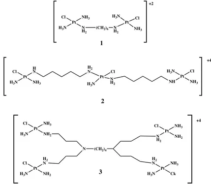
By the structural point of view, polynuclear platinum complexes are a different group. These innovative complexes have two or more platinum centers linked by various types of ligand (aromatic, aminoalkane, etc.). Thus, polynuclear complexes contravene many structure–activity rules for platinum complexes. Different SARs rules were created for polynuclear complexes based on their in vitro cytotoxicity results. One of the conditions to obtain good antitcancer activity of the complexes is the location of the leaving group, preferably chlorido, in trans position to the bridging linker. In the meanwhile, many trans and cis polynuclear complexes were synthesized, mainly by Farrell et al. [162-164]. Binuclear platinumII complexes with bifunctional spermine, thiourea and modified tetraamine linkers have been synthesized and studied. The secondary, protonated, uncoordinating amines in these compounds may give additional interaction with DNA by electrostatic interaction and hydrogen bonding and thus provide a stronger activity over the parent dinuclear agents such as BBR3005 [{trans-PtCl (NH3)2}2-µ-[{trans-
Pt(NH3)2(H2N(CH2)6NH2)2}](NO3)2 (Fig. 17.-complex 1) [165,166] .
Trinuclear and tetranuclear platimun complexes have also been eavaluated [167,168].The trinuclear complex BBR3464 (Fig. 17.- complex 2) is the first example of multinuclear compound which entered clinical trials in late 1997. Its preclinical antitumor profile was accentuated by remarkable potency, therapeutic doses approximately ten-folds lower that cisplatin, and activity in a wide set of solid human cancer [169,170]. Currently, BBR3464 is being entered in phase II clinical trials under the auspices of Novuspharma SpA. The investigations of phase I trials demonstrated that neutropenia and diarrhea were present as dose-limiting toxicities. The efficacy of this complex was not limited by neurotoxicity nephrotoxicity, or pulmonary toxicity [171]. The complex interacts with DNA with a different mechanism respect to cisplatin or other
mononuclear platinum complexes [172]. The tetranuclear platinum complex (Fig. 17.- complex 3), had low cytotoxic activity perhaps due to problems during trasport across the cell membranes [168].
Figure 17 Structure of polynuclear platinum complexes
1.10 Palladium Complexes as Alternative Potential
Antitumor Agents
The strong analogy between the coordination chemistry of PtII and PdII compounds has encouraged studies of PdII complexs as anticancer compounds[173,174]. A crucial factor that might explain is the ligand-exchange kinetics. The hydrolysis of leaving ligands in palladium complexes is too rapid, 105 times faster than their corresponding platinum complexes. They dissociate promptly in solution giving very reactive species that are unable to reach their pharmacological targets. In addition, some of them acquire an inactive trans-conformation. This
Pt Cl NH3 N H2 H3N (CH2)6 N H2 Pt NH3 Cl H3N +2 1 Pt Cl HN NH3 H3N H2 N Pt Cl N H2 H3N NH Pt H3N Cl NH3 +4 Pt Cl NH3 NH2 H3N N (CH2)4 Pt Cl HN2 NH2 H3N Pt Cl NH3 NH2 N H2 Pt H2 N NH3 Ck H3N +4 2 3
considerably stronger activity of palladium complexes suggests that in order to develop an antitumor palladium drug it must be stabilized by a strongly coordinated nitrogen ligand and a suitable leaving group. If this group is acceptably non labile, the drug can maintain its structural integrity in vivo for an enough long time. Many simple PdII compounds
with interesting cytotoxic features have been previously reported such as [cis-(NH3)2PdCl2] (Fig. 18- complex 1), [trans-(NH3)2PdCl2] (Fig. 18-
complex 2), [(1,5-COD)PdCl2], [(Π-C3H5)PdCl2]2 and
[(cyclopentyl)2PdCl2] Recent progresses in this field have also been based
on PdII compounds having bidendate ligands as a way to prevent any possible cis-trans isomerism [175-177] .
Figure 18 Two palladium (II) compounds with biological properties
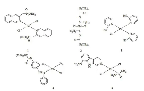
1.10.1 Trans-Palladium(II) Complexes Containing Bulky Monodentate Ligands
Many trans-Pd complexes with interesting activity against cancer cells have been prepared. Generally, the strategies that have been used to design these compounds were on the window of reactivity usually employed for the potential platinum anticancer drugs. A comparative study on anticancer activity was carried out between the diethyl-2- quinolmethylphosphonate (2-dmqmp) and dihalide PdII complexes of monoethyl-2-quinolmethylphosphonate (2-Hmqmp)[178]. The diester ligand has two potential donors, the the O from phosphoryl giving the complex [trans-(2-dqmp)2PdCl2] and N from quinoline and (Fig. 19-
complex 1). The complexes of the diester 2-dmqmp were found to be more active than those of the monoester-based ligand (2-Hmqmp). This may partly be due to the greater leaving activity of the halogen ligands in the complex having the 2-dmqmp ligand and to their greater solubility or lipophilicity. Pd H3N Cl H3N Cl Pd H3N NH3 Cl Cl 1 2
Figure 19 Trans-palladium (II) complexes containing bulky monodentate ligands
Fulrani et al., reported the synthesis and cytotoxic studies of some trans-[(L)2Pd(X)2] complexes (Figure 19 complex 2) (L=
N,N-dimethyl-O-ethylthiocarbamate: DMTC or N-methyl-O-N,N-dimethyl-O-ethylthiocarbamate: MTC, X= Cl, Br). Other palladium complexes based on 2-Mercaptopyridines (MP) were also synthesized. The [(MP)3Pd(Br)]Br (Fig. 19- complex 3) is
of potential clinical use since it shows lower IC50 values on LoVo cell lines than cisplatin and about the same as its PtII analogue [179].
PalladiumII complexes containing alkyl phosphonates derived from
quinoline and aniline were reported. Most of the aniline compounds (e.g. (Fig.19, complex 4)) had cyctotoxic properties in vitro against animal and human tumor cell lines. Complexes with naturally occurring compounds have also been used. The palladium complex which contains the bulky nitrogen ligand harmine (7-methoxy-1-methyl- 911-pyrido[3,4-b]indole, trans-[Pd(harmine) (DMSO)Cl2] (Figure 19 complex 5) shows a greater
cytotoxic activity against K562 and L1210 cell lines than cisplatin [180]. Abu-Surrah et al., recently, reported the synthesis and molecular structure of a new chiral, enantiometrically pure, trans-palladium(II) complex, trans-[2 {(R)-(+)- bornyl-amine}2Cl2] (Fig. 10) that has the bulky amine
activity against HeLa cells when compared to the activity of the standard drugs, cisplatin, carboplatin and oxaliplatin.
Figure 20 Structure of the palladium (II) complex
1.10.2 PalladiumII Complexes with Bidentate N∩N Ligands
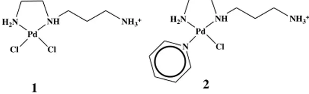
Dichloro palladiumII complexes with spermine and spermidine were
reported by Navarro-Ranninger et al. [182]. This type of chelating ligands have been utilized because of their relevant biological activity; they are involved in differentiation and proliferation of cells in membrane stabilization and DNA replication. Complexes of spermidine (Fig. 21- complex 1) show values of IC50 similar to cisplatin, whereas those of spermine (Fig. 21- complex 2) have low anticancer activity. Ethylendiamine palladiumII complexes with pyridine or its derivatives were also reported[183]. The increase of the electron donor properties of the substituents mainly lead to an increase of the donor strength of the coordinated pyridines, which directly lead to the increase of the cytotoxic activity of the palladium compounds.
Figure 21 Palladium(II) complexes with bidentate N∩N Ligands
Abu-Surrah et al., recently, used an alternative method to prepare the enantiometrically pure DACH-based palladiumII complexes [184,185]. In this procedure, the desired organic bidentate ligand was allowed to react
H2N Pd NH Cl Cl NH3+ H2N Pd NH Cl NH3+ N 1 2
with [cis-Pd(PhNC)2Cl2], a palladiumII starting material that is soluble at
25°C in most organic solvents, in CH2Cl2. Following this method, the
nucleophilic substitution reaction of the complex [cis-Pd(PhNC)2Cl2]
with (1R,2R)-(-)-1,2-diaminecyclohexane afforded the square planar Pd(II) complex [(1R,2R)-(-)-(DACH)PdCl2] (Fig. 22- complex 1) with an
high yield. The corresponding cationic, aqua complex, [(DACH)Pd(H2O)2](NO3)2 (Fig. 22- complex 2) and the oxalate complex
[(DACH)Pd(C2O4)] (Fig. 22- complex 3) have also been synthesized and
characterized [186]. A series of other oxaliplatin like complexes such as [(DACH)Pd(O-O)] has also been synthesized by Khokhar et al. [187] (O-O: malonate, methylmalonate, phenylmalonate, xylate). Unluckly, the influence of the different dicarboxylate ligands could not be focused since the complexes lack the anticancer activity. This could be related to the low stability and solubility of the above complexes in solution.
Figure 22 DACH-based palladium (II) complexes
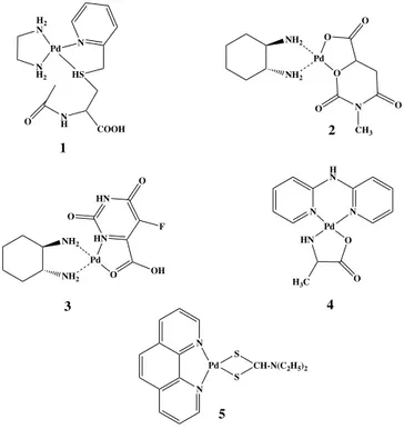
In order to increase the stability of the palladiumII complexes, two chelates forming two rings around the central atom were synthesized and studied. Modified amino acids such as py-CH2-accys (Fig. 23- complex 1)
(accys: N-acetyl-S-methylene-2-(2-pyridine)-L-cysteine) have been used
[188]. The S,N-chelation mode of these ligands is important, since only the
side chain of the aminoacid is involved in metal coordination, whereas the aminoacid function remains uncoordinated, leaving this functional group accessible for the attachment of other aminoacid or peptides. It has been demonstrated that the reactivity of these palladium complexes compete with some platinumII complexes. Another study investigated compounds having NO3 as a chelate in addition to a bidentate nitrogen ligand [189]. A
comparison among [(bipy)Pd(NO3)2], [(AMP)Pd(NO3)2],
[(AEP)Pd(NO3)2], [(DACH)Pd(Meorot)] (bipy = 2,2´-bipyridyl, AMP =
2-aminomethylpyridine, AEP = 2-aminoethylpyridine, Meorot = 3-methylorotate) proved that only [(DACH)Pd(Meorot)] (Fig. 23- complex 2) was active, showing an high activity against sarcoma 180 but a low one
H2 N Pd Cl N H2 Cl 1 H2 N Pd(OH2)2 N H2 2 H2 N PdC2O4 N H2 3 K2C2O4 AgNO3 H2O
against P388 leukemia. Similarly, [(DACH)Pd(5-fluroorot)] (Fig. 23- complex 3) later reported [190], showed a significant anticancer activity. These strong chelating ligands, replacing nitro or chloro ligands, induce a reduction in the percentage of hydrolysis.
Figure 23 Palladium II complexes bearing two bidentate chelates or tetradentate
nitrogen ligands
2-2’-bipyridylamine-based palladiumIIcomplexes having L-alanine or glycine have been reported and evaluated [191]. The alanine based complex
(Fig. 23- complex 4) dysplaied stronger cytotoxic activity against P388 lymphocytic leukemia cells than the glycine based one. Other aromatic ligands such as 1,10-phenanthroline, which is one of the most widely used ligands in coordination chemistry, has been utilized in the field of antitcancer-transition metal chemistry. Its planar nature allows its participation as a DNA intercalator. Several derivatives of it were synthesized and used as tetradentate ligands. The activities of [N,N-dialkyl-1,10- phenanthroline-2,9-dimethamine)Pd(II)] (Fig. 23- complex 5) (alkyl: Me, Ethyl, propyl, cyclohexyl) are significantly dependent on
H2 N N H2 Pd N HS N H O COOH NH2 NH2 O Pd O N O O O CH3 NH2 NH2 HN Pd O OH HN O O F HN O Pd N N H N O H3C N N Pd S S CH-N(C2H5)2 1 2 3 4 5
the nature of the alkyl substituents. The complexe having the bulkiest groups showed lower IC50values than cisplatin [192].
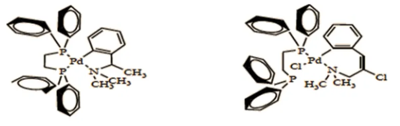
1.10.3 Palladium complexes with Biphosphine P∩P Ligands
Many of the prepared palladiumII complexes showed a moderate anticancer activity in vitro compared to the platinum-based drugs because of their very high lability in biological fluids [193]. Therefore, it has been suggested that the organometallic biphosphine-based cyclopalladated complexes which are more stable and less toxic could have in vivo a more specific anticancer activity[194]. Many cyclopalladate complexes based on biphosphine ligands (Fig. 24) have been synthesized and evaluated for their anticancer properties in a murine syngeneic B16F10 melanoma model. The ionic complex killed 100% tumor cells even at very low concentration (<1.25 µM). Other PdII complexes containing bidentate phosphine ligands of the general formula [L2PdXm]n+nX (L= Ph2
P-A-PPh2, A= (CH2)2, (CH2)3, X= Cl, Br, NO3) were synthesized and studied
for in vitro cytotoxicity, anticancer activity in murine tumor model and mechanism of action. The mode of action of these complexes seems tp be different from that of cisplatin in relation to the effects on DNA.
Figure 24 Palladium (II) complexes with biphosphine P∩P Ligands
1.10.4 Palladium(II) complexes with N∩S Mixed Donor Ligand
Khan et al. reported some palladiumII complexes with mixed sulfur-nitrogen ligands such as substituted pyrimidines (mercapto or amino) and methionine [195]. Methionine coordinates to PdII through sulfur and amino nitrogen, thus leaving a carboxylic group free. It has been demonstrated