Research Article
Whole Exome Sequencing Uncovers Germline Variants of
Cancer-Related Genes in Sporadic Pheochromocytoma
Milena Urbini
,
1Margherita Nannini
,
2Annalisa Astolfi
,
1Valentina Indio,
1Valentina Vicennati,
3Matilde De Luca,
1Giuseppe Tarantino,
1Federica Corso,
2Maristella Saponara,
2Lidia Gatto,
2Donatella Santini,
4Guido Di Dalmazi,
3Uberto Pagotto,
3Renato Pasquali,
3Andrea Pession,
2Guido Biasco,
1,2and Maria A. Pantaleo
1,21“Giorgio Prodi” Cancer Research Center, University of Bologna, Bologna, Italy
2Department of Specialized, Experimental and Diagnostic Medicine, S. Orsola-Malpighi Hospital, University of Bologna,
Bologna, Italy
3Endocrinology Unit, Department of Medical and Surgical Sciences, Center for Applied Biomedical Research,
S. Orsola-Malpighi Hospital, University of Bologna, Bologna, Italy
4Pathology Unit, S. Orsola-Malpighi Hospital, University of Bologna, Bologna, Italy
Correspondence should be addressed to Margherita Nannini; [email protected] Received 6 February 2018; Revised 8 May 2018; Accepted 29 May 2018; Published 19 August 2018 Academic Editor: Atsushi Kurabayashi
Copyright © 2018 Milena Urbini et al. This is an open access article distributed under the Creative Commons Attribution License, which permits unrestricted use, distribution, and reproduction in any medium, provided the original work is properly cited. Background. Pheochromocytomas (PCCs) show the highest degree of heritability in human neoplasms. However, despite the wide number of alterations until now reported in PCCs, it is likely that other susceptibility genes remain still unknown, especially for those PCCs not clearly syndromic. Methods. Whole exome sequencing of tumor DNA was performed on a set of twelve PCCs clinically defined as sporadic. Results. About 50% of PCCs examined had somatic mutations on the known susceptibility VHL, NF1, and RET genes. In addition to these driver events, mutations on SYNE1, ABCC10, and RAD54B genes were also detected. Moreover, extremely rare germline variants were present in half of the sporadic PCC samples analyzed, in particular variants of MAX and SAMD9L were detected in the germline of cases wild-type for mutations in the known susceptibility genes. Conclusions. Additional somatic passenger mutations can be associated with known susceptibility VHL, NF1, and RET genes in PCCs, and a wide number of germline variants with still unknown clinical significance can be detected in these patients. Therefore, many efforts should be aimed to better define the pathogenetic role of all these germline variants for discovering novel potential therapeutic targets for this disease still orphan of effective treatments.
1. Introduction
Pheochromocytomas (PCCs) are rare tumors of the auto-nomic nervous system that arise from the chromaffin tissue of the adrenal medulla [1]. Most of PCCs are benign; however, approximately 10% of cases are malignant and can develop metastases either at the time of diagnosis or even later after several years, with a highly variable clinical course and a 5-year overall survival rate of 50% [2]. PCCs show the highest degree of heritability in human neoplasms and almost 40% of cases occur within heritable syndromes, including multiple endocrine neoplasia type 2 (MEN2), neurofibromatosis
type 1 (NF1), von Hippel Lindau (VHL) disease, and hered-itary paraganglioma and familial pheochromocytoma [3]. Germline mutations have been identified in more than 15 well-characterized genes, such as VHL, SDHB, SDHD, NF1, and RET [4].
Despite this well-known inherited basis of PCCs and paragangliomas (PGLs), during the past decades somatic mutations with variable frequency in many genes, including
EPAS1 (HIF2α), RET, VHL, RAS, NF1, ATRX, and CSDE1
recurrent somatic copy number alterations and several fusion genes, involving MAML3, BRAF, NGFR, and NF1, have been progressively identified [5–16]. Recently a multiplatform
Volume 2018, Article ID 6582014, 9 pages https://doi.org/10.1155/2018/6582014
integrated analysis classified PCCs/PGLs into four clinically relevant molecular subtypes: a kinase signaling subtype, a pseudohypoxia subtype, a Wnt-altered subtype driven by MAML3 and CSDE1, and a cortical admixture subtype [16]. Therefore, given this molecular complexity of PCCs, it is likely that other alterations remain still unknown, especially for those PCCs not clearly syndromic. For this purpose, in the present study, we performed whole exome sequencing on a set of 12 clinically sporadic PCCs, with a family history negative for PCCs/PGLs.
2. Materials and Methods
2.1. Patients and Tumor Samples. Fresh tissue specimens of PCC from 12 patients with a family history negative for PCCs/PGLs were collected during the surgical operation,
snap-frozen in liquid nitrogen, and stored at −80°C until
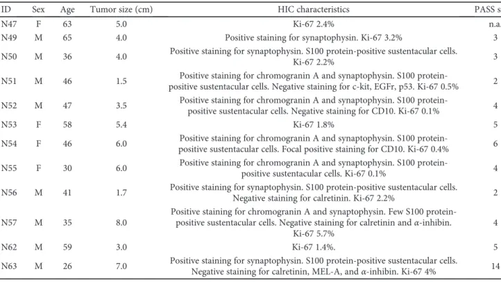
analysis. Patient characteristics are listed in Table 1. Whole exome sequencing was performed on biological tumor sam-ples on matched peripheral blood samsam-ples obtained from all patients. This study was approved by the local institutional ethical committee of S. Orsola-Malpighi hospital (approval number 95/2013/U/Tess). All patients provided written informed consent.
2.2. Whole Exome Sequencing. DNA was extracted from peripheral blood and fresh frozen tissue with DNA mini kit (Qiagen, Milan, Italy) following manufacturer’s instructions. Whole exome sequencing of tumor DNA was performed on HiScanSQ platform in accordance with Nextera Rapid Exome Enrichment protocol (Illumina, San Diego, California,
USA). Briefly, 100 ng of genomic DNA was tagged and
fragmented by the Nextera transposome. The Nextera
transposome simultaneously fragments the genomic DNA and adds adapter sequences to the ends. The products were then amplified and exome regions were enriched. The enriched libraries were amplified by PCR and quantified using PicoGreen assay (Life Technologies, Milan, Italy).
Paired-end libraries were sequenced at 2× 100 bp
read length using Illumina Sequencing by synthesis (SBS) technology.
2.3. Bioinformatic Analysis. After demultiplexing and FASTQ generation performed with bcltofastq function developed by Illumina, the paired-end reads were trimmed using AdapterRemoval (https://github.com/MikkelSchubert/ adapterremoval) with the aim of removing stretches of low-quality bases (<Q10) and Truseq/Nextera rapid capture adapters present in the sequences. The paired-end reads were then aligned on human reference genome hg38 (http://www. genome.ucsc.edu). Data from WES were mapped with Burrows-Wheeler Aligner with the default setting; the PCR and optical duplicates were removed, and Genome Analysis Toolkit (https://software.broadinstitute.org/gatk) was used to locally realign, recalibrate, and call the Ins/del
variants, while point mutations were identified with the
algorithm MuTect (https://www.broadinstitute.org/cancer/ cga/mutect). Single nucleotide variants (SNV) and ins/del were annotated with gene and protein alteration using Annovar (http://annovar.openbioinformatics.org); nonsy-nonymous and nonsense SNV, frameshift/nonframeshift Indels, and splice site mutations were selected with a
threshold read depth≥ 10x and a variant allele
fre-quency≥ 0.2. All the variants were filtered to select novel
or rare events basing on database of human variability
Table 1: Patient characteristics.
ID Sex Age Tumor size (cm) HIC characteristics PASS score
N47 F 63 5.0 Ki-67 2.4% n.a.
N49 M 65 4.0 Positive staining for synaptophysin. Ki-67 3.2% 3 N50 M 36 4.0 Positive staining for synaptophysin. S100 protein-positive sustentacular cells.
Ki-67 2.2% 3
N51 M 46 1.5 Positive staining for chromogranin A and synaptophysin. S100
protein-positive sustentacular cells. Negative staining for c-kit, EGFr, p53. Ki-67 0.5% 2 N52 M 47 3.5 Positive staining for chromogranin A and synaptophysin. S100
protein-positive sustentacular cells. Negative staining for CD10. Ki-67 0.1% 4
N53 F 58 5.4 Ki-67 1.8% 5
N54 F 46 6.0 Positive staining for chromogranin A and synaptophysin. S100
protein-positive sustentacular cells. Focal protein-positive staining for CD10. Ki-67 0.4% 6 N55 F 30 6.0 Positive staining for chromogranin A and synaptophysin. S100
protein-positive sustentacular cells. Ki-67 0.1% 4 N56 M 41 1.7 Positive staining for synaptophysin. S100 protein-positive sustentacular cells.
Negative staining for calretinin. Ki-67 2.2% 2
N57 M 35 8.0
Positive staining for chromogranin A and synaptophysin. Few S100 protein-positive sustentacular cells. Negative staining for calretinin andα-inhibin.
Ki-67 5.7%
4
N62 M 59 3.0 Ki-67 1.4%. 5
N63 M 26 7.0 Positive staining for synaptophysin. S100 protein-positive sustentacular cells.
dbSNP (http://www.ncbi.nlm.nih.gov/SNP), 1000Genomes (http://www.1000genomes.org), ExAC (http://exac.broad institute.org), and EVS (http://evs.gs.washington.edu/EVS).
In-depth evaluation of high confidence somatic variants was
performed by verifying the presence of alternate allele on the normal counterpart and manually visualizing each variation with the tview function of SAMmtools. Both somatic muta-tions and germline variants were searched in COSMIC (Catalog of Somatic Mutations in Cancer; http://cancer. sanger.ac.uk/cosmic), ClinVar (https://www.ncbi.nlm.nih. gov/clinvar), and HGMD (http://www.hgmd.org), and their effect on protein structure and function was predicted with SNPeff, a software that uses three different prediction algo-rithms (SIFT, Polyphen2, and LRT).
Moreover, based on WES data, the analysis of ampli
fica-tions and large delefica-tions were performed making a consensus between Control FREEC (http://boevalab.com/FREEC) and ADTEX (http://adtex.sourceforge.net) with paired tumour/
matched normal samples. Afiltering procedure was applied
taking into account the uncertainty value given by Control FREEC (<80%) and the polymorphic copy number variants from the Database of human Genomic Variants (http://dgv. tcag.ca/dgv/app/home).
For germline variants prioritization, all rare (MAF< 0.01) alterations occurring on the known susceptibility genes of PCC and PGL were considered. Moreover, variants with an
evident effect on the protein (nonsense and splicing mutations
or frameshift ins/del) were prioritized and manually anno-tated using HGMD and ClinVar database and with literature. 2.4. Sanger Sequencing. Sequencing of the DNA extracted from tumors and matched peripheral blood samples was per-formed to validate candidate mutations. Specific PCR assay for the amplification and sequencing of selected genes was designed with Primer Express 3.0 Software (Applied
Biosys-tems, Monza, Italy). PCR products were purified with the
Qia-quick PCR purification kit (Qiagen) and sequenced on both
strands using the Big Dye Terminator v1.1 Cycle Sequencing kit (Applied Biosystems). Sanger Sequencing was performed on ABI 3730 Genetic Analyzer (Applied Biosystems). 2.5. Real-Time PCR. Total RNA was extracted from fresh fro-zen tissues using the RNeasy spin-column method (Qiagen). RNA was reverse transcribed to cDNA using the Transcrip-tor First-Strand cDNA Synthesis Kit (Life Technologies) with
oligo dT primers. qPCR amplification of genes of interest was
performed with real-time LightCycler 480 instrument (Roche). Fold-change was estimated by DDCt method, using ATPS, HPRT, and HMBS genes as housekeeping controls. Primers used were: MAX_FW 5′- GCGATAACGATGAC ATCGAGGT-3′ and MAX_RV 5′-CCCGCAAACTGTGA AAGCTGT-3′, SAMD9L_FW 5′-AAAGTGAGTGAGTG AGCCCAG-3′ and SAMD9L_RV 5′-CATGCTCTTTGGTC CAGTCT-3′, ATPS_FW 5′-GTCTTCACAGGTCATATGG GGA-3′ and ATPS_RV 5′-ATGGGTCCCACCATATAGA AGG-3′, HMBS_FW 5′-GGCAATGCGGCTGCAA-3′ and HMBS_RV 5′-GGGTACCCACGGAATCAC-3′, HPRT_FW 5′-TGACACTGGCAAAACAATGCA-3′ and HPRT_RV
5′-GGTCCTTTTCACCAGCAAGCT-3′. For detection of cortical admixture profile, expression levels of STAR, CYP2W1, CYP11B2, CYP21A2, and CLND2, genes were eval-uated using the following primers: STAR_Fw 5′-TGGGCA TCCTTAGCAACCAA-3′ and STAR_Rev 5′-GCCCACATC TGGGACCACTT-3′; CYP2W1_Fw 5′-GTCATGGTCCT CTTGGGGTC-3′ and CYP2W1_Rev 5′-CTCCAGGAGGG TCCTCAGAA-3′; CYP11B2_Fw 5′-TGCATCCCTGCAGG ATGAT-3′ and CYP11B2_Rev 5′-GCGACAGCACATCT GGGT-3′; CYP21A2_Fw 5′-AGCCCGACCTCCCCAT-3′ and CYP21A2_Rev 5′-CACCACCACATCTTGCAGCC-3′;
CLND2_Fw 5′-CCCCTTGTACTTCGCTCCCC-3′ and
CLND2_Rev 5′-AAGCAGCCTCAAGAAGGCATC-3′.
3. Results
Exome sequencing generated a minimum of 45.5 million reads/sample with a mean coverage of RefSeq regions of 44x. Few somatic mutations were identified (an average of 7 mutations per sample) while several copy number alterations were detected, with losses of chr1, chr3, and chr17 being the most recurrent.
3.1. Analysis of Somatic Mutations. The biological effect of
somatic mutations was predicted with three bioinformatic tools (Suppl. Table 1). Mutations on susceptibility genes were detected in 6 out of 12 cases and were annotated in COSMIC and HGMD databases. VHL missense mutations were identi-fied in two cases: a p.S65A identiidenti-fied in N51, a mutation already reported in PCC (COSM144970), and a p.Y98H in N56, a mutation reported in ClinVar and HMGD as patho-genic in association with Von Hippel-Lindau syndrome. Two novel somatic alterations were detected in NF1: N62
carried a splicing mutation c.480-1G>C in exon 5 and N55
had a frameshift deletion (p.W784fs) in exon 20. Interest-ingly, the abovementioned mutations on NF1 and VHL were
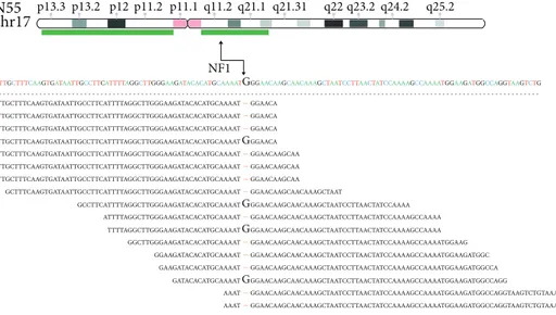
in regions affected by loss of the wild-type allele (Figure 1).
Heterozygous loss of the chromosomal region covering NF1 was detected also in N54; however, no additional mutational event was detected on this gene. RET was found mutated in heterozygosis in two other tumors: a novel exon 11 nonfra-meshift INDEL (p.L633delinsLCR) was detected in N57 and the hotspot mutation p.M918T (COSM965) in N53.
In addition to these driver mutations on known sus-ceptibility genes, other somatic passenger mutations were detected in these 6 tumors. In particular, SYNE1 and ABCC10 were mutated, respectively, in the two RET-mutated cases (N53 and N57), while a missense p.G460S mutation of RAD54B, a gene involved in DNA repair process, affected a NF1 mutated case (N55). On the other side, few somatic mutations were identified in the 6 remaining PCC cases and none was recurrent between samples. N63 carried a p.L114X nonsense mutation in CDC14B, a protein phos-phatase involved in DNA damage response. N52 carried two heterozygous missense mutations (SMARCC2 p.P1092R and PRKG1 p.F387) both predicted as pathogenic by bioinformatic predictors.
chr3 GCAATCGCAGTCCGCGCGTCGTGCTGCCCGTATGGCTCAACTTCGACGGCGAGCCGCAGCCCTACCCAACGCTGCCGCCTGGCACGGGCCGCCGCATCCACAGCTACCGAGGTACGGGCCCGGCG ATCGCAGTCCGCGCGTCGTGCTGCCCGTATGGCTCAACTTCGACGGCGAGCCGCAGCCCCACCCAACGTGCCGCC GTCCGCGCGTCGTGCTGCCCGTATGGCTCAACTTCGACGFCGAGCCGCAGCCCCACCCAACGCTGCCGCCTGGCAC CCGCGCGTCGTGCTGCCCGTATGGCTCAACTTCGACGGCGAGCCGCAGCCCCACCCCAACGTGCCGCCTGGCACGG CGCGTCGTGCTGCCCGTATGGCTCAACTTCGACGGCGAGCCGCAGCCCCACCCCAACGTGCCGCCTGGCACGGGC GTGCTGCCCGTATGGCTCAACTTCGACGGCGAGCCGCAGCCCCACCCAACGCTGCCGCCTGGCACGGGCCGCCGCA TGCTGCCCGTATGGCTCAACTTCGACGGCGAGCCGCAGCCCCACCCAACGCTGCCGCCTGGCACGGGCCGCCGCA TG TGCCCGTATGGCTCAAACTTCGACGGCGAGCCGCAGCCCCACCCAACGCTGCCGCCTGGCACGGGCCGCCGAT CCCGTATGGCTCAACTTCGACGGCGAGCCGCAGCCCTACCCAACGCTGCCGCCTGGCACGGGCCGCCGCATCCACA GTATGGCTCAACTTCGACGGCGAGCCGCAGCCCCACCCAACGCTGCCGCCTGGCACGGGCCGCCGCATCCACAG GGCTCAACTTCGACGGCGAGCCGCAGCCCCACCCAACGCTGCCGCCTGGCACGGGCCGCCGCATCCACAGCTACC TGGCTCAACTTCGACGGCGAGCCGCAGCCCTACCCAACGCTGCCGCCTGGCACGGGCCGCCGCATCCACAGCTACC GCTCAACTTCGACGGCGAGCCGCAGCCCTACCCAACGCTGCCGCCTGGCACGGGCCGCCGCATCCACAGCTACCGA CAACTTCGACGGCGAGCCGCAGCCCCACCCAACGCTGCCGCCTGGCACGGGCCGCCGCATCCACAGCTACCGAGGTA GCAATCGCAGTCCGCGCGTCGTGCTGCCCGTATGGCTCAACTTCGACGGCGAGCCGCAGCCCCACCCAACGCTGCCGC GCAATCGCAGTCCGCGCGTCGTGCTGCCCGTATGGCTCAACTTCGACGGCGAGCCGCAGCCCCACCCAACGC GCAATCGCAGTCCGCGCGTCGTGCTGCCCGTATGGCTCAACTTCGACGGCGAGCCGCAGCCCTACCCAACGC GCTCAACTTCGACGGCGAGCCGCAGCCCCACCCAACGCTGCCGCCTGGCACGGGCCGCCGCATCCACAGCTACCGA Y N56 p26.1 p24.3 p22.1 p21.2p14.2 p11.2q12.3 q13.32 q22.1 q25.1q26.1 q27.2 q29 VHL (a) N51 chr3 p26.1 p24.3 p22.1 p21.2 p14.2 p11.2 q12.3 q13.32 q22.1 q25.1 q26.1 q27.2 q29 VHL K GAGGAACTGGGCGCCGAGGAGGAGATGGAGGCCGGGCGGCCGCGGCCCGTGCTGCGCGCGGTGAACTCGCGCGAG CGGAGGAACTGGGCGCCGAGGAGGAGATGGAGGCCGGGCGGCCGCGGCCCGTGCTGCGCGCGGTAACTCACGC GAGGAGGAGATGGAGGCCGGGCGGCCGCGGCCCGTGCTGCGCTCGGTGAACTCGCGCGAGCCCTCCCAGGTCAT GAGGAGATGGAGGCCGGGCGGCCGCGGCCCGTGCTGCGCGCGGTGAACTCGCGCGAGCCCTCCCAGGTCATCTT GCCCGGAGGAACTGGGCGCCGAGGAGGAGATGGAGGCCGGGCGGCCGCGGCCCGTGCTGCGCTCGGTGAACTCGCG GAGGAACTGGGCGCCGAGGAGGAGATGGAGGCCGGGCGGCCGCGGCCCGTGCTGCGCGCGGTGAACTCGCGCGAGT GCCGCGGCCCGTGCTGCGCGCGGTGAACTCGCGCGAGCCCTCCCAGGTCATCTTCTGCAATCGCAGTCCGCGCGTC GCCGCGGCCCGTGCTGCGCGCGGTGAACTCGCGCGAGCCCTCCCAGGTCATCTTCTGCAATCGCAGTCCGCGCGTC GCCGCGGCCCGTGCTGCGCGCGGTGAACTCGCGCGAGCCCTCCCAGGTCATCTTCTGCAATCGCAGTCCGCGCGTC GTCCGGCCCGGAGGAACTGGGCGCCGAGGAGGAGATGGAGGCCGGGCGGCCGCGGCCCGTGCTGCGCGCGG GTCCGGCCCGGAGGAACTGGGCGCCGAGGAGGAGATGGAGGCCGGGCGGCCGCGGCCCGTGCTGCGCTCGG GTCCGGCCCGGAGGAACTGGGCGCCGAGGAGGAGATGGAGGCCGGGCGGCCGCGGCCCGTGCTGCGCGCGGTGAA GAACTGGGCGCCGAGGAGGAGATGGAGGCCGGGCGGCCGCGGCCCGTGCTGCGCTCGGTGAATCGCGCGAGCC GAACTGGGCGCCGAGGAGGAGATGGAGGCCGGGCGGCCGCGGCCCGTGCTGTGCTCGGTGAAC AACTGGGCGCCGAGGAGGAGATGGAGGCCGGGCGGCCGCGGCCCGTGCTGCGCGCGGTGAACTCGCGCGAGCCCTC ACTGGGCGCCGAGGAGGAGATGGAGGCCGGGCGGCCGCGGCCCGTGCTGCGCGCGGTGAACTCGCGCGAGCCCTC ACTGGGCGCCGAGGAGGAGATGGAGGCCGGGCGGCCGCGGCCCGTGCTGCGCTCGGTGAACTCGCGCGAGCCCTC TCGGTGAACTCGCGCGAGCCCTCCCAGGTCATCTTCTGCAATCGCAGTCCGCGCGTC GTCCGGCCCGGAGGAACTGGGCGCCGAGGAGGAGATGGAGGCCGGGCGGCCGCGGCCCGTGCTGCGC (b) NF1 chr17 N62
GAGATACCACACCTGTCCCCTAATACTTAATTTGATAAGTTAATTTTGGTTTTTACTTTTTAGGTTACAGGAATTAACTGTTTGTTCAGAAGACAATGTTGATGTTCATGATATAGAATTGTTAC
S p13.3 p13.2 p12 p11.2 p11.1 q11.2 q21.1 q21.31 q22 q23.2 q24.2 q25.2 GTCCCCTAATACTTAATTTGATAAGTTAATTTTGGTTTTTACTTTTTACGTTACAGGAATTAACTGTTTGTTCAGA GTCCCCTAATACTTAATTTGATAAGTTAATTTTGGTTTTTACTTTTTAGGTTACAGGAATTAACTGTTTGTTCAGA GTCCCCTAATACTTAATTTGATAAGTTAATTTTGGTTTTTACTTTTTAGGTTACAGGAATTAACTGTTTGTTCAGA CCCCTAATACTTAATTTGATAAGTTAATTTTGGTTTTTACTTTTTACGTTACAGGAATTAACTGTTTGTTCAGAAG AGTTAATTTTGGTTTTTACTTTTTACGTTACAGGAATTAAACTGTTTGTTCAGAAGACAATGTTGATGTTCATG GAGATACCACACCTGTCCCCTAATACTTAATTTGATAAGTTAATTTTGGTTTTTACTTTTTACGTTACAGGAATTAA GAGATACCACACCTGTCCCCTAATACTTAATTTGATAAGTTAATTTTGGTTTTTACTTTTTACGTTACAGGAATTAA GAGATACCACACCTGTCCCCTAATACTTAATTTGATAAGTTAATTTTGGTTTTTACTTTTTAGGTTACAGGAATTAA GAGATACCACACCTGTCCCCTAATACTTAATTTGATAAGTTAATTTTGGTTTTTACTTTTTAGGTTACAGGAATTAA GAGATACCACACCTGTCCCCTAATACTTAATTTGATAAGTTAATTTTGGTTTTTACTTTTTACGTTACAGGAATTAACTGTT CACCTGTCCCCTAATACTTAATTTGATAAGTTAATTTTGGTTTTTACTTTTTAGGTTACAGGAATTAACTGTTTGT CCCCTAATACTTAATTTGATAAGTTAATTTTGGTTTTTACTTTTTAGGTTACAGGAATTAACTGTTTGTTCAGAAG CCCTAATACTTAATTTGATAAGTTAATTTTGGTTTTTACTTTTTAGGTTACAGGAATTAACTGTTTGTTCAGAAG AGTTAATTTTGGTTTTTACTTTTTACGTTACAGGAATTAACTTGTTTGTTCAGAAGACAATGTTGATGTTCATG TTGGTTTTTACTTTTTACGTTACAGGAATTAACTGTTTGTTCAGAAGACAATGTTGATGTTCATGATATAGAAT TTGGTTTTTACTTTTTACGTTACAGGAATTAACTGTTTGTTCAGAAGACAATGTTGATGTTCATGATATAGAATTG (c) Figure 1: Continued.
3.2. Analysis of Germline Rare Variants. In addition to the
abovementioned somatic alterations, rare (ExAc< 0.01)
germline variants were identified in five sporadic PCC
samples (Suppl. Table 2), among which those occurring in MAX and SAMD9L seem to play an important role in PCC pathogenesis, completing the picture of relevant alterations identified in our cohort (Table 2). In N63, a novel germline variant of MAX (c.397-2A>G), affecting splice site and path-ogenic for the protein function, was detected. Noticeably, this patient is a young adult (age 26) and showed the loss of the wild-type allele in the tumor, thus following the Knudson two-hit model (Figure 2(a)), and mRNA level of MAX was found downregulated with respect to the other PCC samples
(Figure 2(b)). According to the findings of Fishbein et al.
[16], we evaluated the expression level of STAR, CYP11B2, CYP2W1, CYP21A2, and CLND2 with the aim to assess whether a cortical admixture profile was present in this MAX mutated sample. However, a general low expression level of these genes was found in our cohort and no difference between the MAX mutated case and the other PCC samples was detected (data not shown). Conversely, 2 germline variants of SAMD9L were found in other 2 PCC, both wild-type for mutations in known susceptibility genes: a novel p.N769fs frameshift deletion in N47 and a rare (ExAc = 0.2%) nonsense p.R406X in N50 (Figure 2(c)). Evaluation of
mRNA level of SAMD9L showed a significant
downre-gulation of the transcript in this two PCC samples (Figure 2(d)). Interestingly, the same gene was found somat-ically mutated in N54 (p.L1016S), increasing to 3 the number of PCC wild-type cases of our cohort that carried alterations on SAMD9L (Table 2).
On the contrary, the role of the remaining germline variants (suppl. Table 2) is not clear. In N50, in addition to SAMD9L alteration, a heterozygous stop gain of BRCA2 (p.K3326X) was detected as constitutive. This variant is
recorded in ClinVar as “benign”; however, it is found
at low frequency in healthy individuals (ExAc allele fre-quency = 0.7%). BRCA2 mRNA expression level was evalu-ated, but it was found not altered in this case with respect to the other PCC cases (data not shown). Finally, the rare
germ-line variants of ATRX and KTM2D, identified in N56, and of
MDH2, in N53, are missense variants occurring in
associa-tion with a well-defined somatic mutation on a susceptibility
gene (VHL in N56 and RET in N53) and then it is not clear whether they could have a role on the tumor onset.
4. Discussion
In this study, we performed whole exome sequencing on a set
of twelve PCCs, clinically defined as sporadic, and we found
that 50% of PCCs examined had somatic mutations on the known susceptibility VHL, NF1, and RET genes. In addition to these driver mutations, other somatic passenger mutations were detected. In particular, SYNE1 and ABCC10 were mutated, respectively, in the two RET-mutated cases, while a missense p.G460S mutation of RAD54B affected a NF1 mutated case, suggesting these other events may play a potential role on PCCs pathogenesis and development.
Nuclear envelope 1 (SYNE1) gene encodes several different
isoforms involved in a variety of cellular processes including cytokinesis, Golgi function, and nuclear organization and structural integrity and positioning of the nucleus [17, 18]. Mutations of SYNE1 have been found in colorectal cancer, glioblastoma, and ovarian cancer, and methylation of the gene was also frequently found in lung adenocarcinoma and colorectal cancer [19–23]. On the other side, RAD54B is a telomere-related gene involved in DNA repair process, and coding-missense changes of this gene have been found in familial breast cancer cases not explained by mutations in the best-known high susceptibility genes BRCA1 and
BRCA2 [24]. Thesefindings may be even more relevant in
PCCs, given the highest degree of heritability of this disease.
p13.3 p13.2 p12 p11.2 p11.1 q11.2 q21.1 q21.31 q22 q23.2 q24.2 q25.2
NF1
N55 chr17
TTGCTTTCAAGTGATAATTGCCTTCATTTTAGGCTTGGGAAGATACACATGCAAAATGGGAACAAGCAACAAAGCTAATCCTTAACTATCCAAAAGCCAAAATGGAAGATGGCCAGGTAAGTCTG
TTGCTTTCAAGTGATAATTGCCTTCATTTTAGGCTTGGGAAGATACACATGCAAAAT–GGAACA TTGCTTTCAAGTGATAATTGCCTTCATTTTAGGCTTGGGAAGATACACATGCAAAAT–GGAACA TTGCTTTCAAGTGATAATTGCCTTCATTTTAGGCTTGGGAAGATACACATGCAAAAT–GGAACA TTGCTTTCAAGTGATAATTGCCTTCATTTTAGGCTTGGGAAGATACACATGCAAAAT–GGAACAAGCAA TTGCTTTCAAGTGATAATTGCCTTCATTTTAGGCTTGGGAAGATACACATGCAAAAT–GGAACAAGCAA TTGCTTTCAAGTGATAATTGCCTTCATTTTAGGCTTGGGAAGATACACATGCAAAAT–GGAACAAGCAA GCTTTCAAGTGATAATTGCCTTCATTTTAGGCTTGGGAAGATACACATGCAAAAT–GGAACAAGCAACAAAGCTAAT TTGCTTTCAAGTGATAATTGCCTTCATTTTAGGCTTGGGAAGATACACATGCAAAATGGGAACA GCCTTCATTTTAGGCTTGGGAAGATACACATGCAAAATGGGAACAAGCAACAAAGCTAATCCTTAACTATCCAAAA ATTTTAGGCTTGGGAAGATACACATGCAAAAT–GGAACAAGCAACAAAGCTAATCCTTAACTATCCAAAAGCCAAAA TTTTAGGCTTGGGAAGATACACATGCAAAATGGGAACAAGCAACAAAGCTAATCCTTAACTATCCAAAAGCCAAAA GGCTTGGGAAGATACACATGCAAAAT–GGAACAAGCAACAAAGCTAATCCTTAACTATCCAAAAGCCAAAATGGAAG GGAAGATACACATGCAAAAT–GGAACAAGCAACAAAGCTAATCCTTAACTATCCAAAAGCCAAAATGGAAGATGGC GAAGATACACATGCAAAAT–GGAACAAGCAACAAAGCTAATCCTTAACTATCCAAAAGCCAAAATGGAAGATGGCCA GATACACATGCAAAATGGGAACAAGCAACAAAGCTAATCCTTAACTATCCAAAAGCCAAAATGGAAGATGGCCAGG AAAT–GGAACAAGCAACAAAGCTAATCCTTAACTATCCAAAAGCCAAAATGGAAGATGGCCAGGTAAGTCTGTAAAG AAAT–GGAACAAGCAACAAAGCTAATCCTTAACTATCCAAAAGCCAAAATGGAAGATGGCCAGGTAAGTCTGTAAAG (d)
Figure 1: Deletion of the wild-type allele occurred in PCC samples that carry NF1 or VHL somatic mutations. Alignments of sequencing reads located on NF1 or VHL mutated bases are shown. Green bars represent deleted regions of chr3 for VHL (a, b) and chr17 for NF1 (c, d) detected by copy number analysis. A black arrow indicates the chromosome position of NF1 or VHL and the mutated base on the sequencing reads.
Table 2: Somatic and germline alterations identi fied in our cohort that could support PCC pathogenesis. Annotations concerning Exac frequency, COSMIC, ClinVar, and biological eff ect predictions are shown for each var iant. Sampl e Gene Somatic/ germline Position Exon cDna Protein Type dbSNP ExAC freq COSMIC ClinVar Eff ect prediction § N47 SAMD9L Germline chr7:93133665 5 c.2307delC p.N769fs Frameshift Novel — Yes — Pathogenic N50 SAMD9L Germline chr7:93134756 6 c.1216C > T p.R406X Stop gain rs150070697 0.002 Yes ∗ — Pathogenic N54 SAMD9L Somat ic chr7:93132925 4 c.3047T > C p.L1016S Missense Novel na —— Pathogenic N63 MAX Germline SNV + somatic loss chr14:65076665 7 c.397-2A > G na Splicing Novel — Yes — Pathogenic N51 VHL Somatic + loss chr3:10142040 1 c.193T > G p.S65A Missense Novel — Yes ∗ — Pathogenic N56 VHL Somatic + loss chr3:10142139 1 c.292T > C p.Y98H Missense rs5030809 — Yes Pathogenic Pathogenic N55 NF1 Somatic + loss chr17:31227548 20 c.2351delG p.W784fs Frameshift Novel — Yes — Pathogenic N62 NF1 Somatic + loss chr17:31169890 5 c.480-1G > C na Splicing Novel — Yes — Pathogenic N53 RET Somat ic chr10:43121968 16 c.2753T > C p.M918T Missense rs74799832 — Yes ∗ Pathogenic Pathogenic N57 RET Somat ic chr10:43114499 11 c.1899_1900insTGCCGC p.L633delinsLCR Nonframeshift insertion Novel — Yes ∗ — Pathogenic ∗COSM IC rec ord that totall y mat ch with the mutat ion ide nti fied; §de fined pathog enic if the mutat ion has bee n cl assi fied as pathog enic or deleter ious by at least tw o out three predict ors used (SIFT, Polyph en2, and LRT ).
Of note on this topic, the identification of germline variants in half of the sporadic PCC samples was analyzed, among which those occurring in MAX and SAMD9L genes may be extremely interesting. Indeed, some reports disclose the importance of screening for germline variants also in sporadic cases, especially on susceptibility genes [25, 26], and besides the potential role played in PCCs pathogenesis, the detection of these germline variants in patients clinically defined as sporadic may suggest the existence of unknown multineoplasia hereditary diseases.
In our study, we have identified a variant of MAX
(c.397-2A>G), affecting splice site and pathogenic for the protein
function, with the loss of the wild-type allele in the tumor as second hit. Of note, this patient is a young adult of 26 years old, and the presence of a germline variant in a known cancer susceptibility gene may suggest that the PCC in this patient
could be thefirst clinical expression of a hereditary disease
still undefined. It is already known that around 1% of PCC patients were negative for mutations in the other known susceptibility genes carried a germline mutation affecting MAX [26, 27]. About 20 variants affecting MAX have been already described distributed along the gene, but more fre-quently involving exons 3 and 4, matching some of the most important residues within the conserved bHLH-Zip domain of MAX. Most mutations lead to truncated proteins, with
the expected LOH affecting the remaining wild-type allele
of the MAX tumor suppressor gene [26, 27]. Two
truncat-ing MAX mutations affecting exon 3 (c.97C>T) and 4
(c.185_186delA) and three missense variants (c.67G>A,
c.281T>C, and c.425C>T) located in exons 3, 4, and 5,
respectively, have been identified [27]. Furthermore, other mutations affecting the initial methionine (c.2T>A), creat-ing a premature stop codon (c.25del, c.97C>T, c.223C>T, and c.244C>T) or affecting a donor/acceptor splice site (c.171 + 1G>A and c.295 + 1G>T), have been subsequently reported [26]. In addition, 2 deletions were identified: the first caused an inframe loss of 6 highly conserved amino
acids within the first helix of the protein (c.140_157del),
and the second, spanned the whole gene (c.1-?_483+?del) [26]. The MAX mutant case of our cohort showed a marked downregulation of MAX at mRNA level; however, it did not show a significant modulation of the genes involved in
cortical admixture phenotype. Conversely to this finding,
two cases of MAX mutant PCC were described to overexpress adrenal cortex markers (including CYP11B2, CYP21A2, and STAR) supporting an association between MAX mutation and the cortical admixture PCC subtype [16]. Further studies on larger cohort will be needed to assess this association.
Moreover, we have found three germline variants in BRCA2 and SAMD9L in other two PCC patients wild-type
Tum N63 MAX c.397-2A > G G C C T T T T G C C T T T T G G T C C G T G C PB G G T C C G T G C G C C T T T T G C C T T T T RR GG T C C G T G CT C C G T G C (a) 1.5 1.0 mRN A r ela tiv e exp ressio n 0.5 0.0
MAX mut MAX WT
MAX ⁎⁎ (b) N47 germline N50 germline SAMD9L c.2307delC SAMD9L c.1216C > T T A G G A A A C Y G AG A C T C A A A A A A A A R A C C A A A A A A A M R M T T A AC C A M T M T A G G A A A C Y G A G A C CA (c) 1.5 2.0 1.0 mRN A r ela tiv e exp re ssio n 0.5 0.0
SAMD9L mut SAMD9L WT
SAMD9L ⁎⁎
(d)
Figure 2: (a) Loss of heterozygosity of germline MAX mutation detected on N63 tumor. Chromatograms showing homozygous status of c.397-2A>G mutation in the tumor of N63 (upper panel), while in the germline it is heterozygous (lower panel). (b) MAX mRNA relative expression of MAX mutant case (N63) in comparison to the other PCC cases.P value was estimated with unpaired t-test (∗∗P < 0 01). (c) Validation of SNV detected on SAMD9L gene in 2 PCC samples. Germline c.2307delC and c.1216C>T heterozygous mutations detected in both tumor and peripheral blood of N47 and N50, respectively. (d) SAMD9L mRNA relative expression of the 2 SAMD9L mutant cases (N47 and N50) in comparison to the other PCC cases.P value was estimated with unpaired t-test (∗∗P < 0 01).
for mutations in susceptibility genes. Specifically, even if it is a rare variant (ExAc allele frequency = 0.7%) recently described as associated with an increased risk of developing breast and ovarian cancers [28], the heterozygous stop gain
of BRCA2 (p.K3326X) is considered as“benign” in ClinVar.
In support of this consideration, we did not detect any varia-tion of BRCA2 mRNA expression level. Thus, we cannot draw any conclusion on the role of this variant and PCC onset. On the contrary, the 2 rare germline variants of SAMD9L detected in two sporadic PCC cases were both producing a premature STOP of the protein (a p.N769fs frameshift deletion and a nonsense p.R406X) and were associated with significant downregulation of SAMD9L mRNA, supporting the pathogenicity of these alterations. The function of this gene is not well characterized; however, evidence has accumulated supporting the role of SAMD9L in cell proliferation and tumor suppression. In particular, somatic mutations have been found in hepatitis B-related hepatocellular carcinomas [29] and inactivation of SAMD9L has been recently correlated with myeloid transformation [30]. Interestingly, a somatic p.L1016S mutation of SAMD9L was detected in another PCC sample of our cohort, and additionally a p. P636S (COSM3412478) was reported in one case of TCGA PCC/PGL dataset (http://cancergenome. nih.gov/). Taken together, these data could support a possible role of SAMD9L in PCC biology; however, functional studies will be needed to further assess this hypothesis.
5. Conclusions
Taken together, the discovery of novel germline variants of known cancer-related genes in sporadic PCC patients, wild-type for mutations in susceptibility genes, may suggest that the likely existence of other multineoplasia syndrome. On the contrary, the role of the other rare germline variants
ATRX, KTM2D, and MDH2 genes identified in two cases
having a well-defined somatic mutation on a susceptibility
gene (RET or VHL) remains still unclear.
Although the pathogenetic role of all these variants is still not known, due to the large number of susceptibility genes implicated in the diagnosis of inherited PCCs and PGLs, we confirm also what other authors stated that the next-generation sequencing technology is ideally suited for carry-ing out genetic screencarry-ing of these individuals [31]. Moreover, the high degree of heritability of PCCs and the wide number of germline variants described suggest the likely need of a more extended genetic counselling and the type and duration
of the surveillance program of patients affected by PCCs with
variants of unknown significance. Finally, these findings may underlie the possible occurrence of novel hereditary diseases that remain still undefined.
In conclusion, additional somatic passenger mutations can be associated to known susceptibility VHL, NF1, and RET genes in PCCs and a wide number of germline variants with still unknown clinical significance can be detected in
these patients. Therefore, many efforts should be aimed to
better define the pathogenetic role of all these germline
variants for discovering novel potential therapeutic targets
for this disease still orphan of effective treatments.
Data Availability
All sequencing data are available upon request to the corresponding author.
Conflicts of Interest
The authors declare no conflict of interest.
Authors
’ Contributions
Milena Urbini and Margherita Nannini contributed equally to this work.
Supplementary Materials
Supplementary Table 1: list of somatic mutations identified
in our cohort. Annotations concerning ExAC frequencies,
COSMIC, and effect prediction are shown for each variant.
In yellow are highlighted mutations affecting PCC suscepti-bility genes. Supplementary Table 2: list of rare germline variants identified. Annotations concerning Exac frequency, COSMIC, ClinVar, and biological effect predictions (SIFT, Polyphen2, and LRT) are shown for each variant. (Supplementary Materials)
References
[1] J. W. M. Lenders, G. Eisenhofer, M. Mannelli, and K. Pacak, “Phaeochromocytoma,” Lancet, vol. 366, no. 9486, pp. 665– 675, 2005.
[2] G. Eisenhofer, S. R. Bornstein, F. M. Brouwers et al., “Malignant pheochromocytoma: current status and initiatives for future progress,” Endocrine-Related Cancer, vol. 11, no. 3, pp. 423–436, 2004.
[3] P. L. M. Dahia, “Pheochromocytoma and paraganglioma pathogenesis: learning from genetic heterogeneity,” Nature Reviews Cancer, vol. 14, no. 2, pp. 108–119, 2014.
[4] J. Favier, L. Amar, and A. P. Gimenez-Roqueplo, “Paragan-glioma and phaeochromocytoma: from genetics to personal-ized medicine,” Nature Reviews. Endocrinology, vol. 11, no. 2, pp. 101–111, 2015.
[5] N. Burnichon, L. Vescovo, L. Amar et al.,“Integrative genomic analysis reveals somatic mutations in pheochromocytoma and paraganglioma,” Human Molecular Genetics, vol. 20, no. 20, pp. 3974–3985, 2011.
[6] N. Burnichon, A. Buffet, B. Parfait et al., “Somatic NF1 inacti-vation is a frequent event in sporadic pheochromocytoma,” Human Molecular Genetics, vol. 21, no. 26, pp. 5397–5405, 2012.
[7] N. H. Cho, H. W. Lee, S. Y. Lim, S. Kang, W. Y. Jung, and C. S. Park,“Genetic aberrance of sporadic MEN 2A compo-nent tumours: analysis of RET,” Pathology, vol. 37, no. 1, pp. 10–13, 2005.
[8] I. Comino-Méndez, A. A. de Cubas, C. Bernal et al.,“Tumoral EPAS1 (HIF2A) mutations explain sporadic pheochromocy-toma and paraganglioma in the absence of erythrocytosis,” Human Molecular Genetics, vol. 22, no. 11, pp. 2169–2176, 2013.
[9] J. Crona, A. Delgado Verdugo, R. Maharjan et al.,“Somatic mutations in H-RAS in sporadic pheochromocytoma and
paraganglioma identified by exome sequencing,” The Journal of Clinical Endocrinology and Metabolism, vol. 98, no. 7, pp. E1266–E1271, 2013.
[10] L. Fishbein, S. Khare, B. Wubbenhorst et al.,“Whole-exome sequencing identifies somatic ATRX mutations in pheochro-mocytomas and paragangliomas,” Nature Communications, vol. 6, no. 1, p. 6140, 2015.
[11] R. Hrasćan, N. Pećina-Slaus, T. N. Martić et al., “Analysis of selected genes in neuroendocrine tumours: insulinomas and phaeochromocytomas,” Journal of Neuroendocrinology, vol. 20, no. 8, pp. 1015–1022, 2008.
[12] P. Komminoth, E. Kunz, O. Hiort et al.,“Detection of RET proto-oncogene point mutations in paraffin-embedded pheo-chromocytoma specimens by nonradioactive single-strand conformation polymorphism analysis and direct sequencing,” The American Journal of Pathology, vol. 145, no. 4, pp. 922– 929, 1994.
[13] R. A. Toledo, Y. Qin, Z. M. Cheng et al.,“Recurrent mutations of chromatin-remodeling genes and kinase receptors in pheochromocytomas and paragangliomas,” Clinical Cancer Research, vol. 22, no. 9, pp. 2301–2310, 2016.
[14] Z. Zhuang, C. Yang, F. Lorenzo et al.,“Somatic HIF2A gain-of-function mutations in paraganglioma with polycythemia,” The New England Journal of Medicine, vol. 367, no. 10, pp. 922– 930, 2012.
[15] A. Flynn, D. Benn, R. Clifton-Bligh et al., “The genomic landscape of phaeochromocytoma,” The Journal of Pathology, vol. 236, no. 1, pp. 78–89, 2015.
[16] L. Fishbein, I. Leshchiner, V. Walter et al., “Comprehen-sive molecular characterization of pheochromocytoma and paraganglioma,” Cancer Cell, vol. 31, no. 2, pp. 181–193, 2017.
[17] J. Fan and K. A. Beck,“A role for the spectrin superfamily member Syne-1 and kinesin II in cytokinesis,” Journal of Cell Science, vol. 117, no. 4, pp. 619–629, 2004.
[18] L. L. Gough, J. Fan, S. Chu, S. Winnick, and K. A. Beck,“Golgi localization of Syne-1,” Molecular Biology of the Cell, vol. 14, no. 6, pp. 2410–2424, 2003.
[19] M. Tessema, R. Willink, K. Do et al.,“Promoter methylation of genes in and around the candidate lung cancer susceptibility locus 6q23-25,” Cancer Research, vol. 68, no. 6, pp. 1707– 1714, 2008.
[20] K. E. Schuebel, W. Chen, L. Cope et al.,“Comparing the DNA hypermethylome with gene mutations in human colorectal cancer,” PLoS Genetics, vol. 3, no. 9, pp. 1709–1723, 2007. [21] T. Sjoblom, S. Jones, L. D. Wood et al.,“The consensus coding
sequences of human breast and colorectal cancers,” Science, vol. 314, no. 5797, pp. 268–274, 2006.
[22] D. L. Masica and R. Karchin,“Correlation of somatic mutation and expression identifies genes important in human glioblas-toma progression and survival,” Cancer Research, vol. 71, no. 13, pp. 4550–4561, 2011.
[23] J. A. Doherty, M. A. Rossing, K. L. Cushing-Haugen et al., “ESR1/SYNE1 polymorphism and invasive epithelial ovarian cancer risk: an Ovarian Cancer Association Consortium Study,” Cancer Epidemiology Biomarkers & Prevention, vol. 19, no. 1, pp. 245–250, 2010.
[24] K. Yanowsky, A. Barroso, A. Osorio, M. Urioste, J. Benitez, and B. Martinez-Delgado,“Mutational analysis of telomere genes in BRCA1/2-negative breast cancer families with very short
telomeres,” Breast Cancer Research and Treatment, vol. 134, no. 3, pp. 1337–1343, 2012.
[25] J. Welander, A. Andreasson, C. C. Juhlin et al.,“Rare germline mutations identified by targeted next-generation sequencing of susceptibility genes in pheochromocytoma and paragan-glioma,” The Journal of Clinical Endocrinology and Metabo-lism, vol. 99, no. 7, pp. E1352–E1360, 2014.
[26] N. Burnichon, A. Cascon, F. Schiavi et al.,“MAX mutations cause hereditary and sporadic pheochromocytoma and paraganglioma,” Clinical Cancer Research, vol. 18, no. 10, pp. 2828–2837, 2012.
[27] I. Comino-Méndez, F. J. Gracia-Aznárez, F. Schiavi et al., “Exome sequencing identifies MAX mutations as a cause of hereditary pheochromocytoma,” Nature Genetics, vol. 43, no. 7, pp. 663–667, 2011.
[28] H. D. Meeks, H. Song, K. Michailidou et al.,“BRCA2 Polymor-phic Stop Codon K3326X and the Risk of Breast, Prostate, and Ovarian Cancers,” Journal of the National Cancer Institute, vol. 108, no. 2, article djv315, 2015.
[29] Q. Wang, Y. Y. Zhai, J. H. Dai, K. Y. Li, Q. Deng, and Z. G. Han, “SAMD9L inactivation promotes cell proliferation via facilitating G1-S transition in hepatitis B virus-associated hepatocellular carcinoma,” International Journal of Biological Sciences, vol. 10, no. 8, pp. 807–816, 2014.
[30] A. Nagamachi, H. Matsui, H. Asou et al.,“Haploinsufficiency of SAMD9L, an endosome fusion facilitator, causes myeloid malignancies in mice mimicking human diseases with mono-somy 7,” Cancer Cell, vol. 24, no. 3, pp. 305–317, 2013. [31] NGS in PPGL (NGSnPPGL) Study Group, R. A. Toledo,
N. Burnichon et al., “Consensus statement on next-generation-sequencing-based diagnostic testing of hereditary phaeochromocytomas and paragangliomas,” Nature Reviews Endocrinology, vol. 13, no. 4, pp. 233–247, 2017.
Hindawi www.hindawi.com International Journal of Volume 2018
Zoology
Hindawi www.hindawi.com Volume 2018 Anatomy Research InternationalPeptides
Hindawi www.hindawi.com Volume 2018 Hindawi www.hindawi.com Volume 2018 Journal of Parasitology ResearchGenomics
International Journal of Hindawi www.hindawi.com Volume 2018Hindawi Publishing Corporation
http://www.hindawi.com Volume 2013 Hindawi www.hindawi.com