Role of Mitochondria-Associated ER
Membranes in Calcium Regulation
in Cancer-Specific Settings
1
Giampaolo Morciano
*†, Saverio Marchi
*, Claudia
Morganti
*, Luigi Sbano
*, Mart Bittremieux
‡,
Martijn Kerkhofs
‡, Mariangela Corricelli
*, Alberto
Danese
*, Agnieszka Karkucinska-Wieckowska
§,
Mariusz R. Wieckowski
¶, Geert Bultynck
‡, Carlotta
Giorgi
*and Paolo Pinton
*† #*
Department of Morphology, Surgery and Experimental
Medicine, Section of Pathology, Oncology and Experimental
Biology and LTTA center, University of Ferrara, Ferrara, Italy;
†
Cecilia Hospital, GVM Care & Research, E.S.: Health
Science Foundation, Cotignola, Italy;
‡KU Leuven, Lab.
Molecular and Cellular Signaling, Dept. Cellular and
Molecular Medicine and Leuven Kanker Instituut, Campus
Gasthuisberg O&N 1 Box 802, Herestraat 49, 3000 Leuven,
Belgium;
§Department of Pathology, The Children's
Memorial Health Institute, Warsaw, Poland;
¶Department of
Biochemistry, Nencki Institute of Experimental Biology,
Warsaw, Poland;
#CNR Institute of Cell Biology and
Neurobiology, Monterotondo, Italy
Abstract
Mitochondria-associated endoplasmic reticulum (ER) membranes (MAMs) are highly specialized subcellular
compartments that are shaped by ER subdomains juxtaposed to mitochondria but are biochemically distinct from
pure ER and pure mitochondria. MAMs are enriched in enzymes involved in lipid synthesis and transport, channels
for calcium transfer, and proteins with oncogenic/oncosuppressive functions that modulate cell signaling
pathways involved in physiological and pathophysiological processes. The term
“cancer” denotes a group of
disorders that result from uncontrolled cell growth driven by a mixture of genetic and environmental components.
Alterations in MAMs are thought to account for the onset as well as the progression and metastasis of cancer and
have been a focus of investigation in recent years. In this review, we present the current state of the art regarding
MAM-resident proteins and their relevance, alterations, and deregulating functions in different types of cancer
from a cell biology and clinical perspective.
Neoplasia (2018)20, 510–523
Introduction
In the early 1990s, although scientists had experimental proof of the
existence of mitochondria-associated membranes (MAMs), they were
not aware of the multiple functions of this specialized subcellular
compartment in cell physiology and human disease. The unique
MAM microdomain between the endoplasmic reticulum (ER) and
the mitochondria was initially identified as fraction X
[1]
after the
separation of a crude rat liver mitochondrial preparation. This
fraction harbored the specific phospholipid biosynthetic enzyme
activity that was present in the crude mitochondrial fraction but
absent from the pure mitochondrial fraction. At that time, fraction X
was thought to account for the mechanism of action of phospholipid
trafficking between organelles
[1,2]
. This fraction corresponded to a
well-defined region of continuity between donor and acceptor
membranes, specifically the mitochondrial and reticular membranes.
www.neoplasia.com
1
Conflict of Interest: The authors declare no conflict of interest.
Address all correspondence to: Paolo Pinton or Carlotta Giorgi, Dept. of Morphology, Surgery and Experimental Medicine, Section of Pathology, Oncology and Experimental Biology, University of Ferrara, Italy. E-mail:[email protected],[email protected]
Received 18 December 2017; Revised 25 February 2018; Accepted 1 March 2018 © 2017 Published by Elsevier Inc. on behalf of Neoplasia Press, Inc. This is an open access article under the CC BY-NC-ND license (http://creativecommons.org/licenses/by-nc-nd/4.0/). 1476-5586
Astonishingly, although the MAM microdomain was observed via
electron microscopy in the years 1952-1959 as packed zones of ER
membranes and mitochondria
[3–5]
, further insights about the
microdomain were not revealed for the next 30 years.
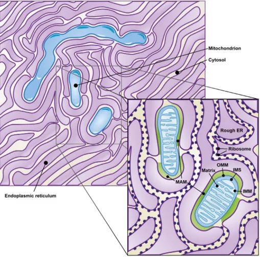
Today, we know that ER-mitochondria contact sites are 10- to
25-nm-wide regions
[6]
(this distance is expected to increase in the rough
reticulum) of juxtaposed membranes tethered by proteins, without
complete fusion or loss of organelle identity (
Figure 1
).
These sites have been fully described from many functional points
of view, and their roles include i) the regulation of lipid synthesis and
transport, serving as the sites where enzymes in lipid synthesis and
transport pathways are located
[7]
, both at the ER and mitochondrial
membranes (e.g., phosphatidylserine synthase 1-2
[8]
), and ii)
calcium (Ca
2+) transport and signaling
[9]
. Ca
2+is known to be
released from the ER through 1,4,5-trisphosphate (IP3) and
ryanodine receptors (IP3Rs, RyRs) as a consequence of the functional
interaction of agonists on the plasma membrane receptors and the
intracellular second messenger IP3; then, Ca
2+is taken up into
mitochondria in a
“quasi-synaptic” manner
[10–12]
through
voltage-dependent anion channels (VDACs) in the outer mitochondrial
membrane (OMM) at ER-mitochondria contact sites
[13]
.
Furthermore, mitochondrial Ca
2+uptake is facilitated by the
highly negative mitochondrial membrane potential and finely tuned
by the proteins in the mitochondrial Ca
2+uniporter (MCU) complex
[14]
. The accumulation of Ca
2+in the mitochondrial matrix has
important implications for several processes, including autophagy,
metabolism, and apoptosis
[15,16]
. In many cell types, a ubiquitous
Ca
2+signaling mechanism represented by the dynamic variation in
free cytosolic Ca
2+concentrations ([Ca
2+]
c) is utilized to sustain
multicellular responses, and it is commonly termed
“Ca
2+oscilla-tions”. These intracellular transient and local [Ca
2+]
c
elevations are
generated by Ca
2+release channels located either in the ER (like
IP3Rs, RyRs, Polycystin-2
[17]
, and two-pore channels
[18]
) or in
the plasma membrane (Orai channels
[19]
) and can be propagated
inside and through cells
[20]
by a complex network of Ca
2+releasing
effectors (like IP3, cADPR, and NAADP) that, individually or in
combination, orchestrate the conversion of local [Ca
2+]
csignals to
global Ca
2+oscillations to achieve a well-defined spatiotemporal
signaling pattern
[21]
. Whereas Ca
2+oscillations are critical to fuel
mitochondrial metabolism, a persistent increase in mitochondrial Ca
2 +triggers cell death, e.g., through opening of the mitochondrial
permeability transition pore (mPTP)
[15,16]
. Another relevant
finding involves the GPX8 protein, a glutathione peroxidase enriched
in MAMs, where it selectively regulates Ca
2+storage and flux via its
transmembrane domain
[22]
. MAMs also play roles in iii)
mitochondrial bioenergetics and iv) mitochondrial morphology and
motility
[23]
, in which the close proximity of the organelles regulates
the machinery responsible for mitochondrial dynamics. It has been
reported that Miro-1, which is anchored to the OMM by its
transmembrane domain and protrudes into the cytosol where it
Figure 1. Mitochondria-associated ER membranes. Membranes juxtaposition of both ER and mitochondria organelles in the cytosol gives
origin to the highly specialized MAM compartment (green zone in the zoom of the figure), here represented as a cartoon on the basis of a
transmission electron microscopy acquisition.
interacts with milton and kinesin proteins
[24]
, organizes
mitochon-drial movement along microtubules, possibly in a calcium-dependent
manner
[25]
; additionally, Fun14 domain-containing 1 (FUNDC1)
together with dynamin-related protein 1 (DRP1) regulates fission and
mitophagy under hypoxic conditions (21). MAMs are also reported to
be involved in v) inflammation signaling
[26]
and vi) ER stress
[27]
.
The interaction between the ER and the mitochondria in cancer,
which is the focus of this review, has been described in many studies
discussing the function of oncogenes and oncosuppressors in the
modulation of Ca
2+and reactive oxygen species (ROS) transfer at
MAMs
[28–30]
. In particular, the most recent report by Sassano et al.
outlines the role of MAMs in cancer growth
[31]
. Thus, MAMs play a
pivotal role in cellular adaptation and cell death pathways, impacting
cancer cell function
[32]
.
In this manuscript, we summarize past and recent findings
regarding MAM-resident proteins and intracellular calcium
modula-tion, categorized by their investigation in specific cancer types (
Table
1
). Although we do not rule out the possible engagement of the
discussed proteins in other tumor environments, assuming that some
mechanisms might apply to multiple cancer types, we collected
information on the highest incidence and best-studied cancers, such
as breast, lung, and prostate cancer.
Alterations at the ER-Mitochondria Interface in
Breast Cancer
Breast cancers (as well as lung cancers) represent one of the most
common types of cancer worldwide
[33]
. As stated in the
Introduction, ER-mitochondria contact sites play a crucial role in
the onset of cancer, participating in mechanisms involved in rewiring
normal cell signaling toward malignancy. In this context, aberrant
expression or localization of MAM-resident proteins is widely
reported. For instance, in breast cancers, the expression of the
stress-activated chaperone sigma-1 receptor (Sig1R), which primarily
acts at the ER-mitochondria interface, is higher in metastatic potential
cancer cells than in normal tissues
[34,35]
. The regulatory role of
Sig1R in MAMs in cell survival was defined in a seminal paper by
Hayashi and Su
[36]
. Under basal conditions, Sig1R binds the MAM
chaperone BiP/GRP78; however, upon activation of IP3Rs, Sig1R
dissociates from BiP and binds IP3R3, thereby stabilizing IP3R3 at
the MAMs and enhancing IP3R3-mediated Ca
2+fluxes to the
mitochondria
[36]
(
Figure 2
). Importantly, it has been demonstrated
[37]
that during conditions of chronic ER stress involving prolonged
ER Ca
2+depletion, Sig1R translocates from MAMs to the peripheral
ER and attenuates cellular damage, thereby preventing cell death
[36]
. Another mechanism through which Sig1R expression is a critical
determinant of cell invasiveness in breast cancer was recently revealed:
Sig1R regulates Ca
2+homeostasis by forming a functional molecular
platform with the calcium-activated K
+channel SK3 and Orai1, thus
driving Ca
2+influx and favoring the migration of cancer cells
[35]
.
These findings support the protumorigenic functions of Sig1R, which
are related to the regulation of Ca
2+dynamics at the
ER-mitochondria zone.
According to the most recent findings, Ca
2+signaling appears to be
an event that is remodulated several times during the malignant
transformation pathway of a cell. Indeed, if an initial reduction of
mitochondrial Ca
2+uptake allows escape from apoptosis, Ca
2+fluxes
towards the mitochondria via MCU are decisive for tumor growth
and metastatic behavior
[35,38]
. For instance, Tosatto et al. showed
that in a set of triple-negative breast cancer cell lines, depletion of
MCU impaired cell migration and invasion and hampered tumor
progression in MDA-MB-231 xenografts, regulating metastasis
through hypoxia-inducible factor 1 (HIF1)–controlled gene
repro-gramming
[38]
. Although MCU is not localized at the
ER-mitochondria interface, its activity and that of its modulators at the
inner mitochondrial membrane finely regulate the cooperativity of
Ca
2+accumulation inside the matrix (please refer to
[39–41]
for
further details) and could be tuned in a cancer-specific manner
[42]
.
Compared with IP3R isoforms 1 and 2, which are located at the
ER membranes, IP3R3 is highly enriched at the MAMs (and is
considered a MAM marker
[43]
), where it conveys Ca
2+-mediated
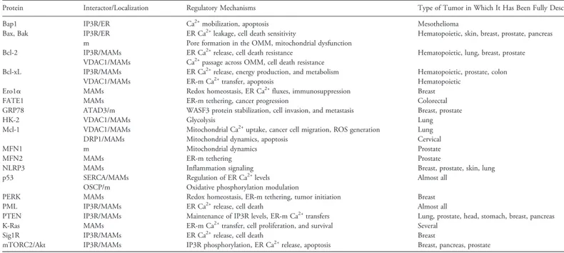
Table 1. Summary of Proteins Discussed in the Review and Their Regulatory Activities at MAMs
Protein Interactor/Localization Regulatory Mechanisms Type of Tumor in Which It Has Been Fully Described
Bap1 IP3R/ER Ca2+mobilization, apoptosis Mesothelioma
Bax, Bak IP3R/ER ER Ca2+leakage, cell death sensitivity Hematopoietic, skin, breast, prostate, pancreas
m Pore formation in the OMM, mitochondrial dysfunction
Bcl-2 IP3R/MAMs ER Ca2+release, cell death resistance Hematopoietic, lung, breast, prostate
VDAC1/MAMs Ca2+passage across OMM, cell death resistance
Bcl-xL IP3R/MAMs ER Ca2+release, energy production, and metabolism Hematopoietic, prostate, colon
VDAC1/MAMs ER-m Ca2+transfer, apoptosis Hematopoietic
Ero1α MAMs Redox homeostasis, ER Ca2+fluxes, immunosuppression Breast
FATE1 MAMs ER-m tethering, cancer progression Colorectal
GRP78 ATAD3/m WASF3 protein stabilization, cell invasion, and metastasis Breast, prostate
HK-2 VDAC1/MAMs Glycolysis Lung
Mcl-1 VDAC1/MAMs Mitochondrial Ca2+uptake, cancer cell migration, ROS generation Lung
DRP1/MAMs Mitochondrial dynamics, apoptosis Cervical
MFN1 m Mitochondrial dynamics Prostate
MFN2 MAMs ER-m tethering Prostate
NLRP3 MAMs Inflammation signaling Breast, prostate, skin, lung
p53 SERCA/MAMs Regulation of ER Ca2+levels Almost all
OSCP/m Oxidative phosphorylation modulation
PERK MAMs Redox homeostasis, ER-m tethering, tumor initiation Breast
PML IP3R/MAMs ER Ca2+release, cell death Almost all
PTEN IP3R/MAMs Maintenance of IP3R levels, ER-m Ca2+transfers Lung, prostate, head, stomach, breast, pancreas
K-Ras MAMs ER-m Ca2+transfer, cell proliferation, and survival Several
Sig1R IP3R/MAMs ER Ca2+release, cell death Breast
mTORC2/Akt IP3R/MAMs IP3R phosphorylation, ER Ca2+release, apoptosis Breast, pancreas, prostate
proapoptotic signals to the mitochondria
[44]
(
Figure 2
). This
channel is also responsible for the regulation of cellular bioenergetics
and metabolism in breast cancer, as its inhibition induces autophagic
death
[45]
and/or mitotic catastrophe in tumorigenic cells, but not in
nontumorigenic cells
[46,47]
. In addition, depletion or
pharmaco-logical blocking of this channel increases the level of LC3-II, an
autophagy marker, via autophagy protein 5 (Atg5) upregulation and
ROS generation, which lead to arrested tumor growth in a related
mouse model
[45]
. These findings correlate with high expression of
IP3R3 in human malignant tissues and high concentrations of
metabolites in serum samples from patients
[48]
. In an independent
study, inhibition of all IP3Rs using xestospongin B resulted in cell
death in cancer cells, without involvement of autophagy. In this case,
IP3R inhibition caused a bioenergetics crisis due to halted
ER-mitochondrial Ca
2+flux
[47]
. While nontumorigenic cells halt their
cell cycle, tumorigenic cells display uncontrolled cell cycle
progres-sion, independent of the presence of mitochondrial substrates for
anabolic pathways, leading to mitotic catastrophe
[32,46,47]
. The
function of IP3R3 is impacted by a wide range of oncogenes and
tumor suppressors that target the receptor
[49–51]
, including the
oncogene Akt kinase
[52,53]
. The PI3K/Akt/mTOR pathway is
frequently altered in human breast cancers
[54,55]
. In 2012, our
group showed that Akt preferentially phosphorylates IP3R3, which
reduces ER-mitochondrial Ca
2+transfer and inhibits apoptotic
responses
[56]
(
Figure 2
). These results were based on previous
findings indicating the capacity of Akt to phosphorylate IP3Rs at their
C-termini
[52,53]
, thereby decreasing Ca
2+release and sensitivity to
apoptosis
[52,57]
. Akt is activated at the ER-mitochondria interface,
where the mechanistic TOR complex 2 (mTORc2) is located
[58]
,
which in turn phosphorylates/activates Akt at position S473
[59]
.
mTORc2-Akt signaling is fundamental for maintaining proper MAM
functionality, and mTORc2 deficiency induces loss of MAM
architecture and a wide range of mitochondrial defects
[58]
.
Importantly, in invasive breast cancer specimens, expression of the
mTORc2 core component Rictor appears to be significantly
upregulated compared with nonmalignant tissues
[60]
. This change
contributes to Akt-dependent tumor progression in HER2-amplified
breast cancers
[60]
.
Due to the role of the MAM region in decoding a wide range of
physiological and danger signals, it seems logical that this region
would host a large number of molecular chaperones to regulate
various intracellular functions. Among these chaperones, the
previously cited GRP78 plays a key role in cancer. In both breast
and colon tumor cells, GRP78 cooperates with ATAD3a, a
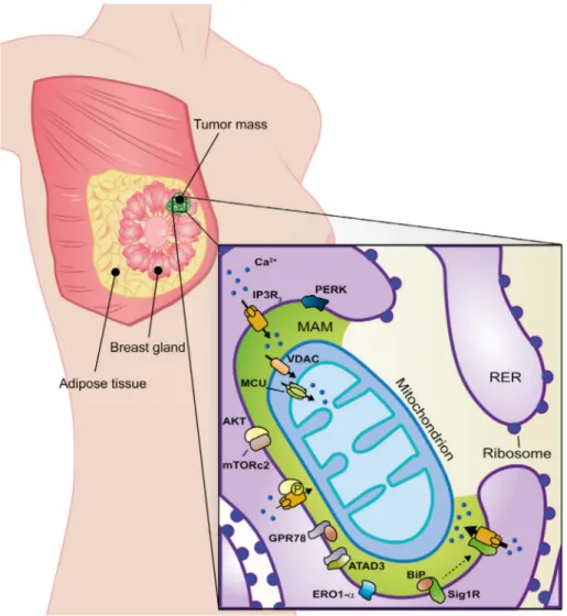
Figure 2. MAM alterations in breast cancer. MAM-resident proteins (green zone) strictly involved in breast cancer onset, progression, and
metastasis are shown in the figure. Black arrows highlight calcium homeostasis where their thickness is proportional to the entity of
calcium fluxes. See text for further details.
Ca
2+, calcium;
RER, rough endoplasmic reticulum.
mitochondrial protein with unknown function, to stabilize WASF3, a
protein that facilitates actin polymerization, thereby promoting
invasion and metastasis
[61]
. Interestingly, ATAD3 may play a role in
ER-mitochondria contact site formation and cholesterol substrate
delivery to the mitochondria
[62]
. Among all organelles inside cells,
mitochondria have a singular lipid composition; the presence of
phosphatidylglycerol, cardiolipin, and phosphatidylethanolamine
confers unique features to mitochondrial membranes. Mitochondria
require that a large amount of lipids be imported, and this is allowed
by the MAM fractions; accordingly, dysregulation of this pathway or
the lipid composition of MAMs has important consequences
[63,64]
.
Since lipid composition and related enzyme activity are essential for
the regulation of Ca
2+homeostasis, they affect ER-mitochondria
contact sites and modify mitochondrial functions
[65]
and may be
essential in regulating apoptotic signaling in tumors. Notably, several
papers have reported both enhanced lipogenesis in cancer cells and
lipolysis from exogenous fatty acids to allow mass growth
[66,67]
and,
in prostate tumors tissues, alterations in the expression of genes
encoding for enzymes designated to produce cholesterol and lipids
[68]
. Thus, these considerations outline an important picture in
which lipid enzyme activities and transport at MAMs are subjected to
cancer-specific variations, but further studies are necessary to unveil
the exact link among all these actors.
As noted in the Introduction section, MAMs are a molecular
platform for the regulation of many oxidoreductase events. In this
context, endoplasmic reticulum oxidoreductin 1-α (ERO1-α) is
extensively studied because of its enrichment at ER-mitochondria
contact sites
[69]
and its high expression in various types of tumors
[70]
. Notably, the expression of ERO1-α in breast cancer is
associated with a poor prognosis
[71]
. ERO1-α controls oxidative
folding and ER redox homeostasis and regulates ER Ca
2+fluxes and
consequent mitochondrial Ca
2+accumulation
[69]
. These ERO1-α–
mediated functions are key events in the cell death mechanism
induced by the procaspase-activating compound-1 (PAC-1), which is
able to promote apoptosis in a variety of cancer cell types
[72]
.
Moreover, in triple-negative breast cancer cells, the expression of
ERO1-α is positively correlated with that of programmed cell
death-ligand 1 (PD-L1), while knockout of ERO1-α results in a significant
attenuation of PD-L1–mediated T-cell apoptosis, suggesting a putative
role for ERO1-α in tumor-mediated immunosuppression
[73]
.
RNA-dependent protein kinase (PKR)–like ER kinase (PERK) is a
critical ER stress sensor of the unfolded protein response at MAMs
[74]
.
PERK has been identified as a key MAM component for maintaining
the ER-mitochondria juxtaposition and ROS-mediated mitochondrial
apoptosis
[75]
. Thus, loss of PERK is expected to cause defects in cell
death processes. PERK-dependent signaling is involved in tumor
initiation and expansion to preserve redox homeostasis and to promote
tumor growth in the MDA-MB-468 and T47D cell lines
[76]
. Silencing
of PERK was shown to reduce tumor growth and restore sensitivity to
chemotherapy in resistant tumor xenografts
[77]
. Moreover, PERK can
regulate the translation of angiogenic factors in the development of
functional microvessels in tumor cells; thus, it plays a fundamental role
in adapting to hypoxic stress and tumor progression
[78]
.
Alterations at the ER-Mitochondria Interface in
Hematopoietic Cancers
B-cell lymphoma 2 (Bcl-2) family proteins were originally discovered
in the context of hematopoietic and lymphoid systems
[79]
, where
antiapoptotic Bcl-2 is upregulated via mechanisms that involve gene
translocation and miRNA deregulation
[80,81]
. It is reported that
upregulation of Bcl-2 enables cancer cells to survive despite high
expression of proapoptotic Bcl-2-family members, whose levels are
elevated by ongoing oncogenic stress
[82,83]
. For example, in
childhood acute lymphoblastic leukemia, apoptosis is avoided in this
way in leukemic cells
[84]
.
Because these proteins are principally localized at the
mitochon-dria, ER, and MAMs, their action strongly reflects their intracellular
localization. Indeed, antiapoptotic Bcl-2 proteins can suppress
ER-mitochondrial Ca
2+transfer via different mechanisms
[85,86]
. Bcl-2
directly targets all three IP3R isoforms, suppressing their Ca
2+-flux
properties
[87–89]
and thereby suppressing Ca
2+accumulation in the
mitochondria. An IP3R-derived peptide corresponding to the
Bcl-2-binding site on IP3R1 was able to overcome the ability of Bcl-2 to
suppress IP3R-channel function
[90]
. A cell-permeable variant of this
peptide that potentially interferes with the Bcl-2-IP3R interaction at
the MAM interface has been proposed to stimulate IP3R-dependent
Ca
2+elevation and cell death in chronic lymphocytic leukemia and
diffuse large B-cell lymphoma models
[91]
. In contrast, Bcl-2 can
sensitize IP3R1 channels to basal IP3, accounting for a decrease in ER
Ca
2+loading and thus reduced ER-mitochondrial Ca
2+transfer
[91]
.
Bcl-2 can also target the N-terminus of the MAM-resident VDAC
isoform 1 (VDAC1)
[92,93]
. Importantly, VDAC1 is the
mitochon-drial channel responsible for Ca
2+passage across the OMM and is
particularly involved in mediating proapoptotic Ca
2+transfer
[94]
.
Thus, Bcl-2 can suppress proapoptotic Ca
2+transfer to the
mitochondria by inhibiting VDAC1
[93]
.
Another apoptosis-inhibiting member of the same family, Bcl-XL
[95]
, is detectable in the MAM compartment
[96]
, where it can target
IP3R3. Previous work has indicated that Bcl-XL can promote
IP3R-driven Ca
2+oscillations
[97,98]
. Therefore, Bcl-XL enhances Ca
2+transfer from the ER to the mitochondria, promoting mitochondrial
energy production and cellular metabolism
[99]
. In addition, via its
BH4 domain, Bcl-XL at MAMs can also target VDAC1
[96]
, and
inhibition of VDAC1 by Bcl-XL prevents mitochondrial Ca
2+overload
and protects against apoptosis
[100]
. Notably, Bcl-XL has also been
reported to enhance VDAC1-mediated Ca
2+flux, promoting basal
prosurvival Ca
2+signaling in particular
[101]
. Furthermore, the
evaluation of myeloid cell leukemia-1 (Mcl-1, whose role in MAMs is
indicated later in the text) expression in mantle cell lymphoma revealed
that high levels of this protein were related to a highly proliferative state
and high-grade morphology
[102]
. In addition, increased levels of
Mcl-1 have been observed in B-cell chronic lymphocytic leukemia and
linked to complete remission failure after single-agent therapy
[103]
.
The Bcl-2-associated X protein (Bax) and Bcl-2-homologous
antagonist killer (Bak), which are proapoptotic members of Bcl-2
family, function at the ER, where they are involved in preserving Ca
2 +homeostasis and ensuring proper cell death sensitivity through Ca
2+dynamics
[104]
. Thus, while Bcl-2 overexpression suppresses
ER-mitochondrial Ca
2+fluxes, Bax overexpression will do the opposite by
increasing ER Ca
2+loading
[105]
. Additionally, in T cells, Bax/Bak
proteins exert critical roles in antigen-induced proliferation through
regulation of IP3R-driven Ca
2+dynamics
[106]
. These proteins also
function at the mitochondria, where they initiate mitochondrial
dysfunction during apoptosis
[104,107]
. Thus, Bax and Bak can be
considered tumor suppressors, either alone or in cooperation with
other alterations. Bax loss-of-function mutations derived from
nucleotide insertions/deletions or single amino acid substitutions
have been observed in human hematopoietic malignancies
[108]
.
As mentioned above, IP3R/Ca
2+channels exhibit a specialized,
crucial function in cancer onset and progression. In acute myeloid
leukemia, IP3R2 expression is upregulated and associated with
dramatically shorter survival
[109]
. However, the mechanisms linking
IP3R2 to adverse clinical events are unknown.
Alterations at the ER-Mitochondria Interface in
Lung Cancer
Although Bcl-2 family proteins have been well described in
hematopoietic malignancies
[79]
, their mitochondrial control in the
MAM platform has mainly been elucidated by studies on solid
tumors, such as prostate
[110]
and lung cancers (
Figure 3
), both of
which are associated with high mortality. The involvement of MAMs
in this pathology is not as well documented in the literature as it is for
breast cancer, although it can be assumed that the underlying
mechanisms are similar. For instance, as noted for hematopoietic
cancers, Bcl-2 acts at the contact sites between the ER and the
mitochondria
[111]
to reduce apoptosis by modulating ER Ca
2+levels
[112,113]
, but the increase in Bcl-2 expression in lung cancer appears to
depend on environmental factors, such as nicotine consumption
[114,115]
. Differential expression of Bcl-2-family proteins occurs in
non–small cell lung cancer–affected patients and in a related mouse
model system
[116,117]
. Indeed, Bcl-XL
[118]
and Bcl-2
overexpres-sion has been associated with a poor prognosis
[116,117]
. However,
recent studies on Bcl-2 expression in clinical specimens have provided
conflicting data; increased Bcl-2 levels were found to be associated with
a better prognosis in lung cancer
[119]
, while there was no correlation
with response to anticancer treatments
[120]
. To exclude possible bias
from these studies, further research is recommended.
Mcl-1, another member of the Bcl-2 family, is also overexpressed
both in lung cancer cell lines, such as H1299, A549, and non–small
cell lung cancer, and in specimens from patients compared with its
expression levels in control cell lines and normal adjacent lung tissues
[121]
. Targeting this protein by reducing its intracellular levels may
improve the clinical management of patients
[122]
. In addition, one
study indicated that Mcl-1 promoted lung cancer cell migration by
directly interacting with VDAC1, thereby increasing mitochondrial
Ca
2+uptake and ROS generation
[123]
(
Figure 3
). VDAC1-derived
peptides that can interfere with the ability of Mcl-1 to bind VDAC1
can counteract lung cancer cell migration.
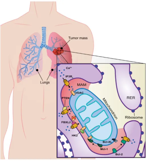
Figure 3. MAM alterations in lung cancer. MAM-resident proteins (red zone) strictly involved in lung cancer onset, progression, and
metastasis are shown in the figure. Among all proteins, a novel and complex role for PTEN has been reported; it counteracts FBXL2 binding to
promote IP3R3- and Ca
2+-mediated apoptosis limiting tumor growth. Indeed, FBXL2 protein binds IP3R3 and targets it for degradation
to limit Ca
2+influx into mitochondria. Black arrows highlight calcium homeostasis where their thickness is proportional to the entity of
calcium fluxes. See text for further details.
Another protein associated with lung cancer pathology that is
expressed at higher levels in cancer cells than in normal tissues
[124,125]
is hexokinase 2 (HK2). Hexokinases are enzymes that
catalyze the first step of glucose metabolism, and they are necessary
for tumor initiation and development, as demonstrated in mouse
models of KRas-driven lung cancer and ErbB2-driven breast cancer
[126]
. Following the phosphorylation of HK2 by Akt, HK2 can
associate with VDAC1 at the MAM site
[127,128]
(
Figure 3
). Here,
HK2 phosphorylates glucose using ATP exiting the mitochondria
through VDAC1 to generate glucose-6-P and stimulate glycolysis.
Thus, HK2 is critical for the Warburg effect in humans, and HK2
depletion restores sensitivity to cell death inducers and oxidative
glucose metabolism
[129]
. Moreover, 2-deoxy-D-glucose, an
inhib-itor of HK2, has been reported to inhibit human and mouse lung
cancer cell growth by inducing cell apoptosis and autophagy.
Regarding the Akt protein network, the phosphatase and tensin
homologue (PTEN), deleted on chromosome 10, is considered a
canonical tumor suppressor, directly counteracting PI3K/Akt/mTOR
pathway activity
[130,131]
. A fraction of this protein is highly
enriched at MAM sites, where its function influences Ca
2+transfer
from the ER to the mitochondria and involves apoptotic behaviors.
Depletion of PTEN impairs Ca
2+release and lowers Ca
2+concentrations in the mitochondria, creating an antiapoptotic
environment. Thus, PTEN can interact with IP3Rs and modify
Ca
2+signaling at MAMs
[132]
. These findings have many
implications for oncology, and PTEN loss of function occurs in
many human cancers
[133,134]
through mutations, deletions,
transcriptional silencing, or protein instability. By focusing on the
protein phosphatase activity of PTEN (directly implicated in its
localization to MAMs), the PTEN
Y138Cmutation was identified in
SCLC, demonstrating that selective loss of protein phosphatase
activity decreases cellular PIP3 levels and Akt phosphorylation
[135]
.
Phosphatase-independent mechanisms in which PTEN acts at the
molecular level can also occur, as demonstrated recently
[136]
,
providing another means of fighting cancer, because PTEN can
compete with the E3-ubiquitin ligase F-box protein FBXL2 for
IP3R3 binding to limit its degradation. FBXL2-dependent
degrada-tion of IP3R3 is increased in cells devoid of PTEN, which results in
the inhibition of apoptosis in cells and tumor masses (in lung and
prostate cancer as a consequence of reduced Ca
2+transfer from ER to
mitochondria)
[136]
(
Figure 3
). Thus, proper maintenance of IP3R3
protein levels is critical for preventing oncogenesis by enabling
tumor-suppressive ER-mitochondrial Ca
2+transfer.
Alterations at the ER-Mitochondria Interface in
Prostate Cancer
In early stages, a high percentage of prostate tumors are dependent on
androgens and, thus, sensitive to their ablation, which leads to cell
death. These tumors rapidly evolve to an androgen-independent
pathological stage that is unavoidable, and therapies must therefore be
improved. Experiments in LNCaP cells (androgen-responsive prostate
cancer) have shown that Bcl-2 overexpression promotes a high rate of
cell replication in vitro and tumor growth in vivo despite hormone
deprivation. However, Bcl-2 depletion via antisense oligonucleotide
therapy was found to improve cell death due to cytotoxic agents in the
same cell line. In addition, immunohistochemistry analysis of 88
neoplastic prostate adenocarcinoma specimens revealed an increase in
the protein levels of Bcl-2, Bcl-XL, and Mcl-1 throughout cancer
development
[137]
. Interestingly, a role in mitochondrial dynamics has
recently been attributed to Mcl-1, which is associated with apoptotic
cell death in a Drp-1–dependent manner, and Mcl-1 was found to be
enriched at the mitochondria, ER, and MAMs
[138]
.
Involvement of the fusion-fission machinery in apoptosis or cancer
development has been observed
[139]
. Enhancement of
mitochon-drial fusion by increasing mitochonmitochon-drial GTPases mitofusin 1
(MFN-1) and mitofusin 2 (MFN-2) levels has been associated with prostate
cancer progression
[140]
. MFN-1 and MFN-2 are essential
components of the physical tethering between the ER and
mitochondria at the MAM compartment, and they are involved in
mitochondrial Ca
2+homeostasis (
Figure 4
).
The tethering role of MFN-2 must be further established. Initially,
MFN-2 was chosen as a putative candidate with tethering functions
[141,142]
for Ca
2+transfer because its depletion reduces
IP3R-mediated mitochondrial Ca
2+uptake
[143]
, leading to a decrease in
contact sites
[143]
in hypothalamic neurons
[144]
; indeed, MFN-2
appears to be a crucial mediator of the energy balance by acting on the
synergy of the mitochondria-ER membrane juxtaposition
[144]
. The
experimental evidence indicating the tethering function of MFN-2
has been questioned and was not shared by Filadi and coworkers, who
reported that there is a high percentage of membrane juxtaposition
between the ER and MAMs when MFN-2 is silenced; the ablation of
MFN-2 initiates Ca
2+signaling at contact sites
[145]
and supports
cell death (
Figure 4
). This hypothesis was confirmed by other
independent groups, as referenced in
[146,147]
. This topic remains
controversial, although the localization of MFN-2 and its pivotal roles
at MAMs are indisputable.
Finally, the MAM chaperone GRP78, which exhibits Ca
2+-binding properties
[36]
, is able to bind the ER antiapoptotic factor
clusterin (CLU) during ER stress to facilitate its redistribution at the
mitochondria and to minimize the detrimental effects provoked by
ER stress, thus inducing prostate cancer cell survival
[148]
.
MAMs in Other Types of Cancer and the Impact of
Tumor Suppressors/Oncogenes
In the following paragraphs, the general understanding of and recent
discoveries regarding oncogene and tumor suppressor functions at
MAMs are summarized. Although oncogenes and tumor suppressors
are involved in almost all types of cancer as transcription factors, new
transcriptional-independent properties have been revealed in healthy
and disease conditions due to their intracellular localization at the
ER-mitochondria interface.
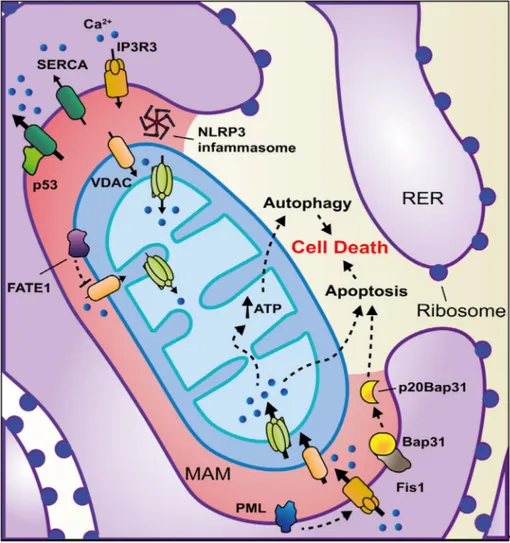
i) p53
The p53 protein exhibits excellent tumor-suppressive properties and is altered in most human cancers, including colon, breast, lung, bladder, brain, pancreatic, stomach, and esophageal cancer[149]. p53 is a nuclear transcription factor that is activated by a variety of stimuli and subsequently transactivates genes involved in apoptosis, cell cycle regulation, and prevention of cell transformation and cancer progression. Additional p53 activities occur in the cytoplasm, where p53 triggers apoptosis and inhibits autophagy[150], and in the mitochondrial matrix, where it promotes F1FO-ATP synthase assembly[151], suggesting an ability to increase oxidative phosphorylation activity by interacting with the oligomycin sensitivity-conferring protein (OSCP) subunit in a transcriptional-independent manner. Interestingly, a fraction of p53 has also been found to be associated with the ER and MAMs, where it modulates Ca2+homeostasis[152](Figure 5). In particular, p53 binds and simulates the sarco/ER Ca2+-ATPase (SERCA) pumps at the ER, increasing ER Ca2+levels. As a consequence, during apoptotic stimulation, a greater amount of Ca2+is releasable from the ER versus the mitochondria, promoting mitochondrial Ca2+overload, mPTP opening, release of caspase cofactors, and induction of apoptosis via the intrinsic pathway [153]. In cancer cells, this
mechanism can easily become impaired due to either inactivation of p53 or missense mutations in the coding gene, contributing to disease progression and resistance to chemotherapy[154,155]. Moreover, by combining the dorsal skinfold chamber technique with intravital microscopy, Giorgi et al. elucidated the involvement of p53 in controlling intracellular Ca2+signals and apoptosis in three-dimensional tumor masses in mice. Dysregulation of p53-dependent Ca2+ homeostasis led to reduced ER Ca2+release and, consequently, low responsiveness to apoptotic stimulation[156].
ii) PML and Bap1
Similar to p53, the promyelocytic leukemia protein (PML) is a potent tumor suppressor that stabilizes the p53 protein and improves its function[157]. PML was originally associated with the pathogenesis of acute promyelocytic leukemia. However, loss of PML has been linked to several human cancers, including prostate, breast, and central nervous system tumors [158]. In addition to its well-characterized nuclear activity, an extranuclear transcription-independent function of PML was identified at MAMs, where it controls cell survival. PML regulates apoptosis in MAMs by modulating Ca2+release through its physical interaction with IP3R3[159](Figure 5). Moreover, it was recently demonstrated that the localization of PML at MAMs is fundamental for apoptosis control and autophagy regulation[160](Figure 5). IP3R3 has emerged as being involved in gastric cancer peritoneal dissemination[161], and its expression is directly associated with the aggressiveness of colorectal carcinomas[162]. Recent findings have provided new insights regarding mesothelioma malignances in which an important tumor suppressor, Bap1, is reduced or inactivated by mutations[163]. ER-localized Bap1
has been shown to bind and stabilize IP3R3, hence modulating Ca2+mobilization in favor of apoptosis. Thus, the depletion of Bap1, along with its nuclear-dependent function, allows cellular transformation and leads to prevalence of mesothelioma onset rather than other type of cancers[163].
iii) FATE1
A direct link between cancer progression and alteration of the correct ER-mitochondria distance is presented by the oncoprotein fetal and adult testis-expressed 1 (FATE1). Analysis of The Cancer Genome Atlas colorectal dataset revealed that FATE1 is frequently co-expressed with the ER-resident E3 ligase RNF183, which is correlated with a poor clinical outcome[164], suggesting that these proteins function in human tumors to inhibit cell death. FATE1 localizes at the outer surface of the mitochondria and is associated with MAMs [165]. Moreover, an ER-mitochondria antitethering function has been attributed to FATE1, which is consistent with a decrease in mitochondrial Ca2+uptake and cell survival [165] (Figure 5). FATE1 protects cells from apoptosis induced by mitotane, a compound that promotes the accumulation of toxic cholesterol esters and triggers ER stress by inhibiting the MAM-resident enzyme SOAT1[166]. Most importantly, high FATE1 expression is an indicator of poor prognosis in adrenocortical carcinoma patients[165].
iv) Mitochondrial fission factors
Fission factors are also mitochondrial regulators of cell death, thus representing pivotal components of apoptosis signaling pathways[167]. Downregulation of a fission 1 homologue (Fis1) and Drp1 was demonstrated to reduce apoptosis[168].
Figure 4. MAM alterations in prostate cancer. MAM-resident proteins (yellow zone) strictly involved in prostate cancer onset, progression.
and metastasis are shown in the figure. MFN-2 protein function has been illustrated with quite attention (question marks), as its role at
MAMs is far to be established; see text for further details.
Interestingly, Fis1 is responsible for the formation of a platform at MAM sites, which triggers a feedback loop by releasing Ca2+from the ER, which stimulates mitochondria-mediated apoptosis. Specifically, Fis1 interacts with Bap31 at the ER, creating a bridge that enhances the cleavage of Bap31 in its proapoptotic form, p20Bap31[169]. Thus, alterations of Fis1 contribute to tumor development. It has been reported that Fis1 is overexpressed in oncolytic cell tumors[170], and Fis1 deletion can drive the selection of compensatory mutations, resulting in defective growth control and cell death regulation, which are characteristics of human tumor cells[171].
v) NLRP3 inflammasome
Although inflammation is not the focus of this review, we would like to briefly refer to the NOD-like receptor family pyrin domain-containing 3 (NLRP3) inflamma-some due to its crucial association with cancer onset. NLRP3 is a multimeric complex that induces innate inflammatory responses with apoptosis-associated speck-like protein containing a CARD complex (ASC), which recruits procaspase-1. Procaspase-1 is then cleaved to caspase-1, which converts 1β and pro–IL-18 to the active forms that are responsible for additional recruitment of other inflammatory cells [172]. Interestingly, a considerable fraction of NLRP3 is associated with MAMs following inflammasome activation, suggesting that NLRP3 checks and modulates mitochondrial activity[173]. Cancer-associated inflamma-tory responses are directly involved in cancer biology, including tumor initiation, progression, metastasis, and treatment. For example, NLRP3 is markedly upregulated in macrophages in pancreatic ductal adenocarcinoma, where NLRP3 directs the polarization of tumor-associated macrophages and, hence, controls immunogenic or tolerogenic CD4+ T-cell differentiation and CD8+ T-cell
activation[174]. Furthermore, in NLRP3 KO mice, inflammasome components have been shown to exacerbate liver colorectal cancer metastatic growth[175]. Moreover, the activation of the NLRP3 inflammasome and the expression and secretion of active IL-1β in melanomas cause disease progression[176].
Oncogenic Ras Signaling
Ras proteins, which belong to the family of small GTPases
controlling cell proliferation, cell cycling, and cell survival, are
frequently deregulated in several types of human cancers
[177]
. One
of the family members K-Ras has recently been found to engage in
cross talk with Ca
2+signaling and to impact ER-mitochondrial Ca
2+transfer
[178]
. By comparing two isogenic colorectal cancer cell lines,
one expressing mutated oncogenic K-Ras
G13D/wild-typeand one in
which the oncogenic allele was deleted (K-Ras
-/wild-type), it was found
that the presence of oncogenic K-Ras
G13Dsuppressed IP3-induced
Ca
2+release due to a decrease in ER Ca
2+store contents. These
functional aberrations in Ca
2+signaling could be linked to
remodeling of the expression of ER Ca
2+transport systems, revealing
that K-Ras
G13D–expressing cells express less SERCA2b and switch
their IP3R-isoform expression profile compared with K-Ras
G13D–
deleted cells. In particular, cells with oncogenic K-Ras display a
decrease in IP3R3 expression levels and an increase in IP3R1
expression levels, establishing a decrease in susceptibility to apoptotic
Figure 5. MAM alterations and other types of cancer. Proteins with key functions (see text for details) in a wide range of tumors are
represented in the figure. A pink zone between the mitochondrion and the ER outlines MAM subcellular compartment.
ATP, adenosine
triphosphate.
stimuli and an increase in the ability to generate prosurvival Ca
2+oscillations that sustain cell proliferation. As such, upon the deletion
of K-Ras, IP3R3 expression is elevated, and the likelihood of IP3R3
accumulating in MAMs is therefore also increased and is correlated
with restoration of ER-mitochondrial Ca
2+transfer and apoptosis
sensitivity. Hence, beyond the direct regulation of oncogenes and
tumor suppressors of Ca
2+-transport systems in MAMs, it is clear that
these genes can also impact the expression levels and, thus, the overall
number of channels available for the MAM compartment, affecting
ER-mitochondrial Ca
2+transfer and cell death susceptibility (or other
cancer-related hallmarks).
Conclusions
An increasing number of studies have identified important roles of
MAMs in cancer processes, although further study is required to
completely elucidate the molecular mechanisms involved. Changes in
the ER-mitochondrial tethering distance and morphology have
dramatic effects on the health of a cell, which communicates with
the
“outside world” via lipids, Ca
2+, ROS, and the exchange of other
mediators among organelles. There are likely to be many
ER-mitochondria contact sites, leading to many unanswered questions.
For example, how will increased knowledge of MAMs impact human
studies and clinical therapies? Are there any properties of
MAM-resident proteins that could be attributed to one specific type of
cancer rather than another? Are there different types of MAMs with
specific functions and protein populations? In addition to Ca
2+, are
there other crucial mediators that could modulate the plasticity and
function of MAMs in the cell? Are lipid synthesis and transfer at
ER-mitochondria membrane contact sites involved in cancer, and what
are the underlying mechanisms? If the proteins that localize or
relocalize to MAMs vary, how do these various proteins regulate their
localization, and under what specific condition are they active?
Acknowledgements
P. P. is grateful to Camilla degli Scrovegni for continuous support. P.
P. is supported by the Italian Ministry of Education, University and
Research, the Italian Ministry of Health, Telethon (GGP15219/B),
the Italian Association for Cancer Research (IG-18624), and local
funds from the University of Ferrara. C. G. is supported by local
funds from the University of Ferrara, the Italian Association for
Cancer Research, the Italian Ministry of Health, and a Cariplo grant.
G. B. is supported by grants from the Research Foundation–Flanders
(FWO) and Research Council KU Leuven. G. B. and P. P. are part
of the FWO Scientific Research Network CaSign. M. B. and M. K.
are supported by PhD fellowships obtained from the FWO. A. K.
W. and M. R. W. are supported by the Internal Project of The
Children’s Memorial Health Institute (no. S141/2014) and by
Statutory Funding from Nencki Institute of Experimental Biology.
References
[1] Vance JE (1990). Phospholipid synthesis in a membrane fraction associated with mitochondria. J Biol Chem265(13), 7248–7256.
[2] Rusinol AE, Cui Z, Chen MH, and Vance JE (1994). A unique mitochondria-associated membrane fraction from rat liver has a high capacity for lipid synthesis and contains pre-Golgi secretory proteins including nascent lipoproteins. J Biol Chem.269(44), 27494–27502.
[3] Copeland DE and Dalton AJ (1959). An association between mitochondria and the endoplasmic reticulum in cells of the pseudobranch gland of a teleost. J Biophys Biochem Cytol.5(3), 393–396.
[4] Bernhard W, Haguenau F, Gautier A, and Oberling C (1952). Submicrosco-pical structure of cytoplasmic basophils in the liver, pancreas and salivary gland; study of ultrafine slices by electron microscope. Z Zellforsch Mikrosk Anat37(3), 281–300.
[5] Bernhard W and Rouiller C (1956). Close topographical relationship between mitochondria and ergastoplasm of liver cells in a definite phase of cellular activity. J Biophys Biochem Cytol2(4 Suppl), 73–78.
[6] Csordas G, Renken C, Varnai P, Walter L, Weaver D, and Buttle KF, et al (2006). Structural and functional features and significance of the physical linkage between ER and mitochondria. J Cell Biol174(7), 915–921.
[7] Vance JE (2015). Phospholipid synthesis and transport in mammalian cells. Traffic16(1), 1–18.
[8] Stone SJ and Vance JE (2000). Phosphatidylserine synthase-1 and -2 are localized to mitochondria-associated membranes. J Biol Chem 275(44), 34534–34540.
[9] Szabadkai G, Bianchi K, Varnai P, De Stefani D, Wieckowski MR, and Cavagna D, et al (2006). Chaperone-mediated coupling of endoplasmic reticulum and mitochondrial Ca2+ channels. J Cell Biol175(6), 901–911.
[10] Naon D and Scorrano L (2014). At the right distance: ER-mitochondria juxtaposition in cell life and death. Biochim Biophys Acta1843(10), 2184–2194.
[11] Csordas G, Thomas AP, and Hajnoczky G (1999). Quasi-synaptic calcium signal transmission between endoplasmic reticulum and mitochondria. EMBO J 18(1), 96–108.
[12] Rizzuto R, Pinton P, Carrington W, Fay FS, Fogarty KE, and Lifshitz LM, et al (1998). Close contacts with the endoplasmic reticulum as determinants of mitochondrial Ca2+ responses. Science280(5370), 1763–1766.
[13] De Pinto VD and Palmieri F (1992). Transmembrane arrangement of mitochondrial porin or voltage-dependent anion channel (VDAC). J Bioenerg Biomembr24(1), 21–26.
[14] Marchi S and Pinton P (2014). The mitochondrial calcium uniporter complex: molecular components, structure and physiopathological implications. J Physiol 592(5), 829–839.
[15] Bonora M, Morganti C, Morciano G, Pedriali G, Lebiedzinska-Arciszewska M, and Aquila G, et al (2017). Mitochondrial permeability transition involves dissociation of F1FO ATP synthase dimers and C-ring conformation. EMBO Rep18(7), 1077–1089.
[16] Morciano G, Giorgi C, Bonora M, Punzetti S, Pavasini R, and Wieckowski MR, et al (2015). Molecular identity of the mitochondrial permeability transition pore and its role in ischemia-reperfusion injury. J Mol Cell Cardiol78, 142–153.
[17] Koulen P, Cai Y, Geng L, Maeda Y, Nishimura S, and Witzgall R, et al (2002). Polycystin-2 is an intracellular calcium release channel. Nat Cell Biol4(3), 191–197.
[18] Zhu MX, Ma J, Parrington J, Calcraft PJ, Galione A, and Evans AM (2010). Calcium signaling via two-pore channels: local or global, that is the question. Am J Physiol Cell Physiol298(3), C430–41.
[19] Frischauf I, Schindl R, Derler I, Bergsmann J, Fahrner M, and Romanin C (2008). The STIM/Orai coupling machinery. Channels (Austin)2(4), 261–268.
[20] Leybaert L and Sanderson MJ (2012). Intercellular Ca(2+) waves: mechanisms and function. Physiol Rev92(3), 1359–1392.
[21] Wu W, Lin C, Wu K, Jiang L, Wang X, and Li W, et al (2016). FUNDC1 regulates mitochondrial dynamics at the ER-mitochondrial contact site under hypoxic conditions. EMBO J35(13), 1368–1384.
[22] Yoboue ED, Rimessi A, Anelli T, Pinton P, and Sitia R (2017). Regulation of calcium fluxes by GPX8, a type-II transmembrane peroxidase enriched at the mitochondria-associated endoplasmic reticulum membrane. Antioxid Redox Signal27(9), 583–595.
[23] Rowland AA and Voeltz GK (2012). Endoplasmic reticulum-mitochondria contacts: function of the junction. Nat Rev Mol Cell Biol13(10), 607–625.
[24] Glater EE, Megeath LJ, Stowers RS, and Schwarz TL (2006). Axonal transport of mitochondria requires milton to recruit kinesin heavy chain and is light chain independent. J Cell Biol173(4), 545–557.
[25] Wang X and Schwarz TL (2009). The mechanism of Ca2+ -dependent regulation of kinesin-mediated mitochondrial motility. Cell136(1), 163–174.
[26] Horner SM, Liu HM, Park HS, Briley J, and Gale Jr M (2011). Mitochondrial-associated endoplasmic reticulum membranes (MAM) form innate immune synapses and are targeted by hepatitis C virus. Proc Natl Acad Sci U S A108(35), 14590–14595.
[27] Haile Y, Deng X, Ortiz-Sandoval C, Tahbaz N, Janowicz A, and Lu JQ, et al (2017). Rab32 connects ER stress to mitochondrial defects in multiple sclerosis. J Neuroinflammation14(1), 19.
[28] Gutierrez T and Simmen T (2014). Endoplasmic reticulum chaperones and oxidoreductases: critical regulators of tumor cell survival and immunorecogni-tion. Front Oncol4, 291.
[29] Gutierrez T and Simmen T (2017). Endoplasmic reticulum chaperones tweak the mitochondrial calcium rheostat to control metabolism and cell death. Cell Calcium70, 64–75.
[30] Booth DM, Enyedi B, Geiszt M, Varnai P, and Hajnoczky G (2016). Redox nanodomains are induced by and control calcium signaling at the ER-mitochondrial interface. Mol Cell63(2), 240–248.
[31] Sassano ML, van Vliet AR, and Agostinis P (2017). Mitochondria-associated membranes as networking platforms and regulators of cancer cell fate. Front Oncol7, 174.
[32] Ivanova H, Kerkhofs M, La Rovere RM, and Bultynck G (2017). Endoplasmic reticulum-mitochondrial Ca2+ fluxes underlying cancer cell survival. Front Oncol7, 70.
[33] Barrdahl M, Rudolph A, Hopper JL, Southey MC, Broeks A, and Fasching PA, et al (2017). Gene-environment interactions involving functional variants: Results from the Breast Cancer Association Consortium. Int J Cancer141(9), 1830–1840.
[34] Aydar E, Onganer P, Perrett R, Djamgoz MB, and Palmer CP (2006). The expression and functional characterization of sigma (sigma) 1 receptors in breast cancer cell lines. Cancer Lett242(2), 245–257.
[35] Gueguinou M, Crottes D, Chantome A, Rapetti-Mauss R, Potier-Cartereau M, and Clarysse L, et al (2017). The SigmaR1 chaperone drives breast and colorectal cancer cell migration by tuning SK3-dependent Ca2+ homeostasis. Oncogene36(25), 3640–3647.
[36] Hayashi T and Su TP (2007). Sigma-1 receptor chaperones at the ER-mitochondrion interface regulate Ca(2+) signaling and cell survival. Cell131(3), 596–610.
[37] Hayashi T and Su TP (2003). Intracellular dynamics of sigma-1 receptors (sigma (1) binding sites) in NG108-15 cells. J Pharmacol Exp Ther306(2), 726–733.
[38] Tosatto A, Sommaggio R, Kummerow C, Bentham RB, Blacker TS, and Berecz T, et al (2016). The mitochondrial calcium uniporter regulates breast cancer progression via HIF-1alpha. EMBO Mol Med8(5), 569–585.
[39] Kamer KJ, Grabarek Z, and Mootha VK (2017). High-affinity cooperative Ca2 + binding by MICU1-MICU2 serves as an on-off switch for the uniporter. EMBO Rep18(8), 1397–1411.
[40] Paillard M, Csordas G, Szanda G, Golenar T, Debattisti V, and Bartok A, et al (2017). Tissue-specific mitochondrial decoding of cytoplasmic Ca2+ signals is controlled by the stoichiometry of MICU1/2 and MCU. Cell Rep18(10), 2291–2300.
[41] Patron M, Checchetto V, Raffaello A, Teardo E, Vecellio Reane D, and Mantoan M, et al (2014). MICU1 and MICU2 finely tune the mitochondrial Ca2+ uniporter by exerting opposite effects on MCU activity. Mol Cell53(5), 726–737.
[42] Marchi S, Lupini L, Patergnani S, Rimessi A, Missiroli S, and Bonora M, et al (2013). Downregulation of the mitochondrial calcium uniporter by cancer-related miR-25. Curr Biol23(1), 58–63.
[43] Wieckowski MR, Giorgi C, Lebiedzinska M, Duszynski J, and Pinton P (2009). Isolation of mitochondria-associated membranes and mitochondria from animal tissues and cells. Nat Protoc4(11), 1582–1590.
[44] Mendes CC, Gomes DA, Thompson M, Souto NC, Goes TS, and Goes AM, et al (2005). The type III inositol 1,4,5-trisphosphate receptor preferentially transmits apoptotic Ca2+ signals into mitochondria. J Biol Chem280(49), 40892–40900.
[45] Singh A, Chagtoo M, Tiwari S, George N, Chakravarti B, and Khan S, et al (2017). Inhibition of inositol 1, 4, 5-trisphosphate receptor induce breast cancer cell death through deregulated autophagy and cellular bioenergetics. J Cell Biochem118(8), 2333–2346.
[46] Bultynck G (2016). Onco-IP3Rs feed cancerous cravings for mitochondrial Ca (2.). Trends Biochem Sci41(5), 390–393.
[47] Cardenas C, Muller M, McNeal A, Lovy A, Jana F, and Bustos G, et al (2016). Selective vulnerability of cancer cells by inhibition of Ca(2+) transfer from endoplasmic reticulum to mitochondria. Cell Rep15(1), 219–220.
[48] Singh A, Sharma RK, Chagtoo M, Agarwal G, George N, and Sinha N, et al (2017). 1H NMR metabolomics reveals association of high expression of inositol 1, 4, 5 trisphosphate receptor and metabolites in breast cancer patients. PLoS One12(1)e0169330.
[49] Marchi S, Patergnani S, Missiroli S, Morciano G, Rimessi A, and Wieckowski MR, et al (2017). Mitochondrial and endoplasmic reticulum calcium homeostasis and cell death. Cell Calcium69, 62–72.
[50] Vervliet T, Clerix E, Seitaj B, Ivanova H, Monaco G, and Bultynck G (2017). Modulation of Ca2+ signaling by anti-apoptotic B-cell lymphoma 2 proteins at the endoplasmic reticulum-mitochondrial interface. Front Oncol7, 75.
[51] Bittremieux M, Parys JB, Pinton P, and Bultynck G (2016). ER functions of oncogenes and tumor suppressors: modulators of intracellular Ca(2+) signaling. Biochim Biophys Acta1863(6 Pt B), 1364–1378.
[52] Khan MT, Wagner II L, Yule DI, Bhanumathy C, and Joseph SK (2006). Akt kinase phosphorylation of inositol 1,4,5-trisphosphate receptors. J Biol Chem 281(6), 3731–3737.
[53] Szado T, Vanderheyden V, Parys JB, De Smedt H, Rietdorf K, and Kotelevets L, et al (2008). Phosphorylation of inositol 1,4,5-trisphosphate receptors by protein kinase B/Akt inhibits Ca2+ release and apoptosis. Proc Natl Acad Sci U S A105(7), 2427–2432.
[54] Gonzalez-Angulo AM, Ferrer-Lozano J, Stemke-Hale K, Sahin A, Liu S, and Barrera JA, et al (2011). PI3K pathway mutations and PTEN levels in primary and metastatic breast cancer. Mol Cancer Ther10(6), 1093–1101.
[55] Stemke-Hale K, Gonzalez-Angulo AM, Lluch A, Neve RM, Kuo WL, and Davies M, et al (2008). An integrative genomic and proteomic analysis of PIK3CA, PTEN, and AKT mutations in breast cancer. Cancer Res68(15), 6084–6091.
[56] Marchi S, Marinello M, Bononi A, Bonora M, Giorgi C, and Rimessi A, et al (2012). Selective modulation of subtype III IP(3)R by Akt regulates ER Ca(2) (+) release and apoptosis. Cell Death Dis3e304.
[57] Marchi S, Rimessi A, Giorgi C, Baldini C, Ferroni L, and Rizzuto R, et al (2008). Akt kinase reducing endoplasmic reticulum Ca2+ release protects cells from Ca2+-dependent apoptotic stimuli. Biochem Biophys Res Commun375(4), 501–505.
[58] Betz C, Stracka D, Prescianotto-Baschong C, Frieden M, Demaurex N, and Hall MN (2013). Feature Article: mTOR complex 2-Akt signaling at mitochondria-associated endoplasmic reticulum membranes (MAM) regulates mitochondrial physiology. Proc Natl Acad Sci U S A110(31), 12526–12534.
[59] Sarbassov DD, Guertin DA, Ali SM, and Sabatini DM (2005). Phosphorylation and regulation of Akt/PKB by the rictor-mTOR complex. Science307(5712), 1098–1101.
[60] Morrison Joly M, Hicks DJ, Jones B, Sanchez V, Estrada MV, and Young C, et al (2016). Rictor/mTORC2 drives progression and therapeutic resistance of HER2-amplified breast cancers. Cancer Res76(16), 4752–4764.
[61] Teng Y, Ren X, Li H, Shull A, Kim J, and Cowell JK (2016). Mitochondrial ATAD3A combines with GRP78 to regulate the WASF3 metastasis-promoting protein. Oncogene35(3), 333–343.
[62] Issop L, Fan J, Lee S, Rone MB, Basu K, and Mui J, et al (2015). Mitochondria-associated membrane formation in hormone-stimulated Leydig cell steroido-genesis: role of ATAD3. Endocrinology156(1), 334–345.
[63] van Vliet AR, Verfaillie T, and Agostinis P (2014). New functions of mitochondria associated membranes in cellular signaling. Biochim Biophys Acta 1843(10), 2253–2262.
[64] Szymanski J, Janikiewicz J, Michalska B, Patalas-Krawczyk P, Perrone M, and Ziolkowski W, et al (2017). Interaction of mitochondria with the endoplasmic reticulum and plasma membrane in calcium homeostasis, lipid trafficking and mitochondrial structure. Int J Mol Sci18(7).
[65] Vance JE (2014). MAM (mitochondria-associated membranes) in mammalian cells: lipids and beyond. Biochim Biophys Acta1841(4), 595–609.
[66] Zaidi N, Lupien L, Kuemmerle NB, Kinlaw WB, Swinnen JV, and Smans K (2013). Lipogenesis and lipolysis: the pathways exploited by the cancer cells to acquire fatty acids. Prog Lipid Res52(4), 585–589.
[67] Kuemmerle NB, Rysman E, Lombardo PS, Flanagan AJ, Lipe BC, and Wells WA, et al (2011). Lipoprotein lipase links dietary fat to solid tumor cell proliferation. Mol Cancer Ther10(3), 427–436.
[68] Ettinger SL, Sobel R, Whitmore TG, Akbari M, Bradley DR, and Gleave ME, et al (2004). Dysregulation of sterol response element-binding proteins and downstream effectors in prostate cancer during progression to androgen independence. Cancer Res64(6), 2212–2221.
[69] Anelli T, Bergamelli L, Margittai E, Rimessi A, Fagioli C, and Malgaroli A, et al (2012). Ero1alpha regulates Ca(2+) fluxes at the endoplasmic reticulum-mitochondria interface (MAM). Antioxid Redox Signal16(10), 1077–1087.
[70] Kakihana T, Nagata K, and Sitia R (2012). Peroxides and peroxidases in the endoplasmic reticulum: integrating redox homeostasis and oxidative folding. Antioxid Redox Signal16(8), 763–771.
[71] Kutomi G, Tamura Y, Tanaka T, Kajiwara T, Kukita K, and Ohmura T, et al (2013). Human endoplasmic reticulum oxidoreductin 1-alpha is a novel predictor for poor prognosis of breast cancer. Cancer Sci104(8), 1091–1096.