to
allow
independent
verification.
Second, comparing crude rates with
age-standardized rates is invalid. This
is
because
melanoma
rates
have
been increasing in older people but
decreasing in younger people during
a period in which more Australians are
surviving to old age. This means that
crude melanoma rates calculated for
the year 1982 relate to an entirely
different population structure versus
those calculated for 2011. Without
properly accounting for these changes,
the differences between crude and
standardized rates are uninterpretable,
and no valid comparisons can be made.
We considered the issue of
popula-tion dilupopula-tion in our article, and we
referenced Dr. Czarnecki’s original
article
positing
his
hypothesis
(
Czarnecki, 2014
). We also referenced
the subsequent paper by
Baade et al.
(2015)
that elegantly disproved it. In
their article, Baade et al. modeled
melanoma incidence in Australia under
the full range of hypothetical scenarios
that might explain Australia’s
popula-tion growth between 1982 and 2011—
that is, from being 100% attributable
to migration to 0% attributable to
migration. Regardless of the assumed
level of migration, the decline in
age-standardized melanoma incidence in
Australia was apparent across all
scenarios, from which the authors
concluded that there is “strong
evi-dence against the hypothesis that the
observed decrease in melanoma
inci-dence among young Australians since
the mid-1990s can be explained solely
by the increasing overseas migration
and any resultant lowering of the ‘at
risk’ population in Australia.” We agree
with their conclusion.
In summary, we agree that
popula-tion dilupopula-tion is of interest and may
explain some of the decline in the
Australian melanoma incidence rates,
but we disagree with the assertion that
melanoma incidence is rising in young
susceptible Australians. As argued by
others (
Baade et al., 2015
), the timing
of the changes in melanoma incidence,
coupled with the divergent trends
among younger and older Australians,
are consistent with birth cohort and
period effects that are best explained
by primary prevention campaigns that
commenced nationally in the 1980s.
ORCIDsDavid C. Whiteman: http://orcid.org/0000-0003-2563-9559
Ade`le C. Green: http://orcid.org/0000-0002-2753-4841
CONFLICT OF INTEREST
The authors state no conflict of interest.
David C. Whiteman
1,2,*, Ade`le
C. Green
1,2,3and Catherine
M. Olsen
1,21QIMR Berghofer Medical Research Institute, 300 Herston Road, Herston, QLD 4006, Australia;2The University of Queensland, School of Public Health, Herston Road, Herston, QLD 4006, Australia; and3Cancer Research UK Manchester Institute and Institute of Inflammation and Repair, University of Manchester, Manchester, UK
*
Corresponding author e-mail:david. [email protected]
REFERENCES
Baade PD, Youlden DR, Youl P, Kimlin M, Sinclair C, Aitken J. Assessment of the effect of migration on melanoma incidence trends in Australia between 1982 and 2010 among people under 30. Acta Derm Venereol 2015;95:118e20.
Czarnecki D. The incidence of melanoma is increasing in the susceptible young Australian population. Acta Derm Venereol 2014;94: 539e41.
Czarnecki D. The relentless rise in the incidence of melanoma in susceptible Australians. J Invest Dermatol 2016;136:1912e3.
Whiteman DC, Green AC, Olsen CM. The growing burden of invasive melanoma: projections of incidence rates and numbers of new cases in six susceptible populations through 2031. J Invest Dermatol 2016;136: 1161e71.
Association of Melanocortin-1 Receptor
Variants with Pigmentary Traits in Humans:
A Pooled Analysis from the M-Skip Project
Journal of Investigative Dermatology (2016) 136, 1914e1917;doi:10.1016/j.jid.2016.05.099
TO THE EDITOR
Skin pigmentation is due to the
accu-mulation
of
eumelanin,
which is
brown-black
pigment
and
photo-protective, and pheomelanin, which is
yellow-red pigment and may promote
carcinogenesis (
Valverde et al., 1995
).
The melanocortin-1 receptor (MC1R)
gene regulates the amount and type of
pigment production and is a major
determinant of skin phototype (
Garcia-Borron et al., 2005; Valverde et al.,
1995
). Binding of
a
-melanocyte
stimu-lating hormone to MC1R stimulates the
enzymatic activity of adenylate cyclase
enzyme, thereby elevating intracellular
cyclic
adenosine
monophosphate
(cAMP) levels. MC1R is highly
poly-morphic, especially in Caucasians:
more than 200 coding region variants
have been described to date (
Garcia-Borron et al.,
2014; Gerstenblith
et al., 2007; Perez Oliva et al., 2009
).
Six variants—D84E, R142H, R151C,
I155T, R160W, and D294H—have
been designated as “R” alleles because
of their strong association with the “red
hair color” phenotype characterized by
red hair, fair skin, freckles, and sun
sensitivity. The V60L, V92M, and
R163Q variants are found to have a
weaker association with the red hair
color phenotype and have been
desig-nated as “r” alleles (
Garcia-Borron
et al., 2014; Raimondi et al., 2008
).
Previous studies demonstrated that
several alleles are associated with
phenotypic characteristics and that
MC1R variants are associated with both
Abbreviations: cAMP, cyclic adenosine monophosphate; MC1R, melanocortin-1 receptor; SOR, summary odds ratio; WT, wild-type
Accepted manuscript published online 29 May 2016
ª 2016 The Authors. Published by Elsevier, Inc. on behalf of the Society for Investigative Dermatology.
E Tagliabue et al.
MC1R and Pigmentary Traits: A Pooled Analysis
Journal of Investigative Dermatology (2016), Volume 136
melanoma and nonmelanoma skin
cancer (
Han et al., 2006; Pasquali et al.,
2015; Scherer et al., 2008; Tagliabue
et al., 2015
) with a stronger role
for darker-pigmented populations,
sug-gesting that nonpigmentary pathways
link MC1R with skin cancer
develop-ment. Because the role and strength of
each MC1R variant in determining
specific phenotypic characteristics and
the red hair color phenotype remains
unclear, we performed a pooled
anal-ysis of individual-level data from the
M-SKIP project, described in full
else-where (
Raimondi et al., 2012
). We
selected from the M-SKIP database all
5,366 cancer-free controls with MC1R
gene sequenced and information on at
least one of the following phenotypic
characteristics: hair color, eye color,
skin type, and freckles, thus including
16
independent
studies
from
18
publications (
Supplementary Table S1
online).
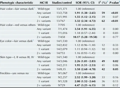
We found greater summary odds
ratios (SORs) for carriers of two MC1R
variants compared with carriers of only
one
variant
allele
(
Table
1
).
Furthermore carriage of any MC1R
variant, one variant and two or more
variants, compared with not having
such variants (i.e., wild-type [WT]
subjects), was significantly associated
with fair hair color, skin type I/II, and
presence of freckles. Red hair color was
significantly associated with carrying
any MC1R variant (SOR; 95%
confi-dence interval: 3.54; 1.91e6.55) and
with carrying two or more variants
(SOR; 95% confidence interval: 10.17;
5.28e19.58), but not with carrying one
MC1R variant (SOR; 95% confidence
interval: 1.18; 0.57e2.44). No
signifi-cant association was observed for light
eye color and MC1R. Sensitivity
ana-lyses indicated that the observed
between-study heterogeneity may be
attributable to single studies: when we
excluded the studies that were outliers,
we obtained similar pooled odds ratios
as the original ones, but no longer with
evidence of heterogeneity (results not
shown). No evidence of publication
bias was found by Egger’s test. All the
investigated MC1R variants compared
with WT subjects were positively
associated with skin type I/II and
freckles (
Supplementary Table S2
on-line). The three variants that seemed to
play the most important role in skin
type determination and the presence of
freckles were D84E, R151C, and
D294H. Red hair color was
signifi-cantly associated with all MC1R
vari-ants except for V92M and R163Q.
We
visualized
the
associations
between hair color, eye color, skin type,
freckles, and the three main studied
geographical areas by multiple
corre-spondence
analysis
(
Supplementary
Figure S1
a or b online). A
two-dimensional multiple correspondence
analysis solution, with dimension 1 on
the horizontal axis and dimension 2 on
the vertical axis, was considered the
most adequate because the first and
second dimension presented
Benzecri-adjusted
inertias
of
85.31%
and
11.31%, respectively (
Supplementary
Table
S3
online),
accounting
for
96.62% of the total association. The
extreme red hair color phenotype (red
hair, skin type I, and freckles) was
associated either with carrying at least
two MC1R variants (
Supplementary
Figure S1
a) or with the presence
of
major
penetrant
(“R”)
alleles
(
Supplementary Figure S1
b). We
sug-gest that dimension 1 can be
inter-preted as a “pigmentation score”
because it differentiates well between
dark and fair phenotypic
characteris-tics. The median pigmentation score
increased with increasing number of
MC1R variants, and for single MC1R
variants it was higher (P
< 0.0001)
compared
with
WT
subjects
(
Supplementary Figure S2
online).
Seven of the nine MC1R variants
analyzed in this study, V60L, D84E,
R142H, R151C, I155T, R160W, and
D294H, are clearly hypomorphic with
significant reduction in cAMP signaling
potential (
Beaumont et al., 2007;
Herraiz et al., 2012; Kadekaro et al.,
2010; Scott et al., 2002
). Within this
group of variants, the lowest SOR for
red hair, skin type I/II, or freckles
cor-responds to V60L. Interestingly, this
variant was also the one with the
smallest functional impairment in terms
of coupling to the cAMP pathway,
when the seven variants analyzed here
were compared under identical
exper-imental conditions (
Herraiz et al.,
2012
).
Table 1. Summary odds ratios for the association between combined
MC1R variants and phenotypic characteristics
Phenotypic characteristic MC1R Studies/control SOR (95% CI) I2(%)3P-value3 Hair color—fair versus dark1 Wild-type 13/1,371 1.00 (reference)
Any variant 13/2,758 1.91 (1.38e2.65) 59 <0.01 1 variant 13/1,991 1.55 (1.12e2.15) 39 0.07 2þ variants 13/767 3.32 (2.34e4.72) 62 <0.01 Hair color—red versus others Wild-type 7/705 1.00 (reference)
Any variant 7/1,474 3.54 (1.91e6.55) 0 0.80 1 variant 7/1,016 1.18 (0.57e2.44) 0 0.83 2þ variants 7/458 10.17 (5.28e19.58) 0 0.77 Eye color—fair versus dark2 Wild-type 14/1,530 1.00 (reference)
Any variant 14/2,832 1.12 (0.96e1.30) 12 0.33 1 variant 14/2,079 1.11 (0.94e1.32) 10 0.35 2þ variants 14/753 1.16 (0.93e1.45) 0 0.80 Skin type—I, II versus III, IV Wild-type 14/1,540 1.00 (reference)
Any variant 14/3,046 2.26 (1.81e2.83) 49 0.02 1 variant 14/2,211 1.95 (1.51e2.53) 41 0.06 2þ variants 14/8,35 3.58 (2.68e4.78) 42 0.05 Freckles—yes versus no Wild-type 9/1,067 1.00 (reference)
Any variant 9/2,257 2.52 (1.99e3.20) 33 0.16 1 variant 9/1,528 2.00 (1.52e2.64) 36 0.13 2þ variants 9/729 4.47 (3.25e6.15) 38 0.12 Significant ORs and P-values are in bold.
Abbreviations: CI, confidence intervals; MC1R, melanocortin-1 receptor; OR, odds ratio; SOR, summary odds ratio.
1Fair hair colors were red, blond, dark blonde, light brown. Dark hair colors were brown, black, dark brown.
2Fair eye colors were blue, green, gray, hazel. Dark eye colors were brown, black.
3I2and Q test P-value are measures of between-study heterogeneity (seeSupplementary Methodsonline).
E Tagliabue et al.
MC1R and Pigmentary Traits: A Pooled Analysis
Results also showed that V92M and
R163Q behave as “r” alleles, with a
weak albeit significant association with
cutaneous phenotypic traits. In
heter-ologous systems, V92M has been
reported to display either a slight
functional impairment (
Herraiz et al.,
2012
) or normal coupling to the
cAMP pathway (
Beaumont et al.,
2007
), whereas R163Q apparently
signals as efficiently as WT. Therefore,
it appears that the ability of V92M or
R163Q to activate the cAMP pathway
is similar, if not identical to WT.
This suggests that other mechanisms
account for their association with
cutaneous phenotypic characteristics,
for example, V92M or R163Q might
impair functional coupling to signaling
module(s) different from the cAMP
cascade. MC1R promiscuously binds
to a variety of intracellular partners
with signaling potential and this ability
might depend on WT conformation.
However, little is known as to the
ef-fects of other variants on MC1R
bind-ing to its various protein partners, and
the phenotypic consequences of such
molecular interactions also remain
largely unknown. Further research is
needed to understand the scaffolding
properties of MC1R, the functional
consequences of the formation of
signaling complexes orchestrated by
the receptor, and the effects on these
processes of the myriad of natural
variants in the MC1R gene.
ORCID
Leah Ferrucci: http://orcid.org/0000-0001-9488-7586
CONFLICT OF INTEREST
The authors state no conflicts of interest.
ACKNOWLEDGMENTS
This work was supported by the Italian Association for Cancer Research (grant number: MFAG 11831). The Melanoma Susceptibility Study (PAK) was supported by the National Cancer Institute [CA75434, CA80700, and CA092428]. The Nurses’ Health Study and the Health Professionals Follow-Up Study (JH) were supported by NIH R01 CA49449, P01 CA87969, UM1 CA186107, and UM1 CA167552. We would like to thank the participants and staff of the Nurses’ Health Study, the Health Professionals Follow-Up Study for their valuable contributions as well as the following state cancer registries for their help: AL, AZ, AR, CA, CO, CT, DE, FL, GA, ID, IL, IN, IA, KY, LA, ME, MD, MA, MI, NE, NH, NJ, NY, NC, ND, OH, OK, OR, PA, RI, SC, TN, TX, VA, WA, WY. Genoa study was supported by AIRC IG 15460 to PG. The M-SKIP study group consists of the following members: Principal Investigator (PI): Sara Rai-mondi (European Institute of Oncology, Milan,
Italy); Advisory Committee members: Philippe Autier (International Prevention Research Institute, Lyon, France), Maria Concetta Fargnoli (University of L’Aquila, Italy), Jose´ C. Garcı´a-Borro´n (Univer-sity of Murcia, Spain), Jiali Han (Brigham and Women’s Hospital and Harvard Medical School, Boston, MA), Peter A. Kanetsky (Department of Cancer Epidemiology, H. Lee Moffitt Cancer Center and Research Institute, Tampa, FL), Maria Teresa Landi (National Cancer Institute, NIH, Bethesda, MD), Julian Little (University of Ottawa, Canada), Julia Newton-Bishop (University of Leeds, UK), Francesco Sera (UCL Institute of Child Health, London, UK); Consultants: Saverio Caini (ISPO, Florence, Italy), Sara Gandini and Patrick Maisonneuve (European Institute of Oncology, Milan, Italy); Participant Investigators: Albert Hofman, Manfred Kayser, Fan Liu, Tamar Nijsten, and Andre G. Uitterlinden (Erasmus MC Univer-sity Medical Center, Rotterdam, The Netherlands), Rajiv Kumar and Dominique Scherer (German Cancer Research Center, Heidelberg, Germany), Tim Bishop, Julia Newton-Bishop, and Faye Elliott (University of Leeds, UK), Eduardo Nagore (Insti-tuto Valenciano de Oncologia, Valencia, Spain), DeAnn Lazovich (Division of Epidemiology and Community Health, University of Minnesota, MN), David Polsky (New York University School of Medicine, New York, NY), Johan Hansson and Veronica Hoiom (Karolinska Institutet, Stockholm, Sweden), Paola Ghiorzo and Lorenza Pastorino (University of Genoa, Italy), Nelleke A. Gruis and Jan Nico Bouwes Bavinck (Leiden University Medical Center, The Netherlands), Paula Aguilera, Celia Badenas, Cristina Carrera, Pol Gimenez-Xavier, Josep Malvehy, Miriam Potrony, Susana Puig, Joan Anton Puig-Butille, Gemma Tell-Marti (Hospital Clinic, IDIBAPS and CIBERER, Barce-lona, Spain), Terence Dwyer (Murdoch Childrens Research Institute, Victoria, Australia), Leigh Blizzard and Jennifer Cochrane (Menzies Institute for Medical Research, Hobart, Australia), Ricardo Fernandez-de-Misa (Hospital Universitario Nues-tra Sen˜ora de Candelaria, Santa Cruz de Tenerife, Spain), Wojciech Branicki (Institute of Forensic Research, Krakow, Poland), Tadeusz Debniak (Pomeranian Medical University, Polabska, Poland), Niels Morling and Peter Johansen (Uni-versity of Copenhagen, Denmark), Susan Mayne, Allen Bale, Brenda Cartmel and Leah Ferrucci (Yale School of Public Health and Medicine, New Haven, CT), Ruth Pfeiffer (National Cancer Insti-tute, NIH, Bethesda, MD), Giuseppe Palmieri (Istituto di Chimica Biomolecolare, CNR, Sassari, Italy), Gloria Ribas (Fundacio´n Investigacio´n Clı´nico de Valencia Instituto de Investigacio´n Sanitaria- INCLIVA, Spain), Chiara Menin (Veneto Institute of Oncology, IOV-IRCCS, Padua, Italy), Alexander Stratigos and Katerina Kypreou (Uni-versity of Athens, Andreas Sygros Hospital, Ath-ens, Greece), Anne Bowcock, Lynn Cornelius, and M. Laurin Council (Washington University School of Medicine, St. Louis, MO), Tomonori Motokawa (POLA Chemical Industries, Yokohama, Japan), Sumiko Anno (Shibaura Institute of Technology, Tokyo, Japan), Per Helsing and Per Arne Andresen (Oslo University Hospital, Norway), Gabriella Guida and Stefania Guida (University of Bari, Bari, Italy), Terence H. Wong (University of Edinburgh, UK), and the GEM Study Group. Participants in the GEM Study Group are as follows: Coordinating Center, Memorial Sloan-Kettering Cancer Center, New York, NY: Marianne Berwick (PI, currently at the University of New Mexico), Colin Begg (Co-PI), Irene Orlow (Co-Investigator), Urvi Mujumdar (Project
Coordinator), Amanda Hummer (Biostatistician), Klaus Busam (Dermatopathologist), Pampa Roy (Laboratory Technician), Rebecca Canchola (Lab-oratory Technician), Brian Clas (Lab(Lab-oratory Tech-nician), Javiar Cotignola (Laboratory TechTech-nician), Yvette Monroe (Interviewer). Study Centers: The University of Sydney and The Cancer Council New South Wales, Sydney (Australia): Bruce Armstrong (PI), Anne Kricker (co-PI), Melisa Litchfield (Study Coordinator). Menzies Institute for Medical Research, University of Tasmania, Hobart (Australia): Terence Dwyer (PI), Paul Tucker (Dermatopathologist), Nicola Stephens (Study Coordinator). British Columbia Cancer Agency, Vancouver (Canada): Richard Gallagher (PI), Teresa Switzer (Coordinator). Cancer Care Ontario, Toronto (Canada): Loraine Marrett (PI), Beth Theis (Co-Investigator), Lynn From (Derma-topathologist), Noori Chowdhury (Coordinator), Louise Vanasse (Coordinator), Mark Purdue (Research Officer). David Northrup (Manager for CATI). Centro per la Prevenzione Oncologia Tor-ino, Piemonte (Italy): Roberto Zanetti (PI), Stefano Rosso (Data Manager), Carlotta Sacerdote (Coor-dinator). University of California, Irvine, CA: Hoda Anton-Culver (PI), Nancy Leighton (Coordinator), Maureen Gildea (Data Manager). University of Michigan, Ann Arbor, MI: Stephen Gruber (PI), Joe Bonner (Data Manager), Joanne Jeter (Coordi-nator). New Jersey Department of Health and Senior Services, Trenton, NJ: Judith Klotz (PI), Homer Wilcox (Co-PI), Helen Weiss (Coordi-nator). University of North Carolina, Chapel Hill, NC: Robert Millikan (PI), Nancy Thomas (Co-Investigator), Dianne Mattingly (Coordinator), Jon Player (Laboratory Technician), Chiu-Kit Tse (Data Analyst). University of Pennsylvania, Philadelphia, PA: Timothy Rebbeck (PI), Peter Kanetsky (Co-Investigator), Amy Walker (Laboratory Techni-cian), Saarene Panossian (Laboratory Technician). Consultants: Harvey Mohrenweiser, University of California, Irvine, Irvine, CA; Richard Setlow, Brookhaven National Laboratory, Upton, NY.
Elena Tagliabue
1, Sara Gandini
1,
Jose´ C. Garcı´a-Borro´n
2,
Patrick Maisonneuve
1,
Julia Newton-Bishop
3, David Polsky
4,
DeAnn Lazovich
5, Rajiv Kumar
6,
Paola Ghiorzo
7,8, Leah Ferrucci
9,
Nelleke A. Gruis
10, Susana Puig
11,
Peter A. Kanetsky
12,
Tomonori Motokawa
13, Gloria Ribas
14,
Maria Teresa Landi
15,
Maria Concetta Fargnoli
16,
Terence H. Wong
17,
Alexander Stratigos
18, Per Helsing
19,
Gabriella Guida
20, Philippe Autier
21,
Jiali Han
22, Julian Little
23,
Francesco Sera
24and Sara Raimondi
1,*,
for the M-SKIP Study group
1
Division of Epidemiology and Biostatistics, European Institute of Oncology, Milan, Italy; 2Department of Biochemistry, Molecular Biology and Immunology, University of Murcia and IMIB-Arrixaca, Murcia, Spain;3Section of Epidemiology and Biostatistics, Institute of Cancer and Pathology, University of Leeds, Leeds, UK;4The Ronald O. Perelman Department of Dermatology, New York
E Tagliabue et al.
MC1R and Pigmentary Traits: A Pooled Analysis
Journal of Investigative Dermatology (2016), Volume 136
University School of Medicine, NYU Langone Medical Center, New York, New York, USA; 5Division of Epidemiology and Community Health, University of Minnesota, Minnesota, USA;6Division of Molecular Genetic Epidemiology, German Cancer Research Center, Heidelberg, Germany;7Department of Internal Medicine and Medical Specialties, University of Genoa, Italy;8IRCCS AOU San Martino-IST, Genoa, Italy;9Department of Chronic Disease Epidemiology, Yale School of Public Health, Yale Cancer Center, New Haven, Connecticut, USA;10Department of Dermatology, Leiden University Medical Center, Leiden, The Netherlands;11Melanoma Unit, Dermatology Department, Hospital Clinic Barcelona, University of Barcelona, CIBER de Enfermedades Raras, Spain; 12Department of Cancer Epidemiology, H. Lee Moffitt Cancer Center and Research Institute2, Tampa, Florida, USA;13Skin Research Department, POLA Chemical Industries, Yokohama, Japan;14Department of medical oncology and hematology, Fundacio´n Investigacio´n Clı´nico de Valencia Instituto de Investigacio´n Sanitaria- INCLIVA, Valencia, Spain;15Division of Cancer Epidemiology and Genetics, National Cancer Institute, NIH, Bethesda, Maryland, USA;16Department of Dermatology, University of L’Aquila, L’Aquila, Italy;17NHS Forth Valley, UK;18First Department of Dermatology, Andreas Sygros Hospital, Medical School, National and Kapodistrian University of Athens, Athens, Greece;19Department of Pathology, Oslo University Hospital, Oslo, Norway; 20Department of Basic Medical Sciences, Neuroscience and Sense Organs, University of Bari, Bari, Italy;21International Prevention Research Institute, Lyon, France;22Department of Epidemiology, Richard M. Fairbanks School of Public Health, Melvin & Bren Simon Cancer Center, Indiana University, Indianapolis, Indiana, USA;23School of Epidemiology, Public Health and Preventive Medicine, University of Ottawa, Ottawa, Canada; and 24Department of Social and Environmental
Health Research, London School of Hygiene & Tropical Medicine, London, UK
*Corresponding author e-mail:sara.raimondi@ ieo.it
REFERENCES
Beaumont KA, Shekar SN, Newton RA, James MR, Stow JL, Duffy DL, et al. Receptor function, dominant negative activity and phenotype cor-relations for MC1R variant alleles. Hum Mol Genet 2007;16:2249e60.
Garcia-Borron JC, Sanchez-Laorden BL, Jimenez-Cervantes C. Melanocortin-1 receptor structure and functional regulation. Pigment Cell Res 2005;18:393e410.
Garcia-Borron JC, Abdel-Malek Z, Jimenez-Cervantes C. MC1R, the cAMP pathway, and the response to solar UV: extending the horizon beyond pigmentation. Pigment Cell Melanoma Res 2014;27:699e720.
Gerstenblith MR, Goldstein AM, Fargnoli MC, Peris K, Landi MT. Comprehensive evaluation of allele frequency differences of MC1R variants across populations. Hum Mutat 2007;28: 495e505.
Han J, Kraft P, Colditz GA, Wong J, Hunter DJ. Melanocortin 1 receptor variants and skin cancer risk. Int J Cancer 2006;119:1976e84. Herraiz C, Journe F, Ghanem G,
Jimenez-Cervantes C, Garcia-Borron JC. Functional sta-tus and relationships of melanocortin 1 receptor signaling to the cAMP and extracellular signal-regulated protein kinases 1 and 2 pathways in human melanoma cells. Int J Biochem Cell Biol 2012;44:2244e52.
Kadekaro AL, Leachman S, Kavanagh RJ, Swope V, Cassidy P, Supp D, et al. Melano-cortin 1 receptor genotype: an important determinant of the damage response of mela-nocytes to ultraviolet radiation. FASEB J 2010;24:3850e60.
Pasquali E, Garcia-Borron JC, Fargnoli MC, Gandini S, Maisonneuve P, Bagnardi V, et al. MC1R variants increased the risk of sporadic cutaneous melanoma in darker-pigmented Caucasians: a pooled-analysis from the M-SKIP project. Int J Cancer 2015;136:618e31.
Perez Oliva AB, Fernendez LP, Detorre C, Herraiz C, Martinez-Escribano JA, Benitez J, et al. Identification and functional analysis of novel variants of the human melanocortin 1 receptor found in melanoma patients. Hum Mutat 2009;30:811e22.
Raimondi S, Sera F, Gandini S, Iodice S, Caini S, Maisonneuve P, et al. MC1R vari-ants, melanoma and red hair color pheno-type: a meta-analysis. Int J Cancer 2008;122: 2753e60.
Raimondi S, Gandini S, Fargnoli MC, Bagnardi V, Maisonneuve P, Specchia C, et al. Melano-cortin-1 receptor, skin cancer and phenotypic characteristics (M-SKIP) project: study design and methods for pooling results of genetic epidemiological studies. BMC Med Res Meth-odol 2012;12:116.
Scherer D, Bermejo JL, Rudnai P, Gurzau E, Koppova K, Hemminki K, et al. MC1R variants associated susceptibility to basal cell carci-noma of skin: interaction with host factors and XRCC3 polymorphism. Int J Cancer 2008;122: 1787e93.
Scott MC, Wakamatsu K, Ito S, Kadekaro AL, Kobayashi N, Groden J, et al. Human mela-nocortin 1 receptor variants, receptor func-tion and melanocyte response to UV radiation. J Cell Sci 2002;115(Pt 11): 2349e55.
Tagliabue E, Fargnoli MC, Gandini S, Maisonneuve P, Liu F, Kayser M, et al. MC1R gene variants and non-melanoma skin cancer: a pooled-analysis from the M-SKIP project. Br J Cancer 2015;113:354e63.
Valverde P, Healy E, Jackson I, Rees JL, Thody AJ. Variants of the melanocyte-stimulating hor-mone receptor gene are associated with red hair and fair skin in humans. Nat Genet 1995;11:328e30.
Low Levels of Genetic Heterogeneity in
Matched Lymph Node Metastases from
Patients with Melanoma
Journal of Investigative Dermatology (2016) 136, 1917e1920;doi:10.1016/j.jid.2016.05.103
TO THE EDITOR
In our previous experience, a high
consistency of BRAF and NRAS
muta-tion patterns was observed between
primary tumors and lymph node
me-tastases in patients with advanced
melanoma (
Colombino et al., 2012
).
Conversely, increasing rates of
discrep-ancies in BRAF/NRAS mutation
pat-terns were found between primary
melanomas and metastases in other
sites (brain or, mostly, skin) (
Colombino
et al., 2012
). When the distribution of
BRAF/NRAS mutations was evaluated
in a larger cohort, the high rate of
consistency in sequence variations of
these two genes was further confirmed
between
primary
melanomas
and
lymph node metastases (142/156; 91%)
(
Colombino et al., 2013
; unpublished
data). However, intraindividual
hetero-geneity of BRAF mutations has been
SUPPLEMENTARY MATERIALSupplementary material is linked to the online version of the paper atwww.jidonline.org, and at http://dx.doi.org/10.1016/j.jid.2016.05.099.
Accepted manuscript published online 2 June 2016; corrected proof published online 16 July 2016 ª 2016 The Authors. Published by Elsevier, Inc. on behalf of the Society for Investigative Dermatology.