Decrease in C-reactive protein levels in rabbits after
vaccination with a live attenuated myxoma virus vaccine
L. Ondruska
1, V. Parkanyi
1, J. Vasicek
1, R. Jurcik
1, E. Hanusova
1,
D. Vasicek
1, A. Balazi
1, F. Vizzarri
21National Agricultural and Food Centre, Research Institute for Animal Production Nitra, Luzianky, Slovak Republic
2University of Molise, Campobasso, Italy
ABSTRACT: The aim of this study was to evaluate the acute phase reaction and immune response of rabbits triggered by vaccination with a live attenuated myxoma virus (MXT) vaccine. Thirteen adult and 11 juvenile New Zealand white rabbit-based crossbreed rabbits, were used. Samples of rabbit peripheral blood were collected from vena auricularis centralis into heparinised tubes before vaccination and 48 h after vaccination. All animals were vaccinated by subcutaneous injection (0.5 ml) with a MXT vaccine. The blood plasma C-reactive protein level was measured by an ELISA kit using a double-antibody sandwich. For phenotyping of lymphocytes the fresh cells were stained with the following anti-rabbit monoclonal antibodies: anti-IgM, anti-CD4, anti-CD8 and anti-pan T2. Our results show that the use of attenuated myxoma virus vaccine significantly decreases the level of C-reactive protein in blood plasma of adult rabbits by 38.14% (P < 0.05) and of juvenile rabbits by 37.63% (P < 0.001), within 48 h. The rabbit C-reactive protein after MXT vaccination is a negative acute phase protein. In the group of adult rabbits the immune response to MXT vaccination was accompanied by a non-significant decrease in CD4+, pT2+, IgM+ subsets. On the other hand the values of CD8+, CD4+CD8+ and CD4+/CD8+ were non-significantly higher after MXT vaccination.
Keywords: rabbit; Oryctolagus cuniculus; myxomatosis; ELISA
Myxoma virus, a member of the Poxviridae family, is the agent responsible for myxomatosis, a highly lethal disease of the European rabbit (Oryctolagus
cuniculus). Since this virus is not a natural pathogen
of Oryctolagus cuniculus, it is able to subvert the host rabbit immune system defences and cause a highly lethal systemic infection. A number of strat-egies used by myxoma virus to modulate the host immune response have already been identified and are well characterised (Petit et al. 1996; Messud-Petit et al. 1998; Jackson et al. 1999; Nash et al. 1999). Myxoma virus encodes a number of proteins that have been experimentally shown to function as secreted viroceptors or virokines. Myxoma virus uses intracellular strategies to inhibit various
apop-totic pathways that are triggered by viral infection in certain cell types (McFadden and Barry 1998; Turner and Moyer 1998).
Currently, vaccination represents the most wide-ly used and most effective way to protect rabbits against myxoma virus. Inflammatory reactions are one of the potential safety concerns that are evaluated in the framework of vaccine safety test-ing. In non-clinical studies, the assessment of inflammation relies heavily on the measurement of biomarkers. The acute phase response is a complex systemic early defence system of reactions activated by trauma, infection, tissue damage, inflamma-tion, stress or neoplasia. C-reactive (CR) protein is an acute-phase plasma protein of hepatic origin
Supported by the Slovak Research and Development Agency (Contract No. APVV-0044-12) and by the Operational Programme Research and Development funded from the European Regional Development Fund (Projects REVITAL No. 26210120038 and LAGEZ No. 26220120070).
that could be used as a biomarker in toxicity stud-ies with rabbits (Destexhe et al. 2013). Although a wide range of studies have been carried out to determine the usefulness of acute phase proteins in several human and animal diseases reports of their application in veterinary medicine remain scarce, and are predominantly from farm animals (Tothova et al. 2014). The acute phase response is characterised by the altered expression of specific acute phase proteins. By definition, an acute phase protein increases (positive acute phase protein) or decreases (negative acute phase protein) in plasma concentration by at least 25% during inflammatory disorders (Gabay and Kushner 1990). CR protein be-longs to the group of positive acute phase proteins. The main characteristic feature that determines the biological effects of CR protein is its ability to bind phosphocholine, which enables CR protein to recognise foreign pathogens and the phospholipid components of damaged cells. From an immuno-logical perspective CR protein acts as an opsonin. After binding to foreign particles and phagocytic cells, the complement system is activated via the classical pathway and by interaction with the hu-moral and cellular effector systems of inflammation triggered by the removal of the target cells. The CR protein ligand complex interacts directly with neutrophils, macrophages and other phagocytic cells and stimulates an inflammatory response and the production of cytokines. The rapid induction of the CR protein-dependent response suggests that CR protein is a component of the innate immune response. CR protein has both pro-inflammatory and anti-inflammatory effects.
Lymphocyte subsets occupy a critical position in the reaction cascade of the immune response, and the determination of their distribution in the peripheral blood is a routine part of laboratory tests in human patients suspected of immunodeficiency. At present, the importance of flow cytometry in veterinary medicine is increasing, particularly in small animal practice. The knowledge of physiolog-ical values is necessary for recognition of changes in lymphocyte subset distribution (Faldyna et al. 2001). The most important distinction is that CD4+
cells see antigenic peptides in association with ma-jor histocompatibility complex class II molecules, whereas CD8+ cells react with peptide plus major
histocompatibility complex class I (Nossal 1997). CD4+ T cells, when activated, develop into T cells
secreting a large variety of cytokines (Kelso et al.
1991). However, as the immune response matures, there are many instances where either a T helper 1 response or a T helper 2 response becomes domi-nant (Mosmann and Coffman 1989). The T helper 1 response leads to inflammatory phenomena and the T helper 2 response to antibody formation, includ-ing IgG1 and IgE formation (Finkelman et al. 1990). Injections of vaccines into animals may induce inflammatory reactions, either local or systemic. Evaluation of the acute phase reaction and immune response of rabbits triggered by vaccination against myxomatosis has, to the best of our knowledge, not yet been performed, and could help to better predict the influence of commonly used as well as new vaccines.
MATERIAL AND METHODS
Animals. The trial was performed on the
experi-mental farm at the National Agricultural and Food Centre – Research Institute for Animal Production Nitra, Slovak Republic and was conducted on clini-cally healthy 24 crossbreed rabbits of a line based on New Zealand white rabbits.
Animals were divided into two groups: an adult rabbit group (three males and ten females, 12-month-old) and a juvenile rabbit group (six males and five females, 3-month-old). All females in the adult rab-bit group were after the third kindling.
Animals were individually housed in wire cages arranged in flat-decks on one level. Cages were equipped with a hopper for food. The rabbits were fed with a commercial diet (pellets of 3 mm in di-ameter). All animals were given access to the feed
ad libitum. Drinking water was provided with
nip-ple drinkers ad libitum. A cycle of 16 h of light and 8 h of dark (minimum light intensity of 80 lux) was used throughout the trial. Temperature and humid-ity in the building were recorded continuously by a digital thermograph positioned at the same level as the cages. Heating and forced ventilation systems allowed the building temperature to be maintained at 18 ± 4 °C throughout the trial. Relative humidity was about 70 ± 5%.
In this animal study, institutional and national guidelines for the care and use of animals were fol-lowed, and all experimental procedures involving animals were approved by an ethical committee.
Immunisation and blood plasma samples. One
sam-ples from adult and juvenile rabbits were collect-ed from vena auricularis centralis to hepariniscollect-ed tubes before vaccination. Subsequently, all animals were vaccinated by subcutaneous injection (0.5 ml) with a live attenuated myxoma virus vaccine, min. 103 50% tissue culture infective dose – TCID
50
(Pharmavac MXT, Pharmagal Bio, Slovak Republic). Further blood samples were collected 48 h after vaccination, in the same way as the control samples.
Enzyme-linked immunosorbent assay (ELISA) test. The heparinized tubes with blood were
cen-trifuged for 20 min at the speed of 850 g for plas-ma isolation. The levels of rabbitCR protein in the blood plasma were quantified using a commercial
rabbit ELISA kit (SunRed Bio, Shanghai, China, catalogue No. 201-09-0003, http://www.sunredbio. com/eindex.asp). The kit uses a double-antibody sandwich ELISA to assay the level of rabbit frag-ment CR protein in 40 µl of blood plasma. This commercial ELISA kit does not express the whole protein, but just a fragment. We determined the lev-els of whole CRPafter conversion in accordance with the instructions of the manufacturer. The standard of this ELISA kit corresponds to a concentration of 32 mg/l of whole CR protein (SunRedBio, per-sonal communication). Totallevels of CR protein are presentedin the Resultssection.
The measurements of samples, blanks and standards on a BioTekTM EonTM Microplate
Spectrophotometer (BioTek Instruments, USA) were carried out by measuring optical density at a wavelength of 450 nm within 15 min of adding the stop solution.
Flow cytometry analysis. Mononuclear cells from
peripheral blood were isolated using Ficoll centrifu-gation according to the original protocol: Isolation of mononuclear cells from human peripheral blood by density gradient centrifugation (Miltenyi Biotec, 2008). The freshly isolated mononuclear cells from peripheral blood were divided into prepared tubes
and stained with different clones of anti-rabbit monoclonal antibodies: anti-IgM (IgG1; NRBM; Bio-Rad AbD Serotec GmbH, Germany), anti-CD4 (IgG1; RTH1A; WSU, Pullman, WA), anti-CD8 (IgG2a; ISC27A; WSU, Pullman, WA), anti-pan T2 (pT2; IgG1; RTH21A; WSU, Pullman, WA) and anti-CD45 (IgG1; L12/201; Bio-Rad AbD Serotec GmbH, Germany) according to the producer’s manual. As the secondary immunoreagent, FITC- or R-PE-labelled anti-mouse conjugates of appropriate subi-sotypes (eBioscience, Austria) were used. To assess contamination of the lymphocyte gate by other cell types, a cross-reactive FITC-labelled mAb against human CD14 antigen (IgG2a; TUK4; Bio-Rad AbD Serotec GmbH, Germany) was used. In each sam-ple, 10 000–50 000 cells were measured using a FACS Calibur flow cytometer (Becton Dickinson, Mountain View, CA). 7-AAD staining solution (BD Biosciences, USA) was used to exclude dead cells from analysis. Expression of the common leukocyte antigens CD45 and CD14 was used for the “lympho-gate” setup and lymphocyte purity determination as described by Jeklova et al. (2007). Results obtained for the other surface markers were recalculated to 100% of CD45+ and CD14- cells in the “lymphogate”.
Statistical analyses. Statistical analysis of the
results was performed using one-way analysis of variance (ANOVA) with Scheffe’s test and the t-test with the level of significance set at P-values of less than 0.05, 0.01 and 0.001. The results are presented as means ± standard deviation.
RESULTS
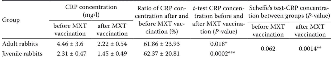
The results of the present experiments have shown that the plasma CR protein levels after MXT vaccination were significantly lower (4.46 ± 3.6 vs 2.22 ± 0.54; P < 0.05) compared with before vacci-nation in the adult rabbits. In the group of juvenile
Table 1. Concentration of whole C-reactive protein in rabbit plasma before and after attenuated myxoma virus vaccination Group
CRP concentration
(mg/l) centration after and Ratio of CRP con-before MXT
vac-cination (%)
t-test CRP concen-tration before and after MXT
vaccina-tion (P-value)
Scheffe’s test-CRP concentra-tion between groups (P-value) before MXT
vaccination vaccinationafter MXT before MXT vaccination vaccinationafter MXT Adult rabbits 4.46 ± 3.6 2.22 ± 0.54 61.86 ± 23.93 0.018*
0.062 0.0014** Jivenile rabbits 2.31 ± 0.47 1.45 ± 0.49 62.37 ± 20.81 0.0002***
CRP = C-reactive protein, MXT = attenuated myxoma virus min. 103 TCID
50; the values shown are the means ± SD *P < 0.05, **P < 0.01, ***P < 0.001
rabbits the CR protein values after MXT vaccina-tion decreased even more significantly (2.31 ± 0.47 vs 1.45 ± 0.49; P < 0.001). Comparing the groups of adults and young rabbits a significantly (P < 0.01) lower level of CR protein after vaccination in the group of young rabbits was observed.
The ratio of CR protein concentration (mg/l) in adult rabbit plasma before and after MXT vaccina-tion was in the range of 92.73% to 17.70% (average 61.86 ± 23.93). Approximately the same ratio of CR protein concentration (mg/l) was also calcu-lated in the plasma of juvenile rabbits, where the range was from 97.95% to 20.14% (average 62.37 ± 20.8; Table 1). Our results showed that the use of the MXT vaccine downregulated the level of CR protein in blood plasma of adult rabbits by 38.14% and juvenile rabbits by 37.63%, on average. Thus, in the rabbit CR protein is a negative acute phase protein after MXT vaccination. The immune re-sponse of the juvenile and adult rabbits to attenu-ated myxoma virus was very similar.
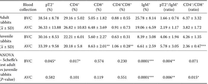
In this study, we also evaluated the changes in lymphocyte subsets of rabbit peripheral blood col-lected from adult and juvenile rabbits before and after immunisation with a vaccine against myxoma-tosis. After immunisation no significant decreases in the percentages of pT2+, CD4+, IgM+
lympho-cytes nor in the CD4+/CD8+ ratio in the group of
adult rabbits were observed. On the other hand, the percentage of T lymphocyte subsets (CD8+ cells,
CD4+CD8+ and pT2+/IgM+ ratio) in this category
of rabbits increased non-significantly (Table 2). A significant increase (P < 0.01) after immunisa-tion in the percentage of T lymphocytes (CD8+,
CD4+CD8+ cells) was observed in juvenile rabbits,
resulting in a lower (P < 0.001) CD4+/CD8+ ratio
value after immunisation.
Comparing adult and juvenile rabbits before vac-cination, significant differences in the percentage of pT2+ (P < 0.05), CD4+ (P < 0.05), IgM+ (P < 0.001)
and in the ratio of pT2+/IgM+ lymphocytes (P < 0.01)
have been observed. However after vaccination sig-nificant differences in the ratio of CD4+/CD8+ (P <
0.05), pT2+/IgM+ (P < 0.01) and percentage of IgM+
lymphocytes (P < 0.001; Table 2) were recorded.
DISCUSSION
CR protein was originally identified as a clinical marker of inflammation (Du Clos and Mold 2003). Watzinger et al. (2011) reported the increase in CR protein levels observed after injection of antigens (Complete Freund’s Adjuvant, typhoid vaccine, sa-line solutions) to rabbits, what is in contrast with our study with MXT vaccination. On the other hand our results are in agreement with the literature, in which myxoma virus has shown an inherent ability to infect and cause immunosuppression in rabbits after infec-tion (Jeklova et al. 2008; Spiesschaert et al. 2011).
Table 2. Lymphocyte subsets in peripheral blood of adult and juvenile rabbits before and after attenuated myxoma virus vaccination Blood collection pT2 + (%) CD4 + (%) CD8 + (%) CD4 +CD8+ (%) IgM + (%) pT2 +/IgM+ (ratio) CD4 +/CD8+ (ratio) Adult rabbits (–x ± SD) BVC 38.54 ± 8.78 29.16 ± 5.02 5.05 ± 1.82 0.88 ± 0.55 25.78 ± 8.14 1.66 ± 0.74 6.37 ± 3.32 AVC 36.33 ± 13.88 26.82 ± 10.83 6.48 ± 3.69 0.91 ± 0.73 19.06 ± 6.59 2.19 ± 1.17 3.82 ± 1.72 Juvenile rabbits (–x ± SD) BVC 30.16 ± 8.53 22.21 ± 6.01 5.60 ± 2.27 0.63 ± 0.31 8.39 ± 3.08 4.06 ± 1.94 4.26 ± 1.35 AVC 33.39 ± 9.58 20.18 ± 5.8 8.63 ± 2.01** 1.06 ± 0.28** 6.61 ± 2.59 5.78 ± 3.05 2.36 ± 0.47*** ANOVA – Scheffe’s test adult vs juvenile rabbits (P-value) BVC 0.045* 0.017* 0.574 0.230 0.0001*** 0.004** 0.071 AVC 0.582 0.101 0.119 0.551 0.0001*** 0.006** 0.015*
AVC = after MXT vaccination, BVC = before MXT vaccination, MXT = attenuated myxoma virus min. 103 TCID 50 *P < 0.05, **P < 0.01, ***P < 0.001
Many viruses encode virulence factors to fa-cilitate their own survival by modulating a host’s inflammatory response. One of these factors, se-creted from cells infected with myxoma virus, is the serine proteinase inhibitor SERP-1 (Brahn et al. 2014). Lomas et al. (1993) and Nash et al. (1998) found that SERP-1 is a myxoma virus-encoded ser-pin, secreted from infected cells, that is required for virulence and has anti-inflammatory activity. These findings suggest that SERP-1 contributes to viral pathogenesis by interacting with and in-hibiting host proteins involved in the regulation of inflammation. The findings of the present trial confirm the down-regulation of CR protein. Jain et al. (2011) reported that CR protein belongs to the group of positive acute phase proteins in human medicine. In contrast, we found that CR protein in rabbits is a negative acute phase protein after MXT vaccination with attenuated myxoma virus.
The average percentages of specific lymphocyte subsets (pT2+, CD4+, CD8+, CD4+CD8+) in the
peripheral blood of rabbits before immunisation were similar to those observed by Jeklova et al. 2007 (40.1%, 29.4%, 10.4%, 2.0%, respectively), thus confirming the normal health status of the rabbits used for the experiments. Although these authors found a more than 10% higher value of B cells in comparison to our results, that could be due to the use of a CD79α antibody for enumeration of B cells. Anti-rabbit IgM monoclonal antibodies have been successfully used as B cell markers also in other studies (Vajdy et al. 1998; Lanning et al. 2000; Tokarz-Deptula and Deptula 2005).
The decrease in the percentage of CD4+ cells
after vaccination in our results are in agreement with an earlier study focused on a characterisa-tion of immunosuppression in rabbits after in-fection with myxoma virus (Jeklova et al. 2008) and also with in vitro studies of malignant rabbit fibroma virus (Strayer et al. 1987; Strayer 1992). This leporipoxvirus, a hybrid of myxoma virus and rabbit fibroma virus, replicates preferentially in mature T lymphocytes of the spleen but is able to grow in B lymphocytes as well. The ability of the Lausanne MXV strain to downregulate CD4 expression on the surface of a T cell line (RL-5), for up to 24 h after infection, has been observed
in vitro (Barry et al. 1995). CD4+ molecules on
T helper cells play a pivotal role in T cell on-togeny and T cell activation. Downregulation of CD4 expression on infected T helper cells may be
involved in inhibition of the survival of infected cells and in disseminating the virus within the host (Best and Kerr 2000).
On the other hand, we found an increased per-centage of T cells (CD8+) in the rabbit peripheral
blood after immunisation, a result which is discord-ant with those of Jeklova et al. (2008). An increase in lymphocyte concentration is usually a sign of a viral infection, in this case caused by the attenuated myxoma virus. The increased percentage of CD8+
cells in the studied rabbits might be indicative of an adequate immune response.
To the best of our knowledge, the present find-ings constitute the first in vivo study reporting that MXT (attenuated myxoma virus min. 103 TCID
50
vaccine) significantly downregulates CR protein levels in rabbit blood plasma. The exact upstream signalling pathway responsible for these effects re-mains to be determined.
Acknowledgement
We would like to thank the company Pharmagal-Bio Ltd. for the provision of vaccines Pharmavac MXT (http://www.pharmagalbio.sk/en/).
REFERENCES
Barry M, Lee SF, Boshkov L, McFadden G (1995): Myxoma virus induces extensive CD4 downregulation and disso-ciation of p56lck in infected rabbit CD41 T lymphocytes. Journal of Virology 69, 5243–5251.
Best SM, Kerr PJ (2000): Coevolution of host and virus: the pathogenesis of virulent and attenuated strains of my-xoma virus in resistant and susceptible European rabbits. Virology 267, 36–47.
Brahn E, Lee S, Lucas AR, McFadden G, Macaulay C (2014): Suppression of collagen-induced arthritis with a serine proteinase inhibitor (serpin) derived from myxoma virus. Clinical Immunology 153, 254–263.
Destexhe E, Prinsen MK, van Scholl I, Kuper FC, Garcon N, Veenstra S, Segal L (2013): Evaluation of C-reactive protein as an inflammatory biomarker in rabbits for vac-cine nonclinical safety studies. Journal of Pharmacologi-cal and ToxicologiPharmacologi-cal Methods 68, 367–373.
Du Clos TW, Mold C (2003): 3 C-reactive protein: structure, synthesis and function. In: Wong S, Arsequell G (eds): Immunobiology of Carbohydrates. Kluwer Academic/ Plenum Publishers, New York. 47–61.
Faldyna M, Leva L, Knotigova P, Toman M (2001): Lympho-cyte subsets in peripheral blood of dogs – a flow cytomet-ric study. Veterinary Immunology and Immunopathology 82, 23–37.
Finkelman FD, Holmes J, Katona IM, Urban Jr JF, Beckmann MP, Park LS, Schooley KA, Coffman RL, Mosmann TR, Paul WE (1990): Lymphokine control of in vivo immu-noglobulin isotype selection. Annual Review of Immunol-ogy 8, 303–333.
Gabay C, Kushner I (1990): Acute phase proteins and other systemic responses to inflammation. New England Jour-nal of Medicine 340, 448–454.
Jackson RJ, Hall DF, Kerr PJ (1999): Myxoma virus encodes an alpha 2,3-sialyltransferase that enhances virulence. Journal of Virology 73, 2376–2384.
Jain S, Gautam V, Naseem S (2011): Acute phase proteins: As diagnostic tool. Journal of Pharmacy and Bioallied Sciences 3, 118–127.
Jeklova E, Leva L, Faldyna M (2007): Lymphoid organ de-velopment in rabbits: Major lymphocyte subsets. Devel-opmental and Comparative Immunology 31, 632–644. Jeklova E, Leva L, Matiasovic J, Kovarcik K, Kudlackova H,
Nevorankova Z, Psikal I, Faldyna M (2008): Characterisa-tion of immunosuppression in rabbits after infecCharacterisa-tion with myxoma virus. Veterinary Microbiology 129, 117–130. Kelso A, Troutt AB, Maraskovsky E, Gough NM, Morris L,
Pech MH, Thomson JA (1991): Heterogeneity in lymphokine profiles of CD4+ and CD8+ T cells and clones activated in vivo and in vitro. Immunological reviews 123, 85–113. Lanning D, Sethupathi P, Rhee KJ, Zhai SK, Knight KL
(2000): Intestinal microflora and diversification of the rabbit anti-body repertoire. Journal of Immunology 165, 2012–2019.
Lomas DA, Evans DL, Upton C, McFadden G, Carrell RW (1993): Inhibition of plasmin, urokinase, tissue plasmino-gen activator, and C1S by a myxoma virus serine protein-ase inhibitor. Journal of Biological Chemistry 268, 516–521. McFadden G, Barry M (1998): How poxviruses oppose
ap-optosis. Seminars in Virology 8, 429–442.
Messud-Petit F, Gelfi J, Delverdier M, Amardeilh MF, Py R, Sutter G, Bertagnoli S (1998): Serp2, an inhibitor of the interleukin-1β-converting enzyme, is critical in the patho-biology of myxoma virus. Journal of Virology 72, 7830–7839. Mosmann TR, Coffman RL (1989): Th1 and Th2 cells: dif-ferent patterns of lymphokine secretion lead to difdif-ferent
functional properties. Annual Review of Immunology 7, 145–173.
Nash P, Whitty A, Handwerker J, Macen J, McFadden G (1998): Inhibitory specificity of the anti-inflammatory myxoma virus serpin SERP-1. Journal of Biological Chem-istry 273, 20982–20991.
Nash P, Barrett J, Cao JX, Hota-Mitchell S, Lalani AS, Ev-erett H, Xu XM, Robichaud J, Hnatiuk S, Ainslie C (1999): Immunomodulation by viruses: the myxoma virus story. Immunological Reviews 168, 103–120.
Nossal GJV (1997): Host immunobiology and vaccine de-velopment. The Lancet 350, 1316–1319.
Petit F, Bertagnoli S, Gelfi J, Fassy F, Boucraut-Baralon C, Milon A (1996): Characterization of a myxoma virus-en-coded serpin-like protein with activity against interleukin-1β-converting enzyme. Journal of Virology 70, 5860–5866. Spiesschaert B, McFadden G, Hermans K, Nauwynck H,
Van de Walle GR (2011): The current status and future directions of myxoma virus, a master in immune evasion. Veterinary Research 76, 1–18.
Strayer DS (1992): Determinants of virus-related suppres-sion of immune responses as observed during infection with an oncogenic poxvirus. Progress in Medical Virology 39, 228–255.
Strayer DS, Skaletsky E, Leibowitz JL, Dombrowski J (1987): Growth of malignant rabbit fibroma virus in lymphoid cells. Virology 158, 147–157.
Tokarz-Deptula B, Deptula W (2005): Values of selected immune and haematological parameters in healthy rab-bits. Polish Journal of Veterinary Sciences 8, 107–112. Tothova C, Nagy O, Kovac G (2014): Acute phase proteins
and their use in the diagnosis of diseases in ruminants: a review. Veterinarni Medicina 59, 163–180.
Turner PC, Moyer RW (1998): Control of apoptosis by pox-viruses. Seminars in Virology 8, 453–469.
Vajdy M, Sethupathi P, Knight KL (1998): Dependence of an-tibody somatic diversification on gut-associated lymphoid tissue in rabbits. Journal of Immunology 160, 2725–2729. Watzinger M, Nguyen A, Gervais F, Lemarchand T, Legrand
J, Descotes J (2011): Evaluation of inflammation biomark-ers for safety assessment of vaccines and adjuvants in the rabbit. 47th Congress of the European Societies of Toxi-cology. Toxicology Letters 205, S113–S114.
Received: 2015–06–16 Accepted after corrections: 2016–08–05 Corresponding Author:
Lubomir Ondruska, National Agricultural and Food Centre, Research Institute for Animal Production Nitra, Hlohovecka 2, 951 41 Luzianky, Slovak Republic