1 23
Journal of Assisted Reproduction and
Genetics
Official Publication of ALPHA, Scientists
in Reproductive Medicine
ISSN 1058-0468
J Assist Reprod Genet
DOI 10.1007/s10815-015-0436-z
Morphokinetic parameters of early embryo
development via time lapse monitoring and
their effect on embryo selection and ICSI
outcomes: a prospective cohort study
Charalampos Siristatidis, Maria Aggeliki
Komitopoulou, Andreas Makris,
Afrodite Sialakouma, Mitrodora
Botzaki, George Mastorakos, et al.
1 23
Your article is protected by copyright and all
rights are held exclusively by Springer Science
+Business Media New York. This e-offprint is
for personal use only and shall not be
self-archived in electronic repositories. If you wish
to self-archive your article, please use the
accepted manuscript version for posting on
your own website. You may further deposit
the accepted manuscript version in any
repository, provided it is only made publicly
available 12 months after official publication
or later and provided acknowledgement is
given to the original source of publication
and a link is inserted to the published article
on Springer's website. The link must be
accompanied by the following text: "The final
publication is available at link.springer.com”.
ASSISTED REPRODUCTION TECHNOLOGIES
Morphokinetic parameters of early embryo development via time
lapse monitoring and their effect on embryo selection and ICSI
outcomes: a prospective cohort study
Charalampos Siristatidis&Maria Aggeliki Komitopoulou&Andreas Makris&
Afrodite Sialakouma&Mitrodora Botzaki&George Mastorakos&
George Salamalekis&Stefano Bettocchi&Giles Anthony Palmer
Received: 8 October 2014 / Accepted: 13 January 2015 # Springer Science+Business Media New York 2015 Abstract
Purpose To compare the outcomes of embryos selected via time lapse monitoring (TLM) versus those selected with con-ventional methods of selection in subfertile women undergo-ing ICSI.
Methods The study population (239 women) was classified into two groups, based on the monitoring method used: Group 1 (TLM) and Group 2 (conventional monitoring). Groups were compared according to the clinical and ICSI cycle char-acteristics and reproductive outcomes, while transfers were performed at day 2 or 3. Subgroup analyses were performed,
in women of both groups according to age and clinical param-eters, and in embryos of Group 1 based on their cellular events.
Results There was a statistically significant difference be-tween the two study groups with regard to the outcome pa-rameters, favoring Group 1 and especially in women >40 years of age. No differences were found in subgroup analyses in participants of both groups, regarding the stimulation protocol used, number of the oocytes retrieved and type of subfertility, while in Group 1 the percentages of“in range” cellular events were higher in certain divisions in ages 35–40, non-smokers, and the GnRH-agonist group, and in embryos that resulted in pregnancy.
Conclusion Morphokinetic parameters of early embryo de-velopment via TLM are related to the characteristics of subfertile patients and associated with ICSI outcomes.
Keywords Assisted reproductive techniques . ICSI . IVF . Time-Lapse Monitoring . Pregnancy
Introduction
Embryo staging, evaluation and selection remain a challenge in order to increase the current success rates in Assisted Reproduction Techniques (ART), with pregnancy and live birth rates to be around 35 % and 25 %, respectively [1]. Until recently, and even today in most ART centers rely on the use of the inverted microscope, which offers information based on morphological and developmental characteristics from the early cleavage till the blastocyst stage. However, this method has several limitations [2,3]. Notably, transfer of top grade morphologically embryos often fails to result in clinical pregnancy, while embryos with poor scores sometimes result Capsule This prospective cohort study showed that the morphokinetic
parameters of early embryo development observed via time lapse monitoring in subfertile women undergoing ICSI are related to the special characteristics of these women and are associated with ICSI outcomes. Electronic supplementary material The online version of this article (doi:10.1007/s10815-015-0436-z) contains supplementary material, which is available to authorized users.
C. Siristatidis (*)
:
G. SalamalekisAssisted Reproduction Unit, 3rd Department of Obstetrics and Gynecology, University of Athens, Rimini 1, Chaidari, Athens, Greece 12642
e-mail: [email protected]
M. A. Komitopoulou
:
A. Makris:
A. Sialakouma:
M. Botzaki:
G. A. Palmer
Assisted Reproduction Unit, MITERA Hospital, Erythrou Stavrou 6, Marousi, Athens, Greece 15123
G. Mastorakos
2nd Department of Obstetrics and Gynecology, University of Athens, Aretaieion Hospital, Athens, Greece
S. Bettocchi
First Unit of Obstetrics and Gynecology, Department of Biomedical
Sciences and Human Oncology, University“Aldo Moro”,
70124 Bari, Italy J Assist Reprod Genet
DOI 10.1007/s10815-015-0436-z
in live births. Apart from morphological assessment, invasive and non-invasive methods of selection have been developed. Single cell biopsy at the cleavage stage has been shown to not affect embryo progression to blastocyst [4]. Therefore, preimplantation genetic screening (PGS) has been used for embryo selection and aneuploidy screening, however its effectiveness has recently been placed under scrutiny [5]. Non-invasive techniques such as embryo oxygen consumption, testing of soluble HLA-G, amino acid turnover, proteomics and metabolomics, cumulus cell gene expression analysis, and time-lapse microscopy (TLM) are consequently becoming more popular options for embryo selection [6].
The consensus behind the use of TLM is based on the improved assessment of the embryos and their early cellular divisions, enabling to determine the timing of specific mor-phological occurrences and permit comparison between them, while also identifying anomalies (such as fragmentations and multinucleations), which otherwise would not be detected. This information can lead to more objective selection of em-bryos for transfer and/or cryopreservation, the reduction of the number of embryos to be biopsied in PGS and the improve-ment of the success rates for ART patients, and for those diagnosed with repeated implantation failures [7,8]. More-over, the potential undesirable shock or stress due to sudden changes in environmental parameters, such as temperature, associated with the use of a microscope are avoided [9–11].
The technology is easily assimilated into the ART labora-tory and is used with any culture medium and environment. Images of the embryos are recorded at regular intervals with-out their removal from the culture environment, and images can be viewed instantly or merged to form a video showing complete development from the oocyte to blastocyst stage: their review can assist in identifying and selecting embryos with normal developmental profiles, and in deselecting those with abnormal phenotypes [12–14].
Reports on the modality have been numerous during the last 10 years or more. The target was to explore the dynamics of an embryo to implant successfully along with its various cleavage patterns and characteristics, by observing it at vari-ous stages of development [12,15–21]. Recent studies report-ed information on the association of the type of fertilization [22], the IVF protocol used [23], female obesity [24], and smoking [25] on embryo kinetics and development. Interest-ingly, the sensitivity and specificity of the modality in terms of predicting the progression to the blastocyst stage was >90 % by measuring parameters at day 2 after fertilization, before embryonic genome activation [26,27], while the bad progno-sis of good-performing but unviable embryos reaches a spec-ificity of 100 % [28]; moreover, the area under the receiver operating characteristic curve has been reported to be 0.74 for live birth [29] with high intra- and inter-observer correlation [30].
Regarding the use of TLM with pregnancy and implanta-tion rates compared to the convenimplanta-tional incubator, literature data are conflicting, reporting both positive– even after ac-counting for confounding factors- [31,32] and negative asso-ciations [33–35]. To date, most reports conclude that further studies are warranted to elucidate the relationship, either in the form of a large, age-adjusted data set or in a randomized con-trolled trial [36,37]. We performed a prospective cohort trial to compare the effects of the TLM versus the conventional methods of selection on embryo implantation potential and reproductive outcome in subfertile women undergoing ICSI.
Materials and methods
Patient population and study design
This is a prospective, cohort trial performed at the Assisted Reproductive Unit of the MITERA Private Hospital with the scientific support of the Assisted Reproductive Unit of 3rd Department of Obstetrics and Gynecology,“Attikon” Hospi-tal, of the Athens University School of Medicine. The study was approved by the Scientific and Ethics Committee of MITERA Hospital. A written informed consent was obtained from all patients enrolled in the current study.
Subjects
Two hundred thirty-nine cycles of ICSI in 239 women with primary or secondary subfertility were analyzed. Subfertility factors were recorded through the typical processes of the Unit, categorized as follows: female (tubal subfertility and ovulatory dysfunction), male factor and unexplained infertili-ty. Exclusion criteria for the participation in the study were: age>42 years old, basal hormonal levels of FSH at day 3 of the menstrual cycle>15 IU/L, other protocols towards oocyte retrieval (natural or mild IVF cycles) and signs of ovarian hyperstimulation syndrome, as well as cases where fresh em-bryo transfers were a priori excluded. In addition, women with known previous poor ovarian response to ovarian stimulation were excluded.
Patients were enrolled consecutively during a 4 months period and participated in the study only once. The study population was classified into two groups, according to the monitoring modality, Group 1 (TLM) and Group 2 (conven-tional monitoring). Classification was achieved immediately after oocyte retrieval (OR): if more than five oocytes were retrieved, TLM or conventional monitoring was offered ac-cording to the number of the file of each patient (0 to 2 vs. 3 to 9 as a last digit), respectively. A consensus between the at-tending physician, the embryologist and the couple/ patient was placed in either groups. Of note, two patients in Group J Assist Reprod Genet
1 and three in Group 2 did not agree with the allocated method and therefore were not included in the study.
Stimulation protocol
For the GnRH-agonist protocol, Triptorelin [Gonapeptyl, 0.1 mg (Ferring Pharmaceutical HellasΑ.Ε.) or Arvekap, 0.1 mg (Ipsen, EPE)] was administered subcutaneously daily during the midluteal phase or the second day of the menstrual cycle. For the GnRH antagonist protocol, ovarian stimulation began on the second day of the cycle and the antagonist, either Cetrorelix (Merck Serono Europe Limited, UK) or Orgalutran (Merck Sharp & Dohme Limited, UK) was initiated as soon as the leading follicle reached a diameter of 14 mm. Once pitu-itary down-regulation was achieved, ovarian stimulation with exogenous gonadotropins was started while GnRH agonist administration was continued concomitantly until the day of human chronic gonadotrophin (hCG) administration. Recom-binant FSH in the form of either follitropin alpha (Gonal-F; Merck Serono Europe Ltd) or follitropin beta (Puregon Merck Sharp & Dohme Ltd) was administered subcutaneously.
The patients were monitored with serial transvaginal ultra-sound and E2 levels every 2~4 days to monitor follicular growth and endometrial thickness. Starting doses were adjust-ed individually according to the age, FSH, AMH levels and previous response to IVF/ICSI cycles of each participant, while further adjustments and monitoring frequency were de-pendent upon women’s response to stimulation. When ≥2 fol-licles reached a diameter of 18 mm, human chorionic gonad-otrophin [(10.000 IU Pregnyl (N. V. Organon, Netherlands) or 250mcg Ovitrelle (Merck Serono Europe Ltd, Germany)] was administered prior to transvaginal ultrasound–guided oocyte retrieval (OR) 36 h later.
According to the embryo quality, embryo transfer (ET) was performed either 2 or 3 days after the OR. Luteal phase sup-port was achieved by transvaginal administration of proges-terone in the form of either Utrogestan vaginal suppositories (Angelini Pharma HellasΑ.Β.Ε.Ε.) or Vasclor gel (Verisfield; U.K. Ltd) or a combination of both.
Fertilisation and preparation and embryo culture methods All oocytes were stripped of cumulus cells and all M2 oocytes were microinjected through ICSI, 40 h post-hCG injection. The inseminated oocytes were placed in standard culture dishes containing 1 mL Universal IVF Medium™ (Origio, Denmark), overlaid with oil and left overnight in a standard Thermo Forma™ incubator (Thermo Scientific, Thermo Fish-er Scientific, USA) at 37 °C and 6%CO2. Fertilisation was
assessed 18 h post-insemination based on the presence of two pronuclei.
All zygotes in Group 1 were then transferred to a PrimoVision Embryo Culture Dish™ (Vitrolife, Sweden)
containing 60–80 μl ISM1™ culture medium (Origio, Den-mark), overlaid with oil and placed under a PrimoVision Time-Lapse Embryo Monitoring System™ (Vitrolife, Swe-den) in a standard Thermo Forma™ incubator at 37 °C and 6%CO2. The monitoring system automatically took
photo-graphs of the embryos using an inverted microscope every 10 min, for the whole duration of culture. Zygotes from the control group were cultured in Nunc™ 4-Well Dishes (Ther-mo Scientific, Ther(Ther-mo Fisher Scientific, USA) containing 500μl ISM1™, placed in a Thermo Forma™ incubator. Em-bryos in both groups were cultured until day 2 or 3, and then transferred, depending on their quality, and consensus be-tween the embryologist and the attending physician of the couple.
Embryo evaluation and selection for transfer
Embryos in Group 2 were evaluated at 44 and 68 h (depending on day of ET) based on classic morphological criteria (number of blastomeres, fragmentation and multi-nucleation) on a scale of 1–5, with 1 representing a perfect morphological embryo, as previously described [38,39]. Embryos in Group 1 were assessed without removing them from the incubator, using the Primo Vision–Analyser program (Vitrolife, Sweden) through which the exact time-points of each embryological event were marked, by viewing the TLM images. Evaluation of embryos was made based on the position of their time-points within the “normal” range, as proposed previously [39]. The events employed as morphological markers for embryo assessment and selection were:: t2: time to 2 cells (24–28 h), ii) cc2a: time between division from 2 to 3 cells (8–12 h), iii) t3: time to 3 cells (30–38 h), iv) s2: time between division from 3 to 4 cells (<45 min), v) t4: time to 4 cells (35–41 h), vi) cc3a: time between division from 4 to 5 cells (13–16 h), vii) t5: time to 5 cells (48–57 h), viii) s3: time between division from 5 to 8 cells (<6 h), ix) t8: time to 8 cells (50–59 h), as described elsewhere [39,40]. The embryos that showed the most opti-mal morphokinetic parameters were chosen for transfer. All embryos were evaluated by a single operator. Missing data, mainly coming from uncertainty regarding the exact timing of cellular events, were avoided by classifying the event at the nearest time point. Up to three embryos were selected and then transferred per patient. Surplus embryos were frozen. Outcome assessment
Groups were compared regarding the reproductive outcomes: clinical pregnancy, ongoing pregnancy and live birth rates. Subgroup analyses were performed according to participants’ age, stimulation protocol used, number of oocytes retrieved and type of subfertility. Clinical pregnancy was confirmed by a transvaginal ultrasound scan at 7 weeks of gestation, ongo-ing pregnancy as a positive heart beat at 12 weeks, while live J Assist Reprod Genet
birth was defied a viable pregnancy after 20 completed gesta-tional weeks.
A further subgroup analysis was performed in embryos of Group 1, based on the timing of cellular events, age of the woman, smoking habit, stimulation protocol used, number of oocytes retrieved and reproductive outcomes.
Statistical methods
The study yielded categorical variables which were presented as percentages and analysed usingχ2test. Student t-test was used to analyse continuous data such as cleavage timing. Fi-nally, the effect of age and infertility type was analysed by ANOVA. All analysis was performed on SPSS v17.0 (SPSS, Inc, Chicago, IL) and p values<0.05 were considered to be significant.
Results
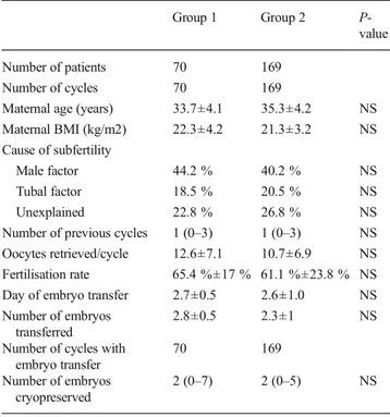
Two hundred thirty-nine women undergoing 239 ICSI cycles were enrolled. Women’s age were 33.7±4.1 (mean±SD) and 35.3±4.2 years for Groups 1 and 2, respectively. Baseline and cycle characteristics in the two study groups were similar (Table 1). In Group 1, half of them underwent the long GnRH-agonist and half the antagonist protocol.
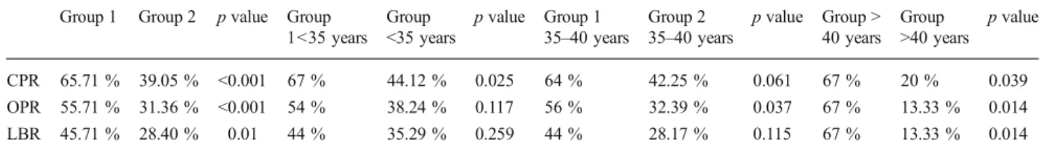
There was a statistically significant difference between the two study groups with regard to the outcome parameters:
Group 1 demonstrated higher clinical (65.7 % vs. 39 %, p<0.001), ongoing (55.7 % vs. 31.3 %, p<0.001) and live birth rates (45.7 % vs. 28.4 %, p=0.01) as compared with Group 2 (Table2). Subgroup analysis according to women’s
age, showed that this difference was retained between Group 1 and 2 regarding clinical pregnancy rates at the age of<35 years (67 % vs. 44.12 %, respectively, p=0.025), in ongoing preg-nancy rates at the age of 35–40 (56 % vs. 32.39 %, respec-tively, p=0.037) and in clinical (67 % vs. 20 %, respecrespec-tively, p<0.039), ongoing (67 % vs. 13.33 %, respectively, p<0.014) and live birth rates (67 % vs. 13.33 %, respectively, p<0.014), in women older than 40 years (Table2). No differences were found among participants of both groups after subgroup anal-yses according to the stimulation protocol used, number of the oocytes retrieved and type of subfertility (all p values>0.05) (Suppl Figures1,2,3).
In Group 1, the percentages of“in range” cellular events in the embryos, expressed by the timings of the second and third cell cycles (2c–3c and 3c–4c), were found to be significantly higher in women aged 35–40 compared to those>40 years of age (59.6 % vs. 38.1 %, p=0.03 and 63.6 % vs. 44.4 %, p= 0.008, respectively) (Fig. 1). The percentage of “in range” cellular events was higher in non-smokers than in smokers, expressed by an increase in“in range” divisions in the fourth cell cycle (4c–5c) period (54.5 % vs. 37.9 %, p=0.012) (Supp. Fig.4). With regard to the protocol used, the cellular events were higher in the GnRH-agonist group, as compared to the GnRH-antagonist, in the timing to the eight cell cycle (65.8 % vs. 37.3 %, p=0.001) (Supp. Fig.5). Regarding the effect of the number of oocytes retrieved on the timing of cellular events, there was a higher length in the second cell cycle (2c-3c) in embryos derived from retrievals with>10 oocytes, as compared to those with five to 10 (55 % vs. 43.6 %, p= 0.032); in contrast, a higher length was observed in the fifth cell cycle (5c–8c) in embryos derived from retrievals with>10 oocytes, as compared to those with five to 10 (87.2 % vs. 62.3 %, p=0.004) (data not shown).
Also, the percentage of“in range” events in embryos that were transferred and resulted in successful outcomes was compared to that of embryos that did not. For the first cellular cycles (events 2c, 2c–3c, 3c, 3c–4c, 4c, 4c–5c) embryos that resulted in successful outcomes had 10–20 % higher proba-bility of being“in range” than those that did not. However, this was significant only in the 3c–4c period (73.4 % vs. 59.7 %, p<0.05). This trend was not observed in the more advanced cellular divisions (5c, 5c–8c, 8c) (Fig.2).
Discussion
We performed a prospective cohort study of patients undergo-ing ICSI, comparundergo-ing the reproductive outcomes between em-bryos whose evaluation was performed through TLM and
Table 1 Baseline and cycle characteristics in the two groups studied
Group 1 Group 2
P-value
Number of patients 70 169
Number of cycles 70 169
Maternal age (years) 33.7±4.1 35.3±4.2 NS
Maternal BMI (kg/m2) 22.3±4.2 21.3±3.2 NS
Cause of subfertility
Male factor 44.2 % 40.2 % NS
Tubal factor 18.5 % 20.5 % NS
Unexplained 22.8 % 26.8 % NS
Number of previous cycles 1 (0–3) 1 (0–3) NS
Oocytes retrieved/cycle 12.6±7.1 10.7±6.9 NS
Fertilisation rate 65.4 %±17 % 61.1 %±23.8 % NS
Day of embryo transfer 2.7±0.5 2.6±1.0 NS
Number of embryos transferred
2.8±0.5 2.3±1 NS
Number of cycles with embryo transfer
70 169
Number of embryos cryopreserved
2 (0–7) 2 (0–5) NS
Continuous data are presented as mean ± SD
J Assist Reprod Genet
those whose evaluation was performed with conventional methods. We found higher clinical, ongoing and live birth rates in participants whose embryos were monitored through TLM, as compared to those whose embryos were monitored by morphological assessment. This difference was maintained in women over 40 years. Early cellular events of the embryos monitored by TLM, were more“in range” in women aged 35– 40 compared to those > 40 years (2c–3c, 3c–4c), in non-smokers compared to non-smokers (4c–5c), in the GnRH-agonist group, as compared to the GnRH-antagonist group (8c); also, these events were more“in range” in those embryos which resulted in pregnancy.
There was a statistically significant difference between the two study groups with regard to the outcome parameters, such as higher clinical, ongoing and live birth rates, favoring the use of TLM. These favourable results were maintained with regard to clinical pregnancy rates at the age of<35 years, on-going pregnancy rates at the age of 35–40 and, most interest-ingly, with regard to all outcomes at>40 years of age. It should be noted, however, that in the case of the>40 subgroup the sample size was very low (n=6) causing the misleading im-pression of an extremely high LBR (Table2). These results may be attributed to the detailed embryological assessment and improved embryo selection through the use of morphokinetic parameters for embryo selection under stable culture conditions that TLM offers. Similar reports have linked with high sensitivity and specificity the use of TLM with the ability of prediction of the cleavage stage embryos
and their potential to reach the blastocyst stage, resulting in increased live births [12,16,19,26,31,34,39,41]. On the other hand, other reports showed that TLM parameters are not able to predict live birth when compared with the conventional methods [33,34,42].
In fact, robust evidence in the literature on the effectiveness of TLM in improving success rates in ART has been scarce until recently. In a systematic review, after an initial yield of more than 1000 records, Polanski et al. [43], found only two randomized trials addressing this issue [44,45]: authors cluded that there is no effect of TLM on live birth and con-genital abnormality rates, while it was not correlated with a large change on the chance of achieving clinical and/or ongo-ing pregnancy when transferrongo-ing blastocyst stage embryos. Although the present study is of lesser strength than these trials, it should be noted that cleavage stage transfers were performed and our positive results may be associated with significantly improved selection of high potential embryos which could have a similar effect on results as during blasto-cyst transfers. Following Polanski’s review, Rubio et al. [46] published the largest to date randomized control trial on TLM: authors reported that the use of the integrated EmbryoScope time-lapse monitoring system significantly increases the im-plantation and ongoing pregnancy rates while decreasing early pregnancy loss, when compared to a standard incubator em-bryo culture and selection based exclusively on morphology. Our study independently observed similar ongoing pregnancy rates in the TLM group and a similarly large increase, when
Table 2 Outcome parameters of the two study groups in total and subgroup analysis based on participants’ age (<35, 35–40 and>40 years of age)
Group 1 Group 2 p value Group
1<35 years Group <35 years p value Group 1 35–40 years Group 2 35–40 years p value Group > 40 years Group >40 years p value CPR 65.71 % 39.05 % <0.001 67 % 44.12 % 0.025 64 % 42.25 % 0.061 67 % 20 % 0.039 OPR 55.71 % 31.36 % <0.001 54 % 38.24 % 0.117 56 % 32.39 % 0.037 67 % 13.33 % 0.014 LBR 45.71 % 28.40 % 0.01 44 % 35.29 % 0.259 44 % 28.17 % 0.115 67 % 13.33 % 0.014
CPR clinical pregnancy rate, OPR ongoing pregnancy rate, LBR live birth rate
Fig. 1 Percentage in Group 1
(subgroup analysis) of“in range”
cellular events of the embryos (from 2c up to 8c stage) according
to participants’ age (groups <35,
35–39, and ≥40 years of age). P
values are presented on the top of the columns
J Assist Reprod Genet
compared to the control group. Of note, Rubio’s group per-formed embryo transers mainly at the cleavage stage, as we did in our study, as opposed to the previously mentioned trials. Nevertheless, regardless of its immediate effect on outcomes, all authors have acknowledged that apart from the reproduc-tive outcomes, TLM is associated with advantages in the func-tion of ART laboratories [47].
No significant effect was measured on the outcomes of TLM cycles by clinical/external factors, such as type of subfertility, type of down-regulation protocol used and num-ber of oocytes retrieved (a measure of the effectiveness of stimulation). Smoking habit, BMI, certain types of subfertility (such as PCOS) and the type of ovarian stimulation used have b e e n f o u n d t o a f f e c t e m b r y o d e v e l o p m e n t a n d morphokinetics, as well as reproductive outcomes following IVF/ICSI [25,48,49]. Our observations corroborate at least the morphokinetic conclusions of these studies since age, smoking and type of down-regulation were all found to affect at least one of the embryological events in the early cleavage stage of development. What is interesting is that the most affected events (and the only ones that achieved statistical significance) were the lengths of cell cycles (2c–3c, 3c–4c and 4c–5c) and not their exact timing. This is an encouraging result, as the length of each cell cycle is probably more indic-ative of embryo normality than the exact timing of events, which may vary from patient to patient without necessarily indicating some underlining defect of the embryo.
Moreover, the 3c–4c period was also significantly more likely to be“in range” in transferred embryos that resulted in successful outcomes than those linked with failed cycles. The 3c–4c period is very indicative of the synchronicity of the second cell division and therefore of the embryo’s compe-tence. This result, in addition to the fact that younger women in our study exhibited shorter 3c–4c periods than older women seems to point to the importance of synchronicity in the early embryo development. The same time period (3c–4c) was also found to be an important predictor of implantation potential in
the study of Meseguer et al. [39]: authors attempted to eluci-date the “correct” timing of cellular events in the cleavage stage embryo by comparing the timings of embryos that im-planted with those of embryos that failed to implant; by rank-ing the embryos accordrank-ing to their temporal“normality” they were able to identify embryos with the highest implantation potential. Synchronicity may also be an indicator of normal chromosomal profile, with long delays in divisions being re-lated to aneuploidy [50]. Our study therefore suggests that synchronicity of early divisions can be used to select viable embryos with high sensitivity within the first 48 h post-insem-ination. In this context, TLM might be offered as an alterna-tive to blastocyst culture in certain cases as a means of selec-tion of viable embryos; the later carries the advantage of a high embryo selection potential, and has led to increased success rates coupled with reduced numbers of embryos transferred [51]. However, concerns have been raised on the effect of prolonged culture on embryo epigenetics and overall health of the resulting fetus [52].
The apparent limitations of our study are mainly attributed to its nature. The lack of power calculation and equality of the size of the groups studied, blinding, proper randomization and random allocation of the participants, is known to be linked with known and unknown confounders and selection and mis-classification bias. Specifically, the allocation based on the number of patient’s file does not represent a formal random component in the sequence generation process, as stated in the Cochrane Handbook for Systematic Reviews of Interventions; the latter would necessitate referring to a random number table or generator [53]. Also, although the number of oocytes col-lected in each group was similar, the effect of the slightly higher number in the TLM group cannot be fully discounted and may be linked with the elevated success rate in this group, as compared to the control [54].
In addition, the present study is heavily influenced by that of Meseguer et al. [39] and, being comparable in terms of sample size, depends on the accuracy of the ranges of normal Fig. 2 Percentage of transferred
embryos of Group 1 (TLM) that
exhibited“in range” cellular
events in patients with positive outcomes (Blue lines) and negative outcomes (Red lines)
J Assist Reprod Genet
cleavage events. Comparing our study with Meseguer’s, we observed that synchronicity is possibly the most important parameter in early cleavage embryonic stage (specifically the synchronicity of the second cell cycle) and the most affected by factors such as age. Both studies, however, agree that early cellular events as opposed to later ones are more predictive of successful outcomes.
Conclusion(s)
The present prospective cohort trial on 239 subfertile women undergoing ICSI, found better reproductive outcomes in embry-os whembry-ose evaluation was performed through TLM compared with those with conventional methods. The results were more evident in women aged more than 40 years. In contrast, there was no effect of TLM on outcomes, when clinical/external fac-tors were taken under consideration. Also, we observed more “in range” cellular events in certain embryo cycles in women aged 35–40 compared to those>40 years, in non-smokers than in smokers, in the GnRH-agonist group, as compared to the GnRH-antagonist, as well as in the embryos resulted in preg-nancy compared to those that did not. In conjunction with sim-ilar studies, our results indicate the importance of the timing of cleavage events on embryo competence and the positive effect that embryo selection via TLM has on ICSI outcomes.
The evident selection and attrition bias of the current study make properly powered and conducted prospective studies a must, in order to support or reject these findings.
Acknowledgments The authors wish to thank the clinical, paramedical
and laboratory team of Mitera Assisted Reproduction Unit. There was no finding for the current work. The study is a part of the Msc thesis of the second author.
Conflict of interest All authors declare no conflict of interest.
Authors’ roles CS: Study design, interpretation of findings and
manu-script preparation.
MAK: Study design, data collection and data analysis. AM: Data collection, data analysis and manuscript preparation. AS, MB: Clinical embryology, data acquisition and interpretation of results.
GS, GM: data acquisition and interpretation of results.
GAP: Study concept and design, clinical embryology, data acquisi-tion, data analysis and interpretation.
All authors critically reviewed and approved the final version of the manuscript.
References
1. Ferraretti AP, Goossens V, Kupka M, Bhattacharya S, de Mouzon J, Castilla JA, et al. Assisted reproductive technology in Europe, 2009: results generated from European registers by ESHRE. Hum Reprod.
2013;28:2318–31.
2. Edwards RG, Purdy JM, Steptoe PC, Walters DE. The growth of human preimplantation embryos in vitro. Am J Obstet Gynecol.
1981;141:408–16.
3. Alpha Scientists in Reproductive Medicine and ESHRE Special Interest Group of Embryology. The Istanbul consensus workshop on embryo assessment: proceedings of an expert meeting. Hum
Reprod. 2011;26:1270–83.
4. Palmer GA, Traeger-Synodinos J, Davies S, Tzetis M, Vrettou C, Mastrominas M, et al. Pregnancies following blastocyst stage transfer in PGD cycles at risk for beta-thalassaemic haemoglobinopathies.
Hum Reprod. 2002;17:25–31.
5. Mastenbroek S, Twisk M, van der Veen F, Repping S. Preimplantation genetic screening: a systematic review and
meta-analysis of RCTs. Hum Reprod Update. 2011;17:454–66.
6. Nel-Themaat L, Nagy ZP. A review of the promises and pitfalls of
oocyte and embryo metabolomics. Placenta. 2011;32 Suppl 3:S257–
63.
7. Swain JE. Could time-lapse embryo imaging reduce the need for
biopsy and PGS? J Assist Reprod Genet. 2013;30:1081–90.
8. Simon A, Laufer N. Assessment and treatment of repeated
implanta-tion failure (RIF). J Assist Reprod Genet. 2012;29:1227–39.
9. Anifandis G. Temperature variations inside commercial IVF
incuba-tors. J Assist Reprod Genet. 2013;30:1587–8.
10. Zhang JQ, Li XL, Peng Y, Guo X, Heng BC, Tong GQ. Reduction in exposure of human embryos outside the incubator enhances embryo
quality and blastulation rate. Reprod Biomed Online. 2010;20:510–5.
11. Calzi F, Papaleo E, Rabellotti E, Ottolina J, Vailati S, Vigano P, et al. Exposure of embryos to oxygen at low concentration in a cleavage stage transfer program: reproductive outcomes in a time-series
anal-ysis. Clin Lab. 2012;58:997–1003.
12. Lemmen JG, Agerholm I, Ziebe S. Kinetic markers of human embryo quality using time-lapse recordings of IVF/ICSI-fertilized oocytes.
Reprod Biomed Online. 2008;17:385–91.
13. Conaghan J. Time-lapse imaging of preimplantation embryos. Semin
Reprod Med. 2014;32:134–40.
14. Chen AA, Tan L, Suraj V, Reijo Pera R, Shen S. Biomarkers identi-fied with time-lapse imaging: discovery, validation, and practical
ap-plication. Fertil Steril. 2013;99:1035–43.
15. Hardarson T, Löfman C, Coull G, Sjögren A, Hamberger L, Edwards RG. Internalization of cellular fragments in a human embryo:
time-lapse recordings. Reprod Biomed Online. 2002;5:36–8.
16. Rubio I, Kuhlmann R, Agerholm I, Kirk J, Herrero J, Escriba MJ, et al. Limited implantation success of direct-cleaved human zygotes:
a time-lapse study. Fertil Steril. 2012;98:1458–63.
17. Athayde Wirka K, Chen AA, Conaghan J, Ivani K, Gvakharia M, Behr B, et al. Atypical embryo phenotypes identified by time-lapse microscopy: high prevalence and association with embryo
develop-ment. Fertil Steril. 2014;101:1637–48.
18. Dal Canto M, Coticchio G, Mignini Renzini M, De Ponti E, Novara PV, Brambillasca F, et al. Cleavage kinetics analysis of human em-bryos predicts development to blastocyst and implantation. Reprod
Biomed Online. 2012;25:474–80.
19. Hashimoto S, Kato N, Saeki K, Morimoto Y. Selection of high-potential embryos by culture in poly (dimethylsiloxane) microwells
and time-lapse imaging. Fertil Steril. 2012;97:332–7.
20. Stecher A, Vanderzwalmen P, Zintz M, Wirleitner B, Schuff M, Spitzer D, et al. Transfer of blastocysts with deviant morphological and morphokinetic parameters at early stages of in-vitro
develop-ment: a case series. Reprod Biomed Online. 2014;28:424–35.
21. Yang Z, Zhang J, Salem SA, Liu X, Kuang Y, Salem RD, et al. Selection of competent blastocysts for transfer by combining time-lapse monitoring and array CGH testing for patients undergoing pre-implantation genetic screening: a prospective study with sibling oo-cytes. BMC Med Genomics. 2014;7:38.
22. Joergensen MW, Agerholm I, Hindkjaer J, Bolund L, Sunde L, Ingerslev HJ, et al. Altered cleavage patterns in human tripronuclear J Assist Reprod Genet
embryos and their association to fertilization method: a time-lapse
study. J Assist Reprod Genet. 2014;31:435–42.
23. Munoz M, Cruz M, Humaidan P, Garrido N, Perez-Cano I, Meseguer M. The type of GnRH analogue used during controlled ovarian stim-ulation influences early embryo developmental kinetics: a time-lapse
study. Eur J Obstet Gynecol Reprod Biol. 2013;168:167–72.
24. Bellver J, Mifsud A, Grau N, Privitera L, Meseguer M. Similar morphokinetic patterns in embryos derived from obese and normoweight infertile women: a time-lapse study. Hum Reprod.
2013;28:794–800.
25. Freour T, Dessolle L, Lammers J, Lattes S, Barriere P. Comparison of embryo morphokinetics after in vitro fertilization-intracytoplasmic sperm injection in smoking and nonsmoking women. Fertil Steril. 2013;99:1944–50.
26. Wong CC, Loewke KE, Bossert NL, Behr B, De Jonge CJ, Baer TM, et al. Non-invasive imaging of human embryos before embryonic genome activation predicts development to the blastocyst stage. Nat Biotechnol. 2010;28:1115–21.
27. Wang Y, Moussavi F, Lorenzen P. Automated embryo stage classifi-cation in time-lapse microscopy video of early human embryo
devel-opment. Med Image Comput Comput Assist Interv. 2013;16:460–7.
28. Hlinka D, Kaľatová B, Uhrinová I, Dolinská S, Rutarová J, Rezáčová
J, et al. Time-lapse cleavage rating predicts human embryo viability.
Physiol Res. 2012;61:513–25.
29. Campbell A, Fishel S, Bowman N, Duffy S, Sedler M, Thornton S. Retrospective analysis of outcomes after IVF using an aneuploidy risk model derived from time-lapse imaging without PGS. Reprod
Biomed Online. 2013;27:140–6.
30. Sundvall L, Ingerslev HJ, Breth Knudsen U, Kirkegaard K. Inter- and intra-observer variability of time-lapse annotations. Hum Reprod.
2013;28:3215–21.
31. Meseguer M, Rubio I, Cruz M, Basile N, Marcos J, Requena A. Embryo incubation and selection in a time-lapse monitoring system improves pregnancy outcome compared with a standard incubator: a
retrospective cohort study. Fertil Steril. 2012;98:1481–9.
32. Findikli N, Oral E. Time-lapse embryo imaging technology: does it improve the clinical results? Curr Opin Obstet Gynecol. 2014;26:
138–44.
33. Cruz M, Gadea B, Garrido N, Pedersen KS, Martinez M, Perez-Cano I, et al. Embryo quality, blastocyst and ongoing pregnancy rates in oocyte donation patients whose embryos were monitored by
time-lapse imaging. J Assist Reprod Genet. 2011;28:569–73.
34. Kirkegaard K, Kesmodel US, Hindkjaer JJ, Ingerslev HJ. Time-lapse parameters as predictors of blastocyst development and pregnancy outcome in embryos from good prognosis patients: a prospective cohort study. Hum Reprod. 2013;28:2643–51.
35. Kirkegaard K, Hindkjaer JJ, Grondahl ML, Kesmodel US, Ingerslev HJ. A randomized clinical trial comparing embryo culture in a con-ventional incubator with a time-lapse incubator. J Assist Reprod Genet. 2012;29:565–72.
36. Montag M. Morphokinetics and embryo aneuploidy: has time come
or not yet? Reprod Biomed Online. 2013;26:528–30.
37. Ottolini C, Rienzi L, Capalbo A. A cautionary note against embryo aneuploidy risk assessment using time-lapse imaging. Reprod
Biomed Online. 2014;28:273–5.
38. Veeck LL. An Atlas of Human Gametes and Conceptuses: An Illustrated Reference for Assisted Reproductive Technology. In: Preembryo grading and degree of cytoplasmic fragmentation. New
York: Parthenon Publishing; 1999. p. 46–51.
39. Meseguer M, Herrero J, Tejera A, Hilligsøe KM, Ramsing NB, Remohí J. The use of morphokinetics as a predictor of embryo
im-plantation. Hum Reprod. 2011;26:2658–71.
40. Ciray HN, Campbell A, Agerholm IE, Aguilar J, Chamayou S, Esbert M, et al. Time-lapse user group. Proposed guidelines on the nomen-clature and annotation of dynamic human embryo monitoring by a
time-lapse user group. Hum Reprod. 2014;29:2650–60.
41. Conaghan J, Chen AA, Willman SP, Ivani K, Chenette PE, Boostanfar R, et al. Improving embryo selection using a computer-automated time-lapse image analysis test plus day 3 morphology: results from a
prospective multicenter trial. Fertil Steril. 2013;100:412–9.
42. Azzarello A, Hoest T, Mikkelsen AL. The impact of pronuclei mor-phology and dynamicity on live birth outcome after time-lapse
cul-ture. Hum Reprod. 2012;27:2649–57.
43. Polanski LT, Coelho Neto MA, Nastri CO, Navarro PA, Ferriani RA, Raine-Fenning N, et al. Time-lapse embryo imaging for improving reproductive outcomes: a systematic review and meta-analysis.
Ultrasound Obstet Gynecol. 2014. doi:10.1002/uog.13428.
44. Kovacs P, Matyas S, Forgacs V, Sajgo A, Rarosi F, Pribenszky C. Time-lapse embryo selection for single blastocyst transfer-results of a multicenter, prospective, randomized clinical trial. Fertil Steril. 2013;100:S90.
45. Kahraman S, Cetinkaya M, Pirkevi C, Yelke H, Kumtepe Y. Comparison of blastocyst development and cycle outcome in patients with eSET using either conventional or time lapse incubators. A prospective study of good prognosis patients. J Reprod Stem Cell Biotechnol. 2013;3:55–61.
46. Rubio I, Galán A, Larreategui Z, Ayerdi F, Bellver J, Herrero J, et al. Clinical validation of embryo culture and selection by morphokinetic analysis: a randomized, controlled trial of the EmbryoScope. Fertil Steril. 2014;102:1287–94.
47. Aparicio B, Cruz M, Meseguer M. Is morphokinetic analysis the answer? Reprod BioMed Online. 2013;27:654–63.
48. Muñoz M, Cruz M, Humaidan P, Garrido N, Pérez-Cano I, Meseguer M. Dose of recombinant FSH and oestradiol concentration on day of HCG affect embryo development kinetics. Reprod BioMed Online. 2012;25:382–9.
49. Wissing ML, Hoest T, Mikkelsen AL. Slower early embryo develop-ment in women with polycystic ovary syndrome (PCOS) compared to regularly cycling women (controls). Fertil Steril. 2012;98 Suppl 3: S109.
50. Davies S, Christopikou D, Tsorva E, Karagianni A, Handyside AH, Mastrominas M. Delayed cleavage divisions and a prolonged transi-tion between 2-and 4-cell stages in embryos identified as aneuploid at the 8-cell stage by array CGH. Hum Reprod. 2012;27 Suppl 2:ii84. 51. Mercader A, Garcia-Velasco JA, Escudero E, Remohi J, Pellicer A, Simon C. Clinical experience and perinatal outcome of blastocyst transfer after coculture of human embryos with human endometrial
epithelial cells: a 5-year follow-up study. Fertil Steril. 2003;80:1162–8.
52. Kallen B, Finnstrom O, Lindam A, Nilsson E, Nygren KG, Olausson PO. Blastocyst versus cleavage stage transfer in in vitro fertilization:
differences in neonatal outcome? Fertil Steril. 2010;94:1680–3.
53. Higgins JPT, Green S. Cochrane Handbook for Systematic Reviews of Interventions Version 5.1.0 [updated March 2011]. The Cochrane
Collaboration. <http://handbook.cochrane.org/> (2011). Accessed on
31 Dec 2014.
54. Sunkara SK, Rittenberg V, Raine-Fenning N, Bhattacharya S, Zamora J, Coomarasamy A. Association between the number of eggs and live birth in IVF treatment: an analysis of 400 135 treatment
cycles. Hum Reprod. 2011;26:1768–74.
J Assist Reprod Genet