Landscape of somatic single-nucleotide and
copy-number mutations in uterine serous carcinoma
Siming Zhaoa, Murim Choia, John D. Overtona, Stefania Belloneb, Dana M. Roqueb, Emiliano Coccob, Federica Guzzob, Diana P. Englishb, Joyce Varugheseb, Sara Gasparrinib, Ileana Bortolomaib, Natalia Buzac, Pei Huic, Maysa Abu-Khalafd, Antonella Ravaggie, Eliana Bignottie, Elisabetta Bandierae, Chiara Romanie, Paola Todeschinie, Renata Tassie,
Laura Zanottie, Luisa Carrarae, Sergio Pecorellie, Dan-Arin Silasib, Elena Ratnerb, Masoud Azodib, Peter E. Schwartzb, Thomas J. Rutherfordb, Amy L. Stieglerf, Shrikant Manea, Titus J. Boggonf, Joseph Schlessingerf, Richard P. Liftona,1, and Alessandro D. Santinb
aDepartment of Genetics, Howard Hughes Medical Institute, Yale University School of Medicine, New Haven, CT 06510;bDepartments of Obstetrics, Gynecology, and Reproductive Sciences,cPathology,dInternal Medicine and Oncology, andfPharmacology, Yale University School of Medicine, New Haven, CT 06510; andeDepartment of Obstetrics, and Gynecology,“Angelo Nocivelli” Institute of Molecular Medicine, University of Brescia, 25123 Brescia, Italy Contributed by Richard P. Lifton, December 27, 2012 (sent for review December 12, 2012)
Uterine serous carcinoma (USC) is a biologically aggressive subtype of endometrial cancer. We analyzed the mutational landscape of USC by whole-exome sequencing of 57 cancers, most of which were matched to normal DNA from the same patients. The distribution of the number of protein-altering somatic mutations revealed that 52 USC tumors had fewer than 100 (median 36), whereas 5 had more than 3,000 somatic mutations. The mutations in these latter tumors showed hallmarks of defects in DNA mismatch repair. Among the remainder, we found a significantly increased burden of mutation in 14 genes. In addition to well-known cancer genes (i.e.,TP53, PIK3CA, PPP2R1A, KRAS, FBXW7), there were frequent mutations in CHD4/ Mi2b, a member of the NuRD–chromatin-remodeling complex, and TAF1, an element of the core TFIID transcriptional machinery. Addi-tionally, somatic copy-number variation was found to play an im-portant role in USC, with 13 copy-number gains and 12 copy-number losses that occurred more often than expected by chance. In addi-tion to loss ofTP53, we found frequent deletion of a small segment of chromosome 19 containingMBD3, also a member of the NuRD– chromatin-modification complex, and frequent amplification of chromosome segments containingPIK3CA, ERBB2 (an upstream ac-tivator of PIK3CA), and CCNE1 (a target of FBXW7-mediated ubiq-uitination). These findings identify frequent mutation of DNA damage, chromatin remodeling, cell cycle, and cell proliferation pathways in USC and suggest potential targets for treatment of this lethal variant of endometrial cancer.
endometrial carcinoma
|
uterine serous papillary cancer|
cancer genomicsE
ndometrial cancer is the most prevalent gynecologic tumor inwomen, with an annual incidence of 47,130 new cases and 8,010 deaths in 2012 in the United States (1). On the basis of clinical and histopathological features, endometrial cancer is classified into type I and type II disease groups (2). Type I tumors, which constitute the majority of cases, are generally diagnosed at an early stage, are low grade and endometrioid in histology, are associated with a history of hyperestrogenism, and typically have a good prognosis. In contrast, type II cancers are poorly differ-entiated, often with serous papillary [uterine serous carcinoma (USC)] or clear cell histology. Although these tumors account for a minority of endometrial cancers, the majority of relapses and deaths occur in this group of patients (2).
Among type II cancers, USC represents the most biologically aggressive subtype (3, 4). Classically, the neoplastic epithelium is characterized by serous differentiation with psammoma bodies and a predominantly papillary architecture (3). Pleomorphic cytology with nuclear atypia, prominent nucleoli, a vescicular chromatin pattern, and high mitotic activity are seen. Clinically, USC has a propensity for early intra-abdominal and lymphatic spread (3) and is more commonly diagnosed in women of African ancestry (3– 5). The overall 5-y survival of USC is only 30± 9% for all stages,
and the recurrence rate after surgery is extremely high (50–80%) (3–5). This poor prognosis motivates determination of the molec-ular basis of this tumor’s aggressive behavior in hope of developing new effective treatment modalities.
To address these issues, we have sequenced the exomes of a large cohort of USC and have identified genes with increased numbers of somatic single-nucleotide and copy-number variants. The results identify cancer genes and define the pathways in-volved in USC.
Results
Exome Sequencing of USC.Fifty-seven patients with uterine serous carcinoma were studied. Their clinical features are presented inSI Appendix, Table S1. Upon surgical removal of tumors, primary cell lines were prepared (15 tumors) or tumors were frozen (42 tumors). Exome sequencing was performed on all tumors; for 34 of these, DNA samples from normal tissue were available and se-quenced. Exome sequencing was performed using the NimbleGen/ Roche capture reagent followed by 74 base paired-end DNA se-quencing on the Illumina HiSeq platform (6). By design, tumor samples were sequenced to greater depth of coverage to permit detection of somatic mutations in tumors despite admixture of normal and tumor cells in these samples. For tumors and normal DNA, each targeted base was sequenced by a mean of 187 and 100 independent reads, respectively (SI Appendix, Table S2). Of all targeted bases in tumors, 94.5% were read by 20 or more in-dependent reads; mean per-base per read error rates were 0.42% for normal DNA and 0.48% for tumor DNA. Segments of loss of heterozygosity (LOH) were called from the difference in B-allele frequency between tumor-normal pairs (SI Appendix, Fig. S1), allowing estimates of tumor purity, which were above 60% for frozen tumors and higher for primary cell lines. Somatic mutations were identified by finding variant reads in tumors that were sig-nificantly more frequent than expected by chance (Materials and Methods). At the coverage levels studied, there was no significant relationship between tumor purity and the number of somatic variants detected, consistent with sufficient depth of coverage having been achieved to identify the vast majority of somatic mutations. Variants in genes implicated in the pathogenesis of
Author contributions: J.S., R.P.L., and A.D.S. designed research; J.D.O., S.B., D.M.R., E.C., F.G., S.G., I.B., N.B., P.H., A.R., E. Bignotti, E. Bandiera, C.R., P.T., R.T., L.Z., L.C., A.L.S., S.M., and T.J.B. performed research; D.P.E., J.V., M.A.-K., S.P., D.-A.S., E.R., M.A., P.E.S., and T.J.R. contributed new reagents/analytic tools; S.Z., M.C., and R.P.L. analyzed data; and S.Z., A.L.S., T.J.B., J.S., R.P.L., and A.D.S. wrote the paper.
The authors declare no conflict of interest.
Freely available online through the PNAS open access option.
1To whom correspondence should be addressed. E-mail: [email protected]. This article contains supporting information online atwww.pnas.org/lookup/suppl/doi:10. 1073/pnas.1222577110/-/DCSupplemental.
USC were verified by direct Sanger sequencing and were found to be expressed in all available USC cell lines.
Tumors with Hypermutator Phenotype.The number of protein-al-tering somatic mutations per tumor markedly deviated from a normal distribution (Fig. 1A). In the discovery set of 34 USC with matched normal DNA, 30 tumors had fewer than 100 protein-altering somatic mutations (median 36), whereas 4 had more than 3,000 somatic mutations each. Only one of these tumors was from a cell line (with limited propagation), and none came from patients who had received chemotherapy before sample acquisition. These tumors with high mutation burden were also notable for having no LOH segments or copy-number variants (CNVs), a feature found in onlyfive other tumors. These features suggest a hypermutator phenotype due to deficiency of mismatch repair (MMR) or poly-merasee (POLE) genes (7, 8). Consistent with this, these
hyper-mutated tumors showed a paucity of T:A> A:T or C:G > A:T transversions (SI Appendix, Fig. S2) (9). Examination of thePOLE and MMR genes showed no germ-line mutations; however, so-matic mutations in these genes were highly prevalent in these tumors (mean of 4 per tumor, including 4 premature termination mutations for MMR genes and a mean of 4.5 per tumor forPOLE) and more frequent than expected by chance (P = 2.23 × 10⁻³) (SI Appendix, Table S3). Among the cancers without matched normal DNA, one showed a similarly high prevalence of rare protein-altering variants (>3,000) and a skewed distribution of rare pro-tein-altering transversions. Thus, 9% of USC in this cohort have a hypermutator phenotype. Because of the skewing effect of the large number of mutations in these tumors, they were not included in subsequent analyses of mutation burden.
Analysis of Single-Nucleotide Variants.Among somatic mutations in the 30 remaining matched tumors, we identified recurrences of somatic mutations at the same positions. Accounting for the rate of protein-altering somatic mutations in these tumors (1.1× 10⁻⁶) and the size of the exome, the likelihood of seeing the mutation twice by chance at any position among these tumors is<10−3. We identified six genes with recurrent somatic mutations (Table 1). These in-cluded well-established activating mutations inPIK3CA (10), the catalytic subunit of phosphoinositide-3 kinase (five tumors); the well-established G12V mutation inKRAS (three tumors) (11); and a mutation at R465 inFBXW7 in four tumors (12). FBXW7 is the targeting component of a SCF-type 3 ubiquitin ligase, and R465 occurs in the WD40 domain involved in substrate recognition; mutation at this site prevents targeting of cyclin E for ubiquitination and degradation (SI Appendix, Fig. S3) (12, 13). Recurrent muta-tions also occurred at two sites inPPP2R1A, the constant regulatory subunit of serine-threonine phosphatase 2a. The P179R and S256F mutations occurred four and two times, respectively, and have been previously reported (14). These mutations occur at the interface where PPP2R1A interacts with regulatory B subunits that target the phosphatase to specific substrates; inhibition of this interaction by SV40 small t antigen plays a role in viral transformation (SI Ap-pendix, Fig. S4). Additional somatic mutations were found on the surface of PPP2R1A that interacts with the B or C (catalytic) subunit (Table 1).TP53, the well-characterized tumor suppressor gene, hadfive different positions mutated two or more times, and there were 19 additional single somatic mutations in this gene. Eighty-two percent of these mutations were in segments of somatic LOH (Table 1 and Fig. 1B).
In addition to these previously described recurrent mutations, a recurrent mutation was found in CHD4/Mi2b (chromodomain-helicase–DNA-binding protein 4), an ATP-dependent chromatin-remodeling protein that is a major subunit of the Mi2b/nucleosome remodeling and deacetylase (NuRD) complex. Mutations inCHD4/ Mi2b have not been previously associated with cancer. In addition, there were 10 other somatic or rare mutations inCHD4 among matched and unmatched tumors (see further discussion below).
We next sought genes with overall increased somatic mutation burden in the 30 matched tumor-normal pairs. In this analysis, we determined the probability of seeing≥n mutations in each gene, taking into account the overall rate of protein-altering somatic mu-tations in the matched tumor normal set (1.1 × 10⁻⁶) and the length of the protein-coding region in each gene. We also adjusted for the level of expression of each gene from expression data in normal human endometrium (15) because we found a higher so-matic mutation rate among genes with lower expression, consistent with effects of transcription-coupled DNA repair reducing the mutation rate among expressed genes (16). We consideredP val-ues< 2.4 × 10⁻⁶to represent a significant increase in mutation burden compared with that expected under the null hypothesis, accounting for the testing of∼21,000 genes. We added to this set variants in the 22 unmatched tumors that occurred in genes that had at least one somatic mutation in the matched set and that had Fig. 1. Somatic variation pattern underlying USC. (A) Distribution of the
number of protein-altering somatic mutations in 34 normal-tumor USC pairs. Subplot left, mutation spectrum in four hypermutator phenotype samples; subplot right, mutation spectrum in 30 moderately mutated samples. (B and C) Thirty tumors with moderate somatic burden are arranged by the total number of somatic point mutations from left to right. The four hyper-mutator phenotype tumors are excluded in this analysis. (B) Significantly mutated genes are listed vertically by the order of damaging or conserved P values shown in Table 1. (C) Genes with significant CNVs and genes of interest are listed. Copy neutral status is shown as lavender rectangles. Five samples without CNV information are marked by crosses.
GENET
never been seen in >7,000 exomes in the Yale University and National Heart, Lung, and Blood Institute exome databases. Be-cause we found no novel variants in any of these genes in the 30 germ-line samples of tumor-normal pairs, we infer that virtually all of these represent somatic mutations.
In the resulting set, the six genes with recurrent mutations were among the most frequently mutated genes. Included in this set was CHD4, which had six somatic mutations and five more novel var-iants found in the 22 unmatched tumors (Table 1 and Fig. 1B). Many of these 11CHD4 mutations (Fig. 2), which all appear to be heterozygous, impair at least some normal CHD4 functions. CHD4 is a SWI2/SNF2 ATPase and part of the larger helicase superfamily
2 whose members share a similar catalytic core containing two RecA-like helicase domains. Conserved catalytic“signature” motifs have been well described and contain many residues required for catalysis of ATP hydrolysis and helicase activity (20). ThreeCHD4 mutations (R957Q, R1127G, and R1162W) alter residues in these signature motifs (motif B, V, and VI, respectively) that are con-served from yeast to humans, and whose mutation has been shown to impair normal function. Similarly, there is a mutation in the second plant homeodomain (PHD) finger that normally binds methylated histone H3K9. This C464Y mutation disrupts one of the key cysteines that coordinate Zn2+binding. In addition, there are four mutations (Q1106R, I1109T, and two instances of F1112L)
Table 1. Genes with significant mutation burden in 52 USC
Gene Recurrent (no.)
Coding length (bp) No. nonsynonymous mutations Nonsynonymous P value No. damaging mutations No. Cons MS Dam+ Cons P value No. silent mutations TP53 R43 (2), C44 (2), R116 (4), G134 (3), R141 (2) 1,222 30 1.34E-75 3 14 1.01E-45 0
PIK3CA E542 (3), N1044 (2) 3,287 13 6.39E-23 0 13 4.26E-27 0
CHD4 F1112 (2) 5,895 11 3.82E-17 1 10 1.05E-20 0
FBXW7 R465 (4) 2,168 9 1.05E-17 0 9 1.08E-20 1
PPP2R1A P179 (4), S256 (2) 1,830 8 1.28E-16 0 8 2.60E-19 0
TAF1 — 5,834 7 2.71E-10 0 7 1.19E-12 0
KRAS G12 (2) 586 3 2.61E-08 0 3 1.66E-09 0
PTEN — 1,248 3 2.28E-07 1 2 1.46E-08 0
HCFC1R1 — 433 2 8.00E-07 0 2 1.01E-07 0
CDKN1A — 503 2 1.25E-06 0 2 1.58E-07 0
CTDSPL — 863 2 7.86E-06 0 2 9.99E-07 0
YIPF3 — 1,089 2 1.25E-05 0 2 1.59E-06 0
SPOP — 1,161 2 1.51E-05 0 2 1.92E-06 0
FAM132A — 941 2 1.51E-05 0 2 1.93E-06 0
Recurrent (no.), positions with recurrent mutations (no. of instances); Cons MS, missense mutations at conserved positions; Dam+Cons, damaging + conserved missense mutations.
Fig. 2. Mapping of USC mutations onto the crystal structure of CHD4. (A) Schematic representation of somatic mutations found in CHD4. The horizontal bar represents full-length CHD4 protein with func-tional domains shown as boxes. Somatic mutations found in USC are marked in red text. All mutations are missense mutations except E1628X, which is a nonsense mutation. (B) C464 locates in the second PHD finger, which binds directly to histone H3 methylated at K9. An NMR structure of the second PHD domain of CHD4 (blue ribbon plot) has been determined [Protein Data Bank (PDB) ID: 2L75] (17). Red dot represents C464Y mutation. (Lower) A close-up view of the zinc–C464 interaction. (C) So-matic mutations in catalytic core of CHD4 mapped to the crystal structure of a related protein, human CHD1 (sequence identity of the ATPase lobes is 42%, homology is 57% over 572 residues; PDB ID: 3MWY) (18). Blue, ATPase lobe 1; light purple, ATPase lobe 2; cyan, chromodomains; green, C-ter-minal bridge. Three mutations (red dots) in CHD4 fall within known conserved motifs (motifs B, V, VI) (19); three mutations were found in unknown helicase motif i, and two mutations were found in unknown motif ii. (D) A close-up view of mutations infive motifs in C. Somatic mutations are labeled in red text; amino acid positions in parentheses rep-resent homologous positions in CHD1.
clustered in a shortα-helix in ATPase lobe 2; we speculate that alteration of this helix might alter interaction with another protein in the complex. Additional mutations include two in a C-terminal bridge that links ATPase lobes 1 and 2 at positions that appear to stabilize this segment. Finally, there is a premature termination near the normal C terminus (Fig. 2). The high prevalence ofCHD4 mutations, the clear implication of disrupted normal function by manyCHD4 mutations, and the common genomic amplifications of theCHD4 gene (see CNV results below) implicate CHD4 muta-tions in USC.
Another gene of interest wasTAF1, an X-linked gene, which had four different somatic mutations and three additional var-iants in unmatched tumors (Table 1). TAF1 is the largest com-ponent and core scaffold of the TFIID basal transcription factor complex and has DNA-binding activity, histone acetyltransferase (HAT) activity, two kinase domains, and ubiquitin-activating/ conjugating activity (21). It is known to be required for pro-gression through the G1 phase of the cell cycle, promoting cyclin D expression (22). Most of the sevenTAF1 mutations lie in the HAT domain at positions that are extraordinarily well conserved —all are conserved in vertebrates and nearly all are conserved in yeasts (Fig. 3). Although the function(s) of these mutations in USC are uncertain, overexpression ofTAF1 has been previously reported in human lung and breast carcinoma and found to be associated with poor tumor differentiation and high mitotic ac-tivity (25).
Additional genes that meet thresholds for significantly in-creased burden in the entire set includePTEN, CDKN1A, and SPOP, as well as HCFC1R1, CTDSPL, YIPF3, and FAM132A, genes not previously implicated in cancer. For each of these genes, mutations are predominantly at highly conserved posi-tions, there are few if any silent mutations in the same gene, and quantitative PCR demonstrated expression of each of these genes in all available USC cell lines.
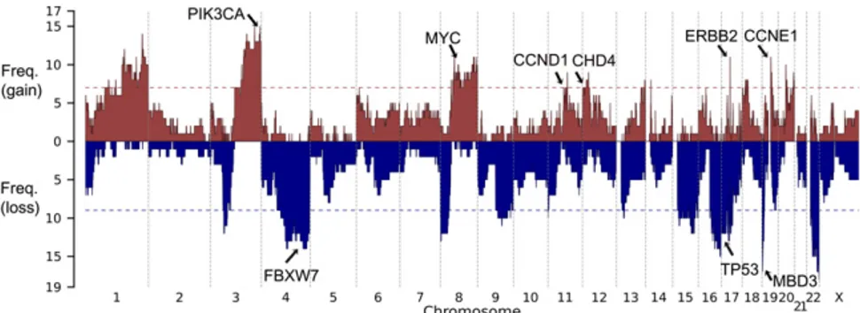
Analysis of CNVs.We next assessed somatic CNVs. For the 25 tumors in which read coverage distribution showed distinct modes (SI Appendix, Fig. S5), CNVs were identified by comparing cov-erage depth of individual capture intervals from tumor and normal samples (Materials and Methods); CNVs were supported by sig-nificant deviation of the B allele frequency from the genome-wide average. The significance of CNVs affecting specific chromosome segments was assessed by Monte Carlo simulation, randomly
distributing CNVs of the empirically observed sizes and numbers in each tumor in 108 permutations to assess the distribution expected by chance alone. A significance threshold was established that provided a false discovery rate<0.25. Within each significant copy-number gain or loss, all CNVs that contained the most fre-quently altered segment were removed, and the remaining CNVs were reassessed to see if independent signals could be detected. We identified 13 chromosome segments with more frequent gains of copy number and 12 with more frequent deletions than expected by chance (Fig. 4). Among these, we found focal amplification of the segment of chromosome 17 that containsERBB2 in 11 of the 25 tumors (44%) (SI Appendix, Fig. S6), large duplications that include the PIK3CA locus in 60%, and a small duplication of chromosome 19 containingCCNE1 in 48% (SI Appendix, Table S4). There was also amplification of a large segment of chromo-some 8 containingMYC in 11 (44%) tumors and amplification of a segment of chromosome 12 that included CHD4 in 7 (28%) tumors (Fig. 1C). Among deletions, TP53 was deleted in 44% of tumors. The most frequent somatic deletions were small (0.5 Mb) deletions on chromosomes 19 and 22, which occurred in 68% and 72% of tumors, respectively (SI Appendix, Table S5 and Fig. S7). Most interestingly, the chromosome 19 interval containsMBD3, which is a component of the same SWI/SNF complex asCHD4 (19). The chromosome 22 interval includes a number of interesting genes, including three in the MAP kinase pathway, HDAC10,
andPPP6R2.
Discussion
We report exome sequencing of a USC cohortfive times larger than those recently reported (26, 27). The results define the genetic hallmarks of uterine serous cancer. We have found significantly increased mutation burden in 14 genes, including the previously identified and well-recognized cancer genes TP53, PIKC3A, PPP2R1A, KRAS, and PTEN, FBXW7, and CDKN1A.
The high frequency of single-nucleotide variants (SNVs) in CHD4, which was mutated in 19% of tumors and was the third most frequently mutated gene, was noteworthy. These mutations were diverse and predominantly at highly conserved positions from yeast to humans, and several have been previously shown to cause loss of function (19). Nonetheless, because CHD4 has many functional domains, it is possible that not all CHD4 functions are lost. Indeed, there appears to be clustering of mutations in particular domains, and there are seven copy-number gains that includeCHD4.
Similarly, mutations inTAF1, a component of the core RNA polymerase II machinery, are found in 13% of tumors, with mutations at positions conserved throughout yeasts. Because TAF1 has diverse biochemical functions, the observed clustering of mutations in the HAT domain does not require that these muta-tions are null for all TAF1 funcmuta-tions. One known function of TAF1 is promotion of cyclin D expression; overexpression of cyclin D is itself known to promote cell cycle progression and proliferation and is frequently amplified in cancers (28). Notably, seven tumors had amplification of the segment of chromosome 11 containing CCND1 (Fig. 4). It will be of interest to determine the biochemical and phenotypic consequences ofTAF1 mutations.
Several other genes show marginal statistical significance; these include known cancer genes such asPTEN and CDKN1A as well as SPOP, which targets proteins for ubiquitination via its MATH do-main. SPOP has recently been shown to have clustered mutations in its MATH domain in prostate cancer (29); the two USC mutations are at different sites in the MATH domain. Additional genes not previously implicated in cancer include HCFC1R1, CTDSPL, YIPF3, and FAM132A. Further efforts will be required to determine the impact of these latter genes on USC and other cancers.
Our results demonstrate that somatic CNVs play a major role in the pathogenesis of USC, one that is likely at least as important as somatic point mutations. Interestingly, the most frequent CNV was a small deletion found in 68% of tumors affecting a short Fig. 3. Schematic representation of TAF1 functional domains and mutation
conservation analysis. (A) Functional domains in TAF1 are represented by col-ored boxes with domain names noted below (23). HAT domain, histone ace-tyltransferase domain. Mutations found in USC are labeled at the top by red text. (B) Multiple sequence alignment across vertebrate and invertebrate species around the seven mutations found in USC. Mutation positions in hu-man TAF1 are labeled in red at the top. Sequence aligned by Clustal W 2.0 (24).
GENET
segment of chromosome 19 that contains only 17 genes. Among these genes is MBD3, which is part of the same chromatin-remodeling complex—NuRD—as CHD4. Additionally, there were seven copy-number gains of the segment of chromosome 12 that includesCHD4, all of which were in samples with MBD3 deletions. This complex deacetylates histones, repressing gene expression. Collectively, thesefindings add to the growing list of genes involved in chromatin remodeling that are mutated in cancer (30, 31).
CNV analysis of USC also identified frequent amplifications, including the well-known cancer genesPIK3CA (60%) and ERBB2 (encoding HER2/neu; 44%) (32). ErbB2 overexpression has been previously reported to be associated with cancer cell proliferation, poor survival, and resistance to therapy in multiple human tumors including USC (5). Moreover, ErbB2 functions as an upstream regulator of the PIK3CA/AKT/mTOR-signaling pathway. These findings suggest common involvement of this pathway in USC and the possible utility of Food and Drug Administration-approved antibodies (i.e., trastuzumab, pertuzumab) or small molecule TK inhibitors used either alone or in combination with anti-mTOR, AKT, and/or PIK3CA active agents (5).
Another frequent somatic amplification (found in 44% of tumors) included a small segment of chromosome 19 that harbors CCNE1. CCNE1 encodes cyclin E1 and is known to regulate the transition from the G1 phase to the S phase. High levels ofCCNE1 accelerate the transition through the G1 phase, and its accumu-lation is common in a number of cancers (33, 34). Most in-terestingly, CCNE1 degradation is mediated by binding to FBXW7
followed by ubiquitination via the SCF complex. Seventeen per-cent of USC harbor recurrent mutations in FBXW7 that abrogate CCNE1 binding (SI Appendix, Fig. S3) (35). These observations suggest that inhibition of CCNE1 activity may have efficacy in patients harboring mutation in this pathway.
Collectively, our results implicate frequent mutations in sev-eral pathways in USC, including specific genes in DNA damage, chromatin remodeling, cell cycle, and cell proliferation pathways (Fig. 5). Analysis of correlation and anticorrelation of all possi-ble pairs of significant mutations did not provide evidence of strong associations (SI Appendix, Fig. S8). Nonetheless, a large fraction of tumors shared mutations affecting genes in different pathways such asTP53, PIK3CA, MBD3, and FBXW7.
We also found that 9% of USC have a very high number of somatic mutations with many somatic mutations in mismatch repair andPOLE genes. This distribution is distinct from the remainder (median 36 protein-altering mutations, all<100). Mutator pheno-types in the absence of germ-line mutations in mismatch repair genes have been seen in several other cancers (7, 8). These USC tumors are notable for being relatively frequent and for having a uniformly very high number of mutations, more than those seen in 90% of colon cancers with the mutator phenotype (36). Despite the remarkable somatic mutation burden, these tumors had no identified CNVs.
The establishment of 15 USC cell lines with different mutation profiles (SI Appendix, Fig. S9) provides the opportunity for in vitro assessment of whether a mutation profile is predictive of Fig. 4. Copy-number profile of 25 USC tumors. Frequency of copy-number gain (red) and copy-number loss (blue) are plotted along the genome. Horizontal dotted line, genome-wide significance level for CNV gain (red) and CNV loss (blue). Genes of interest in significant CNV peak regions are labeled.
Fig. 5. Major altered pathways in USC. The altered percentages shown for genes and pathways come from the 25 matched tumors with CNV information. Genes are colored based on their activity in the pathway diagram. Pink, predicted activated; blue, predicted inactivated; gray, uncertain at this stage; lines with blunt end, inhibiting effect; lines with pointed end, promoting effect; dotted line, uncertain. Mutation and CNV status for each gene across the 25 samples are shown at the bottom following the pathway diagram.
drug response. For example, the finding that MMR-defective colorectal cancers may respond favorably to poly (ADP ribose) polymerase inhibitors (37) raises the question of whether the same may apply to USC with the hypermutator phenotype.
USC and high-grade serous ovarian carcinoma (HG-SOC) are histologically similar gynecological tumors characterized by a highly aggressive biologic behavior. Exome sequencing of HG-SOC has been recently reported by The Cancer Genome Atlas Research Network (38). TP53 was mutated in 95% of these cancers, with no other gene with somatic SNVs in more than 6% and only four (BRCA1, BRCA2, CSMD3, and FAT3) that were mutated in more than 3%. USC shows a lower frequency ofTP53 mutation (59%), 5 genes mutated in 13–23% of tumors, 10 more genes mutated in 3– 10%, and noBRCA1 or BRCA2 mutations (SI Appendix, Table S6). Thesefindings indicate substantial differences in the genetics of USC and HG-SOC.
While this article was in preparation, Khun et al. (26) and Le Gallo et al. (27) reported results from exome sequencing of a small number of USC (10 and 13 USC in discovery sets, respectively). These studies also found mutations inTP53, PIK3CA, PPP2R1A, FBXW7, CHD4, and SPOP genes, but were underpowered to de-tect other genes and CNVs that we report.
Our results define the genetic landscape of USC and identify specific pathways that are frequently mutated in these tumors. Thesefindings will guide further research and targeted therapies against this highly aggressive variant of endometrial cancer.
Materials and Methods
Patients and Specimens. The study protocol was approved by the Yale Uni-versity Human Investigation Committee. DNA and RNA were purified from tumors and normal tissues. Libraries were prepared as described (6). Addi-tional details are provided inSI Materials and Methods.
Exome Sequencing and Analysis. Genomic DNA was captured on the Nim-bleGen 2.1M human exome array and subjected to 74 base paired-end reads on the Illumina HiSeq instrument as described (6). Sequence reads were mapped to the reference genome (hg18) using the ELAND program (6). For matched normal-tumor pairs, somatic mutations were called by comparing reference and nonreference reads from the matched pair as described (39). For unmatched tumors, SAMtools was used to call variant bases appended with quality scores. Additional details are provided in SI Materials and Methods.
Somatic CNV. CNVs were identified by comparing coverage depth of individual capture intervals (0.5-Mb bins) from tumor and normal samples after nor-malization for mean coverage depth of each exome. A permutation-based strategy was used to assess the significance of recurrent CNVs with a false-discovery-rate cutoff of 0.25 after Benjamini–Hochberg correction. Independent significant CNV peaks were called based on a GISTIC-like peel algorithm (40). Quantitative RT-PCR. Quantitative RT-PCR was performed for all genes showing significant mutation burden using RNA from 15 USC cell lines. Additional details are provided inSI Materials and Methods.
ACKNOWLEDGMENTS. This work was supported in part by National Institutes of Health (NIH) Grants R01 CA122728-01A4 and R01 CA154460-01A1 (to A.D.S.) and by Gilead Sciences Inc. This investigation was also supported by NIH Research Grant CA-16359 from the National Cancer Institute.
1. Siegel R, Naishadham D, Jemal A (2012) Cancer statistics, 2012. CA Cancer J Clin 62(1): 10–29.
2. Bokhman JV (1983) Two pathogenetic types of endometrial carcinoma. Gynecol Oncol 15(1):10–17.
3. Hendrickson M, Ross J, Eifel P, Martinez A, Kempson R (1982) Uterine papillary serous carcinoma: A highly malignant form of endometrial adenocarcinoma. Am J Surg Pathol 6(2):93–108.
4. Lee KR, et al. (2003) Tumors of the ovary and peritoneum. World Health Organization Tumours of the Breast and Female Genital System, eds Tavassoéli F, Devilee P (IARC Press, Lyon, France), pp 113–145.
5. El-Sahwi KS, Schwartz PE, Santin AD (2012) Development of targeted therapy in uterine serous carcinoma, a biologically aggressive variant of endometrial cancer. Expert Rev Anticancer Ther 12(1):41–49.
6. Choi M, et al. (2009) Genetic diagnosis by whole exome capture and massively parallel DNA sequencing. Proc Natl Acad Sci USA 106(45):19096–19101.
7. Loeb LA (2011) Human cancers express mutator phenotypes: Origin, consequences and targeting. Nat Rev Cancer 11(6):450–457.
8. Yoshida R, et al. (2011) Concurrent genetic alterations in DNA polymerase proof-reading and mismatch repair in human colorectal cancer. Eur J Hum Genet 19(3): 320–325.
9. Greenman C, et al. (2007) Patterns of somatic mutation in human cancer genomes. Nature 446(7132):153–158.
10. Samuels Y, et al. (2005) Mutant PIK3CA promotes cell growth and invasion of human cancer cells. Cancer Cell 7(6):561–573.
11. Boguski MS, McCormick F (1993) Proteins regulating Ras and its relatives. Nature 366(6456):643–654.
12. Welcker M, Clurman BE (2008) FBW7 ubiquitin ligase: A tumour suppressor at the crossroads of cell division, growth and differentiation. Nat Rev Cancer 8(2):83–93. 13. Hao B, Oehlmann S, Sowa ME, Harper JW, Pavletich NP (2007) Structure of a
Fbw7-Skp1-cyclin E complex: Multisite-phosphorylated substrate recognition by SCF ubiq-uitin ligases. Mol Cell 26(1):131–143.
14. Shih IeM, et al. (2011) Somatic mutations of PPP2R1A in ovarian and uterine carci-nomas. Am J Pathol 178(4):1442–1447.
15. Talbi S, et al. (2006) Molecular phenotyping of human endometrium distinguishes menstrual cycle phases and underlying biological processes in normo-ovulatory women. Endocrinology 147(3):1097–1121.
16. Pleasance ED, et al. (2010) A comprehensive catalogue of somatic mutations from a human cancer genome. Nature 463(7278):191–196.
17. Mansfield RE, et al. (2011) Plant homeodomain (PHD) fingers of CHD4 are histone H3-binding modules with preference for unmodified H3K4 and methylated H3K9. J Biol Chem 286(13):11779–11791.
18. Hauk G, McKnight JN, Nodelman IM, Bowman GD (2010) The chromodomains of the Chd1 chromatin remodeler regulate DNA access to the ATPase motor. Mol Cell 39(5): 711–723.
19. Flaus A, Martin DM, Barton GJ, Owen-Hughes T (2006) Identification of multiple distinct Snf2 subfamilies with conserved structural motifs. Nucleic Acids Res 34(10): 2887–2905.
20. Lai AY, Wade PA (2011) Cancer biology and NuRD: A multifaceted chromatin re-modelling complex. Nat Rev Cancer 11(8):588–596.
21. Wassarman DA, Sauer F (2001) TAF II 250: A transcription toolbox interaction part-ners. J Cell Sci 114(Pt 16):2895–2920.
22. Hilton TL, Li Y, Dunphy EL, Wang EH (2005) TAF1 histone acetyltransferase activity in Sp1 activation of the cyclin D1 promoter. Mol Cell Biol 25(10):4321–4332. 23. Kloet SL, Whiting JL, Gafken P, Ranish J, Wang EH (2012) Phosphorylation-dependent
regulation of cyclin D1 and cyclin A gene transcription by TFIID subunits TAF1 and TAF7. Mol Cell Biol 32(16):3358–3369.
24. Larkin MA, et al. (2007) Clustal W and Clustal X version 2.0. Bioinformatics 23(21): 2947–2948.
25. Wada C, Kasai K, Kameya T, Ohtani H (1992) A general transcription initiation factor, human transcription factor IID, overexpressed in human lung and breast carcinoma and rapidly induced with serum stimulation. Cancer Res 52(2):307–313.
26. Kuhn E, et al. (2012) Identification of molecular pathway aberrations in uterine serous carcinoma by genome-wide analyses. J Natl Cancer Inst 104(19):1503–1513. 27. Le Gallo M, et al. (2012) Exome sequencing of serous endometrial tumors identifies
recurrent somatic mutations in chromatin-remodeling and ubiquitin ligase complex genes. Nat Genet 44(12):1310–1315.
28. Musgrove EA, Caldon CE, Barraclough J, Stone A, Sutherland RL (2011) Cyclin D as a therapeutic target in cancer. Nat Rev Cancer 11(8):558–572.
29. Barbieri CE, et al. (2012) Exome sequencing identifies recurrent SPOP, FOXA1 and MED12 mutations in prostate cancer. Nat Genet 44(6):685–689.
30. Turcan S, et al. (2012) IDH1 mutation is sufficient to establish the glioma hyper-methylator phenotype. Nature 483(7390):479–483.
31. Wang K, et al. (2011) Exome sequencing identifies frequent mutation of ARID1A in molecular subtypes of gastric cancer. Nat Genet 43(12):1219–1223.
32. Yarden Y, Sliwkowski MX (2001) Untangling the ErbB signalling network. Nat Rev Mol Cell Biol 2(2):127–137.
33. Geisen C, Moroy T (2002) The oncogenic activity of cyclin E is not confined to Cdk2 activation alone but relies on several other, distinct functions of the protein. J Biol Chem 277(42):39909–39918.
34. Donnellan R, Chetty R (1999) Cyclin E in human cancers. FASEB J 13(8):773–780. 35. Thompson BJ, et al. (2007) The SCFFBW7 ubiquitin ligase complex as a tumor
sup-pressor in T cell leukemia. J Exp Med 204(8):1825–1835.
36. Cancer Genome Atlas Network (2012) Comprehensive molecular characterization of human colon and rectal cancer. Nature 487(7407):330–337.
37. Takahashi M, Koi M, Balaguer F, Boland CR, Goel A (2011) MSH3 mediates sensiti-zation of colorectal cancer cells to cisplatin, oxaliplatin, and a poly(ADP-ribose) polymerase inhibitor. J Biol Chem 286(14):12157–12165.
38. Cancer Genome Atlas Research Network (2011) Integrated genomic analyses of ovarian carcinoma. Nature 474(7353):609–615.
39. Choi M, et al. (2011) K+ channel mutations in adrenal aldosterone-producing ade-nomas and hereditary hypertension. Science 331(6018):768–772.
40. Beroukhim R, et al. (2007) Assessing the significance of chromosomal aberrations in cancer: Methodology and application to glioma. Proc Natl Acad Sci USA 104(50): 20007–20012.
GENET