Lab Resource: Stem Cell Line
Generation of induced pluripotent stem cells (iPSC) from an atrial
fibrillation patient carrying a PITX2 p.M200V mutation
Cristina Mora
a, Marialaura Serzanti
a, Alessio Giacomelli
a, Silvia Beltramone
b, Eleonora Marchina
b,
Valeria Bertini
b, Giovanna Piovani
b, Lena Refsgaard
c, Morten Salling Olesen
c,
Venusia Cortellini
d, Patrizia Dell'Era
a,⁎
aCellular Fate Reprogramming Unit, Department of Molecular and Translational Medicine, University of Brescia, 25123 Brescia, Italy bDepartment of Molecular and Translational Medicine, University of Brescia, 25123 Brescia, Italy
c
The Heart Centre, Rigshospitalet, Laboratory for Molecular Cardiology, Copenhagen, Denmark
d
Department of Medical and Surgical Specialties, Radiological Sciences and Public Health, Forensic Medicine Unit, University of Brescia, Brescia, Italy
a b s t r a c t
a r t i c l e i n f o
Article history: Received 5 June 2017
Received in revised form 4 August 2017 Accepted 8 August 2017
Available online 10 August 2017
Atrialfibrillation (AF) is the most common sustained arrhythmia associated with several cardiac risk factors, but increasing evidences indicated a genetic component. Indeed, genetic variations of the specific PITX2 gene have been identified in patients with early-onset AF. To investigate the molecular mechanisms underlying AF, we reprogrammed to pluripotency polymorphonucleated leukocytes isolated from the blood of a patient carrying a PITX2 p.M200V mutation, using a commercially available non-integrating expression system. The generated iPSCs expressed pluripotency markers and differentiated toward cells belonging to the three embryonic germ layers. Moreover, the cells showed a normal karyotype and retained the PITX2 p.M200V mutation.
© 2017 The Authors. Published by Elsevier B.V. This is an open access article under the CC BY-NC-ND license (http://creativecommons.org/licenses/by-nc-nd/4.0/).
Resource Table
Unique stem cell line identifier
UNIBSi003-A Alternative name(s) of
stem cell line
MI37
Institution Department of Molecular and Translational Medicine, University of Brescia, 25123 Brescia, Italy
Contact information of distributor
Patrizia Dell'Era;[email protected]
Type of cell line iPSC
Origin Human
Additional origin info Age:24 Sex: male Ethnicity: Caucasian Cell source Blood
Method of reprogramming
CytoTune™-iPS 2.0 Sendai Reprogramming Kit (Thermo Fisher Scientific). The episomal reprogramming vectors include the four Yamanaka factors, Oct, Sox2, Klf4, and c-Myc
Genetic modification NO Type of modification N/A
Associated disease Paroxysmal atrialfibrillation
Gene/locus PITX2 (NM_001204397.1) gene p.M200V (c.598 ANG)
mutation in exon 7. gDNA Chr4(GRCh38): g.110618481TNC
Method of modification N/A Name of transgene or resistance N/A Inducible/constitutive system N/A Date archived/stock date August 2015 Cell line repository/bank NO
Ethical approval Written informed consent was obtained from all study participants. The study was approved by the scientific ethics committee for the Capital Region of Denmark (protocol number H-1-2011-044)
Resource utility
We generated a human cellular atrialfibrillation model starting from a patient carrying PITX2 p.M200V mutation, already described to segre-gate with lone AF (Christophersen et al., 2013). We believe that the analysis of iPSC-derived mutated cardiomyocytes will contribute to elu-cidate the molecular mechanisms underlying AF.
Resource details
UNIBSi003-A cell line was generated from patient's isolated polymorphonuclear leukocytes under feeder-free culture conditions.
Stem Cell Research 24 (2017) 8–11
⁎ Corresponding author at: Cellular Fate Reprogramming Unit, Department of Molecular and Translational Medicine, University of Brescia, Viale Europa, 11, 25123 Brescia, Italy.
E-mail address:[email protected](P. Dell'Era).
http://dx.doi.org/10.1016/j.scr.2017.08.007
1873-5061/© 2017 The Authors. Published by Elsevier B.V. This is an open access article under the CC BY-NC-ND license (http://creativecommons.org/licenses/by-nc-nd/4.0/).
Contents lists available atScienceDirect
Stem Cell Research
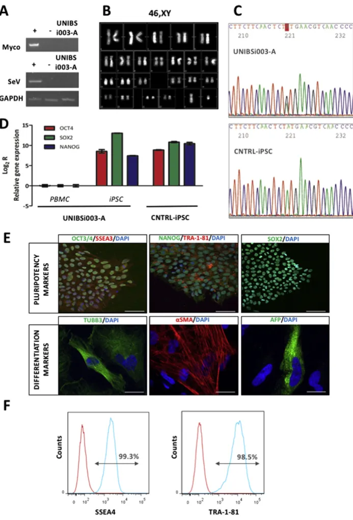
Fig. 1. Molecular and genetical analysis of UNIBSi003-A iPSC line.
9 C. Mora et al. / Stem Cell Research 24 (2017) 8–11
To obtain iPSC cells we infected blood cells using the CytoTune 2.0 iPS Sendai Reprogramming Kit (Thermo Fisher Scientific) which employs the non-integrating Sendai virus (SeV) to deliver Yamanaka's factors OCT4, SOX2, KLF4, and cMYC. Once the cell line was stabilized we evaluate the presence of contaminating mycoplasma by PCR analysis as well as the loss of the SeV used for the reprogramming procedure by reverse transcriptase PCR. Results are illustrated inFig. 1A and indicate that UNIBSi003-A cell line is both mycoplasma- and SeV-free. The presence of numerical or structural chromosome abnormalities was evaluated using standard QFQ-banding that revealed a normal 46, XY karyotype of the cell line (Fig. 1B). Moreover, the persistence of the patient's DNA mutation was confirmed by iPSC Sanger sequencing. As shown inFig. 1C, UNIBSi003-A iPSCs carry the heterozygosity in PITX2 gene that leads to the p.M200V mutation.
Next, the expression of the endogenous pluripotent transcription factors was evaluated by quantitative PCR, using the primers reported inTable 2, while the presence of the related proteins, as well as the presence of additional stem cell markers, was evaluated by immunofluorescence staining of iPSC colonies using the antibodies listed inTable 2. At variance with PBMC, UNIBSi003-A iPSCs express high levels of endogenous OCT4, SOX2, and NANOG, fully comparable to those of a control iPSC line (Fig. 1D). Moreover, the resulting proteins are correctly shown in the nuclear compartment of the cells, while additional SSEA3 and Tra-1-81 markers are properly present on cell surface (Fig. 1E). Flow cytometry using SSEA4 and Tra-1-81 antibodies revealed a pluripotent population higher than 98.5% (Fig. 1F).
Following the verification of pluripotency marker expression, we investigated the UNIBSi003-A iPSC spontaneous differentiation ca-pacity in the three-dimensional structures called embryoid bodies (EBs). EBs were left in suspension for seven days and then allowed to adhere to a standard tissue culture surface. After two weeks, cells werefixed and immunofluorescence was performed. The in vitro EB formation assay confirmed the spontaneous differentiation capacity toward the three germ layers of the UNIBSi003-A iPSC as demonstrated by the expression of ectodermal (TUBB3), mesodermal (alpha-smooth muscle actin,α-SMA) and endodermal (alpha-fetoprotein, AFP) markers (Fig. 1E).
In conclusion, we generated an iPSC line carrying the PITX2 gene p.M200V variant that has been previously described to co-segregate with AF. InTable 1, STR analysis that uniquely identify UNIBSi003-A iPSCs has been reported. Potentially, the study of the molecular basis of lone AF will strongly benefit by the analysis of the cardiomyocytes derived from UNIBSi003-A iPSCs, where the mutation is inserted in the correct pathological genetic background of the patient.
Materials and methods
Reprogramming of peripheral blood mononuclear cells (PBMC)
Peripheral blood was collected from a clinically diagnosed 38-years-old male patient suffering from paroxismal AF harboring the PITX2 p.M200V mutation in exon 7. PBMC were isolated by Ficoll-Paque® PLUS density gradient centrifugation using SepMate™-15 - specialized PBMC Isolation Tube (Stem Cell Technologies) and maintained in StemPro®-34 SFM Medium (Thermo Fisher Scientific) supplemented with StemSpam CC100 cytokines (Stem Cell Technologies) for 4 days.
Then, PBMCs were transduced using the CytoTune 2.0 iPS Sendai Reprogramming Kit (Thermo Fisher Scientific) following manufacturer's instructions. hiPSC colonies were manually picked be-tween day 17 and day 28 post infection, and expanded for further characterization.
iPSC karyotyping
Cells undergoing active cell division were blocked at metaphase by adding 10μg/mL of colcemid (Karyo Max, Gibco Co. BRL) to culture medium for 3 h at 37 °C. Then, cells were trypsinized, swollen by exposure to hypotonic 75 mM KCL solution,fixed with methanol/ glacial acetic acid (3:1), and dropped onto glass slides. Cytogenetic analysis was performed using QFQ-banding at 400–450 bands resolution according to the International System for Human Cytogenetic Nomenclature (ISCN 2016). A minimum of 20 metaphase spreads and 3 karyograms were analyzed.
iPSC genotyping
Genomic DNA from patient-derived iPSC was extracted using DNeasy blood and tissue kit (Qiagen) and the analysis of the PITX2 (NM_001204397.1) mutation was performed using a primer pair, de-scribed inTable 2, spanning the p.M200V (c.598 ANG) mutation in exon 7 of the PITX2 gene.
RNA extraction and RT-PCR analysis
Total RNA was extracted using Quick-RNA MiniPrep (Zymo Re-search), and quantified. Reverse transcriptase-PCR of 1 μg of total RNA was carried out using with iScript cDNA Synthesis Kit (BIO-RAD), followed by specific PCR amplification. Sequences of individual primer pairs are detailed inTable 2. PCR products were visualized by agarose/ ethidium bromide gel electrophoresis.
Table 1
Characterization and validation of UNIBSi003-A iPSC line.
Classification Test Result Data
Morphology Photography Visual record of the line: normal Not shown but
available with author Phenotype Immunocytochemistry Expression of pluripotency markers OCT4, SOX2, NANOG, SSEA3, and Tra-1-81 Fig. 1panel E
Expression of pluripotency markers SSEA4 and Tra-1-81 Fig. 1panel F Flow citometry Expression of pluripotency markers SSEA4: 99.3% and Tra-1-81: 98.5%. Fig. 1panel F Genotype Karyotype
(QFQ-banding) and resolution
46XY, at 400–450 band resolution Fig. 1panel B
Identity STR analysis 22 sites tested Supplementaryfile
Mutation analysis
Sequencing Heterozygous Fig. 1panel C
Microbiology and virology
Mycoplasma Absence of mycoplasma detected by PCR analysis Fig. 1panel A
Differentiation potential
Embryoid body formation
Immunocytochemical expression of alpha fetoprotein (endodermal germ layer), beta III Tubulin (ectodermal germ layer), and alpha Smooth Muscle Actin (mesodermal germ layer)
Fig. 1panel E
Molecular analysis
The expression of endogenous pluripotency-related genes OCT4, SOX2, and NANOG was assessed by quantitative PCR (qPCR) using the primers reported inTable 2. The expression ratio of the target genes was calculated by using the 2^−ΔΔCtmethod, considering GAPDH as
reference gene.
Embryoid body formation assay of pluripotency
For the generation of EBs, UNIBSi003-A cells were resuspended in DMEM/F12 medium supplemented with 20% FBS, 0.1 mM NEAA, 1 mML-Glutamine, 50μM 2-mercaptoethanol, 50 U/mL penicillin and 50 mg/mL streptomycin (all from Thermo Fisher Scientific). Seven days later, EBs were transferred onto 0.1% gelatin-coated glass and cul-tured for additional 14 days. Then, cells werefixed using 4% paraformal-dehyde and stained.
Immunofluorescence staining
The cells were incubated with Blocking Buffer (PBS containing 3% Donkey serum, 0.1% Triton X-100) for 60 min at room temperature. Next, primary antibodies, listed inTable 2, diluted in blocking buffer were added and incubated O/N at 4 °C. After extensive washing, Alexa
Fluor 594- and/or Alexa Fluor 488-conjugated secondary antibodies (Thermo Fisher Scientific) were added 1 h at room temperature. Cellular nuclei were counterstained with DAPI. The cells were observed under an invertedfluorescent microscope (Axiovert, Zeiss).
Flow cytometry
The cells were stained with either anti-SSEA4-PE antibody (BD Bio-science) or with anti Tra-1-81 antibody (Millipore), followed by incuba-tions with a biotinylated anti mouse IgM and a PE-conjugated streptavidine. Samples were analyzed using FACSCanto™ flow cytome-try (BD Bioscience).
Acknowledgement
This work was supported by Fondazione Cariplo (grant number 2014-0822) to PDE.
Reference
Christophersen, I.E., Nielsen, J.B., Holst, A.G., Sajadieh, A., Haunsoe, S., Tveit, A., Svendsen, J.H., Olesen, M.S., 2013.The PITX2 variant p.M200V is associated with early-onset lone atrialfibrillation and co-segregates within a family. Eur. Heart J. 34 (suppl_1), 4557.
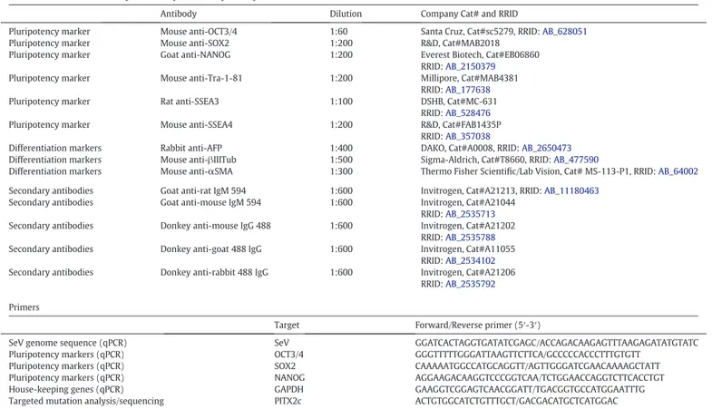
Table 2 Reagents details.
Antibodies used for immunocytochemistry andflow cytometry
Antibody Dilution Company Cat# and RRID
Pluripotency marker Mouse anti-OCT3/4 1:60 Santa Cruz, Cat#sc5279, RRID:AB_628051
Pluripotency marker Mouse anti-SOX2 1:200 R&D, Cat#MAB2018
Pluripotency marker Goat anti-NANOG 1:200 Everest Biotech, Cat#EB06860
RRID:AB_2150379
Pluripotency marker Mouse anti-Tra-1-81 1:200 Millipore, Cat#MAB4381
RRID:AB_177638
Pluripotency marker Rat anti-SSEA3 1:100 DSHB, Cat#MC-631
RRID:AB_528476
Pluripotency marker Mouse anti-SSEA4 1:200 R&D, Cat#FAB1435P
RRID:AB_357038
Differentiation markers Rabbit anti-AFP 1:400 DAKO, Cat#A0008, RRID:AB_2650473
Differentiation markers Mouse anti-βIIITub 1:500 Sigma-Aldrich, Cat#T8660, RRID:AB_477590
Differentiation markers Mouse anti-αSMA 1:300 Thermo Fisher Scientific/Lab Vision, Cat# MS-113-P1, RRID:AB_64002
Secondary antibodies Goat anti-rat IgM 594 1:600 Invitrogen, Cat#A21213, RRID:AB_11180463
Secondary antibodies Goat anti-mouse IgM 594 1:600 Invitrogen, Cat#A21044 RRID:AB_2535713
Secondary antibodies Donkey anti-mouse IgG 488 1:600 Invitrogen, Cat#A21202 RRID:AB_2535788
Secondary antibodies Donkey anti-goat 488 IgG 1:600 Invitrogen, Cat#A11055 RRID:AB_2534102
Secondary antibodies Donkey anti-rabbit 488 IgG 1:600 Invitrogen, Cat#A21206 RRID:AB_2535792
Primers
Target Forward/Reverse primer (5′-3′)
SeV genome sequence (qPCR) SeV GGATCACTAGGTGATATCGAGC/ACCAGACAAGAGTTTAAGAGATATGTATC
Pluripotency markers (qPCR) OCT3/4 GGGTTTTTGGGATTAAGTTCTTCA/GCCCCCACCCTTTGTGTT
Pluripotency markers (qPCR) SOX2 CAAAAATGGCCATGCAGGTT/AGTTGGGATCGAACAAAAGCTATT
Pluripotency markers (qPCR) NANOG AGGAAGACAAGGTCCCGGTCAA/TCTGGAACCAGGTCTTCACCTGT
House-keeping genes (qPCR) GAPDH GAAGGTCGGAGTCAACGGATT/TGACGGTGCCATGGAATTTG
Targeted mutation analysis/sequencing PITX2c ACTGTGGCATCTGTTTGCT/GACGACATGCTCATGGAC
11 C. Mora et al. / Stem Cell Research 24 (2017) 8–11