Translational Medicine @ UniSa, - ISSN 2239-9747 2013, 7(6): 29-30
29
Università degli Studi di Salerno
A 35-year-old woman in the fortieth week of her first pregnancy was admitted into our Department for terminal pregnancy.
She underwent cesarian delivery after forty-eight hours, and eight hours later, she showed cephalea, growing dyspnea and cyanosis.
Pneumonologic and cardiac advice were required. Chest examination showed: damp sounds in the right upper lung zone. Heart examination showed: blood pressure of 95/70mmHg, medium heart rate of 86 beats/min (max heart rate: 129, min heart rate: 54).
Upon physical examination, the woman was afebrile with a body temperature of 36°C.
She was intubated and treated by intravenous inotropic therapy.
An arterial blood-gas analysis gave the following values: pH, 7.462; partial pressure of oxygen, 38.7 mmHg; carbon dioxide pressure, 26,3 mmHg; bicarbonate, 18.5 mmol/L.
Electrocardiography showed: sinus rhythm (86/min), ST-segment changes without necrosis and ischemia; supraventricular ectopic beats of 63SV; no bradycardia; no paroxysmal supraventricular tachycardia.
Echocardiography revealed: severe LV systolic dysfunction with akinesia of the LV base and mid -portion, together with hypercontractility of the apex and ejection fraction of 30%; mild mitral and tricuspid regurgitation; pulmonary hypertension with a pulmonary arterial pressure of 45 mmHg; no pericardium effusion; Inferior Vena Cava congestion.
Coronary angiography revealed that both coronary arteries were intact [1].
Because of the patient’s hemodynamic instability, intra-aortic balloon pumping was begun, and medical treatment that included inotropic agents. The woman was admitted into Cardiac Intensive Care Unit [2].
Laboratory findings included raised levels of: ferritine (506ng/ml; normal 10-291ng/ml), troponin I (1,73ng/ml; normal 0-0,10) and PCR (12,3 mg/dl; normal <0,5); CK muscle and brain (MB) is 63,1 ng/ml (reference range 10-92).
The patient received cardiogenic shock due to takotsubo cardiomyopathy treatment. She responded well to treatment: her hemodynamic state stabilized one day later; intra-aortic balloon pumping was discontinued two days later and tracheal tube was removed.
About two weeks later, echocardiography showed complete recovery of LV systolic and diastolic function,
chest radiography was normal so the patient was discharged [3].
This case further supports the importance of perforrming a pre-conception planning, and proper counselling about the timing and mode of delivery by a coordinated decision between the gynaecologist, anaesthesiologist and cardiologist to balances mother’s and fetus’s risks (Table 1, Table 2). The purpose is the early diagnosis and the best management of the cardiomyopathy[4-6].
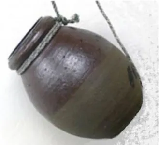
Figure 1. A picture of a real takotsubo, a Japanese octopus fishing pot.
Figure 2. Echocardiography of takotsubo.
A “Broken Heart” syndrome case
after cesarean delivery.
Prevention and risk assessment.
Petta R.
1, Zullo F.
1, Tipaldi A.
1, Guida M.
1* 1Department of Gynecology and Obstetrics, University of Salerno, Salerno, Italy *Corresponding author: [email protected]
Translational Medicine @ UniSa, - ISSN 2239-9747 2013, 7(6): 29-30
30
Università degli Studi di Salerno
Pre-conception evaluation
- Thorough history of cardiac symptoms and physical examination
- 12-lead electrocardiogram
- Baseline exercise tolerance and functional class (exercise testing if needed)
- Baseline echocardiogram
- Assessment of ventricular function (right and left)
- Assessment of pulmonary artery pressure - Presence and degree of valvular dysfunction - Assessment of stability of cardiac
hemodynamic status over time
- Effective contraception until pregnancy desired
- Adjust medications to prevent adverse fetal events
- Genetics referral for patients with heritable cardiac lesion
Table 1. Pregnancy after Tako Tsubo: risk assessment
Decision regarding timing and mode of delivery
- Management during labor and delivery and post-partum concerns
- Short vaginal delivery with excellent anesthesia, with consideration of assisted second stage of labor
- Left lateral decubitus position
- Cesarean section per obstetric indications - Invasive monitoring if needed (right heart
catheterization, invasive arterial blood pressure monitoring)
- Medical therapy optimization of loading conditions
- Consider treatment of severe anemia
- Medical therapy to optimize loading conditions
- Hemodynamic monitoring for 12–24 h - Contraception or sterilization consideration - Future consideration of ICD
Table 2. TakoTsubo prevention during labour and
delivery
REFERENCES
[1] Parsad S. Apical ballooning syndrome: an important differential diagnosis of acute myocardial infarction. Circulation 2007; 115:e56-e59.
[2] Yoshiteru Abe, MD, Makoto Kondo, MD, Ryota Matsuoka, MD, Makoto Araki, MD, Kiyoshi Dohyama, MD, Hitoshi Tanio, MD. Assessment of Clinical Features in Transient Left Ventricular Apical Ballooning. J. Am. Coll. Cardiol. 2003;41;737-742
[3] Sahng Lee, Kyung Jin Lee, Hyeon Soo Yoon, Ki Woon Kang, Young Sook Lee, Jun Wan Lee. Atypical Transient Stress -Induced Cardiomyopathies with an Inverted Takotsubo Pattern in Sepsis and in the Postpartal State. Tex Heart Inst J 2010;37(1):88-91
[4] Ettore Crimi, Aaron Baggish, Lisa Leffert, May C.M. Pian-Smith, James L. Jannuzzi and Yandong Jang. Acute Reversible Stress -Induced Cardiomyopathy Associated with Cesarean Delivery under Spinal Anesthesia. Circulation. 2008;117:3052-3053
[5] Yasir Yaqub, Leigh Ann Jenkins, Kenneth M. Nugent, Warangkana Chokesuwattanaskul. Pastpartum Depression and Apical Ballooning Syndrome (Takotsubo Syndrome). J Obstet Gynaecol Can 2009;31(8):736-739 [6] Stergiopoulos K, Shiang E, Bench T. Pregnancy in patients with pre-existing cardiomyopathies. J Am Coll Cardiol. 2011 Jul 19;58(4):337-50.