UNIVERSITA’ DEGLI STUDI DI PISA
Corso di Dottorato di Ricerca in
Fisiopatologia e Clinica dell’Apparato Cardiovascolare e Respiratorio
Tesi
The risk for fatal pulmonary embolism, recurrence, and bleeding in
patients undergoing 1-year anticoagulation for PE.
The clinical course of 1-year anticoagulated pulmonary embolism: results of a prospective, cohort study.
Candidato Presidente del Corso
Dott. Carmine Ribas Prof. Alfredo Mussi
Tutor
Prof. Antonio Palla
INDICE
ABSTRACT……… pag. 3
BACKGROUND………...pag. 5
AIMS………...pag. 7
MATERIALS AND METHODS...pag. 8
RESULTS...….pag. 13
DISCUSSION………..pag. 20
CONCLUSIONS...pag. 24
ABSTRACT
Background. Pulmonary embolism (PE) is a potentially fatal but treatable disease. Secondary prophylaxis is usually continued for three to six months, but the risk for recurrence or death lasts at least for one year. Knowing the clinical course of 1-year anticoagulated patients and identifying risk factors for adverse events may be of high clinical relevance.
Objective. To investigate the incidence of recurrence, mortality and bleeding in PE patients anticoagulated for 1 year and to identify risk factors for such adverse events.
Design. We evaluated prospectively for 1 year all consecutive in- and out-patients with a final diagnosis of acute PE referred to a single centre in Pisa, Italy, in the years 2001-2005. No exclusion criteria were adopted.
Results. Out of the original 497 patients, 136 (27.4%) died before completing the 1-year follow-up, 36 (26.5%) because of PE. Of them, 31 (86.1%) did it within 10 days of diagnosis. Risk factors for death were presence of idiopathic PE (0.003), of persistently severe dyspnea in spite of treatment (0.002), of high perfusion defect score index ( PDI ). Risk of death increased proportionally when persistently severe dyspnea and high PDI were contemporary present. Recurrence occurred in 48 (9.6%) cases, 39 (81.2%) within 10 days of diagnosis. The 1-year fatality rate was 74%, the 10 days 79%. Risk factors for recurrence were persistently severe dyspnea (0.007), high PDI (0.003), cardiac or pulmonary comorbilities (0.005).
Bleeding occurred in 17 (3.4%) cases and was major in 1 (0.2%); most cases (70.6%) occurred between 30 and 180 days, no case was observed after 180 days. No risk factor could be identified as associated to bleeding.
Conclusions. In patients anticoagulated and carefully followed for one year, death occurred in more than a quarter of cases, a quarter of them because of PE; the vast majority did it in the first 10 days. Recurrence occurred in one every ten patients, again early. Persistently severe dyspnea and high
PDI were risk factors for both mortality and recurrence. Bleeding was rare and minor and no case was observed after 180 and 360 days of treatment.
The risk for fatal pulmonary embolism,
recurrence,
and
bleeding
in
patients
undergoing 1-year anticoagulation for PE.
The clinical course of 1-year anticoagulated pulmonary embolism: results of a
prospective, cohort study.
BACKGROUND
Pulmonary Embolism (PE) is a potentially fatal disease for which treatments are effective, though not free from risk.1-5 After the acute event, efforts must be made to prevent fatal and nonfatal recurrencies; to this end, the knowledge of the natural history of PE may be of help. Insofar, most papers reported results of the 3-months-risk for recurrence, mortality, and bleeding,6-10 other reported on the clinical course of PE until one year or more in patients in whom the anticoagulation was withdrawn after 3 or 6 months11-12 or was not defined since left to the discretion of the attending physician.13-15
Few data are available in patients anticoagulated for longer time, though it is known that the risk of recurrent fatal and non fatal PE is present even far from the acute event, namely during the first year from diagnosis.12,14,16 Moreover, Agnelli et al showed that a clinical benefit was associated with extending the duration of anticoagulant therapy to one year in patients with idiopathic DVT 17 and in those with a first episode of PE;18 however, they did not identify risk factors for recurrence, bleeding and PE mortality during such period of time.
Therefore, we evaluated prospectively all the consecutive patients affected by PE with the purpose of giving novel insight in terms of recurrence, mortality and bleeding, both early after diagnosis and
lately during the whole 1-year-follow-up period under oral anticoagulant treatment. Also, we used the multivariate analysis in order to identify and quantify the relative risk of the above adverse events during the follow-up.
AIMS
We evaluated the clinical outcome during one year in a largegroup of consecutive patients with PE. We aimed to assess the incidence of recurrent VTE, mortality, and bleeding complications in patients with PE and treated with oral anticoagulants during this follow-up period. Second, we aimed to identify risk factors for recurrence, bleeding, and mortality, and to determine the time course of these events within one year of the start of treatment.
MATERIALS AND METHODS
Study design
We followed prospectively all the consecutive patients who had a final instrumental diagnosis of PE made at the Cardio-Thoracic Department of the University of Pisa during the period may 1st 2001 - may 31st 2005; therefore, patients were enrolled in a single academic center for diagnosis and treatment of venous thromboembolism. All patients were anticoagulated for at least 12 months independently on the origin of PE, since this has been our policy since 1969. No exclusion criteria to enter the study was established, in order to remain as adherent as possible to real life conditions; thus, patients with active cancer, permanent immobility, or in anticoagulant prophylaxis before PE were included.
Both in and out patients were included. Inpatients were defined as those who showed symptoms compatible with the disease while they were in the hospital for reasons different from PE. Out patients were defined as those who came to the Emergency Department right because symptoms compatible with PE. Furtherly, patients were divided according to risk factors as having secondary PE in presence of 1 or more transient risk factors and idiopathic PE in absence of any of them. The protocol was approved by the institutional review board for human studies.
Acute embolization
All patients underwent perfusion lung scan (PLS): when it was normal or abnormal but not compatible with PE and clinical probability was low, PE was excluded.19-20 In the remaining cases, diagnosis was made by a positive spiral CT 21 or a positive pulmonary arteriography19. CT instrument was located in the emergency department and dedicated to the emergencies only. When this examination was not promptly available or contraindicated due to hypersensitivity to contrast
medium or renal failure the diagnosis was made or excluded in presence of concordant results of perfusion scintigraphy and clinical probability.19-20
Perfusion lung scintigraphy was obtained according to a previously described method.22 Ventilation lung scan was not performed.22 Perfusion lung scintigraphy was read for the detection of unperfused lung segments (ULSs) and graded in the following way: a score of 1 was assigned to each unperfused area with the size, shape and location of a known lung segment; and a score of 0.5 was assigned to perfusion defects with subsegmental size, shape, and location or when a scintigraphic region corresponding to a lung segment was poorly perfused. The number of ULSs was taken as an index of the severity of PE (Perfusion Damage Index, PDI).22 Perfusion lung scintigraphy was examined by two independent observers and the degree of agreement was tested.
Chest spiral CT images have been acquired with a four-slices multidetector-row CT.23 The examination consisted of an evaluation of the pulmonary arteries up to and including the subsegmental vessels. The patient were examined during a breath hold or shallow breathing. The acquisition parameters for multidetector-row CT were injection of a total volume of 100 to 120 ml of non-ionic contrast material; imaging 9 to 20 seconds after initiation of the contrast-material injection; scanning perfomed at 1.0 to 1.3 mm per section with a pitch of 1.25 to 1.75, 120 kV, and 115 to 260 mA; and reconstruction of images at 0.6 to 0.8 mm intervals.
Pulmonary angiography was obtained according to standardized procedures.19
All patients were investigated for chest pain, dyspnea, presence of syncope, presence of acquired predisposing factors. Dyspnea was subsequently divided in mild, moderate, severe; moreover, we introduced the new definition of persistently severe dyspnea as “the one that did not improve 24 hours of the onset of treatment”. Acquired predisposing factors were: immobilization for longer than 7 consecutive days, major surgical procedure, bone fracture of the lower extremities, history of lower deep vein thrombosis (DVT), or any prior episode of PE (if the patient had at any time documented episodes of DVT or PE that required anticoagulant therapy), estrogen use (defined as use of estrogen containing drugs within the past 3 months), and postpartum period (defined as the
presence of pregnancy within the past 3 months), neoplastic disease. All neoplasms were included independently from disease activity or medical treatment. Also, pre-existing cardiac or respiratory disease were taken into considerations as follows: coronary artery disease, hypertensive cardiomiopaty, valvular cardiomiopaty, heart failure (right, left or both), COPD, bronchiectasis, interstitial lung disease.
Follow-up
All patients were followed by the authors until death, lost from follow-up or 31th may, 2006, whichever came first. Patients were anticoagulated for a period variable within 5 and 10 days with unfractioned heparin monitored by aPTT in most cases; rarely, patients were assigned to low-molecular-weight heparin. In all cases, heparin was followed by oral anticoagulation monitored by the INR (between 2 and 3) at least until 12 months after the acute episode. Patients were regularly followed in the anticoagulation clinic to optimize the oral treatment.
A clinical follow up was performed directly when patients were in hospital, by ambulatory visits every 3 months or by phone calls at predetermined times in dismissed patients. Moreover, all patients were scheduled for a scintigraphic and blood gas follow-up at 7, 30 and 365 days after the acute episode, to investigate the recovery of pulmonary perfusion or the possible recurrence of asymtomatic PE. By definition all deaths occurring during hospitalization with symptoms highly suggestive of PE were considered as due to recurrence. Recurrences were divided in two categories, according to a model reported recently12 tat we slightly modified: a) definite or probable fatal PE if it was confirmed at autopsy, or it was preceded in the immediate period before death by confirmed nonfatal PE, or if patients died during hospitalization with symptoms highly suggestive of PE; possible fatal PE when patients died after hospitalization with symptoms highly suggestive of PE as reported by the attending physician; b) definite or probable non fatal PE if new perfusion defects were detected on a scintigraphic follow-up predefined at 7, 30, 360 days of the acute episode or
extraordinarily made because of new-onset symptoms; possible non fatal PE if patients reported symptoms compatible with recurrence at the clinical follow-up.
Bleedings were sought in all hospitalized patients, at the time of ambulatory visits or by telephone calls when dismissed. Bleedings where categorized as major and minor. We classified as major the following: fatal (death due to haemorrhage), intracranial (documented by imaging), intra-abdominal, required surgery or angiographic intervention and if bleeding led to haemoglobin reduction of 2g/dL or more and/or needed transfusion. Minor bleedings were considered all those not fulfilling criteria for major bleedings.
Statistical Analysis
Continuous variables were expressed as mean ± 1 SD and categorical variables as percentages. The duration of follow-up was expressed in days. We considered clinical outcomes that occurred during follow-up from the time of recruitment until death or the end of patient follow-up. The primary end points were the occurrence of definite or probable fatal PE, definite or probable recurrences, bleedings. To standardize for different lengths of follow-up in the cohort, we used the annual risk for definite or probable fatal PE (the number of fatal events per 100 person-years of follow-up). The case-fatality rate for definite or probable fatal PE was calculated as follows: definite or probable fatal PE / (definite or probable fatal PE + any nonfatal PE). We used the Kaplan–Meier method to determine the time course of events.
The predictors of event-free survival were identified by univariate and multivariate analysis performed using the Cox proportional hazards regression model. Categorical variables were included into the model as dummy variables. The independent predictors of event-free survival were identified using a backward elimination procedure in a multivariate Cox regression analysis. Once the independent predictors of event-free survival were identified, interaction terms among these
predictors were also entered into the Cox proportional hazards regression model to evaluate if they also acted as effect modifiers.
To assess the prognostic value of the final Cox proportional hazards regression models we used the time-dependent ROC curve estimation from censored survival data using the Nearest Neighbor Estimation method.24 The linear prognostic scores obtained from the final Cox models were considered in the analysis. The area under the time-dependent ROC curves was used as prognostic accuracy of the models.
All statistical tests were two-tailed; a p <0.05 was considered as significant. Statistical analysis was performed with commercially and free available software (SPSS 10.0, SPSS Inc., Chicago, Illinois, USA; R: A Programming Environment for Data Analysis and Graphics, Version 2.7.1, R Foundation for Statistical Computing, Vienna, Austria).
RESULTS
Among 1027 patients referred with the suspicion of PE, 497 (48.4%) had a final diagnosis of PE. In particular, 415 (83.5 %) were diagnosed by spiral CT, 4 (0.80 %) by pulmonary arteriography. In a further group of 78 (15.7 %) with abnormal basal perfusion scintigraphy in whom spiral CT or arteriography were contraindicated or not available promptly, the diagnosis was confirmed by the concordance of lung scintigraphy compatible with PE and high clinical probability.19 Basal characteristic of 497 patients are reported in table 1.
Table 1. Baseline Characteristics of the 497 Patients With PE N ( % )
Inpatients 281 (61.8 %) Age** 68.9 ( 17-95 ) Age<60 104 ( 20.9 ) 60≤Age<70 120 ( 24.1 ) Age>70 273 ( 54.9 ) Male gender 223 ( 44.9 ) Smokers 48 ( 9.9 ) Previous smokers 131 ( 26.9 ) Idiopathic PE 212 ( 42.7 ) Risk factors Cancer 127 ( 25.6 ) Surgery 164 ( 33.0 ) Immobilization > 7days 179 ( 36.0 )
Estrogen use ( on 274 female ) 15 ( 5.5 )
Previous PE 35 ( 7.0 )
Previous DVT 40 ( 8.4 )
DVT 172 ( 36.2 )
Vasculitis 2 ( 0.4 )
Cardiopulmonary Comorbilities
Previous cardiopulmonary disease 191( 38.4 )
Symptoms
Dyspnea 310 ( 66.0 )
Persistent Dyspnea 58 ( 12.3 )
Syncope 71 ( 15.4 )
Arterial blood gas analysis and Perfusion parameters mean ± sd/ N(%)
pH 7.5 ± 0.05 PaO2 67.2 ± 15.0 PaO2st 55.5 ± 17.6 PaCO2 33.4 ± 6.6 PDI 4.7 ± 2.5 PDI ≥ 6 148 (29.8 %)
Among them, 281 (61.8 %) were inpatients. Mean age was 68.9 years with a prevalence of subjects of female sex (55,1%). Among the risk factors, surgical procedures (33.0 %), cancer (25.6 %) and cardiopulmonary comorbilities (38.4%) were the most represented. In 212 patients (42.7 %) no identified risk factor was found and PE was classified as idiopathic. Dyspnea was the most frequent symptom (313 cases, 63%) and, most relevant, persistently severe dyspnea was present in 60 cases (12.1%).
Patients showed an average perfusion impairment of 26.1% of the entire vascular bed (PDI 4.7+ 2.5). Moreover, 148 (29.8%) patients had a PDI greater than or equal to 6 unperfused lung segments, that represents 30% of the entire vascular bed. This value was therefore arbitrarily taken as the threshold above which the perfusion impairment may be considered as severe. No significant difference was found between the two readers as for the entity of lung scintigraphic impairment (p>0.5).
Among 497 patients, 172 (36,2 %) had DVT in adjunct to PE, as demonstrated on instrumental examination, 40 (8.4%) reported a previous episode of DVT.
Recurrence and death
Among the original 497 patients, 361 (72.6 %) completed the 1-year-follow-up while 136 (27.4 %) patients died before completing it. In further details, 36 (26.5%) patients died because of recurrent PE, 59 (43.4%) due to neoplasms, 20 (14.7%) to cardiac and 21 (15.4%) to pulmonary causes. We recorded 48 (9.6%) recurrences among all patients; of them 39 (81.2%) occurred early within 10 days of diagnosis and 9 lately during 1 year. Definite or probable fatal PE occurred in 31 (65%) cases, all within 10 days of diagnosis (Fig. 1); possible fatal PE in 5 (10%) cases that occurred sparsely after discharge during the following period until 1 year. Annual risks for fatal PE was 9.4/100 person/years. The case-fatality rate for PE was 75% (31/48) when considered during the
non fatal PE occurred in 10 (21%) cases, two of them found casually at the scintigraphic follow up, possible in 2 (4%).
Risk factors for recurrence and death
Risk factors for both early and late recurrences were the presence of a persistently severe dyspnea (p=0.007), of a high PDI (p=0.003) and of cardiac or pulmonary comorbilities (p=0.005) (table 2).
Table 2. Risk factors for definite or probable recurrences
Recurrence No Recurrence Univariate Analysis Multivariate Analysis
Variables 48 449 p HR (95 % ICI) p HR (95 % ICI)
N ( % )/means±sd N ( % ) /means±ds Inpatients 27 (60.0) 254 (62.0) 0,815 0.93 (0.51-1.69) Age 69.6 ± 14.5 68.8 ± 14.1 0.702 1.00 ( 0.98 - 1.02 ) Male gender 17 ( 35.4% ) 206 ( 45.9% ) 0,191 0.67 ( 0.37 - 1.22 ) Idiopathic PE 25 ( 52.1 ) 187 ( 41.6 ) 0,185 1.47 ( 0.83 – 2.58 ) Risk factors Surgery 12 ( 75.0 ) 152 ( 33.9 ) 0,228 0.67 ( 0.35 - 1.28 ) Cancer 8 ( 16.7 ) 119 ( 26.5 ) 0,194 0.60 ( 0.28 - 1.29 ) Immobilization > 7days 17( 35.4 ) 162 ( 36.1 ) 0.928 0.97 ( 0.54 – 1.76 ) Previous PE 3 ( 6.3 ) 32 ( 7.1 ) 0,821 0.87 ( 0.27 – 2.81 ) DVT 14 ( 29.2 ) 158 ( 37.0 ) 0,223 0.67 ( 0.36-1.27 ) Previous DVT 3 ( 6.3 ) 37 ( 8.7 ) 0.429 0.62(0.19-2.03) Cardiopulmonary Comorbilities Previous Cardiopulmonary disease 33( 68.8 ) 158 ( 35.2 ) 0,000 3.80 ( 2.06 – 7.00 ) 0.005 2.59 (1.34-5.02) Symptoms Dyspnea 29( 60.4 ) 284( 63.3 ) 0,227 1.66 ( 0.73 – 3.79 ) Persistent Dyspnea 12 ( 25.0 ) 48 ( 10.7 ) 0,005 3.83 ( 1.51-9.73) 0.007 3.64 (1.42-9.37) Syncope 5 ( 10.4 ) 66 ( 14.7 ) 0,447 0.70 ( 0.28 – 1.76 )
Arterial blood gas analysis and Perfusion parameters
means±sd/N (%) means±sd/N (%) p p HR ( 95% C.I. )
pH 7.46±0.04 7.46±0.05 0,602 0.17 ( 0.00-126.5 ) PaO2 68.7±12.4 67.0±15.2 0,524 1.01 ( 0.99-1.03 ) PaO2st 58.5±14.4 55.2±17.9 0,284 1.01 ( 0.99-1.03 ) PaCO2 34.0±4.2 33.4±6.8 0,585 1.01 ( 0.97-1.05 ) PDI 6.2±2.3 4.5±2.4 0,000 1.24 ( 1.12-1.37 ) 0.003 1.18(1.06-1.31) PDI ≥ 6 31 ( 64.6 ) 117 ( 26.1 ) 0,000 4.71 ( 2.60-8.51 )
Risk factors for overall PE death were the presence of idiopathic PE (p=0.003), of a persistently severe dyspnea (p=0.002) and of a high PDI (p=0.000) (table 3).
Table 3. Risk factors for definite or probabile fatal PE Patients died due to PE
Living patient/patient died due to others disease
Univariate Analysis Multivariate Analysis
Variables 36 461 p HR (95 % ICI) p HR (95 % ICI)
N ( % )/means±sd N ( % ) /means±ds Inpatients 20 (55.5) 261 (56.6) 0.892 0.95 (0.47-1.91) Age 70.0 ± 14.9 68.81 ± 14.3 0.616 1.01 ( 0.98 - 1.03) Male gender 15 ( 41.7% ) 208 ( 45.1% ) 0.714 0.88 ( 0.45 - 1.71 ) Idiopathic PE 22 ( 61.1 ) 190 ( 41.2 ) 0.028 2.12 ( 1.08 - 4.15 ) 0.003 2.11 ( 1.07 - 4.15 ) Risk factors Surgery 8 ( 22.2 ) 156 ( 33.8 ) 0.166 0.57 ( 0.26 - 1.26 ) Cancer 6 ( 16.7 ) 121 ( 26.2 ) 0.264 0.61 ( 0.25 - 1.46 ) Immobilization > 7days 11( 30.6 ) 168 ( 36.4 ) 0.487 0.78 ( 0.38- 1.58 ) Previous PE 1 ( 2.8 ) 34 ( 7.4) 0.321 0.37 ( 0.05-2.67 ) DVT 9( 25.0 ) 163 ( 37.1 ) 0.109 0.54 ( 0.25-1.15 ) Previous DVT 2 ( 5.6 ) 38 ( 8.2 ) 0.249 0.51(0.12-2.16) Cardiopulmonary Comorbilities Previous Cardiopulmonary disease 24( 66.7) 167 ( 36.2 ) 0.001 3.39 ( 1.69 – 6.78 ) Symptoms Dyspnea 19 (52.8) 294( 63.8 ) 0.861 1.08 ( 0.45 – 2.57 ) Persistent Dyspnea 10 (27.8) 50 ( 10.8 ) 0.020 3.15 ( 1.20-8.29 ) 0.002 3.55 ( 1.33-9.45 ) Syncope 4 (11.1) 67 ( 14.5 ) 0.593 0.75 ( 0.27 – 2.13 )
Arterial blood gas analysis and Perfusion parameters
means±sd/N (%) means±sd/N (%) p HR ( 95% C.I. ) p HR ( 95% C.I. )
pH 7.46±0.04 7.46±0.05 0.959 0.81 ( 0.00-2247 ) PaO2 66.6±12.9 67.2±15.1 0.997 1.00 ( 0.97-1.02 ) PaO2st 56.0±14.8 55.4±17.8 0.858 1.00 ( 0.98-1.02 ) PaCO2 34.32±4.78 33.4±6.7 0.692 1.01 ( 0.96-1.06 ) PDI 6.5±1.9 4.5±2.4 0.000 1.29 ( 1.15-1.44 ) 0.000 1.30 ( 1.16-1.46 ) PDI ≥ 6 27 ( 75.0 ) 121 ( 26.2 ) 0.000 7.66( 3.60-16.29 )
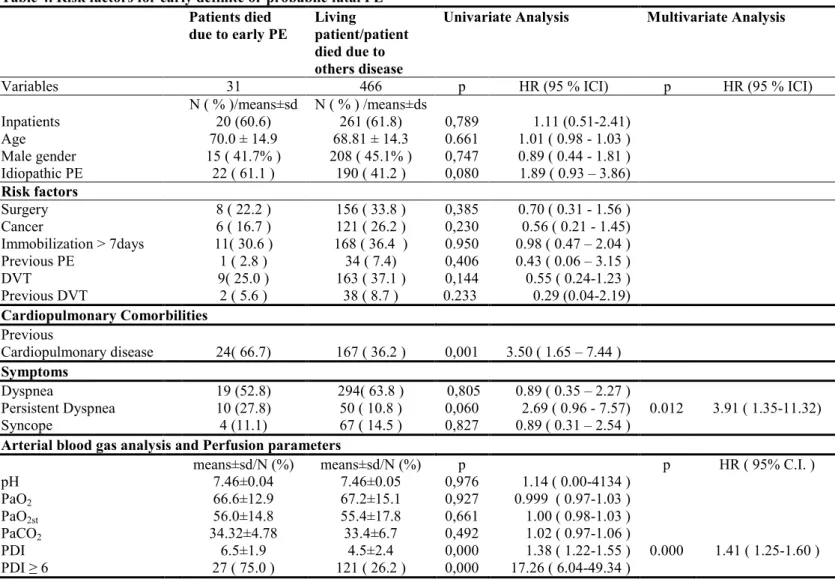
When looking for risk factors for early death only, we found the presence of a persistently severe dyspnea (p=0.012) and of a high PDI (p=0.000) (table 4).
Table 4. Risk factors for early definite or probabile fatal PE Patients died due to early PE Living patient/patient died due to others disease
Univariate Analysis Multivariate Analysis
Variables 31 466 p HR (95 % ICI) p HR (95 % ICI) N ( % )/means±sd N ( % ) /means±ds Inpatients 20 (60.6) 261 (61.8) 0,789 1.11 (0.51-2.41) Age 70.0 ± 14.9 68.81 ± 14.3 0.661 1.01 ( 0.98 - 1.03 ) Male gender 15 ( 41.7% ) 208 ( 45.1% ) 0,747 0.89 ( 0.44 - 1.81 ) Idiopathic PE 22 ( 61.1 ) 190 ( 41.2 ) 0,080 1.89 ( 0.93 – 3.86) Risk factors Surgery 8 ( 22.2 ) 156 ( 33.8 ) 0,385 0.70 ( 0.31 - 1.56 ) Cancer 6 ( 16.7 ) 121 ( 26.2 ) 0,230 0.56 ( 0.21 - 1.45) Immobilization > 7days 11( 30.6 ) 168 ( 36.4 ) 0.950 0.98 ( 0.47 – 2.04 ) Previous PE 1 ( 2.8 ) 34 ( 7.4) 0,406 0.43 ( 0.06 – 3.15 ) DVT 9( 25.0 ) 163 ( 37.1 ) 0,144 0.55 ( 0.24-1.23 ) Previous DVT 2 ( 5.6 ) 38 ( 8.7 ) 0.233 0.29 (0.04-2.19) Cardiopulmonary Comorbilities Previous Cardiopulmonary disease 24( 66.7) 167 ( 36.2 ) 0,001 3.50 ( 1.65 – 7.44 ) Symptoms Dyspnea 19 (52.8) 294( 63.8 ) 0,805 0.89 ( 0.35 – 2.27 ) Persistent Dyspnea 10 (27.8) 50 ( 10.8 ) 0,060 2.69 ( 0.96 - 7.57) 0.012 3.91 ( 1.35-11.32) Syncope 4 (11.1) 67 ( 14.5 ) 0,827 0.89 ( 0.31 – 2.54 )
Arterial blood gas analysis and Perfusion parameters
means±sd/N (%) means±sd/N (%) p p HR ( 95% C.I. )
pH 7.46±0.04 7.46±0.05 0,976 1.14 ( 0.00-4134 ) PaO2 66.6±12.9 67.2±15.1 0,927 0.999 ( 0.97-1.03 ) PaO2st 56.0±14.8 55.4±17.8 0,661 1.00 ( 0.98-1.03 ) PaCO2 34.32±4.78 33.4±6.7 0,492 1.02 ( 0.97-1.06 ) PDI 6.5±1.9 4.5±2.4 0,000 1.38 ( 1.22-1.55 ) 0.000 1.41 ( 1.25-1.60 ) PDI ≥ 6 27 ( 75.0 ) 121 ( 26.2 ) 0,000 17.26 ( 6.04-49.34 )
Among the 48 patients who showed recurrences, the risk factors for mortality were the presence of a high PDI (p=0.000), of a idiopathic PE (p=0.004) and of neoplasms (p=0.056). The area under the time-dependent ROC curves (Fig. 1) showed a good prognostic value of the final Cox models (AUC=0.74).
Figure 1.
No significant interaction between predictors of fatal PE-free survival was observed. Finally, the RR for early death increased proportionally when persistently severe dyspnea and high PDI were contemporary present.
Risk of bleeding
Bleeding occurred in 17 cases (3.4%); it was classified as major in 1 (5.9%) case. In the whole follow-up, bleeding occurred in 1 (5.9%) case in the first 10 days, in 4 (23.5%) cases between 10 and 30 days, in 12 (70.6%) cases between 30 and 180 days. No case was observed between 180 and 360 days.
No risk factor was identified by Cox regression analysis as associated with bleeding in patients of the present series (table 5).
Table 5. Risk factors for Bleeding Univariate
Bleeding No Bleeding Univariate Analysis
Variables 17 480 p HR (95 % ICI) N ( % )/means±sd N ( % ) /means±ds Inpatients 9 (52.9) 272 (56.6) 0,441 0.69 (0.26-1.78) Age 72.1 ± 10.5 68.8 ± 14.2) 0.336 1.02 ( 0.98 - 1.06 ) Male gender 8 ( 47.1% ) 215 ( 44.8% ) 0,823 1.11 ( 0.43 – 2.89 ) Idiopathic PE 9 ( 52.9 ) 203 ( 42.3 ) 0,400 1.50 ( 0.58 – 3.90 ) Risk factors Surgery 5 ( 29.4 ) 159 ( 33.1 ) 0,733 0.83 ( 0.29 – 2.37 ) Cancer 2 ( 11.8 ) 125 ( 26.0 ) 0,275 0.44 ( 0.10 - 1.92 ) Immobilization > 7days 7( 41.2 ) 172 ( 35.8 ) 0.668 1.23 ( 0.47 – 3.24 ) Previous PE 0 ( 0.0 ) 35 ( 7.3 ) 0,448 0.04 ( 0.00 – 139 ) DVT 7 ( 41.2 ) 165 ( 36.0 ) 0,658 1.26 ( 0.46-3.47 ) Previous DVT 2 ( 11.8 ) 38 ( 8.3 ) 0.590 1.53(0.32-7.21) Cardiopulmonary Comorbilities Previous Cardiopulmonary disease 9( 52.9 ) 182 ( 37.9 ) 0,131 2.08 ( 0.80 – 5.40 ) Symptoms Dyspnea 13( 76.5 ) 300( 62.5 ) 0,396 1.72 ( 0.49 – 6.04 ) Persistent Dyspnea 1 ( 5.9 ) 59 ( 12.3 ) 0,872 0.83 ( 0.09-7.98) Syncope 2 ( 11.8 ) 69 ( 14.4 ) 0,801 0.83 ( 0.19 – 3.62 )
Arterial blood gas analysis and Perfusion parameters
means±sd/N (%) means±sd/N (%) p pH 7.46±0.03 7.46±0.05 0,788 0.24 ( 0.00-7032 ) PaO2 65.2±12.3 67.2±15.0 0,600 0.99 ( 0.95-1.03 ) PaO2st 54.2±18.7 55.5±17.6 0,767 0.99 ( 0.96-1.03 ) PaCO2 34.8±9.5 33.4±6.5 0,443 1.02 ( 0.97-1.07 ) PDI 4.4±3.5 4.7±2.4 0,916 1.01 ( 0.83-1.23 ) PDI ≥ 6 6 ( 35.3 ) 142 ( 29.6 ) 0,433 1.49 ( 0.55-4.03 )
DISCUSSION
We evaluated prospectively the 1-year-clinical and scintigraphic outcome under anticoagulant treatment of a large cohort of unselected in and outpatients with confirmed diagnosis of PE, with the aim of investigating incidence, timing and risk factors of adverse events.
Mortality in our patients was as high as 25% after 1 year, causes spanning from neoplasms, to cardiopulmonary causes other than PE, to fatal recurrence of PE. This result reproduces strictly the overall mortality reported by Carlson et al 13 who stopped treatment after 3 or 6 months and had a 24% one-year overall mortality in patients with PE. Also, the underlying comorbidities were similar, being cancer (43.4%) and cardiopulmonary diseases (30.1%) responsible for 73.5% of all deaths. Differently from Carlson, however, we found that death related to PE after 1 year occurred in about 7% ( versus 2.5% of Carlson) of the original sample of patients, 27% of all patients who die. Since patients in our study were all anticoagulated up to 1 year, the difference could be searched in more severe acute pulmonary damage, older age, higher number of in-patients or patients with idiopathic PE in our series. The PE-associated mortality rate of 7% during one year is lower than the 15% found byvan Strijen and colleagues7, who stopped follow-up (and the therapy) at 3 months; it is instead consistent with the 7.7% found in a study by Perrier et al23 that also concluded the follow-up after 3 months. Comparison with both studies seem to underline the maximum risk of death in the early phases; however, our study only allows to define the risk that a population of both in and out-patients has during a one-year anticoagulant treatment. Although most frequently in the early phases of disease (86% of cases died during the first 10 days of the embolization), death due to PE may also occur later, in spite of an appropriate treatment. This underlines the necessity of a early diagnosis and prompt therapy, as well as the need for the knowledge of the early and late risks for fatal PE in each single patient.
We found that risk factors for early mortality were severity of PE as evaluated on perfusion lung scintigraphy and persistently severe dyspnea while presence of idiopathic PE represents a further late risk factor. Severity of disease has previously been described as associated to the mortality of PE14 in adjunct, however, we demonstrated in the current study that this parameter is of great importance also beyond the acute event keeping its value during the whole follow-up. This underline the utility of a simple method to quantify the severity of the damage extension. The persistence of severe dyspnea in spite of the anticoagulant treatment is a powerful risk factor for fatal PE, both in the acute and in the late phases of the disease. Patients complaining this frequent symptom (12.1% of all patients included and 27.8% of patients who died because of PE) have as much as three and half times increased probability of dying because of fatal PE. Furthermore, this symptom increases disproportionally the risk for death in relation to the extension of pulmonary perfusion damage. Indeed, the presence of severe persistent dyspnea makes the risk 3.5 times greater in patients with 6 unperfused segments, 7 times greater when the damage corresponds to 14 unperfused segments. A simple formula to calculate the risk may easily be available. A third risk factor for fatal PE is represented by the idiopathic origin; though this was already known, we defined the risk in as much as more than twice. Considering that in our series the amount of patients affected by idiopathic PE was more than 40% of the total, it is easy to understand the relevance of such variable. The reason for which it plays a role in the late phases of disease is unclear, but it is hypothesizable that the idiopathic origin includes other risk factors largely unknown, whose influence may be played at any time.
The recurrence rate of 9.6% is similar to that observed in most cohort studies.13-14 Carson et al had 33 patients out of a total of 399 (8.3%) with clinically apparent recurrence of PE in the year of follow-up, Miniati et al 30 out of 320 (9.4%). In a recent Dutch study, instead, such a percentage was much lesser (20 patients over 673, 3%); however, in this case they stopped prophylaxis and follow-up 3 months of the acute PE. Thediscrepancy might be due to a differentcomorbidity profile with a higher prevalence of cardiovascular disease, a greater number of older, in-patients in our
series, and the systematic performance of a scintigraphic follow-up which has been able to identify new perfusion abnormalities in 2 asymptomatic patients.
Recurrencies occurred early (within 10 days) in at least 80% of cases and often they were fatal. Indeed, the case fatalityrate for PE in the current study was 75% when considered during the whole 1-year-follow-up, it was 79% when deaths occurred during the first 10 days. Risk factors for early recurrences are again persistently severe dyspnea and severity of PE; later on, presence of cardiopulmonary comorbidities joins the previous two risk factors in determining recurrences. Our results appear rather different from them of an earlier study9 in which the presence of cancer, chronic cardiovascular disease, chronic respiratorydisease, and other clinically significant diseases were risk factors for recurrent VTE in patients with PE.
We found clinically most useful to know that the first 10 days represent the most critical period for recurrences, that the case fatality rate of recurrences is high and even higher in the early phases, and that the knowledge of risk factors (specifically massive damage on perfusion scan, severe persistent dyspnea and idiopathic origin of PE) might help in more rigorous monitoring and more aggressive therapy. Also, it appears relevant that risk factors for mortality among patients with recurrences (severity of PE, idiopathic origin, presence of neoplasms) may be known since the patient' presentation; this should alert physicians on how and where to treat patients with such risk factors. Likewise it seems of interest to know that the risk of recurrences drops after the acute embolization but it is still present months later in spite of an appropriate treatment.
Bleeding occurred in 17 cases (3.4%); it was classified as major in 1 (0.2%) case since it occurred as intracranial bleeding. However, neither this patient or any other died of bleeding. In the whole follow-up, bleeding occurred in 1 (5.9%) case in the first 10 days, in 4 (23.5%) cases between 10 and 30 days, in 12 (70.6%) cases between 30 and 180 days, none thereafter. Our bleeding figures in the first 3-6 months were similar to those of other studies;9,11 no comparison is obviously possible with such studies after 6 months. No risk factor was identified as associated with bleeding in
being an inpatient; a recent paper from the RIETE registry25 has demonstrated that a risk score based on 6 variables (recent bleeding, abnormal creatinine level, anemia, cancer, age >75, pulmonary embolism at baseline) can identify VTE patients at risk at entry. This of course may help in therapeutic decisions. Interestingly enough, no case of bleeding occurred in our patients from 6 to 12 months from incidental PE. Thereason for this might be searched in the careful monitoring of patients in the 3-months ambulatory visits and in the effectiveness of anticoagulation clinic; moreover, the stabilization of therapy after the initial period and the learning capacity of patients and their familiars may have contributed to this excellent result. This occurred in spite of the high percentage of patients aged more than 70 years (54.9%), of high amount of inpatients (61.8%), of the highly incidence of risk factors for bleeding at the entry, such as cancer (25.6%), surgery (33.0%), prolonged immobilization (36.0%). Although we do not have a control group of patients in whom the anticoagulant therapy was withdrawn after 3 or 6 months, it seems reasonable to state that prolonging anticoagulation until 1 year is certainly safe. Moreover, it appears rather effective.
Limitations
We are aware that the study suffers some limitations. We included only patients with a certain diagnosed of PE and therefore patients in whom emboli were never diagnosed were missed. Our follow-up period is limited to one year; however, the recurrence of PE after one year is less frequent, as demonstrated by other recent papers14,26-28 and the detection of pulmonary hypertension that is more frequent beyond a year was not among the aims of our work. Furthermore, we cannot demonstrate definitely that prolonging anticoagulation up to 1 year is cost-effective since we do not have a control group in which therapy had stopped after 3 or 6 month of the embolization. However, this has been our satisfactory policy since more than thirty years and, thus, the data that we report represent what happens in real life.
CONCLUSIONS
Several points make our study unique. First, all consecutive patients with definitive diagnosis of PE were anticoagulated and prospectively followed for one year. This allowed us to report on mortality, recurrencies and bleeding complications for a until now poorly explored lapse of time in patients treated. Moreover, this made it possible to find the risk factors for mortality, recurrence and bleeding in the early stages after embolization and later on until one year of the acute PE; to our knowledge, this has not been reported by other studies. We demonstrated for the first time that the persistence of severe dyspnea in spite of the anticoagulant treatment is a powerful risk factor for both recurrence and mortality due to PE, both in the acute and in the late phases of PE. This newly described symptom occurs in our series associated to the severity of disease and shows an additive effect to the scintigraphic evaluation of perfusion impairment, in predicting both PE-mortality and recurrence. Finally, we systematically performed a 1-year scintigraphic follow-up by predefined (at 7, 30, 360 days of the diagnosis) controls of patients; this allowed to detect cases of non fatal recurrences that otherwise would have passed unrecognized.
REFERENCES
1.Giuntini C, Di Ricco G, Marini C, Melillo E, Palla A. Pulmonary Embolism: Epidemiology. Chest 1995; 107: 3S-9S
2. Anderson FA, Wheeler HB, Goldberg RJ, et al. A population-based perspective of the hospital incidence and case-fatality rates of deep vein thrombosis and pulmonary embolism: The Worcester DVT study. Arch Intern Med 1991; 151: 933-8
3. Silverstein MD, Heit JA; Mohr DN, Petterson TM, O’Fallon WM, Melton LJ III. Trends in the incidence of deep vein thrombosis and pulmonary embolism: a 25-years population-based study. Arch Intern Med 1998; 158: 585-93.
4. Torbicki A, van Beek JR, Charbonnier B, Meyer G, Morpurgo M, Palla A, Perrier A. Task force report. Guidelines on diagnosis and management of acute pulmonary embolism. Eur Heart J 2000; 21: 1301-35.
5. Barritt DW, Jordan SC. Anticoagulant drugs in the treatment of pulmonary embolism: a controlled trial. Lancet 1960; 1: 1309-12.
6. Perrier A, Roy PM, Sanchez, O, et al Multidetector-row computed tomography in suspected pulmonary embolism. N Engl J Med 2005; 352:1760-1768
7. van Strijen, MJ, de Monye, W, Schiereck, J, et al Single-detector helical computed tomography as the primary diagnostic test in suspected pulmonary embolism: a multicenter clinical management study of 510 patients. Ann Intern Med 2003;138:307-314.
8. Aujesky D, Roy PM, Le Manach CP, et al. Validation of a model to predict adverse outcomes in patients with pulmonary embolism. Eur Heart J 2006; 27: 476-81.
9. Nijkeuter M, Sohne M, Lidwine W, et al. The Natural Course of Hemodinamically Stable Pulmonary Embolism. Chest 2007; 131: 517-523
10. Douketis JD, Kearon C, Bates S, et al. Risk of fatal pulmonary embolism in patients with treated venous thromboembolism. JAMA. 1998; 279: 458–462
11. Van Beek EJR, Kuijer PMM, Buller HR, Brandjes DPM, Bossuyt PMM, ten Cate JW. The clinical course of patients with suspected pulmonary embolism. Arch Intern Med 1997; 157: 2593-8.
12. Douketis JD, Gu CS, Schulman S, Ghirarduzzi A, Pengo V, Prandoni P. The risk for fatal pulmonary embolism after discontinuing anticoagulant therapy for venous thromboembolism. Ann Intern Med 2007; 147: 766-74.
13. Carlson, JL, Kelley, MA, Duff, A, et al The clinical course of pulmonary embolism. N Engl J Med 1992;326,1240-1245
14. Miniati M, Monti S, Bottai M et al Survival and Restoration of Pulmonary Perfusion in a Long-Term Follow-up of Patient After Acute Pulmonary Embolism. Medicine 2006; 85:253-26
15. Hansson P-O, Sorbo J, Eriksson H. Recurrent venous thromboembolism after deep vein thrombosis. Arch Intern Med 2000;160:769-74.
16. Salzman EZ, Hirsh J. The epidemiology, pathogenesis and natural history of venous thrombosis. In Coleman RW, Hirsh nJ, Salzman EW, eds. Haemostasis and thrombosis: basic principles and clinical practice. Philadelfia, Pa; JB Lippincott & Co; 1994: 1275-96.
17. Agnelli G, Prandoni P, Santamaria MG, et al. Three months versus one year of oral anticoagulant therapy for idiopathic deep vein thrombosis. N Engl J Med. 2001; 345: 165–169.
18. Agnelli G, Prandoni P, Becattini C, et al. Extended oral anticoagulant therapy after a first episode of pulmonary embolism. Ann Intern Med. 2003; 139: 19–2
19. Miniati M, Prediletto R, Formichi B, et al. Accuracy of clinical assessment in the diagnosis of pulmonary embolism. Am J Med 1999; 159: 864-8.
20. Miniati M, Monti S, Bottai A. A structureted clinical model for predicting the probability of pulmonary embolism Am J Med 2003; 114: 173-179
21. Remy-Jardin M, Pistolesi M, Goodman LR, Gefter WB, Gottschalk A, Mayo JR, Sostman HD. Management of suspected acute pulmonary embolism in the era of CT angiography: a statement from the Fleischner Society. Radiology 2007; 245: 315-29.
22. Palla A, Giuntini C. Imaging of pulmonary embolism. In M. Morpurgo Ed.; M. Dekker, Basel, New York, Hong Kong. Pp 115-51.
23. Perrier, A, Roy, PM, Aujesky, D, et al. Diagnosing pulmonary embolism in outpatients with clinical assessment, d-dimer measurement, venous ultrasound, and helical computed tomography: a multicenter management study. Am J Med 2004;116, 291-299.
24. Heagerty PJ, Lumley T, Pepe MS. Time-dependent ROC curves for censored survival data and a diagnostic marker. Biometrics. 2000 Jun;56:337-44.
25. Ruiz-Gimenez N, Suarez C, Gonzales R, Nieto JA, Todolì JA, Samperiz AL, Monreal M.; RIETE Investigators. Predictive variables for major bleeding events in patients presenting with documented acute venous thromboembolism. Findings from the RIETE Registry. Thromb Haemost. 2008 Jul;100:26-31.
26. Pengo V, Lensing AV, Prins MH, et al. Incidence of chronic thromboembolic pulmonary hypertension after pulmonary embolism. N Engl J Med 2004; 350: 2257-64.
27. Prandoni P, Noventa F, Ghiraduzzi A, et al. The risk of recurrent venous thromboembolism after discontinuing anticoagulation in patients with acute proximal deep vein thrombosis or pulmonary embolism. A prospective cohort study in 1,626 patients. Haematogica 2007; 92:199-205.
28. Heit JA, Mohr DN, Silverstein MD, Petterson TM, O’Fallon WM , Melton LJ III. Predictors of recurrence after deep vein thrombosis and pulmonary embolism. A population-based cohort study. Arch Intern Med 2000; 160: 761-8.