SCUOLA DI SCIENZE
CORSO DI LAUREA MAGISTRALE IN BIOLOGIA MARINA
TESI DI LAUREA in FISIOLOGIA
Bioactivity of extracts from extremophile microalgae Coccomyxa sp.
on chloride cells in operculum tissue of Fundulus heteroclitus
RELATORE
Prof.ssa Elena Fabbri
CORRELATORE
Dott. Juan Fuentes
Prof.ssa Filomena Fonseca
Anno accademico: 2014/2015
PRESENTATA DA
Mirko Pompili
RIASSUNTO
Le microalghe Coccomyxa sp., studiate in questa tesi, sono organismi poliestremofili isolati dalle acque di drenaggio della miniera di S. Domingo, ambiente caratterizzato da bassi valori di pH (< 3) e alta concentrazione di metalli disciolti come rame e ferro.
OBIETTIVI
Valutazione del potenziale bioattivo di estratti cellulari ottenuti da colture di
Coccomyxa sp. coltivate a pH 7 con o senza l'esposizione a rame (0.6mM
Cu2+), nel modello animale ex-vivo (Fundulus heteroclitus), e nell'espressione dei geni selezionati.
Il lavoro è stato sviluppato in tre sezioni principali:
• Confermare la cultura uni-algale di Coccomyxa sp. ceppo VAL007;
• Caratterizzazione funzionale degli estratti di Coccomyxa sp. sul modello animale ex vivo Fundulus heteroclitus attraverso la tecnica elettrofisiologia (Ussing chamber);
• Coltura in vitro del tessuto opercolare di Fundulus heteroclitus alla presenza di estratti di microalghe e analisi dell'espressione genica (NKCC1, NKCC2 e CFTR) successiva a essa.
MATERIALI E METODI
Prima di iniziare la sperimentazione sul modello animale ex vivo Fundulus
heteroclitus, è stata accertata la sola presenza di Coccomyxa sp. ceppo
VAL007 nella cultura algale da cui sono stati ottenuti successivamente gli estratti. Per verificare lo stato di partenza della cultura di Coccomyxa sp., attraverso amplificazione, clonaggio e sequenziamento del frammento di DNA, sono stati utilizzati due set di primers: la prima coppia di primers è universale per il gene 18S, avente come target un frammento del genoma nucleare, la seconda è una coppia primers specifica per il gene 23S presente nel genoma dei cloroplasti. In seguito, la coltura iniziale è stata suddivisa in due colture distinte, in una è stato aggiunto rame (0.6 mM Cu2+), mentre l'altra è rimasta invariata. 8 giorni dopo l'aggiunta di rame, a una delle due colture, è stato effettuato il conteggio delle cellule con camera di Bürker e sono stati preparati gli estratti attraverso omogeneizzazione meccanica.
Gli estratti sono stati somministrati all’epitelio opercolare di Fundulus
heteroclitus, un pesce caratterizzato da un epitelio opercolare ricco di cellule
cloruro-secernenti, in cui sono presenti tre proteine di trasporto ionico: il cotrasportatore basolaterale Na+-K+-2Cl- (NKCC1), il regolatore apicale di conduttanza trans membrana della fibrosi cistica (CFTR) e il cotrasportatore apicale NKCC2.
Le potenzialità bioattive degli estratti di Coccomyxa sp. sono state testate, con la tecnica dell'elettrofisiologia attraverso l'utilizzo del Ussing chamber, sul tessuto epiteliale di Fundulus heteroclitus. Utilizzando questa tecnica è possibile misurare il flusso ionico del tessuto attraverso "voltage clamp to 0"
by "short circuit current" (intensità di corto circuito, Isc), che consente di mantenere il tessuto a un potenziale zero, e di misurare quindi la corrente necessaria per mantenerla tale; questa corrente (Isc) è direttamente correlata al flusso di Cl-, quindi più il valore di Isc si avvicina a zero, minore è il flusso di Cl- e viceversa. Per testare l'effetto degli estratti di Coccomyxa sp., con l'aggiunta di rame e non, sulla variazione dell'Isc, sono stati somministrati in concentrazioni crescenti (millioncell/ml) e in lati differenti, basolaterale e apicaledel tessuto.
L'esperimento successivo valuta l'effetto degli estratti di Coccomyxa sp., sulla regolazione dell'espressione genica di cotrasportatori specifici (NKCC1, NKCC2 e CFTR) sulla coltura in vitro del tessuto opercolare di Fundulus
heteroclitus attraverso Real-Time PCR. Prima di estrarre l'RNA, ogni epitelio
di Fundulus heteroclitus è stato diviso in tre parti, una parte incubata con estratti di Coccomyxa sp.(16x106 cells/ml ), un’altra con estratti con rame e un’altra come controllo, per un periodo di 24 ore. Dopo l'incubazione dei tessuti è stato estratto l’RNA, rimosso il DNA genomico e sintetizzato il cDNA, utilizzato nella Real-Time PCR. Come gene di referenza è stato scelto 18S. RISULTATI
Dai risultati ottenuti dall’analisi della sequenza di DNA è stata confermato lo stato di cultura uni-algale Coccomyxa sp. ceppo VAL007.
I valori basali di Isc (-211.3 ± 24.8 µA/cm²) confermano che in queste condizioni sperimentali Isc è una diretta misura del flusso di Cl-.
Per quanto riguarda l'effetto di estratti di Coccomyxa sp., somministrati al modello animale ex-vivo Fundulus heteroclitus attraverso la tecnica elettrofisiologia, solo la combinazione dei fattori, lato basolaterale ed estratti con aggiunta di rame, hanno causato un decremento dei valori di Isc fino a raggiungere il 100% di inibizione (significatività statistica p=0,0003), con meno di 64×106 cell/ml; mentre per le altre combinazioni di fattori, la stessa concentrazione, non è stata sufficiente a raggiungere il 50% di inibizione. In contrapposizione con i risultati ottenuti con la tecnica dell'elettrofisiologia, non si è verificata nessuna variazione statisticamente significativa per quanto riguarda l'effetto degli estratti di Coccomyxa sp., sulla regolazione dell'espressione genica di co-trasportatori specifici (NKCC1, NKCC2 e CFTR) sulla coltura in vitro del tessuto opercolare di Fundulus heteroclitus.
DISCUSSIONE E CONCLUSIONI
L'effetto degli estratti di Coccomyxa sp., somministrati al modello animale ex
vivo Fundulus heteroclitus, attraverso la tecnica elettrofisiologia, evidenzia
l'importanza del lato in cui vengono somministrati e il pretrattamento che subiscono le microalghe e avendo come target il cotrasportatore NKCC1, questo è stato dimostrato con l'uso del diuretico bumetanide, un inibitore specifico del NKCC1, in quanto addizionando sullo stesso tessuto, in tempi differenti, sia bumetanide che estratti con rame, si instaurava una sorta di competizione tra i due composti. In studi precedenti è stato dimostrato che anche la sola aggiunta di rame al tessuto opercolare causa una diminuzione
dei valori di Isc, ma con danni irreversibili, invece i tessuti trattati con estratti di microalghe con rame, una volta lavati, riprendevano le funzionalità al 100%. Questo può essere dovuto alla capacità delle microalghe di produrre composti che eliminano la tossicità del metallo, a volte traendone vantaggio.
Per quanto riguarda il mancato effetto degli estratti di Coccomyxa sp., sulla regolazione dell'espressione genica di cotrasportatori specifici (NKCC1, NKCC2 e CFTR) sulla coltura in vitro del tessuto opercolare di Fundulus
heteroclitus, si possono formulare tre ipotesi: i) gli estratti di Coccomyxa sp.,
con o senza rame, non hanno influenza sulla regolazione dell'espressione genica. ii) La concentrazione degli estratti di Coccomyxa sp. non era sufficientemente elevata. iii) Il periodo di incubazione del tessuto opercolare di
Fundulus heteroclitus non era sufficiente per conseguire a cambiamenti.
Queste tre ipotesi meritano successivi studi futuri, visto le potenzialità degli estratti di Coccomyxa sp., riscontrate in questo studio.
ABSTRACT
Microalgae have been studied because of their great potential as a source of new compounds with important value for biotechnology and to understand their strategies of survival in extreme environments. The microalgae
Coccomyxa sp., studied in this thesis, is a poly-extremophile witch was
isolated from the acid mine drainage of S. Domingos mine. This environment is characterized by low pH (<3) and high concentration of metals, such as copper and iron. The main purpose of the present work was to evaluate the potential bioactivity in an ex-vivo animal model (Fundulus heteroclitus), and expression on selected genes, of cellular extracts obtained from cultures of
Coccomyxa sp. at pH 7 without or with exposure to copper (0.6mM Cu²+). The extracts of Coccomyxa sp. cultured at pH 7 exposed to copper show a great potential to be used as epithelial NKCC inhibitors, revealing their potential use as diuretics, but did not show significant effects on gene expression.
Coccomyxa sp. could be a good source of cellular extracts with a great
potential to be used in pharmaceutical and biotechnology industries.
Keywords: Coccomyxa, polyextremophile, microalgae, NKCC, Ussing chamber, qPCR, short circuit current
Figure index
Figure Page
Figure 1: geographic position of the Iberian Pyrite Belt and related main mining
districts
4 Figure 2: sequence of steps to verify the unialgal state of the starting cultures of
Coccomyxa sp.
8
Figure 3: sequence of PCR steps 11
Figure 4: Dendrogram obtained based on the alignment of partial 18S
sequences.
17 Figure 5: Dendrogram obtained based on the alignment of partial 23S sequences. 18 Figure 6: scheme of the experimental procedure for obtaining the extracts after 8
days (t8) of the addition of Cu2+
20
Figure 7: Representation of the Bϋrker chamber. 21
Figure 8: Working model for the extrusion of NaCl by the marine teleost gill
epithelium.
24 Figure 9: example of electrode (A), and insert (B) used in the Ussing chamber (C). 26 Figure 10: Original recording of short circuit current (μA/cm²) in the opercular
epithelium of Fundulus heteroclitus mounted in the Ussing chamber in voltage clamp. Response to the extracts of Coccomyxa sp. cultured in the presence of Cu2+,
applied in increasing concentrations (4×10⁶ cell/ml; 8×10⁶ cell/ml; 16×106 cell/ml;
32×106 cell/ml) in the basolateral side of the opercular epithelium.
29
Figure 11: Original recording of short circuit current (μA/cm²) in the opercular
epithelium of Fundulus heteroclitus mounted in the Ussing chamber in voltage clamp. Response to the extracts of Coccomyxa sp. culture, applied in different concentrations (4×10⁶ cell/ml; 8×10⁶ cell/ml; 16×106 cell/ml; 32×106 cell/ml) in the
basolateral side of the opercular epithelium.
29
Figure 12: variation of short circuit current (Isc) in relation to respective control in
the opercular epithelium of Fundulus heteroclitus mounted in the Ussing chamber in voltage clamp. Response to the extracts of Coccomyxa sp. cultureafter the addition of the last dose of extracts (32×106 cell/ml), applied in the basolateral side
of the opercular epithelium.
30
Figure 13: Original recording of short circuit current (μA/cm²) in the opercular
epithelium of Fundulus heteroclitus mounted in the Ussing chamber in voltage clamp. Response to the extracts of Coccomyxa sp. Culture with Cu2+, applied in
different concentrations (4×10⁶ cell/ml; 8×10⁶ cell/ml; 16×106 cell/ml; 32×106
cell/ml) in the apical side of the opercular epithelium.
31
Figure 14: Original recording of short circuit current (μA/cm²) in the opercular
epithelium of Fundulus heteroclitus mounted in the Ussing chamber in voltage clamp. Response to the extracts of Coccomyxa sp. culture, applied in different concentrations (4×10⁶ cell/ml; 8×10⁶ cell/ml; 16×106 cell/ml; 32×106 cell/ml) in the
apical side of the opercular epithelium.
31
Figure 15: variation of short circuit current (Isc) in relation to respective control in
opercular epithelium of Fundulus heteroclitus mounted in the Ussing chamber in voltage clamp. Response to the extracts of Coccomyxa sp. cultureafter the addition of the last dose of extracts (32×106 cell/ml), applied in the apical side of the
opercular epithelium.
Figure 16: variation of short circuit current (Isc) in the opercular epithelium of
Fundulus heteroclitus mounted in the Ussing chamber in voltage clamp. Response to the extracts of Coccomyxa sp. culture after the addition of the last dose of extracts (32×106 cell/ml), applied in the nbasolateral and apical side of the
opercular epithelium. A: addition of extracts with Cu2+, B: addition of extracts
without Cu2+
33
Figure 17: Percentage variation of short circuit current (Isc) in relation to
respective control in the opercular epithelium of Fundulus heteroclitus mounted in the Ussing chamber in voltage clamp. Response to the extracts of Coccomyxa sp. added in different concentrations (1×10⁶ cell/ml; 2×10⁶ cell/ml 4×10⁶ cell/ml; 8×10⁶ cell/ml; 16×106 cell/ml; 32×106 cell/ml; 64×106 cell/ml) and in different conditions: BL Cu+: extracts with Cu2+ applied to the basolateral side of the opercular epithelium; AP Cu+: extracts with Cu2+ applied to the apical side of the opercular epithelium; BL Cu-: extracts applied to the basolateral side of the opercular epithelium; AP Cu-: extracts applied to the apical side of the opercular epithelium.
34
Figure 18: Original recording of short circuit current (µA/cm²) in the
opercular epithelium of Fundulus heteroclitus mounted in the Ussing chamber in voltage clamp. Response to the diuretic bumetanide (200 µM), applied to the basolateral side of the opercular epithelium.
36
Figure 19: Original recording of short circuit current (μA/cm²) in the opercular
epithelium of Fundulus heteroclitus mounted in the Ussing chamber in voltage clamp. Response to the diuretic bumetanide (200 μM), applied to the apical side of the opercular epithelium.
36
Figure 20: Original recording of short circuit current (μA/cm²) in the opercular
epithelium of Fundulus heteroclitus mounted in the Ussing chamber in voltage clamp. Combined response to the extracts of Coccomyxa sp. culture (8×10⁶
cell/ml) with Cu2+, applied to the basolateral side of the opercular epithelium before
the addition of the diuretic bumetanide (200 μM), applied to the basolateral side and then extracts of Coccomyxa sp. Culture (32×106 cell/ml) with Cu2+ applied to
the basolateral side of the opercular epithelium.
37
Figure 21: variation of short circuit current (Isc) in relation to respective control in
opercular epithelium of Fundulus heteroclitus mounted in the Ussing chamber in voltage clamp. Response to the diuretic bumetanide (200 μM), applied to the A basolateral side and B apical side of the opercular epithelium.
37
Figure 22: percentage of short circuit current (Isc) inhibition in the opercular
epithelium of Fundulus heteroclitus mounted in the Ussing chamber in voltage clamp. Response to the diuretic bumetanide (200 μM), applied in the basolateral side of the opercular epithelium n before and after the addition of extracts of
Coccomyxa sp. culture (8×10⁶ cell/ml) with Cu2+.
38
Figure 23: percentage of short circuit current (Isc) inhibition in the opercular
epithelium of Fundulus heteroclitus mounted in the Ussing chamber in voltage clamp. Response to the extracts of Coccomyxa sp. culture with Cu2+ applied in the
basolateral side of the opercular epithelium n before (8×10⁶ cell/ml) and after
(32×106 cell/ml) the addition of the diuretic bumetanide (200 μM).
38
without Cu2+.
Figure 25: NKCC2 relative gene expression in the treatments with Cu2+ and without Cu2+.
49 Figure 26: CFTR relative gene expression in the treatments with Cu2+ and
without Cu2+.
50
Table Index
Table Page
Table 1: unicellular extremophiles classified in several groups based on the
type of environment and relative examples.
2 Table 2: PCR program used for the PCR amplification of a fragment of the
18S gene or the 23S gene.
10 Table 3: Primer pairs used in this work, selected to amplify two regions with
phylogenetic information to molecularly identify phototrophic eukaryotes: 18S gene for 18SrRNA (nuclear DNA) and 23S gene for 23S rRNA (chloroplastidial DNA).
12
Table 4: final concentration of PCR reaction components in a final volume of
50 µl (45 µl of PCR reaction components and 5 µl of DNA sample).
13 Table 5: Reaction Component for final volume of 10 µl (3 µl of PCR product
and 1 µl of the vector).
13
Table 6: Growth medium K9 composition 19
Table 7: constituents of Ussing chamber saline solution (pH=8) 25 Table 8: basal values (average ± standard error) of the biometric
parameters of Fundulus heteroclitus epithelium mounted into the Ussing chamber in voltage clamp.
28
Table 9: culture medium constituents for the incubation of the Fundulus
heteroclitus epithelia used for RNA extraction.
42 Table 10: Quantity of TRK Lysis Buffer for ≤ 15 mg of tissue. 42 Table 11: DNAse reaction components in a total volume of 50 µl. 43
Table 12: cDNA synthesis reagents I. 44
Table 13: cDNA synthesis reagents II. 44
Table 14: List of primers used in the q-PCR process. 45
Table 15: Real-Time PCR reaction components. 46
Table 16: Real-Time PCR program. 46
Table 17: q-PCR parameters (R2, Slope, Efficiency) for the 18S gene amplification.
47 Table 18: q-PCR parameters (R2, Slope, Efficiency) for the NKCC1 gene amplification.
48 Table 19: q-PCR parameters (R2, Slope, Efficiency) for the NKCC2 gene amplification.
49 Table 20: q-PCR parameters (R2, Slope, Efficiency) for the CFTR gene amplification.
I. INTRODUCTION 1. Microalgae
The prokaryotes are the most studied extremophile organisms (Madigan and Oren, 1999; Satyanarayana et al., 2005), however the unicellular eukaryotes, for example microalgae, were also reported to inhabit extreme environments (Chong et al., 2011; Vaquero et al., 2012; Gross, 2000). Microalgae have an important role in different ecosystems, they are responsible for 30-50% of the photosynthetic production of the planet (Field et al., 1998). Recently, microalgae have been the subject of many studies because they can absorb sunlight and carbon dioxide to produce energetic compounds (lipids and carbohydrates) which can be converted into bio fuels (Hu et al., 2008; Ridolfi
et al., 2009) and protein supplements (Becker, 2007). Moreover, recent
studies have been conducted to understand how microalgae survive in extreme environments and to discover new compounds of biotechnological interest (Pulz and Gross, 2004; Vaquero et al., 2012).
1.1. Aquaculture
A range of microalgae species is produced in hatcheries and is used in a variety of ways for commercial purposes. Previous studies have estimated the main factors in the success of a microalgae hatchery system as the dimensions of the container/bioreactor where microalgae are cultured, exposure to light/irradiation and concentration of cells within the reactor (Tredici and Materassi, 1992)
1.2. Extremophiles
An extremophile is an organism that thrives in physically or geochemically extreme conditions that are detrimental to most life forms on Earth (Rampelotto, 2010; Rothschild and Mancinelli, 2001), adapting to unusual limits of one or more abiotic factors in the environment. Some of the extreme conditions are temperature, pH, high salinity, high levels of radiation and high pressure. Most extremophiles are microorganisms such as bacteria and Achaea, since higher organisms generally are less adaptive to wide variations from the norm in environmental conditions. Upper limits of existence for
carbon-based life forms appear to be about 150 degrees Celsius, based upon inherent thermal stabilities of amino acids and polypeptides essential to DNA manufacture. In some cases extremophile metabolism thrives on the exotic variation in environmental conditions; in other situations, there is adaptive behavior such as metabolic diapause (cryptobiosis) or desiccation (endospore
formation). Extremophiles are believed to have been some of the earliest life forms on earth, since such early organisms would have to be adapted to harsh conditions, at least in comparison to present day environments. Some microorganism extremophiles have shown industrial potential, such as the ability to remove sulphure compounds from crude oil at high temperatures (Fred et al., 2006).
1.2.1. Classifications
There are many classes of extremophiles that thrive all around the globe; each corresponding to the way its environmental niche differs from mesophilic
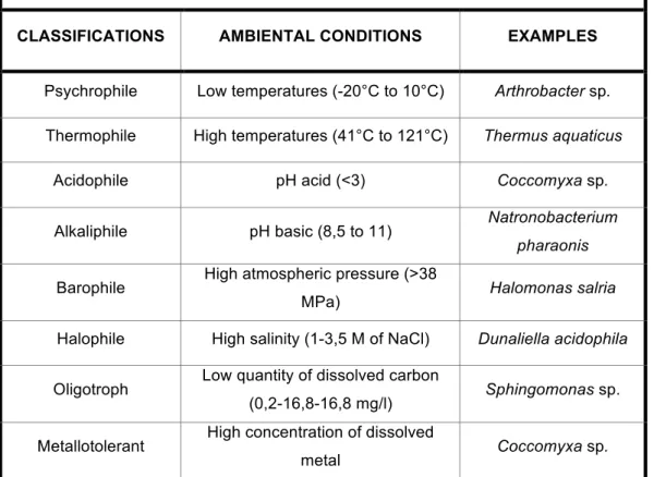
Table 1: unicellular extremophiles classified in several groups based on the type of environment and relative examples.
CLASSIFICATIONS AMBIENTAL CONDITIONS EXAMPLES
Psychrophile Low temperatures (-20°C to 10°C) Arthrobacter sp.
Thermophile High temperatures (41°C to 121°C) Thermus aquaticus
Acidophile pH acid (<3) Coccomyxa sp.
Alkaliphile pH basic (8,5 to 11) Natronobacterium
pharaonis
Barophile High atmospheric pressure (>38
MPa) Halomonas salria
Halophile High salinity (1-3,5 M of NaCl) Dunaliella acidophila
Oligotroph Low quantity of dissolved carbon
(0,2-16,8-16,8 mg/l) Sphingomonas sp.
Metallotolerant High concentration of dissolved
conditions. These classifications are not exclusive. Many extremophiles fall under multiple categories and are classified as polyextremophiles. For example, organisms living inside hot rocks deep under Earth's surface are thermophilic and barophilic such as Thermococcus barophilus (Marteinsson et
al., 1999). A polyextremophile living at the summit of a mountain in the
Atacama Desert might be a radioresistant xerophile, a psychrophile, and an oligotroph. Polyextremophiles are well known for their ability to tolerate both high and low pH levels. The unicellular extremophiles can be classified in several groups based on the type of environment (Madigan and Oren, 1999; Satyanarayana et al., 2005) (tab. 1).
1.2.2. Adaptation to low pH and high concentrations of metals
The microalgae living in acidic environments have had to adapt to this extreme condition. One of the main factors is the low pH due to a high concentration of protons (H+). The plasma membrane of microalgae is not very permeable to the passage of H+ from the external environment to the intracellular fluid, so the cell does not consume a lot of energy to prevent proton entry, the impermeability of the membrane may be due to the presence of sterols, fatty acids and proteins (Gross, 2000; Brake and Hasiotis, 2010). The microalgae also have the capacity to regulate intracellular pH through ionic transporters present in the cell membrane, such as for example, H+ pump (Messelerli et al., 2005). The mine acid waters possess a low concentration of dissolved phosphate due to the presence of aluminum, phosphate is an essential element for the functioning of each type of microalgae, so they have developed the ability to produce phosphatases resistant to an acid environment (Gross, 2000).
Another important factor that microalgae must overcome is the presence of metals such as copper, lead, zinc, aluminum. These metals induce the oxidation of the plasma membrane and possible rupture, in addition to the denaturation of the proteins, blocking of functional groups or decompositions of essential metabolites (Brake and Hasiotis, 2010).
In order to tolerate the high concentration of metals in the water, microalgae must adopt different strategies such as:
- Reduction of the entry of metals or increasing their flow.
- Mechanism of complexation of metals, with precipitation on the extracellular side.
- Grouping in essential biofilms (Brake and Hasiotis, 2010).
1.2.3. Coccomyxa
The microalgae Coccomyxa sp. studied in this work, belongs to the family of
Coccomyxaceae (Eucaryota; Viridiplantae; Clorophyta; Trebouxiophyceae), it
is a polyextremophile isolated in the acid waters of mine S. Domingos (Valente et al., 2013). It includes more than 30 species found in both marine and fresh water, are epiphytic species, symbiotic with lichen, bark and even protozoa parasites. They are unicellular polyextremophiles (acidophilic, metallotolerant) microalgae, able to tolerate low pH levels and high concentration of dissolved metals.
2. The mine of S. Domingos
On Earth there are many acidic environments, generated by geological and anthropogenic activities through the leakage of acid water from mines, which combine low pH to high concentrations of metals. The intense mining activity produced considerable amount of residues, which have caused the environmental deterioration of the zone in all its aspects: soil degradation, water resources pollution, biodiversity
decrease, and even, atmospheric pollution in some moments of the history.
The most important environmental problem derives from the sulphide (mainly
Pyrite) oxidation contained in the aforementioned residues. This process produces an extremely acid leachate with high contents of sulphate, metals and metalloids known as acid mine
Figure 1: geographic position of the
Iberian Pyrite Belt and related main mining district.
drainage (Lowson, 1982; Parker and Robertson, 1999; Younger et al., 2002). The mine of S. Domingos is located in the northern sector of the Iberian Pyrite Belt, about 5 km from the Spanish border (fig. 1). It’s one of the most emblematic Portuguese massive sulphide deposits and one of the world's largest reserves of mineral sulfur. Although mining activity has ceased at present, the large-scale exploitation of this deposit between 1857 and 1966 favored the production of enormous waste dumps, where oxidation of pyrite and associated sulphide is resulting in the acid mine drainage production. The final acid discharge with high contents of metals, as copper, lead, zinc, with amounts of copper which can reach a maximum of 1468 mg/Kg, and low pH (<3), from S. Domingos reaches the Chanza river, main effluent of Guadiana river, causing its partial pollution (Pinho et al., 2014).
The field-bearing secondary minerals within waste deposits indicate that these materials are reactive. Jarosite and other secondary low crystalline minerals as oxides, ox-hydroxides, and iron hydroxyl-sulphates are important for the environment as they play a crucial role in the solubility of the potentially toxic chemical elements (Alvarez-Valero et al., 2007).
3. Fundulus heteroclitus
The Fundulus heteroclitus, is an exceptionally wide ranging cyprinodont fish. It naturally occurs along the east coast of North America from southwestern Newfoundland to northeastern Florida. This species is ubiquitous in North American East Coast salt marshes, being mostly found in sheltered coastal waters. Although occasionally it inhabits freshwater habitats, the species is best known from the tidal salt marsh, a fluctuating physical environment for which they are well adapted due to considerable plasticity in their ecological requirements (Kneib, 1986).
As adults, Fundulus heteroclitus range between 12.7 and 17.8 centimeters in length, the females growing larger than the males. They have flattened heads and the mouth is turned upward, clearly an adaptation to feeding at the surface of the water. This attractive fish is dimorphic, males are darker in color than females and exhibit blue or orange markings during the breeding season, are dark olive green on the dorsal side and lighter yellow on the ventral side. They also display vertical stripes along their sides. Females are silvery yellow
on the ventral side and that color gradually fades to a more distinct yellow on the dorsal side. All Fundulus heteroclitus have a single soft dorsal fin and their pelvic fins are located close to the rear fin(National Oceanic and Atmospheric Administration Coastal Services Center, 2001).
Fundulus heteroclitus is the premier model for environmental biology (Burnett
et al., 2007). The inner opercular epithelium of the killifish Fundulus
heteroclitus is rich in chloride cells and provides a proxy model system to
study chloride cell function (Karnaky et al., 1977). When the membrane is removed and mounted in Ussing chambers, the short circuit current is equivalent to Cl− secretion rates (Degnan et al., 1977)
4. Objectives
The main objectives of the present work was to evaluate the potential bioactivity in an ex-vivo animal model (Fundulus heteroclitus), and expression on selected genes, of cellular extracts obtained from cultures of Coccomyxa sp.at pH 7 without or with exposure to copper (0.6mM Cu2+). The work was developed into three main parts:
• Establishment of unialgal culture of Coccomyxa sp. strain VAL007 (chapter I);
• Functional characterization of Coccomyxa sp. extracts on animal model
ex-vivo Fundulus heteroclitus through epithelial electrophysiology
techniques (chapter II);
• Opercular tissue of Fundulus was cultured in vitro in the presence of microalgae and the expression of NKCC1, NKCC2 and CFTR was analyzed (chapter III).
II. ESTABLISHMENT OF UNIALGAL CULTURES OF Coccomyxa sp. STRAIN VAL007
1. Coccomyxa sp. strain VAL 007
The microalgae Coccomyxa sp. strain VAL007 studied in this work, belongs to the family Coccomyxaceae (Eucaryota; Viridiplantae; Clorophyta; Trebouxiophyceae). It is a unicellular polyextremophile isolated from the acid
waters of the S. Domingos mine (Valente et al., 2013). The genus Coccomyxa includes more than 30 species found in both marine and fresh water. These can be epiphytic species, symbiotic with lichen and tree bark and even protozoa parasites. Some of the species are polyextremophiles (acidophile, metallotolerant) microalgae, able to tolerate low pH levels and high concentration of dissolved metal (Vaquero et al., 2012; Leonardo et al., 2016). Most of the published studies have focused on the characterization of the physiological adaptations to these conditions, the optimization of culture conditions for the production of metabolites of interest and use in bioremediation technology of environments contaminated with metals or radioisotopes (Leonardo et al., 2016). As a way to test the possible application in human medicine, some studies have used extracts of these microorganisms and analyzed their effects in vitro and in vivo. A recent study by Komatsu et al. (2013) evaluated the antiviral activity of Coccomyxa
gloeobotrydiformi on cells infected by the human influenza virus A and
another study by Sun et al., (2013), using rats have as the classic model of myocardial occlusion of the middle cerebral artery, observed the therapeutic effects of Coccomyxa gloeobotrydiformis on the recovery from ischemic stroke.
2. Materials and Methods
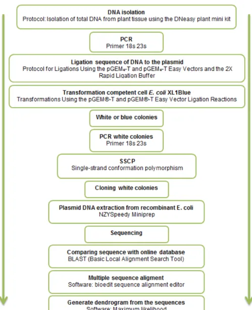
2.1. Verification of unialgal cultures of Coccomyxa sp.
To verify that the starting cultures consisted only of Coccomyxa sp., isolated and maintained in the Laboratory of Microbial Molecular Ecology (UAlg-CIMA), the following steps were performed (fig. 2). Three samples of 2 ml each were collected from each starting culture and the cells recovered after
centrifugation at 3500 g for 1 minute. The culture medium was discarded and the pellet was used for DNA extraction.
2.2. DNA isolation
Protocol: Isolation of total DNA from plant tissue using the DNeasy plant mini kit. (QUIAGEN Companies, DNeasy Plant Mini Kit and DNeasy Plant Maxi Kit Handbook 07/2003).
1. Grind plant tissue under liquid nitrogen to a fine powder using a mortar and pestle. Transfer the tissue powder and liquid nitrogen to an appropriately sized tube and allow the liquid nitrogen to evaporate. Do not allow the sample to thaw. Continue immediately with step 2.
Figure 2: sequence of steps to verify the unialgal state of the starting
2. Add 400 µl Buffer AP1 and 4 µl RNase A stock solution (100 mg/ml) to a maximum of 100 mg disrupted plant and vortex vigorously.
3. Incubate the mixture for 10 min at 65°C. Mix 2 or 3 times during incubation by inverting tube.
4. Add 130 µl Buffer AP2 to the lysate, mix, and incubate for 5 min on ice. Recommended: Centrifuge the lysate for 5 min at 14,000 rpm.
5. Pipet the lysate into the QIAshredder Mini spin column (lilac) placed in a 2 ml collection tube, and centrifuge for 2 min at 14,000 rpm.
6. Transfer the flow-through fraction from step 5 into a new tube (not supplied) without disturbing the cell-debris pellet.
7. Add 1.5 volumes of Buffer AP3/E to the cleared lysate, and mix by pipetting.
8. Pipet 650 µl of the mixture from step 7, including any precipitate that may have formed, into the DNeasy Mini spin column placed in a 2 ml collection tube (supplied). Centrifuge for 1 min at ≥8000 rpm, and discard the flow-through. Reuse the collection tube in step 9.
9. Repeat step 8 with remaining sample. Discard flow-through and collection tube.
10. Place the DNeasy Mini spin column into a new 2 ml collection tube (supplied), add 500 µl Buffer AW2, and centrifuge for 1 min at ≥8000 rpm. Discard the flow-through and reuse the collection tube in step 11.
11. Add 500 µl Buffer AW to the DNeasy Mini spin column, and centrifuge for 2 min at 14,000 rpm to dry the membrane.
12. Transfer the DNeasy Mini spin column to a 1.5 ml or 2 ml microcentrifuge tube (not supplied), and pipet 100 µl Buffer AE directly onto the DNeasy membrane. Incubate for 5 min at room temperature (15–25°C), and then centrifuge for 1 min at ≥8000 rpm to elute.
13. Repeat step 12 once.
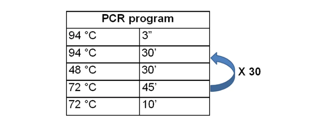
2.3. PCR
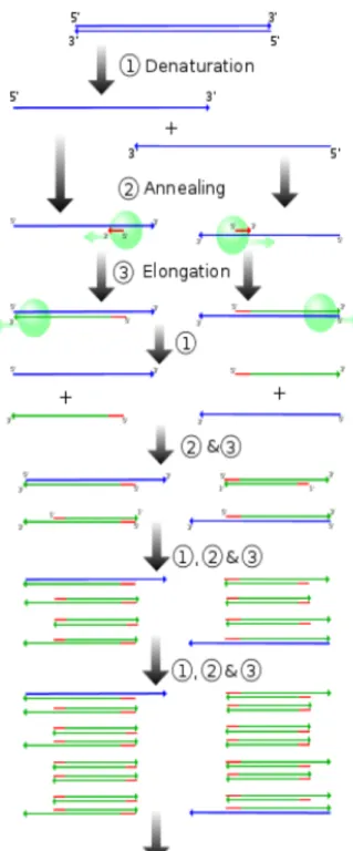
The DNA samples obtained were subjected to PCR (polymerase chain reaction) amplification, using universal primers for the 18S gene (nuclear genome) and the 23S gene (chloroplastidial genome). The PCR is a technique for amplification of DNA in vitro using thermostable DNA
polymerases. It involves repetitive cycling of three steps: denaturation, annealing and elongation (fig.3). The entire cycling process of PCR is automated and can be completed in just a few hours using a thermal cycler (tab.
2).
Table 2: PCR program used for the PCR amplification of a fragment of the 18S gene or the 23S gene.
Figure 3: sequence of PCR steps:
1- Denaturation step: This step consists of heating the reaction to 90–95 °C. It causes DNA separation by disrupting the hydrogen bonds between complementary bases, yielding single strands of DNA.
2- Annealing step: The reaction temperature is lowered to 50–65 °C allowing hybridization of the primers to the single-stranded DNA template.
3- Elongation step: At this step, the Taq polymerase synthesizes a new DNA strand complementary to the DNA template strand by adding dNTPs that are complementary to the template in 5' to 3' direction.
The PCR technique requires the following components and reagents:
- Primers (Forward & Reverse) (tab. 3): short pieces of artificially prepared DNA that will target the gene fragment of interest in the entire genome. They
are complementary to the 3’ ends of each strand of the double stranded target gene i.e. DNA.
Table 3: Primer pairs used in this work, selected to amplify two regions with phylogenetic information to molecularly identify phototrophic eukaryotes: 18S gene for 18SrRNA (nuclear DNA) and 23S gene for 23S rRNA (chloroplastidial DNA).
Gene Primer Sequence 5’→3’ bp References
18s 18s_f GTCAGGGTGAAATTCTTGGATTTA 700 Rasoul-Amini et al., 2009 18s_r AGGGCAGGGACGTAATCAACG 23SrV 23SrV GGACAGAAAGACCCTATGAA 410 Sherwood and Presting, 2007 23SrV TCAGCCTGTTATCCCTAGAG
- Taq polymerase (DNA polymerase): It’s an enzyme whose function is to extend the new DNA strand. Taq polymerase attaches near the 3’ end of the primer and starts adding nucleotides. It requires double stranded DNA to become functional. The DNA polymerase in our bodies breaks down at temperatures below 95 °C, the temperature necessary to separate two complementary strands of DNA in a test tube.
- Deoxynucleotide triphosphate (dNTP’s): They are the building blocks from which the DNA polymerases synthesizes a new DNA strand, Taq polymerase grabs nucleotides that are floating in the liquid around it and attaches them to the 3’ end of a primer.
PCR reactions were done in a final volume of 50 µl (45 µl of PCR reaction components and 5 µl of DNA sample) (tab. 4).
2.4. Ligation of DNA fragments to the plasmid-vector
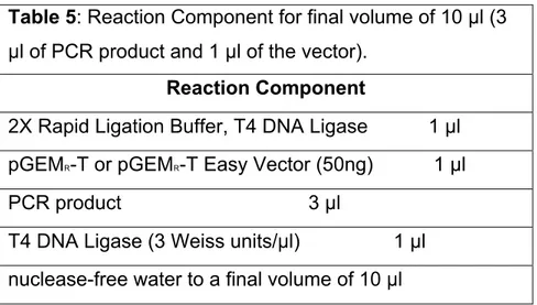
The results of each PCR amplification were visualized under UV light, after electrophoresis on agarose gels (1% in buffer TAE) stained with ethidium bromide. The fragments of the expected size (700 bp for 18S and 410 bp for 23S) were ligated to the plasmid-vector following the Protocol for Ligations Using the pGEM®-T Easy Vector and the 2X Rapid Ligation Buffer (tab. 5).
Table 5: Reaction Component for final volume of 10 µl (3 µl of PCR product and 1 µl of the vector).
Reaction Component
2X Rapid Ligation Buffer, T4 DNA Ligase 1 µl pGEMR-T or pGEMR-T Easy Vector (50ng) 1 µl PCR product 3 µl
T4 DNA Ligase (3 Weiss units/µl) 1 µl nuclease-free water to a final volume of 10 µl
The reactions were mixed by pipetting and incubated for 1 hour at room temperature.
Table 4: final concentration of PCR reaction components in a final volume of 50 µl (45 µl of PCR reaction components and 5 µl of DNA sample).
PCR reaction components
H2O sterile(DNase and RNase free, Sigma-Aldrich, USA) Taq Buffer MgCl2 0,2 mM dNTPs 0,2 µM Primer Fw 0,2 µM Primer Rv 0,2 µM Taq 1U
2.5. Transformation of competent cells - E. coli XL1Blue
The constructs obtained were used to transform E. coli strain XL1Blue competent cells, prepared in the lab and kept at -80°C.
Transformation protocol:
1. Prepare two LB-AGAR/ampicillin/IPTG/X-Gal plates for each transformation,
2. Add 2µl of each ligation reaction to an aliquot of 100 µl of competent cells defrosted and kept on ice. Gently flick the tubes to mix and place them on ice for 30 minutes.
3. Heat-shock the cells for 45–50 seconds in a water bath at exactly 42°C (do not shake).
4. Immediately return the tubes to ice for 2 minutes.
5. Add 250µl room temperature SOC medium (LB liquid with glucose 20 mM) to the tubes and incubate for 45 minutes at 37°C with shaking (~200rpm). 6. Plate 150µl of each transformation culture onto duplicate LB-AGAR/ampicillin/IPTG/X-Gal plates.
7. Incubate the plates overnight (16–24 hours) at 37°C. White colonies will contain the desired inserts.
2.6. Verification of inserts in white colonies
After the cultivation of the transformed cells was carried out, white colonies on each plate were subjected to a PCR using the same primer pair (tab. 3) with which the inserted fragment had been obtained, and the same reaction components (tab. 4). Size of the obtained fragments was verified after electrophoresis on agarose gels (1% agarose in TAE), stained with ethidium bromide and under UV light.
2.7. SSCP (single strand conformation polymorphism) analysis
In order to select recombinant clones for sequencing a single strand conformation polymorphism analysis (SSCP, Orita et al., 1989) was conducted. This analysis allows the detection of a SNP (single nucleotide polymorphism) in single strand sequences of up to 700 nt. For this the double stranded DNA is initially denatured and subjected to electrophoresis in
polyacrylamide gels. Polymorphisms between sequences of the same size are detected as different band patterns on the gels after staining, usually with silver nitrate.
For this analysis, 1 µl of the PCR product was added to 9 µl of denaturing buffer (95% formamide, 20 mM EDTA pH 8.0, stained with bromophenol blue), denaturated at 90 °C for 5 min and placed on ice. The denatured DNA was subjected to electrophoresis in 8% polyacrylamide gels (1X TBE buffer), conducted at 4 °C and 200 V, during 2 hours. After electrophoresis, the polyacrylamide gels were stained with silver nitrate. For this the gels were initially placed in a solution of 10% glacial acid acetic during 20 minutes (fixative) after which they were washed 3 times for 3 minutes with distilled water. Next, the gels were passed into a 1% nitric acid solution for 3 minutes, followed by 3 times 3 minute washes with distilled water. The gels were then immersed in a silver nitrate solution (1g/L AgNO3, 1,5 ml/L formaldehyde) during 30 minutes, after which they were washed with distilled water for 3 times and revealed with a sodium carbonate solution (30 g/L Na2CO3, 1,5 ml/L formaldehyde, 2 mg/ml NaS2O3 + 5H2O). The reaction was stopped placing the gels in the initial acetic solution.
Recombinant clones evidencing different SSCP patterns, were selected for plasmid extraction.
2.8. Plasmid DNA extraction from recombinant E. coli colonies
The colonies of interest were grown over night in LB liquid with ampicillin. Plasmid DNA extraction was carried out using the NZYMiniprep kit (NZYTech, Portugal).
Protocol for plasmid DNA purification (miniprep) from E. coli cells
All centrifugations were carried out at room temperature in a tabletop microcentrifuge at 10000-15000 rpm.
1. Harvest bacterial cells: Pellet 1-5 ml of an E. coli LB culture for 30 s. Discard supernatant. Remove as much media as possible.
2. Cell lysis: Re-suspend cell pellet completely in 150 µl of Buffer A1 by vigorous vortexing/pipetting. Add 250 µl of Buffer A2 and mix gently by inverting the tube for 5 times. Incubate at room temperature (10-25 ºC) for up
to 2 min. Do not vortex. Add 300 µl of Buffer A3. Mix gently by inverting the tube for 6-8 times. Do not vortex.
3. Clarification of lysate: Centrifuge for 3 min at room temperature to pellet precipitate.
4. Bind DNA: Place NZYTech spin column in a 2 ml collecting tube and load the supernatant from step 3 onto the column. Centrifuge for 30 s. Discard flow-through.
5. Wash silica membrane: Add 600 µl of Buffer AS (make sure ethanol was previously added). Centrifuge for 1 min. Discard flow-through.
6. Dry silica membrane: Re-insert the NZYTech spin column into the empty 2 ml collecting tube and centrifuge for 1 min.
7. Elute highly pure DNA: Place the dried NZYTech spin column into a clean 1.5 ml microcentrifuge tube and add 50 µl of Buffer AE. Incubate for 1 min at room temperature (10-25 ºC). Centrifuge for 1 minute and store the purified DNA at -20 °C.
2.9. Sequencing
Sequences were obtained through commercial sequencing (CCMar, Faro, Portugal) by the Sanger Method.
2.10. BLAST analysis and phylogenetic inference
The sequences obtained were subjected to a BLAST analysis at the NCBI (National Center for Biotechnology Information) site (blast.ncbi.nlm.nih.gov). This software makes a search per similarity searches and compares the submitted sequences with sequences from cultured organisms present in the database, thus allowing the identification of the organisms present in the starting cultures.
The homologous sequences (at least 98% homology) were retrieved from the database and aligned with the sequences from this study, using BioEdit (Hall, 1999) and ClustalW (Thompson et al., 1994) softwares. The dendrograms for phylogenetic inference were constructed in MEGA5 (Tamura et al., 2011) and the Maximum Likelihood Method with 1000 bootstraps.
3. Results
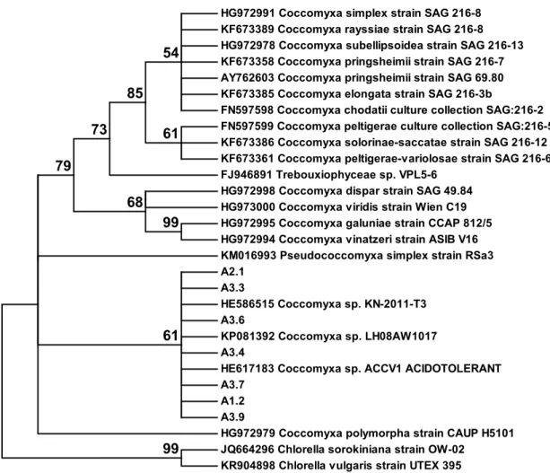
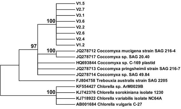
From the SSCP analysis 7 different patterns were obtained for the cloned fragments of the 18S gene. The dendrogram obtained with the corresponding sequences and homologous sequences retrieved from the Genbank database (NCBI), including sequences from Chlorella as anoutgroup (fig. 4), revealed that all sequences from this study are closely related to sequences obtained from Coccomyxa species. No sequence from another organism was obtained. The same was observed with the 8 sequences obtained for the 23S gene fragment (fig. 5).
HG972991 Coccomyxa simplex strain SAG 216-8 KF673389 Coccomyxa rayssiae strain SAG 216-8 HG972978 Coccomyxa subellipsoidea strain SAG 216-13 KF673358 Coccomyxa pringsheimii strain SAG 216-7 AY762603 Coccomyxa pringsheimii strain SAG 69.80 KF673385 Coccomyxa elongata strain SAG 216-3b
FN597598 Coccomyxa chodatii culture collection SAG:216-2 FN597599 Coccomyxa peltigerae culture collection SAG:216-5 KF673386 Coccomyxa solorinae-saccatae strain SAG 216-12 KF673361 Coccomyxa peltigerae-variolosae strain SAG 216-6 FJ946891 Trebouxiophyceae sp. VPL5-6
HG972998 Coccomyxa dispar strain SAG 49.84 HG973000 Coccomyxa viridis strain Wien C19 HG972995 Coccomyxa galuniae strain CCAP 812/5 HG972994 Coccomyxa vinatzeri strain ASIB V16 KM016993 Pseudococcomyxa simplex strain RSa3 A2.1 A3.3 HE586515 Coccomyxa sp. KN-2011-T3 A3.6 KP081392 Coccomyxa sp. LH08AW1017 A3.4
HE617183 Coccomyxa sp. ACCV1 ACIDOTOLERANT A3.7
A1.2 A3.9
HG972979 Coccomyxa polymorpha strain CAUP H5101 JQ664296 Chlorella sorokiniana strain OW-02
KR904898 Chlorella vulgaris strain UTEX 395 99 99 68 61 54 85 73 79 61
Figure 4 : Dendrogram (Maximum Likelihood method) obtained based on the alignment of
partial 18S sequences obtained in this work (A1, A2, A3) and sequences retrieved from GenBank, showing at least 95% homology in a BLAST analysis. Bootstrap values are shown next to each node.
V1.5 V2.7 V3.1 V3.6 V2.2 V2.6 V2.4 V1.2
JQ278712 Coccomyxa mucigena strain SAG 216-4 JQ278717 Coccomyxa sp. SAG 20.40
HQ693844 Coccomyxa sp. C-169 plastid
JQ278713 Coccomyxa pringsheimii strain SAG 216-7 JQ278714 Coccomyxa sp. SAG 49.84
FJ804758 Trebouxia australis strain SAG 2205 KF554427 Chlorella sp. ArM0029B
KJ742376 Chlorella sorokiniana isolate 1230 KJ718922 Chlorella variabilis isolate NC64A AB001684 Chlorella vulgaris C-27
100
100 97
100
Figure 5: Dendrogram (Maximum Likelihood method) obtained based on the alignment of
partial 23S sequences obtained in this work (V1, V2, V3) and sequences retrieved from GenBank, showing at least 95% homology in a BLAST analysis. Bootstrap values are shown next to each node.
III. FUNCTIONAL CHARACTERIZATION OF Coccomyxa sp. EXTRACTS 1. Material and Methods
1.1. Preparation of Coccomyxa sp. cultures
When the study began, the starting cultures of Coccomyxa sp. were being maintained in growth medium K9 (tab. 6) at pH 2,5 in cell culture flasks with a lid fitted with a 0.2 µm filter (to promote the exchange of gases and prevent contamination with microorganisms) at 24 °C temperature and 50 µmol/m2/s light intensity at 12 hours daily. This microalgae is not mobile in the liquid column and forms a film at the bottom of the culture flasks.
Table 6: Growth medium K9 composition
CaCl2 + 2H2O 0,1 mM MgCl2 + 6H2O 2mM KCl 1mM K3PO4 2mM KNO3 20mM K2SO4 20 mM
Solution of the following elements 5 ml/l
H3BO3 70 mM C10H16N2O8 30 mM CoCl2 + 6H2O 3mM MnCl2 · 2H2O 10mM (NH4)6Mo7O24 + 4H2O 0,4mM CuSO4 + 5H2O 3 mM FeSO4 + 7H2O 10mM ZnSO4 + 5H2O 30 mM
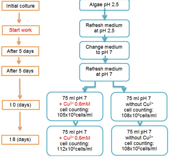
According to results of previous studies (Bras, 2015), the experiment was set up to obtain extracts of this microalgae from cultures at pH 7 without addition of copper (Cu2+)and with addition of Cu2+ at a final concentration of 0.6 mM. At the beginning of this work the growth medium of the starting culture was removed and new growth medium at pH 2.5 was added. The cells were gently resuspended and samples were taken for cell counting with the Bϋrker
chamber. After 5 days the growth medium was removed and new medium was added at pH 7. The culture was left to stabilize for 5 days, after which time samples were taken for cell counting and the final volume of growth medium was adjusted to approx. 100 x 106 cells/ml. Following this, two times 75 ml of culture were taken into separate clean cell culture flasks, to one of which 0,375 ml of copper sulphate (CuSO4) at 120Mm was added, to obtain a final concentration of 0.6 mM in the growth medium. Microalgae samples were taken 8 days after, for extract preparation. The following figure (fig. 6) shows the scheme of the experimental procedure for obtaining each type of culture.
1.2. Determination of culture cell density
The cell density was determined using the Bϋrker chamber (fig. 7) with the use of an optical microscope with incorporated camera (Leica DFC480, Leica Figure 6: scheme of the experimental procedure for obtaining the extracts after 8 days (t8)
Microsystem, Wetzalar, Germany). Prior to counting the algal samples in were diluted 1:100. From this dilution 10 µl were taken and loaded into the Bϋrker chamber. Each sample was counted twice.
After counting the number of cells the following formula was applied to obtain the number of cells per milliliter.
!° !"##$ !" = !""# !" × !"#! !!!° !"##$! × !° !"#$%! ×!"#$%"&' × 10!"
Figure 7: Representation of the Bϋrker chamber. The chamber depth is 0.1 mm. The ruling
shows 9 large squares of 1 mm2 each. The large squares are subdivided into 16 group
squares with 0.2 mm sides. In the central large square, each group square is subdivided into 16 mini squares with 0.05 mm sides (= 0.0025 mm2). To perform the counting were
1.3. Preparation of cell extracts
For the preparation of the microalgae extracts, from the culture at pH 7 and the culture at pH7 with 0.6 mM of Cu2+, samples of 1.8 ml were collected into 2ml Eppendorf tubes and centrifuged at 3500 x g for 1 minute. The growth medium was discarded and 1 ml of phosphate buffered saline (PBS 1 x) was added to wash the cells, followed by centrifugation at 3500 x g for 1 minute. The PBS was discarded and the precipitated cells were stored at -20°C. A total of 48 vials, 24 with microalgae from the culture with addition of Cu2+ and 24 with microalgae from the culture without Cu2+ were obtained. Prior to testing, crude cell extracts were prepared by mechanical homogenization according to the method described below.
Procedure to obtain an extract from cells precipitated from in 1.8 ml of culture: add 600 microliters of Fundulus saline (tab. 7), vortex, take all the contents and put into a 1,5 ml vial with 6 ceramic beads. Insert the tube into the homogenizer at 6800 rpm for 3 cycles of 30 seconds each, freeze the extract at -20 °C. The extracts prepared this way were subsequently used for electrophysiology and molecular biology studies with the opercular epithelium of Fundulus heteroclitus. The precipitates homogenized with Fundulus saline had a final concentration of 106 cell/mL-1.
1.4. Animal model Fundulus heteroclitus
The Fundulus heteroclitus, is an exceptionally wide ranging cyprinodontid fish. It naturally occurs along the east coast of North America from southwestern Newfoundland to northeastern Florida. This species is ubiquitous in North American East Coast salt marshes, being mostly found in sheltered coastal waters. Although occasionally it inhabits freshwater habitats, the species is best known from the tidal salt marsh, a fluctuating physical environment for which they are well adapted due to considerable plasticity in their ecological requirements (Kneib, 1986). As adults, Fundulus heteroclitus range between 12.7 and 17.8 centimeters in length, the females growing larger than the males. They have flattened heads and the mouth is turned upward, clearly an adaptation to feeding at the surface of the water. This attractive fish is
dimorphic, males are darker in color than the females and exhibit blue or orange markings during the breeding season,are dark olive green on the dorsal side and lighter yellow on the ventral side. They also display vertical stripes along their sides. Females are silvery yellow on the ventral side and that color gradually fades to a more distinct yellow on the dorsal side.All
Fundulus heteroclitus have a single soft dorsal fin and their pelvic fins are
located close to the rear fin(National Oceanic and Atmospheric Administration Coastal Services Center, 2001). The first records of Fundulus heteroclitus in the Iberian Peninsula are dated between 1973-1976 (Hernando, 1975; Coelho
et al.,1976). However, the precise date and location of the species’
introduction still remains unclear. Although there have been studies describing its life-history pattern (Arias & Drake, 1986; Drake et al., 1987; Fernández-Delgado, 1989) and density (Arias & Drake, 1987, 1989) little is known about the role of F. heteroclitus in this new European habitat. The introduction of exotic species can have a negative effect on the functioning of native ecosystems (Dowling & Childs, 1992; Barlow et al., 1987; Richardson & Whoriskey, 1992). It is suspected that F. heteroclitus may have already negatively affected some native endemic species like the endangered Lebias
ibera. This introduced species was more or less continuously distributed along
the Atlantic coast of Spain, being more abundant in sites near the coastline (usually < 10 km inland), mainly in extensive marshes. The species preferred marsh related meso-habitats, such as salt lagoons, salt marsh fishponds and marsh channels, both natural and man modified. F. heteroclitus was mostly found at salinities > 25.
1.4.1. Osmoregulation
In the gills of Fundulus heteroclitus, as in all seawater teleost, the basis for ion secretion is attributable, in large part, to the concerted effort of three major ion-transport proteins: the basolateral Na+-K+-ATPase (NKA), the basolateral Na+-K+-2Cl- co-transporter (NKCC1) and the apical cystic fibrosis transmembrane conductance regulator (CFTR) (fig. 8). The primary driving force for ion secretion in ionocytes is the NKA, which maintains the low intracellular Na+ required for secondary import of Na+, K+ and two Cl– ions by the NKCC1. Elevated intracellular Cl– is subsequently extruded through an
apical anion channel, CFTR. Intracellular Na+ is cycled back out of the cell by the basolateral NKA; Na+ ions are then secreted down an apical electrical gradient generated by CFTR, into the environment though shallow tight junctions between ionocytes and accessory cells (McCormick, 1995; Evans et
al., 2005; Marshall and Grosell, 2006; Hwang and Lee, 2007).
1.4.2. Opercular membrane
As in gill tissue in the opercular membrane numerous mitochondria-rich chloride cells are present. The opercular membrane is the inner (bucal) lining of the opercular bone, it is of special interest to fish physiologists because it is an area of scale less skin and unlike the gill tissue, the opercular membrane is a flat epithelium, making the chloride cells more accessible for morphological examination and the study of their ion transport properties. The presence of chloride cells in the opercular epithelium was first observed by Burns and Copeland (1950) in seawater-adapted Fundulus heteroclitus. Degnan et al. Figure 8: Working model for the extrusion of NaCl by the marine teleost gill epithelium.
Plasma Na+, K+, and Cl− enter the cell via basolateral NKCC; Na+ is recycled back to the plasma via Na+-K+-ATPase and K+ via a K+ channel (Kir). Cl− is extruded across the apical membrane via a Cl− channel (CFTR). The transepithelial electrical potential across the gill epithelium (plasma positive to seawater) drives Na+ across the leaky tight junctions between the MRC (mitochondria-rich cells) and the AC (Accessory Cells)
(1997) and Karnaky et al. (1977) mounted the Fundulus heteroclitus opercular membrane in an Ussing chamber and found net chloride secretion; since then the opercular membrane has been widely used to examine the mechanisms and control of ion secretion in teleosts.
1.4.3. Experimental procedures
The Fundulus heteroclitus were collected with fishing nets in the Estero La Leocadia (Cadiz Bay, Spain) were kept in the aquarium with water at a temperature of 21 °C, salinity of 35 ‰ and exposed to a period of 14h of artificial light per day with a luminous intensity of 50 µmol/m²/s. To obtain the opercular tissue the fish were immersed in a beaker with anesthetic 2-Phenoxyethanol (1: 2000) and later beheaded. The head was placed in a beaker with saline solution (tab: 7) and insufflation gas with ratio 99.7:0.3 of O2/CO2, to maintain the tissues alive. All animal manipulations were carried out in compliance with the Guidelines of the European Union Council (86/609/EU) and Portuguese legislation for the use of laboratory animals. All animal protocols were performed under a Group-C license from the Direcção-Geral de Veterinária, Ministerio da Agricultura, do Desenvolvimento Rural e das Pescas, Portugal.
The opercular membrane is removed using tweezers, scalpel, and scissors, under the microscope and stretched over a 0.1-0.3 cm hole in the center of a rectangular piece of Plexiglas (mounting plate). Small pins several mm
Table 7: constituents of Ussing chamber saline solution (pH=8) NaCl 160 mM MgS04 0.93 mM Na2HPO4 3 mM CaCl2 1.5 mM NaHCO3 5 mM KCl 3 mM Glucose 5.5 Mm
outside the hole are used to maintain the membrane flat and stretch it to its original (undissected) dimensions. A circular mounting disc with a central hole (0,126 cm2, same size as that of the mounting plate) and small holes to accommodate the mounting pins is placed over the membrane. Modest amounts of silicone grease are placed on the inner edges of the mounting plate and disk that contact the membrane, prior to placement of the membrane. The mounting plate and disc are then placed into receiving grooves to separate the two halves of the Ussing chamber (fig: 9) which contain 2 ml of saline solution (tab: 7). The salt solutions present in the two halves are separated from each other only by the opercular epithelium. Each chamber is gently bubbled with a gas mixture (O2/CO2 = 99.7:0.3) to maintain oxygen tension and fluid mixture. The chambers are mounted in supports in contact with a liquid coolant which keeps the temperature constant at 21-22 °C. Within this system the tissue can remain alive for 4-5 hours.
The Cl– transport from basolateral to the apical side can be measured through "voltage clamp" to 0 mV, by “short circuit current”, Isc (Hoffmann et al., 2002). The short-circuit current is measured with paired polyethylene 3% agar-3M KCl bridges (two electrodes, fig. 9) each halve) placed 2-3 mm from each side of the membrane, which terminate in 3 M KCl and are in turn connected to a voltmeter by Ag/AgCl electrodes. Current may be passed across the membrane via agar bridges in each chamber that are connected to Ag/AgCl electrodes and a current clamping device (Fuentes et al., 2006). The "voltage clamp" method has its basis on the Ohm law (R=V/I, where R is the Resistance, V the Voltage and I the Current) considering constant the resistance. Thus it was obtained the value of current required to be injected
Figure 9: example of electrode (A), and insert (B) used in the Ussing chamber (C).
so as to clamp the voltage to 0 mV. Isc is directly proportional measurement of the ionic transport, as well as, the secretory tissue activity. The voltage clamp to 0 mV and the current injection were generated by voltage current clamp amplifiers with automatic correction for fluid resistance and voltage asymmetry. DVC-1000 (World Precision Instrument, Sarasota, USA) or VCC600 (Physiologic Instruments, San Diego, USA)
1.4.4. Bioactivity test
When the Isc of the tissue inside the insert was stable, injecting the extracts (previously prepared and stored at -20 °C, section 2.4.) in the saline solution. Each application was performed after the stabilization of the tissue.
The biometric parameters were recorded and saved to a computer via a data acquisition system Lab-Trax-4 (World Precision Instrument, Sarasota, USA). All data obtained were subjected to statistical analysis using the software Prism 5 (GraphPad Software, La Jolla, California, USA). The statistical significance was determined as adequate by two- way ANOVA or Student t-test, considering p values < 0,05.
2. Results
2.1. Basal values
Before adding the cell extracts into the Ussing chamber the basal values of the tissues were recorded (tab. 8). The results presented in Table 8 confirm that in these experimental conditions the Isc is a direct measure of the Cl– ion flow through the opercular epithelium.
2.2. Extracts added to the basolateral side
The extracts of microalgae, with the addition of Cu2+ (Fig. 10, Fig. 12A) and without (Fig. 11, Fig. 12B), were added in increasing concentrations (4×10⁶ cell/ml; 8×10⁶ cell/ml; 16×106 cell/ml; 32×106 cell/ml; 64×106 cell/ml) in the basolateral side in opercular epithelium of Fundulus heteroclitus mounted in the Ussing chamber in voltage clamp. The addition of extracts of Coccomyxa exposed to copper caused a pronounced decrease of Isc coming to inhibit almost completely. In contrast, the addition of copper-free extracts, induced a change in Isc values, but much lower compared to extracts with copper, with values that never exceed the Isc threshold of -100 µA / cm².
Table 8: basal values (average ± standard error) of the biometric parameters of Fundulus heteroclitus epithelium mounted into the Ussing chamber in voltage clamp. Vt is voltage, Isc is the short circuit current, R is the epithelium resistance.
Vt *(mV) 16.1 ± 2.7
Isc (µA/cm²) -211.3 ± 24.8
R (Ω.cm²) 70.4 ± 8.3
Extracts + Cu2+ basolateral side
50 75 100 125 150 175 -150 -125 -100 -75 -50 -25 0Isc (
µ
A/cm
²)
minutes
4×10⁶ cells/ml 8×10⁶ cells/ml 16×106 cells/ml 32×106 cells/mlExtracts basolateral side
25 50 75 100 125 150 175 200 225 250 -250 -225 -200 -175 -150 -125 -100 -75 -50 -25 0 Isc ( µ A/cm ²) minutes 4 ×10⁶ cells/ml 8 ×10⁶ cells/ml 16 ×106 cells/ml 32 ×106 cells/ml
Figure 10: Original recording of short circuit current (µA/cm²) in opercular epithelium of
Fundulus heteroclitus mounted in the Ussing chamber in voltage clamp. Response to the
extracts of Coccomyxa sp. cultured in the presence of Cu2+, applied in increasing concentrations (4×10⁶ cell/ml; 8×10⁶ cell/ml; 16×106 cell/ml; 32×106 cell/ml) in the basolateral side of the opercular epithelium. The current vertical lines with a regular period correspond to the voltage pulse of 1 mV, used to calculate the resistance of the epithelial tissue (Rt, Ωcm2) by Ohm's law.
Figure 11: Original recording of short circuit current (µA/cm²) in opercular epithelium of
Fundulus heteroclitus mounted in the Ussing chamber in voltage clamp. Response to the
extracts of Coccomyxa sp. culture, applied in different concentrations (4×10⁶ cell/ml; 8×10⁶ cell/ml; 16×106 cell/ml; 32×106 cell/ml) in the basolateral side of the opercular epithelium. The current vertical lines with a regular period correspond to the voltage pulse of 1 mV, used to calculate the resistance of the epithelial tissue (Rt, Ωcm2) by Ohm's law.