Index Introduction...page 3 Aims...page 5 Abbreviations...page 6 Papers Paper 1...page 8 Paper 2...page 21 Paper 3...page 28 Paper 4...page 37 Paper 5...page 46 Discussion...page 63 Acknowledgements...page 66 References...page 67 Appendix A...page 77
Introduction
Respiratory syncytial virus (RSV), an enveloped, non-segmented, negative-sense RNA virus belonging to the Paramyxoviridae family, is the most common respiratory pathogen in infants and young children worldwide [1]. Studies have suggested a strong association between RSV and lower respiratory tract infection during infancy, and subsequent development of recurrent airway lability in childhood [2, 3]. Nevertheless, a causal link between RSV infection and chronic airway dysfunction remains a matter of debate. Thus, the identification and validation of novel biomarkers, that would allow to predict and monitor the severity and clinical course of RSV infection, could pave the way for research efforts aimed at establishing a causative relationship between early-life RSV infection and childhood airway dysfunction.
On this regard, host-derived "danger-associated molecular patterns" (DAMPs) contribute to innate immune responses and serve as markers of disease progression and severity for inflammatory and infectious diseases. There is accumulating evidence that generation of DAMPs such as oxidized phospholipids and high-mobility-group box 1 (HMGB1) during RSV virus infection leads to acute lung injury [4]. However, changes in systemic HMGB1 kinetics during the course of RSV infection, both in vitro and in vivo studies, have yet to establish an association of HMGB1 release with RSV infection. To this end, we used HMGB1 gene and protein expression in infected human bronchial epithelial cells (HBEC) in vitro and in the lungs of rat pups RSV-infected in the neonatal period. Furthermore, we selectively inhibited HMGB1 activity in RSV-infected cells using glycyrrhizin, a natural HMGB1 antagonist, and studied its effects on viral replication.
In an experimental model, RSV was able to spread across the placenta from the respiratory tract of the mother to the rat pups, and it was also detected postnatally in the lungs throughout development and into adulthood [3]. Vertical RSV infection was associated with dysregulation of critical neurotrophic pathways during fetal development, leading to aberrant innervation and increased airway reactivity after postnatal reinfection with RSV [3].
Supporting the idea that HMGB1 could probably be involved in the development of vertically transmitted RSV infection, the HMGB1 behaviour was investigated in pregnant rats inoculated intratracheally at midterm using recombinant RSV expressing red fluorescent protein (RFP).
In light of these results, the HMGB1’s role has been evaluated in serum cord blood in a population of neonates, assessing the potential utility of this alarmin also in humans.
Following the description of the first neonatal case of human RSV infection consistent with vertical transmission from a previously infected mother to her unborn son, we have determined the serologic evidence of anti-RSV immunity in fetal cord blood of offspring with a maternal history of respiratory illness occurring during the third trimester of pregnancy, and also characterized the postnatal clinical outcomes associated with RSV seropositivity. Finally, the RSV-HMGB1 relationship in vertically-infected neonates was also investigated.
The last part of this PhD thesis attempts to summarize the clinical manifestations of this infection in order to provide the reader with the background information necessary to fully appreciate the many challenges presented by the clinical management of young children with bronchiolitis. Also, it has been provided an evidence-based review of the pharmacologic strategies currently available and those being evaluated, intentionally omitting highly experimental approaches not yet tested in clinical trials and, therefore, not likely to become available in the foreseeable future.
Aims
As specific aims, we intended to estimate:
1. To investigate the role of HMGB1 for the establishment of productive RSV infection. To this end, we studied its gene and protein expression in human infected bronchial epithelial cells infected in vitro and in the lungs of rat pups RSV-infected in the neonatal period. Furthermore, we selectively inhibited HMGB1 activity in RSV-infected cells using glycyrrhizin and studied its effects on viral replication (Paper 1).
2. We described a case of RSV infection documented at birth in the peripheral blood of a newborn with onset of severe respiratory distress immediately after delivery from a mother with serological and clinical evidence of RSV infection during pregnancy (Paper 2).
3. In order to further investigate the HMGB1’s role, this study evaluated serum cord blood HMGB1 levels in a population of neonates, to investigate the potential utility of alarmin as a novel marker, and its connection with mode of delivery (Paper 3).
4. To determine serologic evidence of anti-RSV immunity in fetal cord blood of offspring with a maternal history of respiratory illness occurring during the third trimester of pregnancy, and also characterized the postnatal clinical outcomes associated with RSV seropositivity (Paper 4).
5. To systematically appraise current guidelines on the management of bronchiolitis, considering the differences and similarities between them and offering recommendations for further research (Paper 5).
Abbreviations
BPD: bronchopulmonary dysplasia CMM: cubic millimeters
CMV: Cytomegalovirus
CPG: clinical practice guidelines CRP: C-reactive protein
CS: caesarean section
DAMP: Damage-Associated Molecular Pattern EBV: Epstein-Barr Virus
ELISA: Enzyme-linked immunosorbent assay F-344: Fischer 344
FR: Free Radicals G: grams
GA: glycyrrhetic acid
GBS: Group B Streptococcus
GDPR: General Data Protection Regulation HBEC: human bronchial epithelial cells HBsAg: Hepatitis B Surface Antigen HCV: Hepatitis C Virus
HHV6: Human Herpes Virus 6
HIPAA: Health Insurance Portability and Accountability Act HIV: Human Immunodeficiency Virus
HMGB1: High Mobility Group Box Type 1 HSV: Herpes Simplex Virus
Ig: immunoglobulin
IIAM: International Institute for the Advancement of Medicine IL: interleukin
IUGR: intrauterine growth restriction mg/dl: milligram/deciliter
Min: minutes N: nucleocapsid
nCPAP: nasal continuous positive airway pressure NGF: nerve growth factor
NICU: Neonatal Intensive Care Unit
NOD: nucleotide-binding oligomerization domain NLRs: NOD- like receptors
OS: oxidative stress
PRRs: pattern recognition receptors PVDF: polyvinylidene difluoride
q-PCR: quantitative Real-Time Polymerase Chain Reaction RAGE: Receptor For Advanced Glycation End-Products RCT: randomised controlled trials
RDS: respiratory distress syndrome RFP: red fluorescent protein
RIG-I: retinoic acid-inducible gene RLRs: RIG-I- like receptors
RSV: Respiratory syncytial virus TTN: transient tachypnea of newborn SD: standard deviation
SVD: spontaneous vaginal delivery Th: T-lymphocyte helper
TLRs: toll like receptors VZV: Varicella-Zoster Virus WBC: white blood cell
Papers Paper 1
Induction of High Mobility Group Box-1 in vitro and in vivo by Respiratory Syncytial Virus
(Published data: Pediatr Res. 2018 May;83[5]:1049-1056)
Introduction
Respiratory syncytial virus (RSV), an enveloped, non-segmented, negative-sense RNA virus of the Paramyxoviridae family, is the most common respiratory pathogen in infants and young children worldwide [1]. Prospective epidemiologic studies have suggested a strong association between RSV lower respiratory tract infection during infancy and subsequent development of recurrent wheezing and asthma in childhood [2]. Recent research in animal models has shown vertical transmission of RSV from the mother’s respiratory tract to the fetal lungs, with postnatal persistence of the virus linked to persistent airway hyperreactivity [3]. Despite many years of research, we still lack reliable biomarkers of disease activity as well as effective vaccines and therapeutic strategies.
Recently, the high mobility group box type 1 (HMGB1) protein has been proposed as a biomarker potentially able to elucidate the link between RSV and chronic airway dysfunction [4, 5]. HMGB1 is an inflammation marker of the alarmins family promoting immediate immune response to tissue damage [6], and is one of the most important damage-associated molecular pattern (DAMP) molecules, initiating and perpetuating immune responses in infectious and non-infectious inflammatory diseases [7]. Its role is to act as a ‘danger signal’ orchestrating homeostatic defensive responses in damaged tissues [6].
Major structural features of HMGB1, a 30 kDa nuclear and cytosolic ubiquitous protein, are its two DNA-binding domains, termed A and B box, and a negatively charged C-terminal acidic region. HMGB1 contains two nuclear localization sequences, resides in the nucleus, and functions as a non-histone chromatin-binding protein [8]. Early work demonstrated that HMGB1 stabilizes
chromatin structure and modulates gene transcription by bending the DNA helical structure [9]. However, HMGB1 can also be localized to the cytosolic compartment, implicating that it might also have important functions outside the nucleus [7].
As a consequence of infection or apoptosis HMGB1 is released in the extracellular compartment either by passive release from necrotic cells or active production by macrophages, dendritic cells, and natural killer cells [10]. By binding to toll like receptors (TLR) 2 and 4, and the receptor for advanced glycation end-products (RAGE) [11], HMGB1 upregulates the synthesis of inflammatory cytokines, elicits chemotaxis of inflammatory cells and supports proliferation, chemotaxis, and synthesis of metalloproteinases by stromal fibroblasts [12], thereby contributing to the pathogenesis of both acute and chronic diseases [13].
Although it has been reported that HMGB1 is critically involved in multiple stages of several DNA (herpes simplex virus type 2) and RNA (West Nile virus, Dengue) viral infections, limited data is available on its role during RSV infection [4, 5]. Hou et al. reported increased HMGB1 levels in the lung tissue of RSV-infected mice [5]. Also, HMGB1 in infants with RSV bronchiolitis tends to reach higher concentrations compared to other viral infections [14]. Thus, we hypothesized that HMGB1 is essential for the establishment of productive RSV infection, and to this end, we studied its gene and protein expression in human bronchial epithelial cells infected in
vitro and in the lungs of rat pups infected in the neonatal period. Furthermore, we selectively
inhibited HMGB1 activity in RSV-infected cells using glycyrrhizin and studied its effect on viral replication.
Materials and Methods
Airway epithelial cell culture
16HBE14o-, SV-40 virus-transformed immortalized human bronchial epithelial (called thereafter 16HBE) cells were seeded on collagen coated Transwell inserts (Costar, Corning, NY, USA) or 12-well cell culture plates, and cultured in D-MEM high glucose containing 10% heat-inactivated FCS,
penicillin (100 U/ml), streptomycin (100 µg/ml), and HEPES (0.015 mol/l) at 37°C in humidified 5% CO2 atmosphere [15, 16]. Primary normal human bronchial epithelial (NHBE) cells isolated
from lungs of de-identified deceased donors, provided by the International Institute for the Advancement of Medicine (IIAM) were grown in defined media and utilized between passages 3-6 [15].
Viral infection of epithelial cell cultures
Recombinant RSV-A2 expressing the red fluorescent protein (RFP) gene (rrRSV) was kindly
provided by Dr. Mark Peeples (Nationwide Children’s Hospital, Columbus, OH) and Dr. Peter Collins (National Institutes of Health, Bethesda, MD) [17, 18]. Expression of viable RFP requires successful full-length RSV replication and the rrRSV strain construct (BN1) used in these experiments is described elsewhere [19]. Stock rrRSV was propagated using HEp-2 cells (ATCC CCL-23; American Type Culture Collection, Manassas, VA) in 1X DMEM with 10% fetal bovine serum. HEp-2 cells at 70% confluence were inoculated, harvested and titrated as described previously [3]. To obtain virus-free inoculum, HEp-2 cells were identically cultured and harvested. Also, cells were treated with glycyrrhizin, ammonium salt 5g, EMB Millipore Corp., Billerica, MA, USA. Glycyrrhizin 50/100µM was applied to 16 hbec and human primary cells, both simultaneously and successively virus.
Animals
Ten-week old, pathogen-free Fischer 344 (F-344) rats housed under barrier conditions in a BSL-2 facility were used. Rats were housed in polycarbonate isolation cages on racks providing positive individual ventilation with class-100 air at the rate of one cage change per minute (Lab Products, Seaford, DE). All manipulations were conducted inside class-100 laminar flow hoods. Ten-day old pups were inoculated with 4.0 × 10⁵
PFU of rrRSV or an equal volume of sterile inoculum by intratracheal instillation, as previously described [3]. Rats were sacrificed 5 days after infection and the lungs were removed for analysis. All experimental protocols and procedures utilized in this study were reviewed and approved prior to implementation by the Cleveland Clinic Institutional
Animal Care and Use Committee, and adhered to the NIH Guide for the Care and Use of Laboratory Animals.
Extraction of RNA and quantitative Real-Time Polymerase Chain Reaction Analysis
Total RNA was isolated from epithelial cells using RNeasy Kit (Qiagen) according to manufacturer’s instructions. RNA was reverse transcribed into cDNA using the High Capacity cDNA Reverse Transcription Kit (Applied Biosystems, Foster City, CA) and cDNA was used to carry out qPCR reaction using the CFX connect Real Time PCR System (Bio-Rad, Hercules, CA). All experiments were carried out in quadruplicate. Relative expression levels of mRNA were calculated with the 2-ΔΔCt method and were normalized to actin. The primers pairs for the viral gene were designed on the basis of previously published protocols to discriminate cDNA-generated PCR products from genomic DNA contamination [20]. Random primers were used for the RT reaction. RSV primers [F: 5′-GCGATGTCTAGGTTAGGA-3′; and R: 5′-GCTATGTCCTTGGGTAGT-3] target the sequence 1303–1712 bp of the RSV serotype A2 genome (GenBank accession number M11486), which encodes for the viral nucleocapsid (N) protein. Because the GenBank sequence is missing 34 nucleotides in the 3′ leader region, the position of the target sequence on the complete RSV-A2 genome is 1347–1756. The N protein is a structural protein located inside the virion tightly bound to the RNA strand, and is typically targeted to detect the presence of whole virus in infected cells. Primers for human HMGB1 (forward: 5’-TCGGCCTTCTTCCTCTTCT-3’; reverse 5’-CCACATCTCTCC CAGTTTCTTC-3’); and rat
HMGB1 (forward: 5’AGTTTCCTGAGCAATCCGTAT-3’; reverse:
5’-TGTATCCCCAAAAGCGTGAG-3’) were designed and synthesized by Integrated DNA Technologies (Coralville, IA).
Protein electrophoresis and immunoblotting
Proteins separated on SDS–PAGE gel (Bio-Rad), transferred onto polyvinylidene difluoride (PVDF) membranes, and blocked in Tris-buffered saline containing 0.1% Tween-20 (TTBS) with 5% nonfat dry milk. Membranes were incubated overnight at 4°C with anti-HMGB-1 (Santa Cruz
Biotechnologies, Dallas, TX), followed by anti-rabbit secondary antibody conjugated with horseradish peroxidase (1:2000). Blots were developed with an enhanced chemiluminescence reagent kit (Pierce West Pico; Thermo Scientific, Waltham, MA) according to the manufacturer’s instructions.
Immunofluorescence staining
Epithelial cells were fixed in 4% PFA, permeabilized, and probed with anti-HMGB1 antibody, followed by staining with a secondary antibody conjugated with Alexa-488. Nuclei were stained with DAPI. Images were acquired using upright fluorescent or confocal microscope [Leica Microsystems, Wetzlar, Germany] with a 405-diode laser to excite DAPI and a HeNe laser to excite the secondary antibody. Cells were visualized using a 40x or 63x/1.4 oil objective. Densitometry and plot profiles were acquired using the NIH ImageJ software [Bethesda, MD].
Enzyme-linked immunosorbent assay (ELISA)
Supernatant, cell lysates, and rat lung concentrations of HMGB1 were measured with a commercially available ELISA kit (LifeSpan BioSciences, Seattle, WA) according to the manufacturer’s instructions. Detection limit for HMGB1 was 0.16-10 ng/ml.
Statistical analysis
All data are expressed as mean ± SEM. Comparisons between 2 groups were performed with unpaired Student’s t-test. Multiple comparisons were performed by ANOVA followed by post-hoc analysis with the Dunnett’s test using the software GraphPad Prism version 5.0 (La Jolla, CA). The densitometry analysis was performed using ImageJ and statistical analysis was performed using Graphpad Prism Version 5 using One Way ANOVA analysis Tukey's Multiple Comparison Test. P values less than 0.05 were considered statistically significant.
Results
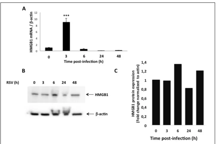
In vitro: 16HBE cells infected with rrRSV at MOI of 1 showed significant upregulation of
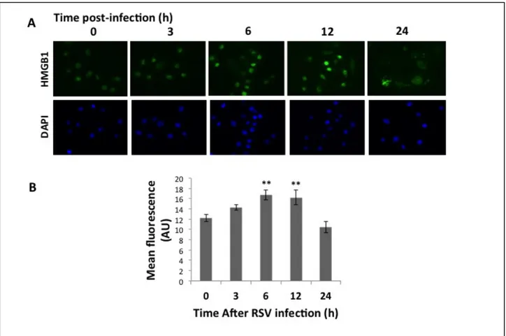
1A). Accordingly, western blot analysis confirmed increased HMGB1 protein in rrRSV-infected 16HBE, peaking at 6 h (p<0.001) and waning by 24 h (Figure 1 B-C). Immunofluorescence of 16HBE cells infected with RSV at MOI of 1 showed localization of HMGB1 into the nuclei with a peak at 6 h (Figure 2°). Densitometry analysis of HMGB1 expression revealed increased overall expression at 6 h before a decline at 24 h (Figure 2B) 16HBE have been frequently used as a model system of the airways for different cell signaling studies. To compare the results obtained with 16HBE, we also used differentiated primary NHBE. Similar to immortalized cells, immunofluorescence staining and confocal microscopy of primary NHBE infected with RSV at MOI of 1 resulted in localization of HMGB1 expression into the nuclei at 3 h and 6 h, and subsequently to the plasma membranes at 24 h (Figure 2C). In particular, densitometry analysis of HMGB1 translocation showed the percentage overlap between nuclear and total HMGB1 translocation at 3, 6, 12, and 24h post-infection (Figures 2D, 2E).
In vivo: qPCR analysis of HMGB1 gene expression in rat lung homogenates measured a
doubling of mRNA transcripts 5 d post inoculation of rrRSV, compared with age-matched mock infected controls dosed with virus-free medium (p<0.01; Figure 3A). ELISA showed significantly higher HMGB1 protein concentrations in the lungs of rrRSV-infected pups (p<0.01; Figure 3B). Western blot analysis confirmed that while HMGB1 protein was weakly detected in pathogen-free lungs, its concentration was 3-fold higher in rrRSV-infected lungs (p<0.001; Figure 3C-D).
In vitro: to determine whether HMGB1 plays a causal role during rrRSV infection, we used
the specific inhibitor glycyrrhizin to block HMGB1 activity in 16HBE cells and NHBE cells infected simultaneously with rrRSV. Glycyrrhizin decreased the number of RFP positive cells in both 16HBE cells (Figure 4 A-B) and NHB cells (Figure 5A-B) in a dose-dependent fashion. This profound effect of HMGB1 on rrRSV-infected cells was confirmed in NHBE cells by qPCR (p<0.001; Figure 5C). Similarly, western blotting confirmed that glycyrrhizin prevents rrRSV-induced HMGB1 protein upregulation (Figure 5D).
Figure and figure legends
Figure 1. HMGB1 expression in rrRSV-infected 16HBE cells. HMGB1 mRNA transcripts and protein
expression were measured by qPCR and Western Blot analysis in human bronchial epithelial cells infected with rrRSV at MOI of 1 for 48 h. [A] HMGB1 gene expression was upregulated at 3 h post-infection, followed by reduced expression at 6, 24 and 48 h. β-actin gene was used as the housekeeping control for transcript normalization. Data are expressed as mean ± SEM. ***P<0.001 compared with non-infected cells.
[B] HMGB1 protein expression was upregulated at 6 h post infection, before returning to baseline at 24 and
48 hours post infection. [C] Densitometric analysis. Data are expressed as mean ± SEM. ***P<0.001 compared with time point 0 h.
Figure 2. HMGB1 localization in rrRSV-infected airway epithelial cells. Human 16HBE and primary
bronchial epithelial cells were infected with rrRSV at an MOI of 1 or with sterile medium at the indicated time points. [A] HMGB1 in 16HBE cells was visualized in cells 24 h post-infection by immunofluorescence showed localization of HMGB1 into the nuclei with a peak at 6 h [B] as evidenced by densitometry, **P<0.01 compared to time zero.
Figure 2. [C] Primary HBE cells by confocal microscopy showed localization of HMGB1 expression into
the nuclei at 3 h and 6 h, in the cytoplasm starting at 12 h, and on plasma membranes at 24 h.
Figure 3. HMGB1 expression in rat lungs. [A] qPCR and [B] ELISA analysis were performed to
determine respectively mRNA and protein expression of HMGB1 in lung tissues of rat pups infected with rrRSV at 10 d of life and killed 5 d later. Both HMGB1 mRNA and protein increased significantly in the lungs of rrRSV-infected rats compared to pathogen-free controls. [C] Increased HMGB1 protein expression in rrRSV-infected rat lungs was confirmed by Western blot. [D] Densitometry analysis. Data are expressed as mean ± SEM. **P<0.01 compared to non-infected cells.
Figure 4. Effect of glycyrrhizin on rrRSV infection in 16HBE cells. [A] Representative pictures
rrRSV-infected 16HBE and non-rrRSV-infected controls treated with the HMGB1 antagonist glycyrrhizin [50 or 100 µM] or vehicle. The density of cells exhibiting red fluorescence produced by active viral replication was
significantly reduced by glycyrrhizin in a dose-dependent fashion. [B] Bar graph showing the number of rrRSV-positive cell in each group. Data are expressed as mean ± SEM. ***P<0.001 compared to RSV group.
Figure 5. Effect of glycyrrhizin on rrRSV infection in human primary cells. [A] Fluorescent microscopy
of human primary cells treated with the HMGB1 antagonist glycyrrhizin [50 µM or 100 µM]. [B] The number of cells exhibiting the red fluorescence produced by active viral replication was significantly reduced by glycyrrhizin in a dose-dependent fashion. [C] Consistently, qPCR measured a significant reduction in RSV f RNA levels in response to treatment with glycyrrhizin. [D] Western blot analysis with corresponding densitometry demonstrated downregulation of HMGB1 protein expression upon glycyrrhizin treatment. Data are expressed as mean ± SEM. ***P<0.001 compared to RSV group.
Discussion
This study shows that HMGB1 expression increases both in vitro and in vivo as a function of RSV infection and its localization reflects different phases of the replicative cycle, which may allow the use of local or systemic levels as a biomarker of disease activity. More importantly, this is the first study indicating that HMGB1 synthesis is an essential step of RSV replication. Consequently,
selective inhibition of this protein in infected human bronchial epithelium drastically reduces the number of RSV-infected cells, thereby providing a novel therapeutic target for this common infection. Through the activity of the G and F glycoproteins, RSV attaches to and enters host cells, and 4-6 h after infection, begins transcription and replication of viral genome, reaching a peak approximately 20 h after infection. Synthesis and release of viral cytoplasmic particles containing RSV RNA and proteins start 12 h after viral entry and persist up to 48 h after infection.
In airway epithelial and immune cells, RSV is detected by 3 types of pattern recognition receptors (PRRs), including retinoic acid-inducible gene (RIG)-I-like receptors (RLRs), nucleotide-binding oligomerization domain (NOD)-like receptors (NLRs), and TLRs, all promoting and sustaining non-specific and/or specific immune response against RSV [21]. TLRs induce immediate release of proinflammatory cytokines and inflammasome components that, in turn, amplify the immune response via late synthesis of alarmins, such as HMGB1 [22]. The different timing of conventional cytokines and HMGB1 expression explains how the former mediate acute inflammation whereas the latter is not only involved in the early inflammatory phase but also maintains chronic inflammatory response [23]. In particular, a previous study reported that HMGB1 expression in vivo starts within 3-8 h after inflammatory and/or infectious stimuli, and then increases progressively from 16 to 32 h [24]. In accordance with these findings, we noted in vitro that: 1) HMGB1 mRNA levels sharply increased at 3 h, during viral entry into living cells; 2) nuclear HMGB1 localization peaked approximately at 6 h, in parallel with viral transcription and replication; and 3) 24 h after infection, during the release of viral cytoplasmic particles, HMGB1 migrated from the nucleus to the cytosol and plasma membranes.
Depending on its localization, HMGB1 exerts different time-dependent effects during viral infections. When expressed in the nucleus, HMGB1 acts as a nuclear enhancer for transcription factors and viral rolling circle replication as seen during adenovirus [25], and parvovirus [26] infections. When in the cytoplasm, HMGB1 is detrimental to the host response, reducing resistance to infections like influenza A [27], and H1N1 [28]. Moving toward the plasma membrane, HMGB1
binds RAGE, a receptor also expressed in epithelial cell cultures [29] and able to exacerbate RSV disease by amplifying the expression of proinflammatory agents. RAGE deficiency has been associated with viral-induced asthma phenotype in a mouse model [30]. Our data also confirmed nuclear HMGB1 localization during the early phase of infection, which has been shown to be a critical initial event for efficient viral cycle [4].
Supporting a pathogenic role of HMGB1 in RSV infection, we found increased HMGB1 expression in lung tissues of rat pups infected with RSV. It has been previously argued that an exogenous or endogenous immunogenic stimulus will activate the innate immune system only if able to induce release of alarmins, like HMGB1 [31]. By binding to its cognate receptors, HMGB1 modifies cell functions not only with direct autocrine/paracrine activity [32], but also through indirect potentiation of other inflammatory pathways, e.g., activation of multiple pattern-recognition receptors like TLR2 and TLR4 [32]; release of proinflammatory cytokines like IL-1, IL-6, and IL-8 [33]; and interference with T cell responses inducing TH1 polarization [34]. Furthermore, Lei et al. [35] reported that increased HMGB1 levels are associated with overexpression of NGF, the prototypical neurotrophic factor known to be responsible for the development of neurogenic inflammation and airway hyperreactivity during and after early-life infection by RSV [36]. Therefore, HMGB1 might play a pivotal role in initiating and amplifying the neurogenic inflammatory cascade. We speculate that HMGB1 promotes neurogenic inflammation in the early stage of the infection and, successively, contributes to the pathogenesis of post-infection airway hyperreactivity by modulating epithelial mesenchymal transition, and airway remodeling [37]. Critical evidence supporting the involvement of HMGB1 in the pathophysiology of RSV infection derives from the novel observation that selective inhibition of this molecule exerts a potent anti-viral effect in human bronchial cells. Glycyrrhizin, a glycoside alkaloid extracted from Glycyrrhiza glabra roots, is made of the biologically active principle glycyrrhetic acid (GA) and two inactive components of glucuronic acid [38]. By binding to the hydrophobic residues of HMGB1 box A and B, glycyrrhizin inhibits the chemotactic and mitogenic function of HMGB1 and prevents
HMGB1-DNA binding thereby exerting anti-inflammatory and immuno-regulatory effects in several non-infectious [39] and non-infectious [40] diseases. In particular, glycyrrhizin inhibits viral gene expression and replication, proinflammatory cells recruitment, and T lymphocyte responses [39].
In conclusion, our findings suggest that HMGB1 plays a critical role in the initiation and maintenance of RSV replication in human bronchial epithelia and it is a promising target for monitoring and managing the infection in infants and children with bronchiolitis and pneumonia. The potential therapeutic effects of glycyrrhizin merit further exploration, especially in light of its safety profile, which lacks cytotoxicity even at high concentrations according to both experimental and clinical studies [41].
Paper 2
Detection of respiratory syncytial virus (RSV) at birth in a newborn with respiratory distress
(Published data: Pediatr Pulmonol. 2017 Oct;52(10):E81-E84)
Introduction
Respiratory syncytial virus (RSV), an enveloped, non-segmented, negative-sense RNA virus belonging to the Paramyxoviridae family, is the most common respiratory pathogen in infants and young children [1], resulting in approximately 24 hospitalizations per 1000 infants and an estimated 66.000-199.000 annual deaths worldwide in children younger than 5 years, with 99% of these deaths occurring in developing countries [42]. After entering the host through the nasopharyngeal or conjunctival mucosa, RSV spreads to the lower respiratory tract where it can cause acute disease characterized by edema and necrosis of the respiratory mucosa leading to airflow obstruction [43]. The incubation period ranges from 2 to 8 days but usually is 4 to 6 days [1]. In addition to its well-documented tropism for the human airway epithelium, RSV is able to spread hematogenously from the primary site of infection to remote extra-pulmonary tissues [44, 45], particularly the bone marrow stromal cells that may provide the virus with an immunologically privileged sanctuary, and allow its persistence in latent state [46]. Expanding on these findings, RSV has been shown recently to spread across the rodent placenta from the respiratory tract of an infected dam to the lungs of the fetus [3, 47, 48]. However, so far vertical transmission of this virus has been demonstrated in animal models, but never in humans. Herein, we describe a case of RSV infection documented at birth in the peripheral blood of a human newborn with onset of severe respiratory distress immediately after delivery from a mother with serological and clinical evidence of RSV infection during pregnancy
Case report
Patient was a baby boy born in September 2016 at 35 weeks gestational age via emergency caesarean section performed for reduced fetal movements. Maternal serologic screening for TORCH infections (Toxoplasmosis, Syphilis, Varicella-Zoster Virus (VZV), Parvovirus B19, Rubella, Cytomegalovirus (CMV), and Herpes Simplex Virus (HSV), Hepatitis C Virus (HCV), Hepatitis B Surface Antigen (HBsAg), and Human Immunodeficiency Virus (HIV), as well as screening test for Group B Streptococcus were all negative. The newborn was second born of non-consanguineous parents and family history was unremarkable. Birth weight was 3550 grams (g) (>95%ile) and Apgar score was 8 at both 1- and 5-min time points. Clinical examination was normal for gestational age, but postnatal cardiorespiratory adaptation appeared suboptimal because of nasal flaring and severe chest retractions. Therefore, positive pressure ventilation was started with FiO2 of 0.21. Physical examination revealed a newborn with grunting respiration, rales in both lungs, tachycardia (185/min), tachypnea (60/min), and oxygen saturation of 92% on room air (Silverman-Andersen score = 4). Umbilical arterial blood analysis revealed a pH of 7.18, partial O2 pressure of 32mm Hg, partial CO2 pressure of 68mm Hg, and a base deficit of −11.8 mmol/L. Chest
radiography at birth showed diffuse fine and granular opacities, and mild perihilar linear opacities bilaterally suggestive of respiratory distress syndrome. Broad-spectrum antibiotic coverage and minimal enteral feeding via gavage were started, and the patient was transferred to the Neonatal Intensive Care Unit for non-invasive ventilation via nasal continuous positive airway pressure (nCPAP) at 5 cm H2O. In light of the clinical and laboratory findings, an umbilical venous catheter
was placed. Complete blood counts and serum chemistry tests yielded normal values, and all blood and urine cultures were negative. After 1 week of nCPAP, the patient still required ventilatory support. Therefore, new chest radiography was obtained at 10 days of life, which revealed the presence of an area of consolidation in the parahilar region of the right lung. At the same time, cultural and serological microbiological tests were undertaken, which detected weakly positive anti-RSV IgM (1/20) and markedly positive anti-anti-RSV IgA (1/60) and anti-anti-RSV IgG (1/160) titers.
Furthermore, RSV RNA was amplified from the newborn’s peripheral blood obtained on the first day of life using standard operating procedures to safeguard the sterility of the sample and analyzed with a high-sensitivity quantitative real-time RT-PCR assay (Argene® Respiratory Multi Well System; BioMérieux, Marcy-l’Etoile, France) according to the manufacturer’s instructions. The analytical sensitivity threshold of this test is 250 copies/mL, and thus results are interpreted as positive if the RSV copy number detected in the sample is above threshold. This test also ruled out the presence of more than 40 other common viral and bacterial respiratory pathogens, including: Influenza (A, B), Human Coronavirus (229E, NL63, HKU1, OC43), Human Metapneumovirus (A, B), Parainfluenza virus (1, 2, 3, 4), Rhinovirus (A, B, C), Enterovirus (A, B, C, D), Adenovirus (A, B, C, D, E, F, G), Bocavirus (1, 2, 3, 4), Chlamydophila pneumoniae, Mycoplasma pneumoniae, Bordetella pertussis, Bordetella parapertussis, Legionella pneumophila, HSV, VZV, CMV, Epstein-Barr Virus (EBV), and Human Herpes Virus 6 (HHV6).
In light of clinical and microbiological findings in the newborn, maternal serologic tests were also performed and showed elevated anti-RSV IgM (1/40), IgA (1/20), and IgG (1/60) titers. Reassessment of maternal prenatal and familial history, revealed that the newborn’s mother and other family members had complained of cough during the second trimester of gestation. All other diagnostic tests performed on the newborn during the course of hospitalization, including brain and renal ultrasound, electrocardiography, echocardiography, and auditory/visual function screening, showed normal findings. At 17 days of life, the patient’s respiratory status improved and in parallel serologic RSV test became negative (IgM: negative; IgA: negative; IgG 1/80). Patient was discharged home in good health on day 22 of life.
Discussion
RSV is the major respiratory pathogen in young children, causing substantial morbidity and mortality worldwide [42]. The clinical course of the first infection varies from mild upper respiratory tract symptoms to severe lower respiratory tract disease (bronchiolitis or pneumonia)
leading to hospitalization in 2-3% of cases [49]. Reinfections are common, but usually less severe or asymptomatic [50]. The mode of transmission of this infection has always been thought to be horizontal (interpersonal) and through direct contact with infected secretions. However, recent experimental evidence has suggested that also vertical transplacental transmission may occur. Due to ethical constrains, vertical transmission of RSV infection so far has been demonstrated only in experimental animal models [3]. The present report is relevant in that it describes a neonatal case of human RSV infection consistent with vertical transmission from a previously infected mother to her unborn son. In this newborn with symptoms consistent with viral pneumonia since birth, microbiological tests revealed high serum titers of anti-RSV IgM, IgA, and IgG, as well as presence of RSV RNA in blood samples obtained with sterile procedure on the first day of life. Serologic tests for RSV were also positive in the mother and correlated with a history of respiratory symptoms during gestation in several members of the immediate family, suggesting an
infectious etiology.
Previously, only the possibility of antenatal RSV sensitization has been investigated [51], showing that RSV-specific neutralizing antibodies are not only efficiently transferred via the placenta to the newborn [52], but also protect the newborn against RSV infection during the first months of life [53, 54]. Piedimonte et al. [3] published an animal study showing that the same RSV strain used to infect pregnant rats was subsequently detected in 30% of fetuses exposed in utero, as well as in lung tissues from 40% of newborn rats and 25% of adult rats born from infected pregnant dams.9 Importantly, prenatal RSV exposure modified the expression of genes encoding growth factors critical for development of the peripheral nervous system, particularly affecting the cholinergic innervation of the airways and leading to bronchial hyperreactivity [55].
The case described in this report may be the first clinical description of vertical transmission of RSV in a newborn, reproducing the experimental conditions used in Piedimonte’s rodent model [3]. Indeed, the newborn’s mother reported cough during the pregnancy, newborn showed evidence of fetal distress in utero and was born in respiratory distress with onset of the respiratory symptoms
immediately after birth in September, that is, before the peak of RSV epidemic generally occurring between October-November and May-June in Southern Italy. As the pre- and peri-natal history of our patient was unremarkable for all possible etiologies of fetal and neonatal respiratory distress, vertical RSV infection appears to be the only plausible explanation of the clinical manifestations. It should be noted that the incubation period for RSV ranges from 2 to 8 days, after which the clinical infection starts with signs and symptoms of mucosal inflammation and irritation of the upper respiratory tract (congestion, rhinorrhea, sneezing). Over the next several days, the clinical status evolves with involvement of the lower respiratory tract manifested by cough and increased work of breathing with use of accessory respiratory muscles to overcome the increased resistance of obstructed airways. In the case described, the lower respiratory tract symptoms were already present at birth, which is not consistent with infection after birth but rather suggest that the infection had occurred before birth. The viral etiology was confirmed in the newborn both by serology and PCR, and corresponded to positive serology for the same virus in the mother. Thus, all historical, clinical, and diagnostic information converge in suggesting vertical transmission of RSV from the mother to the offspring. When considering direct transmission of viral pathogens from mother to offspring, it is essential to understand that the pathological consequences may be significant despite preexisting maternal seroimmunity, as demonstrated by the relevant congenital morbidity, and mortality observed after secondary infections with other viruses, such as CMV and pestivirus [56, 57]. Furthermore, in our previous studies performed in rodent models, all RSV-infected dams developed measurable anti-RSV antibody titers, and yet they were often able to transfer the infection to their fetuses [3], and these models have been highly predictive of human pathology for several other vertically transmitted viruses, such as CMV, arenaviruses, or parvoviruses [58].
Finally, our finding of vertical transmission in animals provides the only plausible explanation for the repeated isolation of blood-borne RSV in the first days of life of unexposed human newborns [59].
As in all case reports, several limitations caution against generalization of our findings. In particular, RSV serology is not standardized and generally not used to make definitive diagnosis of acute infection. Specifically, as IgG cross the placenta, neonatal anti-RSV IgG were likely of maternal origin, and IgM can produce false positives (interference with high titer IgG and other similar viruses). However, several studies have previously demonstrated that the IgA detected in neonatal blood is primarily of fetal origin [60-67]. In normal development, fetal IgA is either undetectable or rises very slowly during gestation and fetal levels at term remain approximately 1000 times lower than concentrations in the maternal circulation.28 Secretory IgA is then transferred to the newborn by breastfeeding [68], but our patient did not receive breast milk in the NICU. Thus, a positive IgG titer indicates that the fetus has “inherited” maternal antibodies, but positive IgA—especially at the high titers found in our patient (1:60) —together with positive IgM at birth suggest strongly that the fetus had been vertically infected. At any rate, these serology results were confirmed in our patient by the detection of clinically relevant amounts of RSV RNA. In addition, to exclude co-infections and/or superinfections, we performed extensive serological tests as well as detection of other RNA viruses (eg, adenovirus, rhinovirus, coronavirus, virus influenza, virus parainfluenza, metapneumovirus, and enterovirus) that resulted all negative.
Another potential limitation is that we do not have a nasopharyngeal swab or bronchoalveolar lavage for RSV detection in the respiratory tract of this newborn. Nevertheless, given the extremely low chance of horizontal transmission, it is reasonable to think that the infection had started prenatally. If so, the studies byPiedimonte et al. [3] indicated that pup sex posed to RSV infection in utero develop strong bronchial hyperreactivity to either electrical nerve stimulation or methacholine challenge after postnatal reinfectionwith the same virus. Therefore, newborns with early respiratory distress associated with evidence of RSV infectionwarrant closer follow-up for the possible recurrence of wheezing. More importantly, the possibility that RSV is transmitted vertically from mother to fetus has the potential of changing our strategies for the prevention and therapy of this highly prevalent infection and its chronic sequelae. The most important implication is that the
passive prophylaxis with humanized monoclonal antibodies currently offered only to infants at high risk for severe infection should probably be anticipated to expecting mothers in order to prevent prenatal infections. Such protection could be provided in a perhaps more effective, safer, and less expensive way by actively immunizing pregnant women with a suitable vaccine, which could be provided to large segments of the population even in third world countries where RSV is still an important cause of infant mortality.
Paper 3
Different concentration of human cord blood HMGB1 according to delivery and labour: A pilot study
(Published data: Cytokine. 2018 Mar 20;108:53-56)
Introduction
The injury mediated by oxidative stress (OS) is one of the major pathogenic protagonists in the onset of several conditions relating to newborns, which are commonly referred to as “oxygen radical diseases of neonatology” [69, 70]. The accumulation of Free Radicals (FR), beyond the capacity of the endogenous antioxidant defence system to scavenge them, results in damage to DNA, proteins and lipids which compromises cellular function, leading to cell death via apoptosis or necrosis. Increasingly emerging evidences from literature reveal that OS-mediated pathways contribute sharply to the preeclampsia, early pregnancy loss, foetal growth restriction and, preterm labour pathogenesis [71]. To date, it is not clear whether also the foetus is subject to OS during the process of labour and birth. Labour is also the result of leukocyte activation which, following uterine invasion, promotes the release of uterotrophins (e.g. cytokines and chemokines) triggering and supporting a myometrial contraction. Whether these events occur early, a preterm labour takes place. Very few literature data are available on changes in OS levels in newborns in relation to delivery mode [71]. When compared to babies born by elective caesarean section (CS), newborns from both spontaneous vaginal delivery (SVD) and emergency CS show more higher oxidative products levels in the umbilical arterial blood, probably due to delivery-related OS [72]. Also, it could be the expression of a condition of prenatal oxidative status [72].
High Mobility Group Box 1 (HMGB1) is a DNA-binding nonhistone protein (25 kDA) expressed both in intracellular site (nucleus, cytosol, mitochondria, and cell surface membranes) and in extracellular space. Following a specific biochemical stimulus (e.g. antigen presentation, OS, cytokines secretion, tissue damage) HMGB1 can be passively secreted from damaged or necrotic
cells [73]. HMGB1 takes part in numerous medical conditions, including pregnancy [74, 75], both in early events, primarily embryo implantation, and in later events, including labour and delivery. The release of alarmin, subsequent to tissue damage, aging cell and/or other stress factors, is known to be involved in pathologies of pregnancy, especially in isolated or recurrent abortion, intrauterine growth restriction (IUGR), and preterm labour [76]. It is known that many inflammatory mediators, among which HMGB1, are involved from the beginning of pregnancy to birth of the infant. Changes in amniotic fluid HMGB1 levels both of non-labouring and labouring (term and preterm) pregnant women have highlighted that HMGB1 can be detected into amniotic fluid [77].
In order to further investigate the HMGB1’s role, this study evaluated serum cord blood HMGB1 levels in a population of neonates, to investigate the potential utility of alarmin as a novel marker, and its connection with mode of delivery, in babies born both by SVD and CS (elective or emergency), as well as the influence of labour.
Materials and Methods
Subjects
The study subjects included 325 newborns delivered at the Department of Obstetrics, at University Hospital “G. Martino” of Messina, Italy, over an 18-month period. Following cord separation, venous blood sampling was performed on umbelical cords.
Exclusion criteria were: born dead, donation of the umbilical cordon and cord preservation.
Pregnant women admitted for labour were informed about the aims of the research and fully informed about study protocol. Participation in this study was voluntary and enrolment occurred at the same moment of admission for delivery. Prior to start the study, written informed consent was obtained from the pregnant women.
The study was conducted in accordance with the Declaration of Helsinki and was approved by the local Ethics Committee of the University Hospital of Messina (approval number 69/17).
The recruited patients were divided into 3 groups related to mode of delivery: SVD (group A), elective CS (group B), emergency CS (group C).
Regarding labour, subjects were divided into 3 groups: spontaneous labour (Group S), induced labour (Group I), absent labour (Group O).
Within 5 minutes after birth, blood samples from the umbilical vein were collected. Blood was left to clot for 5 minutes at room temperature. Then, blood samples were centrifugated and serum aliquots were stored at 80 °C until use. Serum HMGB1 was assessed using ELISA Kit following recommended protocol (Phoenix Pharmaceutical; Belmont, CA. ST51011 HMGB1 ELISA 96). Absorbance was measured using a microplate reader (Bio-Rad, Milan, Italy), and standard curves were constructed by the Bio-Rad Microplate Manager program V.5.1.
Other aliquots of the collected cord blood samples obtained in heparin-washed syringes were used to perform blood gas analysis.
Birth body weight, gestational age, and all maternal characteristics were recorded.
Statistical Analysis
Numerical data are expressed as median and range (minimum and maximum). The non-parametric approach was used since the numerical variables were not normally distributed, as verified by the Kolmogorov Smirnov test. The Kruskall Wallis test was applied to compare HMGB1 among the 3 different kinds of delivery; since it was highly significant, we performed pair wise comparisons between groups using the Mann Whitney test. The same analysis was performed to compare the different kinds of labour. For these multiple comparisons, we had to apply Bonferroni’s correction, for which the significance alpha level 0.050 has to be divided by the number of the possible comparisons; thus, the new “adjusted” significance level for this analysis is equal to 0.050/6 = 0.008. The non-parametric Spearman correlation test was applied in order to assess the existence of any significant interdependence between HMGB1 and numerical parameters. Statistical analyses were performed using SPSS 17.0 for Window package. P<0.05 two sides was considered to be statistically significant.
Results
Clinical findings of enrolled population
From among the enrolled 325 subjects, only 295 were included in this analysis. Thirty neonates with evident hemolysis in umbilical cord blood were excluded from the study.
All neonates were born healthy, as estimated by Apgar scores at 1 and 5 minutes and pH values of blood gas performed at birth. The mean ± standard deviation (±SD) gestational age was 38.6±1.8 weeks, mean birth weight 3100±500 g. Characteristics of infants and their mothers are summarized in Table 1.
HMGB1 levels, obstetric history, clinical and laboratory findings of enrolled population
Statistical analysis performed to identify any potential significant interdependence between umbilical vein blood HMGB1 levels and obstetric history (e.g. personal history of acute and chronic disease or infectious diseases; taking drugs during pregnancy; smoking during pregnancy; weight gain during pregnancy; abortion threats; premature birth threats; presence and length of labour), clinical characteristics (gestational age (p=0.213); Apgar Index at 1’ and 5’; birth weight (p=0.612); need of aspiration of airways, oxygen supplementation, positive pressure ventilation, emergency tracheal intubation, chest compressions, umbilical vein catheterization, and drugs administration), and laboratory findings (arterial blood gas test) of enrolled population failed. Correlations between HMGB1 levels, clinical features, and laboratory findings are summarized in Table 1, but nothing statistical differences were shown.
HMGB1 levels and mode of delivery and labour
Subjects were divided into the following 3 groups related to mode of delivery: SVD (group A, n=196), elective CS (group B, n=49), emergency CS (group C, n=42). Serum HMGB1 levels significantly and directly were correlated with mode of delivery. In the cord venous blood, we found HMGB1 values significantly more elevated in spontaneous vaginal group when compared to elective or emergency caesarean section group (p=0.004). While there was no significant HMGB1
difference between groups of neonates born by caesarean delivery, both elective or emergency (Group B and C) (p=0.046).
Regarding labour, subjects were divided into 3 groups: spontaneous labour (Group S, n=131), induced labour (Group I, n=99), absent labour (Group O, n=55). In the cord venous blood, we found HMGB1 values significantly more elevated in the two groups characterised by presence of labour, both spontaneous and induced (Group S and I), with concentrations significantly higher than the group without labour (Group O) (p=0.010).
Over the years, research on HMGB1 has been quite thorough and other properties have been revealed. HMGB1 has resulted to be a leading mediator in both acute and chronic inflammation, and plays a crucial role in several medical conditions, including autoimmune diseases, cancer, hepatitis, malaria, myocardial ischemia, infection-elicited inflammatory diseases [78].
It is known how inflammation is essential in maintaining uterine homeostasis, for successful embryo implantation and delivery. Even though inflammation is needed for a successful reproduction, early and uncontrolled activation of inflammatory proceedings can cause important adverse effects on childbearing outcomes, including preterm birth [79, 80]. Recent findings by Romero et al. have highlighted the importance of HMGB1 in amniotic fluid sterile inflammation [76]. Authors reported that amniotic fluid levels of HMGB1 were also higher in women who underwent spontaneous preterm labor with intra-amniotic infection or a condition of inflammation, than in those without these clinical conditions [77]. Several pathways have been reported to be initiators of term parturition. Inflammatory overload triggering delivery is a well-documented and known mechanism, and phlogosis may have originated from both foetal and maternal compartments. Alarmin release, caused by tissue injury, oxidative damage, or other injuries, is implicated in several diseases of pregnancy independently of the presence of a coexistent infection. In these years, literature data have associated placental dysfunction with elevated alarmin concentrations, which determines the massive amplification of the effects of placental inflammation, with a high-risk of related complications [81]. Although most of the data regarding a
connection between a rise in alarmin levels and the onset of preterm labour have been reported, causal and mechanistic data have not been documented. Moreover, HMGB1 and its principal receptors, such as RAGE, TLR2 and TLR4 are revealed in the cervix and an increased extra-nuclear fraction of HMGB1 with labour onset, both at term and preterm, has been signalized [82], suggesting that HMGB1 could play a role in the maturation of cervix. Buhimschi et al. [73] proposed that HMGB1, together with sRAGE and S100, are crucial elements involved in cellular damage of foetuses and in preterm birth triggered by inflammation. Authors reported that many stimuli that mimic a situation of infection or pro-inflammatory mediators, such as cytokines, are responsible for a cascade of events that determine the activation of immune cells, cell surface expression of HMGB1 and its secretion via non-classical secretion pathways [73]. Transition into a receptive and active uterus incorporates cellular changes in the endometrium and the modulated expression of different molecules, such as transcription factors, growth factors, endometrial cytokines. These signals are responsible of the transition from a silent myometrium to an active stage, characterized by increasingly close and intense contractions, until the culmination of labour. Our data, supported by the evidence of others more detailed in-depth in vivo and in vitro studies that elucidate molecular and cellular mechanisms of HMGB1, could reveal a possible action of this alarmin related to mode of delivery and to the presence of labour. All these mechanisms, synergistically to others many inflammatory pathways, could take part to determine decidual modifications for the transition of the endometrium from the non-receptive to receptive stage, that lead to moment of birth. Recently, it has been reported that the alarmin HMGB1 mediates its many activities via a reactive oxygen species (ROS)-dependent mechanism [83]. HMGB1 is a known target of such redox reaction molecules, in fact, it contains two redox sensitive cysteine moieties at positions C23 and C45 whose redox states affect strongly HMGB1 activities. However, as OS worsens and these thiols are oxidized to form a disulfide bond, the HMGB1 function shifts to boost inflammation. Increased HMGB1 concentrations was also showed with ischemic reperfusion injury associated with some modes of delivery, suggesting the great usefulness of measuring HMGB1 to
facilitate the early diagnosis of hypoxic–ischemic encephalopathy, an element significantly impacting the prognosis and neurological outcomes of affected neonates [83].
In conclusion, we believe that the dosage of circulating values of HMGB1 could be useful for its potential diagnostic and/or prognostic role in the prediction of the degree of oxidative damage in newborns, although more detailed in-depth in vivo and in vitro studies are needed to better elucidate molecular and cellular mechanisms underpinning the role of HMGB1 in associated FRs-related maternal, foetal and neonatal diseases.
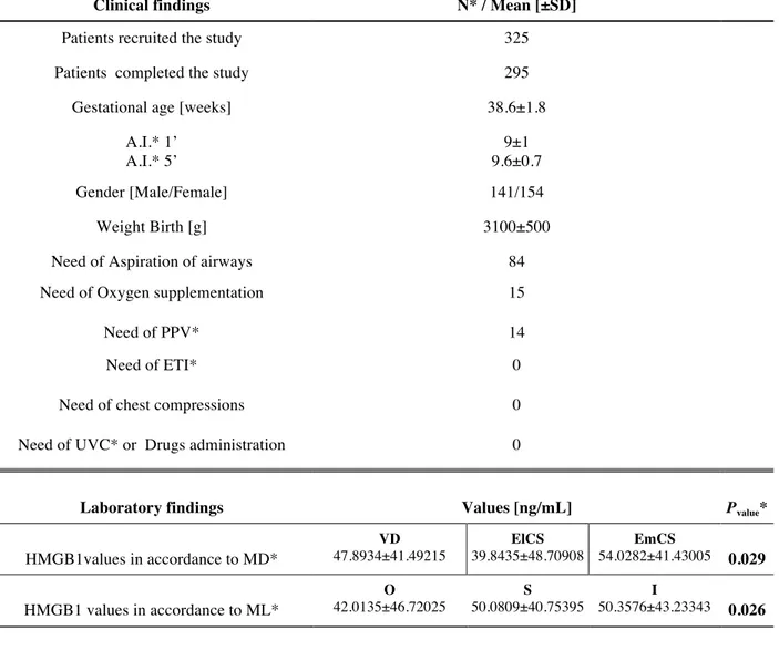
Table 1. Obstetric data, clinical and laboratory findings of enrolled population
Obstetric data N* / Mean [±SD]
Personal history of acute or chronic diseases 47
Smoking during pregnancy 2.5±5.8
Weight gain during pregnancy 12.6±2.8
Abortion threats 26
Premature birth threats 38
Mode of labor [O*, S*, I*] O=55, S=131, I=99
Hours of labor 5.2±5.8
Mode of delivery [VD*/ElCS*/EmCS*] VD=196, ElCS=49, EmCS=42
Clinical findings N* / Mean [±SD]
Patients recruited the study 325
Patients completed the study 295
Gestational age [weeks] 38.6±1.8
A.I.* 1’ A.I.* 5’ 9±1 9.6±0.7 Gender [Male/Female] 141/154 Weight Birth [g] 3100±500
Need of Aspiration of airways 84
Need of Oxygen supplementation 15
Need of PPV* 14
Need of ETI* 0
Need of chest compressions 0
Need of UVC* or Drugs administration 0
Laboratory findings Values [ng/mL] Pvalue*
HMGB1values in accordance to MD* VD 47.8934±41.49215 ElCS 39.8435±48.70908 EmCS 54.0282±41.43005 0.029 HMGB1 values in accordance to ML* O 42.0135±46.72025 S 50.0809±40.75395 I 50.3576±43.23343 0.026
*N: number; O: absent, S: spontaneus; I: induced; VD: vaginal delivery; ElCS: elective caesarean section; EmCS: emergency caesarean section; A.I.: Apgar Index; PPV: positive pressure ventilation; ETI: emergency tracheal intubation; UVC: umbilical vein catheterization; MD: mode of delivery; ML: mode of labor.
Paper 4
Respiratory Syncytial Virus seropositivity at birth associates with adverse respiratory outcomes
(Data under review: Journal of Pediatric Infectious Disease Society)
Introduction
Even with repeated exposure to Respiratory Syncytial Virus (RSV) infection, immunologic protection is usually short-lived and incomplete, allowing RSV to re-infect the host throughout life [84]. In addition, studies of primary RSV infections show the neutralizing antibody response rapidly declines to pre-infection levels within months [85]. Given this evidence, it is not surprising that serological studies of adults generally find neutralizing antibody concentrations below the threshold needed to achieve immune protection. Furthermore, the role of viral respiratory illness during pregnancy is not well-defined outside influenza [86]. Several studies have shown that RSV disease may occur in mothers at any trimester of pregnancy [87-89], but the consequences of this infection for the offspring remain unclear. Yet, mothers with respiratory illness are more likely to have poor maternal and perinatal outcomes than those without respiratory illness [90].
There is growing evidence pointing to extra-pulmonary involvement of RSV infection, including the detection of both viral antigens and genome in peripheral blood mononuclear cells of infected individuals [45, 91-93]. As a result, recent studies have explored whether RSV can cross the placenta leading to vertical transmission of the infection from the mother’s respiratory tract to the fetus [94, 95]. First, Piedimonte et al. used recombinant RSV to infect pregnant rats and detected RSV genome and transgene expression in pulmonary tissues of 40% of their offspring [3]. The virus then persisted into adulthood in 25% of congenitally exposed rats. Importantly, the intrauterine RSV infection influenced expression and function of key neurotrophic pathways and affected the development of cholinergic nerves in the airways and lung tissues, leading to persistent bronchial hyperreactivity [3]. Subsequently, the same group reported the first documented case of
congenital infection caused by vertical transmission of RSV from a mother with history of upper respiratory infection to her son born with acute bronchiolitis [95], while Fonceca et al. reported the presence of RSV genome in cord blood mononucleocytes and identified these cells as a potential cellular reservoir for RSV [96].
Nevertheless, the ability of RSV to cross the human placenta and its impact on the respiratory outcomes of newborns needs further clarification. In this study we sought to determine serologic evidence of anti-RSV immunity in fetal cord blood of offspring with a maternal history of respiratory illness occurring during the third trimester of pregnancy, and also characterized the postnatal clinical outcomes associated with RSV seropositivity.
Methods
Study subjects - Between September 2016 and April 2017, women presenting for delivery to the Departments of Obstetrics and Gynecology at either University of Messina or University of Catania medical centers in Italy were screened for history of respiratory illness. Informed consent was obtained from mothers who reported ≥2 of the following symptoms during the third trimester of pregnancy: fever, influenza-like illnesses, cough, and sore throat. The Institutional Review Boards of the University of Messina, University of Catania, and Cleveland Clinic Foundation in Cleveland, Ohio approved the study protocol. Data were securely stored and managed using the REDCap electronic data capture tools hosted at the Cleveland Clinic. Patient privacy was protected in compliance with the United States Health Insurance Portability and Accountability Act (HIPAA) and European Union General Data Protection Regulation (GDPR).
Participants provided comprehensive medical history information through completion of a detailed demographic and medical questionnaire. Thereafter, all subjects underwent routine obstetric examination. Maternal serologic screening for Hepatitis C Virus (HCV), Hepatitis B Surface Antigen (HBsAg), Human Immunodeficiency Virus (HIV), and Group B Streptococcus (GBS) were obtained. Mothers were excluded if they had diagnosis of any other infection, history of
severe immunosuppression (e.g., HIV infection, transplantation, or malignancy), or used immunosuppressive medications.
Clinical definitions
Demographic, laboratory, radiologic, and clinical data of participating newborns were collected during their entire hospitalization. Previously published definitions for prematurity, low birth weight, intrauterine growth restriction (IUGR), atopy, transient tachypnea of newborn (TTN), respiratory distress syndrome (RDS), and bronchopulmonary dysplasia (BPD) were used [97-105].
RSV serology
After delivery, fetal cord blood were collected for RSV serology as described previously [106]. Briefly, the last 10–15 cm of the umbilical cord was disinfected with iodine prior to removing the clamp and 5 ml of blood was collected by gravity less than 10 min after the cord was sectioned with disinfected scissors. Serum was prepared by centrifugation at 2,650 g for 20 min, and aliquots were stored at -80°C until use. Anti-RSV IgA, IgM, and IgG antibodies were quantified using an immunofluorescence assay (Euroimmun, Padova, Italy) following the manufacturer’s instructions. Positivity for RSV antibodies was determined based on previously published criteria: <1/20 dilution was considered negative, ≥1/20 positive, and ≥1/140 strongly positive [107]. Cord blood serum samples with positive RSV IgM and/or IgA in addition to positive IgG were considered seropositive for this study. This definition of neonatal seropositivity is similar to those used for diagnosis of other congenial infections including rubella, toxoplasmosis and parvovirus [108-110].
Statistical analysis
Data are expressed as median (range) for continuous variables and count (percentage) for categorical variables. The Agresti-Coull method was used to estimate 95% confidence intervals for the prevalence of RSV antibodies. Associations between antibody titers [negative vs. positive] and neonatal clinical outcomes were analyzed using the Kruskal-Wallis and Fisher’s Exact tests for continuous and categorical variables respectively. All tests were two-tailed and performed at a significance level of 0.05. SAS 9.4 software (SAS Institute, Cary, NC) was used for all analyses.
Results
Between September 1, 2016 and April 30, 2017, a total of 22 pregnant women were enrolled in the study with a history of respiratory illness occurring in the third trimester of pregnancy. The majority of infants (82%) were born after 36 weeks gestation with 3 infants born between 34 and 35 weeks gestation and one infant born at 29 weeks gestation. Clinical characteristics and outcomes of the offspring are summarized in Table 1.
Table 1. Newborns’ characteristics and outcomes.
Length of Stay (days) 4 (3, 42)
Gender:
. Female 8 (36)
. Male 14 (64)
Gestational age (weeks) 38 (29, 41)
Multiple Births 2 (9) Mode of Delivery: . Vaginal 5 (23) . C-Section 17 (77) Birth Weight (kg) 2.9 (1.2, 4.1) Birth Length (cm) 49 (36, 54)
Birth Head Circumference (cm) 33.5 (28, 36)
Data are expressed as median (min, max) or N (%).
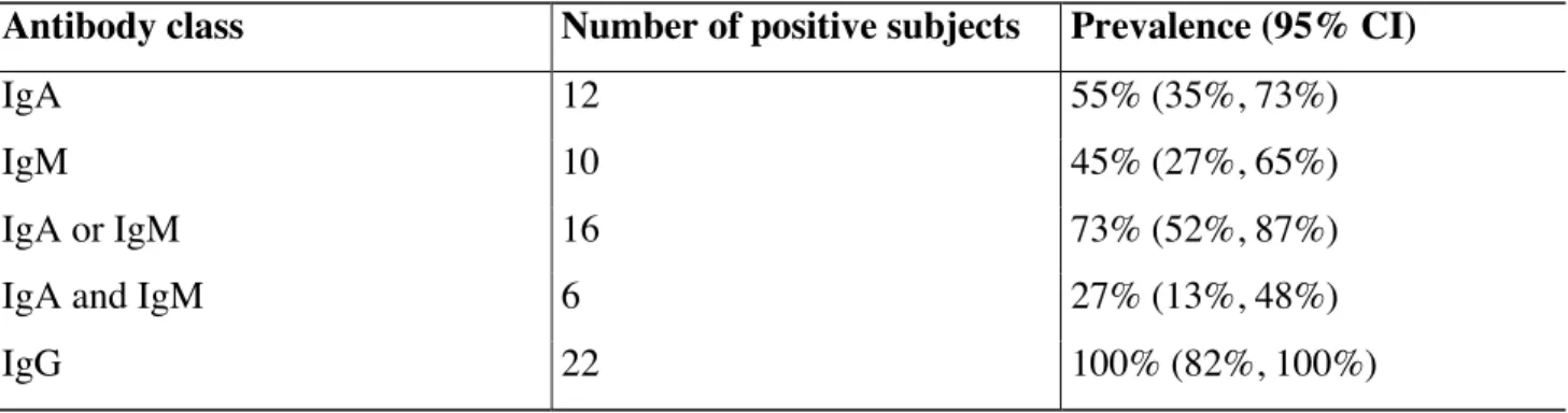
RSV immunity - Newborns born to mothers with a history of respiratory illness during pregnancy had serologic evidence of RSV immunity. All cord blood samples had titers for anti-RSV IgG ≥1:20 (95% CI = 82-100%), while 16 (73%; 95% CI = 52-87%) also had positive titers for either anti-RSV IgA or IgM, thereby meeting our criteria for RSV seropositivity (Table 2).
Table 2. Prevalence of RSV antibodies in cord blood specimens
Antibody class Number of positive subjects Prevalence (95% CI)
IgA 12 55% (35%, 73%)
IgM 10 45% (27%, 65%)
IgA or IgM 16 73% (52%, 87%)
IgA and IgM 6 27% (13%, 48%)
IgG 22 100% (82%, 100%)
The cord blood samples of 6 newborns (27%) were positive for both anti-RSV IgA and IgM. Samples positive for either IgA or IgM were more likely to have strongly positive IgG titers, with 14/16 (88%) having IgG titers ≥1/140, compared to 2/6 (33%) of those negative for IgA and IgM (p=0.025).
Clinical outcomes
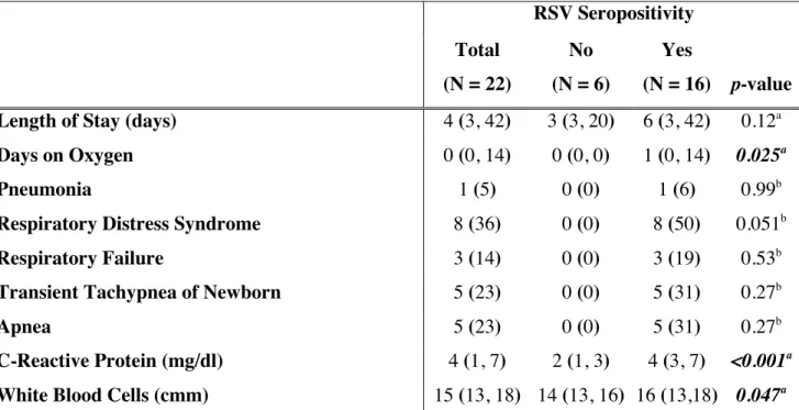
Newborns born to mothers with a history of respiratory illness during pregnancy had adverse clinical and laboratory outcomes. Eight (50%) RSV seropositive newborns developed respiratory problems including RDS (N=8), TTN (N=5), apnea (N=5), respiratory failure (N=3), and pneumonia (N=1), whereas none of the newborns in the RSV seronegative group was diagnosed with any respiratory pathology (Table 3).
![Figure 3. HMGB1 expression in rat lungs. [A] qPCR and [B] ELISA analysis were performed to](https://thumb-eu.123doks.com/thumbv2/123dokorg/4571864.38331/16.892.87.811.108.607/figure-hmgb-expression-lungs-qpcr-elisa-analysis-performed.webp)
![Figure 5. Effect of glycyrrhizin on rrRSV infection in human primary cells. [A] Fluorescent microscopy](https://thumb-eu.123doks.com/thumbv2/123dokorg/4571864.38331/17.892.91.812.214.730/figure-effect-glycyrrhizin-rrrsv-infection-primary-fluorescent-microscopy.webp)