VITREOUS INCARCERATION IN
SCLEROTOMIES AFTER VALVED 23-, 25-,
OR 27-GAUGE AND NONVALVED 23- OR
25-GAUGE MACULAR SURGERY
GIAN MARCO TOSI, MD, PHD,* ALEX MALANDRINI, MD,† GABRIELE CEVENINI, MD,‡
GIOVANNI NERI, MD,* DAVIDE MARIGLIANI, MD,* ARIANNA CERRUTO, MD,* GIANNI VIRGILI, MD§ Purpose: To study the patterns of vitreous incarceration at sclerotomy sites by ultrasound
biomicroscopy in patients subjected to valved or nonvalved small-gauge pars plana vitrectomy. Methods: A prospective comparative study of 88 eyes affected by epiretinal membrane and macular hole. Patients were divided into four groups: valved or nonvalved 23-gauge (16 eyes each) and valved or nonvalved 25-gauge (20 eyes each); their vitreal disposition was compared by ultrasound biomicroscopy. Vitreal disposition was also assessed in 16 eyes of 16 patients subjected to valved 27-gauge pars plana vitrectomy.
Results: Three vitreal patterns were identified: P0 (vitreous not visible or vitreous strand distant from the sclerotomy site), P1 (vitreous strand parallel to and in contact with the sclerotomy site), and P2 (vitreous strand entrapped in the sclerotomy site). The effect of valved trocar use on vitreous incarceration seemed to be somewhat beneficial, but no statistically significant effect could be shown (odds ratio: 0.85, 95% confidence interval: 0.42–1.74, P = 0.657). Similarly, no differences in vitreous incarceration were shown among vitrectomy gauges (23, 25, or 27) both in a model including valved trocars only (P = 0.858) and in a model with all available data (P = 0.935).
Conclusion: In 23- and 25-gauge macular surgeries, postoperative vitreous incarcera-tion does not seem to be reduced using valved cannulas and was similar to that observed in 27-gauge surgery.
RETINA 37:1948–1955, 2017
T
he sclerotomy site remains an object of study even in the era of small-gauge pars plana vitrec-tomy (PPV). Postoperative wound closure, conjunc-tival bleb formation, and vitreous incarceration at thesclerotomy site have all been investigated.1–20 Vitre-ous incarceration may plug the sclerotomy, impact-ing wound closure and conjunctival bleb formation, and consequently the risk of postoperative hypotony and endophthalmitis.6,11,13 Moreover, postoperative contraction of incarcerated vitreous can lead to post-operative retinal break formation and recurrent vitre-ous hemorrhage.11–23 Vitreous shaving around the sclerotomy site in humans and the cannula extraction technique and, recently, the vitrectomy degree in dif-ferent areas of the vitreous cavity in animals have all been found to be related to the amount of vitreous incarceration.11,13,16
The PPV armamentarium has recently been enriched with valved cannulas, thus eliminating the need for plug placement during instrument exchange.24These cannu-las have been proven to boost intraocular safety in animal models by preventing intraoperativefluctuations in both infusion pressure and intraocular pressure.24
From the *Ophthalmology Unit, Department of Medicine, Surgery and Neuroscience, University of Siena, Siena, Italy;†Ophthalmology Unit, Santo Stefano Hospital, Prato, Italy; ‡Department of Medical Biotechnologies, University of Siena, Siena, Italy; and §Department of Surgery and Translational Medicine, University of Florence, Florence, Italy.
None of the authors has any financial/conflicting interests to disclose.
This is an open-access article distributed under the terms of the Creative Commons Attribution-Non Commercial-No Derivatives License 4.0 (CCBY-NC-ND), where it is permissible to download and share the work provided it is properly cited. The work cannot be changed in any way or used commercially without permission from the journal.
Reprint requests: Gian Marco Tosi, MD, PhD, Ophthalmology Unit, Department of Medicine, Surgery and Neuroscience, Siena University Hospital, University of Siena, Viale Bracci 1, 53100 Siena, Italy; e-mail: [email protected]
Recently, valved cannulas have been associated with higherfinal anatomic success after rhegmatogenous ret-inal detachment repair, although single surgery success, functional outcomes, and complications were similar to the nonvalved treated eyes.25 However, the use of valved cannulas may render the insertion of soft tip instruments more difficult and might increase the rate of retained perfluoro-n-octane.25
We wanted to verify whether this increased intra-operative stabilization is associated with different postoperative patterns of vitreous incarceration at the sclerotomy site. This verification is believed to be useful, as recently reported by Oellers et al.25This was performed by studying vitreous appearance in patients subjected to valved or nonvalved 23- or 25-gauge PPV, using postoperative ultrasound biomicroscopy (UBM). Moreover, eyes treated with valved 27-gauge PPV (27-gauge nonvalved cannulas were not available) were compared with 23- and 25-gauge valved PPV treated eyes.
Materials and Methods Subjects
This prospective study included 88 eyes of 88 patients. The inclusion criteria were being affected by idiopathic epiretinal membrane or macular hole (MH) and being subjected to primary 23-, 25-, and 27-gauge PPV without intraocular complications, between February 2015 and March 2016 at the Ophthalmology Section of the Department of Medi-cine, Surgery and Neuroscience, University of Siena, Siena, Italy. The exclusion criteria were as follows: coexisting ocular disorders, except cataract; patients requiring PPV for indications other than those mentioned above; patients previously subjected to intraocular surgery, except cataract surgery; and patients with no identifiable sclerotomy site at slit-lamp examination. The research adhered to the princi-ples of the Declaration of Helsinki, and the institutional review board approved the study. Patients were treated after being informed of the nature of the treatment being offered, its potential risks, benefits, adverse effects, possible outcomes, and after having signed a consent form.
Each patient underwent a complete preoperative evaluation, including the measurement of best-corrected visual acuity, anterior segment examination, dilated fundus examination using indirect ophthalmoscopy with scleral depression, and slit-lamp biomicroscopic observation of the vitreous and the retina. The presence or absence of posterior vitreous detachment (PVD) was defined by biomicroscopic observation
using a 90-diopter lens, optical coherence tomography examination, and B-scan ultrasound. All patients underwent optical coherence tomography evaluation of the macular disorder. We used an optical coherence tomography classification when describing MH characteristics.26
Patients were enrolled consecutively and were assigned on an alternate basis to one of the following groups: or 25-gauge PPV with valved cannula; 23-or 25-gauge PPV with nonvalved cannula; and valved 27-gauge PPV. The patterns of vitreal appearance at the sclerotomy sites were compared between the 23-and 25-gauge valved 23-and nonvalved groups. In addition, the vitreous appearance in valved 27-gauge treated eyes was analyzed (27-gauge nonvalved can-nulas were not available) and compared with the 23-and 25-gauge valved treated eyes.
Surgical Technique
All patients underwent 23-, 25-, or 27-gauge PPV performed by the same surgeon (G.M.T.). The sclerot-omy sites were located in the inferotemporal, super-otemporal, and superonasal quadrants. The conjunctiva was anteriorly displaced away from the intended sclerotomy site with forceps in order to purposefully misalign the conjunctival and scleral incisions. A trocar was inserted 3.5 mm from the corneoscleral limbus at an angle of approximately 30° parallel to the limbus. Once past the trocar sleeve, the angle was changed to be perpendicular to the surface and the cannula was in-serted into the eye. The cannula was held in place with forceps and the trocar was removed. The endoillumina-tor and the vitrecendoillumina-tor were placed through the superior sites. Pars plana vitrectomy was performed using the Constellation vitrectomy instrument (Alcon Laboratories Inc, Fort Worth, TX). A panoramic viewing system with wide-field contact lenses (Advanced Visual Instruments, Inc, New York, NY) and aflat lens were used for intra-operative visualization. In the absence of PVD, it was induced with the vitrectomy probe in the “cutter-off” mode placed over the optic disk. Epiretinal membrane and internal limiting membrane peeling were performed using either triamcinolone or indocyanine green. Fluid/ air exchange was performed in every patient. Air was substituted with gas (C3F8, 8%) in the MH cases. Any leaking sclerotomies were sutured.
Postoperative Examination
Patients were subjected to a complete ophthalmo-logic evaluation the day after surgery and in the subsequent follow-up visits.
Ultrasound biomicroscopy (Model P40, Paradigm Medical Industries Inc, Salt Lake City, UT) was
performed at the sclerotomy sites between 30 and 40 days after surgery, once the tamponade had completely disappeared. To avoid bias, the ultrasonographer (A.M.) was masked to the patient grouping and diagnosis. To identify the precise sclerotomy location, the patients were examined by slit-lamp (small black dot on the sclera and/or presence of converging episcleral ves-sels) and the sclerotomy site was marked with a sterile surgical marking pen. The sutured sclerotomies were not evaluated. Ultrasound biomicroscopy examination was performed using a high-frequency transducer (50-MHz) with the probe oriented both radially and tangentially to the limbus in order to intersect and detect the entire sclerotomy tract. Examinations were performed with the patient in a supine position; drops of topical anesthesia were instilled before examination. A polymethyl methacrylate shell was inserted between the eyelids and filled with 2.5% methylcellulose as a coupling agent. The probe was always perpendicular to the ocular surface. The gain was set at between 60 dB and 80 dB to have a clear display of the structure and simultaneously minimize the ultrasound noise. We found 3 patterns of vitreous/sclerotomy relationship classified as P0 (vitreous not visible or vitreous strand parallel to but distant from the sclerotomy site); P1 (vitreous strand parallel to and in contact with the sclerotomy site); and P2 (vitreous strand entrapped in the sclerotomy site) (Figures 1–3). Statistical Analysis
Linear and logistic regression models were used to investigate the differences among the study groups, including trocar type and vitrectomy gauge, regarding continuous and categorical or nominal variables, respectively.
Multilevel models were used to investigate the effect of covariates on vitreous incarceration pattern, with individuals as random effects to account for the
correlated nature of measurements at multiple sclerot-omy sites. Because of the ordinal nature of the response variable, ordinal logistic regression was used, and the odds ratio (OR) of a higher level of incarcer-ation was obtained.
A sample size calculation was not performed because of the constraints imposed by the time-consuming UBM measurement; hence, a convenience sample was recruited. However, we performed a post hoc calcula-tion of the minimum detectable OR using the actual number of sclerotomies in each group of valved/ nonvalved trocars in relation to the three vitreous pattern groups. This analysis used absolute frequencies recorded in the 23- and 25-gauge vitrectomy groups because the 27-gauge vitrectomies were all valved. A power analysis for a Cochran–Mantel–Haenszel test was used for this purpose.
The statistical significance level for all univariate analyses was always set to 95% (P, 0.05).
All statistic computations were performed using Stata 14.1 statistical packages (StataCorp, College Station, TX).
Results
Table 1 shows well-balanced clinical characteristics across the subgroups at baseline. No differences were found for mean age (P = 0.538) or for the distribution of categorical variables such as sex (0.553), diagnosis (0.999), lens status (0.813), or tamponade (0.989).
In 23-gauge surgery, eight sclerotomies were sutured at the end of the procedure (four in the valved and four in the nonvalved group). In 25-gauge surgery, three sclerotomies were sutured at the end of the procedure (1 in the valved and 2 in the nonvalved group). In valved 27-gauge surgery, no sclerotomy was sutured at the end of the procedure. All the nonsutured sclerotomies were identified and analyzed postoperatively in all groups.
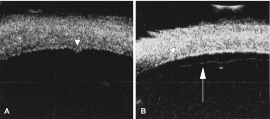
Fig. 1. Pattern 0. Radial scan-ning plane; 25-gauge surgery. A. The internal hole of the scle-rotomy site (arrowhead) shows no vitreous. B. At the internal hole of the sclerotomy site (arrowhead), UBM shows a vit-reous strand (white arrow) par-allel to but distant from the sclera.
Figure 4 presents the distribution of vitreous incar-ceration patterns for each subgroup of trocar type and vitrectomy gauge, summing up all sclerotomy sites (total number: 253 sclerotomies). P0 pattern (no incarceration) seems slightly more prevalent in valved versus non-valved 23- and 25-gauge vitrectomies, P1 slightly more prevalent in nonvalved versus valved 25-gauge vitrec-tomy, however, differences are negligible and figures overlap regarding P2 pattern (more severe incarceration) frequency.
The effect of valved trocar use on vitreous incar-ceration appeared to be somewhat beneficial, but no statistically significant effect could be shown both in a model including all data (OR: 0.85, 95% confidence interval: 0.42–1.74, P = 0.657) and in a model with 23- and 25-gauge vitrectomy data only (OR: 0.78, 95% confidence interval: 0.36–1.66, P = 0.518). Sim-ilarly, no differences in vitreous incarceration were shown among vitrectomy gauges (23, 25, or 27) both in a model including valved trocars only (P = 0.858) and in a model with all available data (P = 0.935). A post hoc power calculation in 205 sclerotomies with complete data in the 23- or 25-gauge vitrectomy
groups yielded a minimum detectable OR of 2.17, meaning that we were able to detect large differences in incarceration patterns between valved and non-valved trocars, but we were still unable to detect small-er diffsmall-erences, as also shown by the width of the 95% confidence interval in our estimates.
All other analyses were based on a model including all data as described hereafter.
Table 2 presents ORs of association with vitreous incarceration pattern for all variables in a model including the whole sample. The frequency of vitreous incarceration halved for every 10 years of age (P = 0.014), possibly related to the effect of PVD, which was a strong protective factor as it reduced incarcera-tion by almost 10 times (P, 0.001). The superotem-poral sclerotomy site was at higher risk of incarceration (P = 0.001). The use of gas tamponade led to an increased frequency of incarceration with respect to air (P, 0.001) as did, to a borderline extent (P = 0.034), phakic compared with pseudophakic status.
No patients experienced postoperative hypotony. No difference was found in the MH closure rate Fig. 2. Pattern 1. Radial scan-ning plane; 25-gauge surgery. A and B. Vitreous strand (white arrow) parallel to and in contact with the sclera without incar-ceration. Arrowheads indicate the internal hole of the scle-rotomy site.
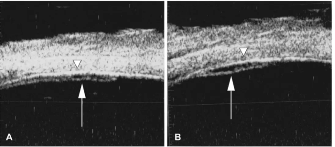
Fig. 3. Pattern 2. Radial scan-ning plane; 25-gauge surgery. Vitreous strand (white arrow) entrapped in the sclerotomy site. The internal hole of the scle-rotomy site (arrowhead) shows two different modalities of scleral wound healing (A, increased amount of tissue on the internal surface of the scle-rotomy site; B, loss of substance on the internal surface of the sclerotomy site).
(all MHs closed) or in rhegmatogenous complications after at least 5 months of follow-up.
Discussion
The use of valved vs. nonvalved cannulas creates a difference in intraocular stabilization during the surgical procedure because we do not routinely insert plugs every time we remove instruments when using nonvalved cannulas.24 Thus, we wanted to verify whether the increased intraoperative stabilization achieved with valved cannulas corresponds to different postoperative patterns of vitreous incarceration at the sclerotomy sites. The patterns of vitreal disposition at sclerotomy sites described in the literature are not uniform. These differences are linked to the method of analysis: direct visualization, UBM, or optical coherence tomogra-phy.1,11,16 However, even considering UBM studies alone, an official UBM classification of vitreal patterns at the sclerotomy site does not exist. Sabty et al16 based their scoring grade on the echographic intensity of the vitreous and its disposition at the sclerotomy
site, whereas other authors17,18only studied the occur-rence of vitreous incarceration ecographically described as“dense lines that radiated from the inter-nal hole of the incision into the vitreous cavity.” In the present series, the classification pattern adopted was related to the three morphologic dispositions that repeatedly came to light during UBM analysis. We classified them as P0 (vitreous not visible or vitreous strand parallel to but distant from the sclerotomy site); P1 (vitreous strand parallel to and in contact with the sclerotomy site); and P2 (vitreous strand entrapped in the sclerotomy site). We decided to differentiate between P0 and P1 because the highly reflective vitreous strand in contact with, although not entrapped in, the sclerotomy site (P1) was considered at greater risk for postoperative complications than the less reflective vitreous strand distant from the sclerotomy site (P0). Analogously, we decided to differentiate between P1 and P2 because vitreous entrapment (P2) was considered at greater risk of postoperative com-plications than simple contact (P1).
The homogeneity of the groups, which represents an important point in evaluating the reliability of our results, has been assessed and confirmed. The pop-ulations showed no differences in the following characteristics: male/female, phakia/pseudophakia, ep-iretinal membrane/MH, and air/gas ratios. Moreover, no difference between groups was present in relation to intraoperative maneuvers that have previously been shown to be associated with vitreal incarceration. In particular, Benitez-Herreros et al11found that cannula extraction with the light probe inserted (instead of removing the cannula with the plug introduced) reduced the amount of vitreous incarceration. In this respect, the characteristics of our populations, valved or nonvalved, at the end of the procedure are very similar, because both valved and nonvalved cannula extraction was performed without pulling the vitreous inside the eye, as the light probe does.11 Moreover, Benitez-Herreros et al13 found that the source of the incarcerated vitreous, core or pericannular, was related to different closure competencies at the 23-gauge Table 1. Baseline Characteristics Across Subgroups of Surgeries With/Without Valved Trocars and 23-, 25-, or 27-Gauge
Vitrectomy
Gauge 23 25 27
Valve Valved (n = 16) Nonvalved (n = 16) Valved (n = 20) Nonvalved (n = 20) Valved (n = 16)
Age (mean) 71.1 72.2 72.0 69.9 72.7
Diagnosis (% MH) 38 38 35 40 38
PVD (% yes) 75 69 80 80 69
Lens status (% pseudophakic) 19 31 20 15 25
Tamponade (% gas) 44 38 35 40 38
Fig. 4. Frequency of vitreous patterns for each subgroup of nonvalved (NV) (23- and 25-gauge) or valved (V) (23-, 25- and 27-gauge) trocars. Numbers within the figure are the absolute number of sclerotomies (total = 253) for each pattern and group, while the y-axis refers to the percentage for each group.
sclerotomy site in their animal model. This implies that the vitrectomy degree may alter the amount of vitreal incarceration13and confirms the previous find-ing in 20-gauge surgery of Sabti et al,16 who found that the vitreous shaving of sclerotomy sites with scleral indentation significantly reduced vitreous incar-ceration. By including only epiretinal membrane and MH eyes, we reduced the bias linked to the extent of vitreous removal. In fact, in our experience, the entity of vitreous removal may partially differ according to the clinical scenario (e.g., MH, vitreous hemorrhage, rhegmatogenous retinal detachment, and proliferative vitreoretinopathy). The homogeneity of the vitreal characteristics at entry is also important when perform-ing a comparison between groups. There were no dif-ferences in the presence of PVD at entrance between our groups. Most of the human studies evaluating vit-reous incarceration have enrolled patients with a vari-ety of diagnoses, which were probably associated with different PVD status at entry and different vitreous base constitutions,3,16–18 whereas in this study only vitreomacular interface disorders were included. Moreover, our evaluation was performed in all groups once the internal tamponade (air or gas) had disap-peared. Only Gutfleisch et al17evaluated patients with-out the tamponade at the end of the procedure, whereas other authors10,18evaluated vitreous incarcer-ation with the tamponade inside, which might have influenced both the amount of vitreous incarceration and its visualization.
In this homogenous clinical scenario, the patterns of vitreal appearance were not significantly different between the valved and nonvalved groups both for 23- and for 25-gauge PPV. In addition, no difference was present when each group was compared with the others, thus yielding equivalent results no matter the caliber or the presence/absence of the valved cannulas.
In terms of caliber, the similarities of postoperative vitreal appearance are confirmed by the results of 27-gauge surgery, which was associated with no different vitreal patterns as compared with valved 23 and 25 gauges.
The similarities in vitreal appearance among our groups were associated with an absence of differences in the MH closure rate or rhegmatogenous complica-tions. No eye had postoperative hypotony nor post-operative retinal breaks in the course of follow-up.
In all groups, vitreous incarceration was correlated with younger age, the absence of PVD at entry, as well as with gas tamponade and the superotemporal sclerotomy site. Younger age and absence of PVD could be seen as interdependent factors, in fact, younger patients are less likely to have a PVD at presentation. In our series, the intraoperative induction of PVD never induced hyaloidal separation up to the extreme periphery (the separation being completed only in the postoperative period), as occurs in patients with an already present PVD. This might influence the postoperative vitreal pattern. As for gas tamponade, it could be linked to the absence of PVD because, in the present series, gas tamponade was used only in MH patients, which presented PVD in only 42.4% of cases. Alternatively, more so than a pushing effect exerted on the residual vitreous by an extended tamponade, which seems unlikely, the gas tamponade might change the structure of the residual vitreous, possibly augmenting vitreous reflectivity and consequently its visualization through UBM. This may be confirmed by the signif-icant correlation between P2 and the superotemporal sclerotomy site. In fact, after the first week spent 12 hours/day face down, our MH patients spend most of their time in a seated position until gas disappearance, thus resulting in a prolonged effect on the superior residual vitreous. The different vitreal patterns in the different quadrants is in disagreement with Sabti et al16and in agreement with Bhende et al,27although in the latter study, the sclerotomy site most frequently associated with vitreal incarceration was the infero-temporal one. However, the present results can hardly
be compared with those obtained by other
series3,6,10,16–18,27 because the indications for PPV in previous studies were highly variable and the surgical techniques often different.
The P2 pattern occurred in 20.5% of the 253 sclerotomies analyzed in this study. Because all the sclerotomies studied were self-sealing at the end of the surgical procedure and because vitreous incar-ceration has been demonstrated to be inversely correlated with incisional leakage in an animal model,1we would have expected a higher incidence of the P2 pattern, as reported by Avitabile et al,18 Table 2. Association of Each Variable With Severity of
Vitreous Incarceration
Covariate OR P
Age (per 10 years) 0.46 (0.24–0.91) 0.014
PVD (vs. none) 0.11 (0.05–0.23) ,0.001
Valved (vs. nonvalved) trocar
0.85 (0.42–1.74) 0.657
Site: inferotemporal (ref) 1 0.025*
Site: superonasal 1.37 (0.74–2.57) 0.319
Site: superotemporal 2.82 (1.51–5.27) 0.001 Lens status (phakic/
pseudophakic) 2.50 (1.08–5.88) 0.034 Gas (vs. air) 4.28 (2.23–8.59) ,0.001 Gauge (vs. 23, ref) 1 0.935* Gauge 25 0.96 (0.44–2.11) 0.920 Gauge 27 1.15 (0.42–2.15) 0.726
López-Guajardo et al,6Benitez-Herreros et al,13and Gutfleisch et al.17 However, Texeira et al3 found a 0% entrapment rate.
In the present series, the improved intraocular stabilization obtained using valved cannulas24 does not seem to correspond to a reduced rate of postoper-ative vitreal incarceration in macular surgery because no significant differences in vitreal patterns were found between the valved and nonvalved cannula groups, and no difference was present when compar-ing each of the 4 groups (23-gauge valved and non-valved, 25-gauge valved and nonvalved) against each other. In addition, the vitreal patterns were similar between 27-gauge (only valved) and 23- and 25-gauge valved groups, meaning equivalent results no matter the caliber. Different vitreal status at entry (i.e., the absence/presence of PVD) and different tam-ponades might influence residual vitreal patterns at the sclerotomy sites.
The conclusions of our study are limited by its inability to detect differences between valved and nonvalved trocars with an OR below approximately 2 because of the small sample size. However, it is difficult to obtain UBM measurements for a large sample of patients and our series represents a signi fi-cant contribution to this issue suggesting that no substantial effects are achieved when valved trocars are used to try to reduce vitreous incarceration in the sclerotomy site.
Our results apply to macular surgery, and we cannot exclude the possibility of different results being achieved under different clinical and vitreal conditions. Key words: 23-gauge pars plana vitrectomy, 25-gauge pars plana vitrectomy, 27-25-gauge pars plana vit-rectomy, valved cannula, nonvalved cannula, vitreous incarceration at sclerotomy site, ultrasound biomicro-scopy, iatrogenic retinal tear.
References
1. Benitez-Herreros J, Lopez-Guajardo L, Camara-Gonzalez C, et al. Influence of incisional vitreous incarceration in sclerot-omy closure competency after transconjunctival sutureless vit-rectomy. Invest Ophthalmol Vis Sci 2013;54:4366–4371. 2. Singh RP, Bando H, Brasil OF, et al. Evaluation of wound
closure using different incision techniques with 23-gauge and 25-gauge microincision vitrectomy systems. Retina 2008;28: 242–248.
3. Teixeira A, Allemann N, Yamada AC, et al. Ultrasound bio-microscopy in recently postoperative 23-gauge transconjuncti-val vitrectomy sutureless self-sealing sclerotomy. Retina 2009; 29:1305–1309.
4. Benitez-Herreros J, Lopez-Guajardo L, Camara-Gonzalez C, et al. Evaluation of conjunctival bleb detection after vitrectomy by ultrasound biomicroscopy, optical coherence tomography and direct visualization. Curr Eye Res 2014;39:390–394.
5. Lin AL, Ghate DA, Robertson ZM, et al. Factors affecting wound leakage in 23-gauge sutureless pars plana vitrectomy. Retina 2011;31:1101–1108.
6. López-Guajardo L, Vleming-Pinilla E, Pareja-Esteban J, Teus-Guezala MA. Ultrasound biomicroscopy study of direct and oblique 25-gauge vitrectomy sclerotomies. Am J Ophthalmol 2007;143:881–883.
7. Taban M, Sharma S, Ventura AA, Kaiser PK. Evaluation of wound closure in oblique 23-gauge sutureless sclerotomies with Visante optical coherence tomography. Am J Ophthalmol 2009;147:101–107.
8. Taban M, Ventura AA, Sharma S, Kaiser PK. Dynamic eval-uation of sutureless vitrectomy wounds: an optical coherence tomography and histopathology study. Ophthalmology 2008; 115:2221–2228.
9. López-Guajardo L, Benitez-Herreros J, Teus-Guezala M. Opti-cal coherence tomography as a method for studying sutureless microincisional vitrectomy sclerotomies. Am J Ophthalmol 2009;148:321–322; author reply 322–323.
10. Chen D, Lian Y, Cui L, et al. Sutureless vitrectomy incision architecture in the immediate postoperative period evaluated in vivo using optical coherence tomography. Ophthalmology 2010;117:2003–2009.
11. Benitez-Herreros J, Lopez-Guajardo L, Camara-Gonzalez C, Silva-Mato A. Influence of the interposition of a non-hollow probe during cannula extraction on sclerotomy vitreous incarceration in sutureless vitrectomy. Invest Ophthalmol Vis Sci 2012;53:7322–7326.
12. López-Guajardo L, Benítez-Herreros J. Vitreous incarceration in sclerotomies. Ophthalmology 2012;119:204–205; author reply 205–206.e1.
13. Benitez-Herreros J, Lopez-Guajardo L, Camara-Gonzalez C, et al. Influence of the source of incisional vitreous incarceration on sclerotomy closure competency after transconjunctival sutureless vitrectomy. Curr Eye Res 2014;39:1194–1199. 14. Lopez-Guajardo L, Benitez-Herreros J, Camara-Gonzalez C,
Silva-Mato A. Assessment of vitreous incarceration in sclerot-omies with OCT, ultrasound biomicroscopy, and direct visual-ization. Ophthalmic Surg Lasers Imaging 2012;43(6 suppl): S117–S122.
15. Nagpal M, Wartikar S, Nagpal K. Comparison of clinical out-comes and wound dynamics of sclerotomy ports of 20, 25, and 23 gauge vitrectomy. Retina 2009;29:225–231.
16. Sabti K, Kapusta M, Mansour M, et al. Ultrasound biomicro-scopy of sclerotomy sites: the effect of vitreous shaving around sclerotomy sites during pars plana vitrectomy. Retina 2001;21: 464–468.
17. Gutfleisch M, Dietzel M, Heimes B, et al. Ultrasound biomi-croscopicfindings of conventional and sutureless sclerotomy sites after 20-, 23-, and 25-G pars plana vitrectomy. Eye 2010; 24:1268–1272.
18. Avitabile T, Castiglione F, Bonfiglio V, Castiglione F. Trans-conjunctival sutureless 25-gauge versus 20-gauge standard vit-rectomy: correlation between corneal topography and ultrasound biomicroscopy measurements of sclerotomy sites. Cornea 2010;29:19–25.
19. Benitez-Herreros J, Lopez-Guajardo L, Vazquez-Blanco M, et al. Assessment of closure competency of sutureless vitrec-tomy sclerotomies after scleral hydration. Curr Eye Res 2015; 22:1–4.
20. Lopez-Guajardo L, Benitez-Herreros J, Silva-Mato A. Experi-mental model to evaluate mechanical closure resistance of sutureless vitrectomy sclerotomies using pig eyes. Invest Ophthalmol Vis Sci 2011;52:4080–4084.
21. Rizzo S, Belting C, Genovesi-Ebert F, di Bartolo E. Incidence of retinal detachment after small-incision, sutureless pars plana vitrec-tomy compared with conventional 20-gauge vitrecvitrec-tomy in macular hole and epiretinal membrane surgery. Retina 2010;30:1065–1071. 22. Tan HS, Mura M, de Smet MD. Iatrogenic retinal breaks in 25-gauge macular surgery. Am J Ophthalmol 2009;148:427–430. 23. Gosse E, Newsom R, Lochhead J. The incidence and distribu-tion of iatrogenic retinal tears in 20-gauge and 23-gauge vitrectomy. Eye 2012;26:140–143.
24. Kim YJ, Park SH, Choi KS. Fluctuation of infusion pressure during microincision vitrectomy using the constellation vision system. Retina 2015;35:2529–2536.
25. Oellers P, Stinnet S, Mruthyunjaya P, Hahn P. Small-gauge valved versus nonvalved cannula pars plana vitrectomy for retinal detachment repair. Retina 2016;36: 744–749.
26. Privat E, Tadayoni R, Gaucher D, et al. Residual defect in the foveal photoreceptor layer detected by optical coherence tomography in eyes with spontaneously closed macular holes. Am J Ophthalmol 2007;143:814–819.
27. Bhende M, Agraharam SG, Gopal L, et al. Ultrasound biomi-croscopy of sclerotomy sites after pars plana vitrectomy for diabetic vitreous hemorrhage. Ophthalmology 2000;107: 1729–1736.