R E S E A R C H A R T I C L E
Open Access
Population diversity of the genetically
determined
TTR expression in human
tissues and its implications in TTR
amyloidosis
Andrea Iorio
1, Flavio De Angelis
1, Marco Di Girolamo
2, Marco Luigetti
3, Luca G. Pradotto
4, Anna Mazzeo
5,
Sabrina Frusconi
6, Filomena My
7, Dario Manfellotto
2, Maria Fuciarelli
1and Renato Polimanti
8*Abstract
Background: Transthyretin (TTR) amyloidosis is a hereditary disease with a complex genotype-phenotype correlation. We conducted a literature survey to define the clinical landscape of TTR amyloidosis across populations worldwide. Then, we investigated whether the genetically determined TTR expression differs among human populations, contributing to the differences observed in patients. Polygenic scores for genetically determined TTR expression in 14 clinically relevant tissues were constructed using data from the GTEx (Genotype-Tissue Expression) project and tested in the samples from the 1,000 Genomes Project.
Results: We observed differences among the ancestral groups and, to a lesser extent, among the investigated populations within the ancestry groups. Scandinavian populations differed in their genetically determined TTR expression of skeletal muscle tissue with respect to Southern Europeans (p = 6.79*10−6). This is in line with epidemiological data related to Swedish and Portuguese TTR Val30Met endemic areas. Familial amyloidotic cardiomyopathy (TTR deposits occur primarily in heart tissues) presents clinical variability among human populations, a finding that agrees with the among-ancestry diversity of genetically determined TTR expression in heart tissues (i.e., Atrial Appendage p = 4.55*10−28; Left Ventricle p = 6.54*10−35).
Conclusions: Genetically determined TTR expression varied across human populations. This might contribute to the genotype-phenotype correlation of TTR amyloidosis.
Keywords: Transthyretin, Genotype-phenotype correlation, Mutation, Amyloid, Gene expression Background
Transthyretin (TTR) amyloidosis (OMIM: 105210) is a rare, life-threatening, progressively debilitating, auto-somal dominant condition characterized by extracellular deposition of TTR-derived amyloid fibrils in peripheral and autonomic nervous system, heart, and other organs, leading to tissue damage and organ failure [1, 2]. The disorder is caused by point mutations in the TTR gene (NM_000371) located in chromosome region 18q12.1 [3]. The disease presents multiple clinical signs, including
peripheral neuropathy (sensory and motor), autonomic neuropathy, gastrointestinal impairment, cardiomyopathy, nephropathy, and ocular deposition [4]. While these symptoms may be present in patients with different TTR mutations, phenotypes are not always concordant and the same point mutation may be associated with different signs/symptoms [5]. The clinical heterogeneity of the car-riers of the TTR amyloidogenic mutations is particularly relevant from a population perspective. The most striking example of this inter-population diversity is the Val30Met mutation [rs28933979, c.148G > A, p.Val50Met], which is one of the leading causes of TTR amyloidosis [6, 7]. In the two European Val30Met endemic areas (i.e., Portugal and Sweden), Val30Met patients show two distinct phenotypic
* Correspondence:[email protected]
8Department of Psychiatry, Yale University School of Medicine and VA CT
Healthcare Center, VA CT 116A2, 950 Campbell Avenue, West Haven, CT 06516, USA
Full list of author information is available at the end of the article
© The Author(s). 2017 Open Access This article is distributed under the terms of the Creative Commons Attribution 4.0 International License (http://creativecommons.org/licenses/by/4.0/), which permits unrestricted use, distribution, and reproduction in any medium, provided you give appropriate credit to the original author(s) and the source, provide a link to the Creative Commons license, and indicate if changes were made. The Creative Commons Public Domain Dedication waiver (http://creativecommons.org/publicdomain/zero/1.0/) applies to the data made available in this article, unless otherwise stated.
presentations. In Val30Met Portuguese families, the dis-ease shows early-onset, strong severity, and high pene-trance [8, 9], whereas Val30Met Swedish patients have late-onset, intermediate severity, and low penetrance [10]. This complex genotype-phenotype correlation indicates that the clinical presentation is not only regulated by the disease-causing mutation. The amyloidogenic mutation is the cause of the disease, but other factors contribute to the modulation of the disease phenotype [11–13]. Our previous investigations focused on the role of non-coding variation in the genotype-phenotype correlation of TTR amyloidosis. We observed an enrichment for non-coding regulatory variants located in heart-related transcription-factor binding sites in African populations, suggesting a contribution to the cardiomyopathy observed in patients of African ancestry [12]. We also investigated the haplo-type structures of Val30Met and Val122Ile [rs76992529, c.424G > A, p.Val142Ile] mutations, observing in both cases independent haplotypes carrying the same disease-causing mutation [14, 15]. Non-coding variation regulates genome functions, especially through its key role in tran-scriptional mechanisms across human tissues [16]. On this basis, we hypothesized that, in presence of an amyloido-genic mutation, the non-coding regulation of TTR gene expression across tissues contributes the distribution of TTR-derived amyloid fibrils and, consequently, the disease presentation. In accordance with this hypothesis, a recent study demonstrated that, although the liver is the main TTR organ source, TTR gene expression in other tissues can also be involved in the processes related to the disease phenotype [17].
Recent studies have focused on how gene expression regulates relevant biological processes and how its alter-ation can lead to the onset of diseases [18]. The Genotype-Tissue Expression (GTEx) Project is investi-gating genetic variation in relation to gene expression in human tissues [19]. GTEx data (available at http:// www.gtexportal.org/) provide information about the rela-tionship between genetic variations and gene expression in 43 different human tissues [20]. The effects of genetic variants can be used to estimate the genetically deter-mined gene expression to investigate the role of gene ex-pression in multiple tissues with respect to disease pathogenesis [21, 22]. Accordingly, the analysis of the genetically determined TTR expression can help to dis-cern its involvement in human tissues with respect to the genotype-phenotype correlation of TTR amyloidosis.
Results
Results from literature review
We identified 88 worldwide disease-causing mutations with information regarding the ancestry (Additional file 1). Our findings indicated that Europeans have the highest number of TTR mutations (N = 60), followed by East
Asians (N = 27), Americans (N = 20), Central-South Asians (N = 8) and Africans (N = 3). The ancestry was not specified for the remaining amyloidogenic muta-tions. Few mutations are reported in multiple ancestry groups (e.g., Val30Met and Val122Ile) and several symptoms are reported for patients with different an-cestries and different mutations (e.g., cardiomyopathy and sensorimotor neuropathy) (Additional file 1). However, clinical signs partially occur in an ancestry-specific manner with respect to the amyloidogenic mutation reported (Additional file 1).
Genetically-predicted TTR expression
As introduced above, we used the data from GTEx Project [19] to build polygenic scores associated with TTR expression in 14 human tissues and tested them in the samples from the 1,000 Genomes Project [23] consid-ering both among-ancestry and within-ancestry analyses. A detailed description of procedures used is reported in the method section.
Among-ancestry comparisons
The among-ancestry comparisons showed very signifi-cant differences (p < 2.89*10−9) for genetically predicted TTR expression scores for all investigated tissue (Additional file 2) with the exception of the Esophagus– Muscularis tissue (p > 0.05). Post-hoc pairwise analysis of the among-ancestry comparisons indicated that these significant differences are generally present across mul-tiple ancestries and are not due to the diversity of a sin-gle population (Additional file 3). The only exception to this general trend is the Colon - Transverse tissue where the significant result is exclusively driven by the differ-ence between African and non-African populations (p = 5.44*10−11).
Within-ancestry comparisons
Within-ancestry comparisons showed less tissue- and ancestry-specific differences than among-ancestry com-parisons (Fig. 1). Significant differences were observed within European ancestry (Colon– Transverse p = 0.002 and Muscle – Skeletal p = 6.79*10−6), within Eastern Asian ancestry (Nerve - Tibial p = 7*10−5), and within American ancestry (Colon - Transverse p = 3.2*10−7, Colon – Sigmoid p = 2*10−4, Muscle – Skeletal p = 9*10
−4, and Skin - Sun Exposed (Lower leg) (p = 6*10−4). The
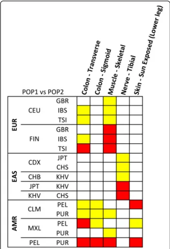
significant results of the post-doc pairwise analysis are reported in Fig. 2. In European populations, the diversity of TTR expression scores in the significant tissues is driven by North–south variability, with the most signifi-cant diversity between Scandinavian populations (i.e., the 1,000 Genomes Project FIN population) and the other European samples (Additional file 4). In Eastern Asian samples, the diversity for TTR expression scores
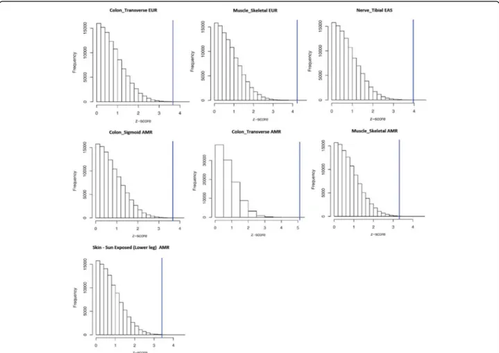
in Nerve - Tibial tissue is driven by differences of Vietnamese populations (i.e., the 1,000 Genomes Project KHV population) with respect to Japanese and Chinese populations (Additional file 5). Regarding the tissues identified in the American samples, the diversity of TTR expression scores is driven by differences between Peruvian population (i.e., the 1,000 Genomes Project PEL population) and other American populations (Additional file 6). Permutation analysis confirmed that all within-ancestry observed differences significantly di-verge from the null distribution of the permuted results (Fig. 3). The observed Z-scores are located in extremely marginal positions with respect to the null distribution of the Z-scores generated by the random permutations.
No significant differences were observed for African ancestry (nominal significance for Colon –Transverse, Esophagus– Muscularis, Liver, Muscle – Skeletal, Stom-ach, and Small Intestine; Additional file 7) and Central-South Asian ancestry (nominal significance for the Skin - Sun Exposed (Lower leg) tissue; Additional file 8).
Discussion
Our literature review indicated that few mutations were observed in multiple ancestral groups (e.g., Val30Met and Val122Ile). However, these are the mutations de-tected in most patients affected by TTR amyloidosis and the corresponding clinical signs mainly occur with ancestry-specific patterns. Although these ancestry dif-ferences are likely biased by the rare disease prevalence and the variability of the clinical practice guidelines across different countries, the inter-population diversity of the molecular mechanisms involved in the
genotype-phenotype correlation surely plays an important role in the clinical presentation observed in patients with differ-ent ancestry backgrounds. Our previous investigations indicated that TTR non-coding regions are affected by human population diversity with potential consequences on gene regulation [12, 14, 15]. Our hypothesis is in agreement with many studies about the regulatory role of non-coding variation on gene expression and other gene functions [24, 25]. TTR gene expression showed a relevant inter-individual variability across human tissues (Fig. 4), and the related tissue-specific regulatory mecha-nisms is likely to be one of the processes involved in the disease genotype-phenotype correlation. Our current findings based on a large multi-ethnic cohort (N = 2,504) and gene expression information from multiple human tissues (N = 14) provide novel insight regarding the regulatory mechanisms of TTR gene. Indeed, very few investigations explored mechanisms related to gene ex-pression in TTR amyloidosis due to limited availability
Fig. 1 Heatmap of the Kruscal-Wallis results related to the within-ancestry comparisons. The colors refer to different significance levels (red: Bonferroni-corrected significance; yellow: Nominal significance). (AFR: Africa, EUR: Europe, EAS: East Asia, SAS: South Asia, AMR: America)
Fig. 2 Heatmap of the Kruscal-Wallis post-hoc analysis results of within-ancestry comparisons. The colors refer to different significance levels (red: Bonferroni-corrected significance; yellow: Nominal significance). Information about population definitions are available at http://www.1000 genomes.org/about (EUR: Europe, EAS: East Asia, AMR: America)
Fig. 3 Distribution of the z-scores generated from 100,000 random permutations with respect to the z-scores observed in the Kruscal-Wallis post-hoc analysis of within-ancestry comparisons. (EUR: Europe, EAS: East Asia, AMR: America)
Fig. 4 TTR gene expression across the 14 clinically relevant tissues investigated the present study. This figure was extracted from the GTEx portal available at http://www.gtexportal.org/home/
of tissue samples from affected patients. In 2014, Norg-ren and colleagues [26] observed that TTR gene expres-sion is significantly higher in patients’ liver than in healthy controls. They hypothesized an impaired endo-plasmatic reticulum-associated degradation and posited that the endoplasmatic reticulum-assisted folding was caused by an overload of mutated TTR protein. Recently, an in vitro study demonstrated that Schwann cells can contribute to neurodegeneration in TTR amyl-oidosis through the local expression of mutated TTR [17]. Accordingly, TTR gene expression patterns across different tissues, including source and target organs, can contribute to the symptoms observed in patients. To provide novel information regarding this topic, we used data from the GTEx project and the 1,000 Genomes Project. Specifically, we calculated tissue-specific scores to link genetic variability to TTR gene expression and analyzed the inter-population variability considering both differences among ancestries and among popula-tions within the same ancestral groups. Our data con-firmed that non-coding variations affect gene expression with tissue-specific patterns and that human populations have significant differences. Due to the very low disease prevalence and relatively few reports regarding TTR amyloidosis, it is difficult to conduct effective compari-sons between epidemiological and molecular data. How-ever, in some cases, we observed consistency between clinical evidences and our computational results.
Since Val30Met is the most recurring mutation in pa-tients with TTR amyloidosis, numerous epidemiological studies investigated its distribution across human popu-lations. As mentioned above, the features of the two en-demic Val30Met foci in Europe are well known: Swedish patients with late-onset, intermediate severity, and low penetrance vs. Portuguese patients with early-onset, se-vere symptoms, and high penetrance [7]. Our molecular outcomes are consistent with these epidemiological data: Scandinavian populations (i.e., the 1,000 Genomes Pro-ject FIN) showed the strongest difference with respect to southern European populations (i.e., the 1,000 Genomes Project IBS and TSI) for Muscle– Skeletal tissue.
The second most recurrent TTR mutation is Val122Ile that reaches 4% in African-Americans and West Africans [27]. This mutation is mainly associ-ated with familial amyloidotic cardiomyopathy due to TTR deposits in heart tissue [28]. In our literature survey, we observed that Val122Ile was reported in multiple ancestry groups with high heterogeneity in the disease features related to the familial amyloidotic cardiomyopathy (e.g., onset and severity). Regarding heart tissues (i.e., Heart Atrial Appendage; Heart -Left Ventricle), we observed significant differences among the ancestry groups investigated that may be in agreement with the epidemiological data collected.
Besides this consistent overlapping of our results with known epidemiological evidences, we observed other strong differences within some ancestry groups that may support future epidemiological investigations. In the East Asian group, our analysis revealed a significant differ-ence between Vietnamese population (i.e., the 1,000 Genomes Project KHV population) and Japanese and Chinese groups (i.e., the 1,000 Genomes Project JPT and CHS populations) that agrees with previous studies on the genetic structure of East Asian populations [29]. Regarding TTR amyloidosis, Japan is one of the endemic foci of the disease with a prevalence of one per million, and different mutations have been identified along with a marked heterogeneity in the disease phenotypic ex-pression [30]. Chinese cases with different TTR muta-tions (e.g., Gly83Arg, Ile107Met) have also been identified [31]. To our knowledge, no reports have been published on Vietnamese or other South-Eastern Asian populations. Further studies on Eastern Asian patients may indicate strong differences within this ancestry group in accordance with our data. Another intriguing result is related to the admixed American populations of the 1,000 Genomes Project (i.e., CLM, MXL, PEL, PUR). These population clusters are an admixture of European, African, and Native American ancestry and a recent study indicated strong differences in the admixture pro-portions [32]. Previous studies demonstrated that haplo-type structure of admixed populations play an important role in gene regulatory mechanisms [33, 34]. Our current data suggested that admixture differences could contribute to the heterogeneity observed among patients from admixed American populations.
Beyond TTR hereditary amyloidosis, non-coding vari-ants associated with TTR expression could be involved in the pathogenesis of the non-inherited form of TTR amyloidosis, known as senile systemic amyloidosis. This disorder is caused by a deposition of fibrils derived from TTR in subjects that do not carry amyloidogenic muta-tions. It occurs as cardiomyopathy in elderly men with European ancestry, and TTR amyloid fibrils can be found in the hearts of the 25% of elderly individuals over 80 years of age [35]. A recent study provided suggestive evidences regarding the role of non-coding regulatory regions in wild-type TTR amyloidosis [36]. Together with these previous findings, our data suggest that TTR non-coding variation and its effect on transcription regulation are strong candidates as casual factors in the non-inherited form of TTR amyloidosis.
Conclusions
In conclusion, the current study advances the knowledge of TTR amyloidosis in terms of both data regarding the inter-population variability of the disease and method-ology that can be applied. However, our results are
affected by some limitations. The GTEx Project investigated a multi-ethnic cohort with limited sample size that cannot completely detect the effects of genetic variability on gene expression across human populations. Although our find-ings provided insights regarding TTR expression regulation, our analysis is based on data from general-population co-hort that included subjects without TTR mutations. There-fore, our findings do not account for interactions between the amyloidogenic mutation and TTR gene regulation, which likely contribute to TTR expression variability in the affected patients. Due to the large sample size (N = 2,504) used to investigate the role of non-coding variation in the regulation of TTR expression across human tissue, the pos-sibility to experimentally confirm our findings is currently limited by cost and sample availability. Finally, in addition to TTR gene expression, other mechanisms can also con-tribute to the genotype-phenotype correlation of the dis-ease, and our data may only reflect one of the molecular processes involved. Further in vivo and in vitro investiga-tions are warranted to follow up our results and confirm the role of genetically determined TTR expression in the disease onset and progression.
Methods
Literature survey
To delineate the genetic and clinical landscape of this disease among worldwide populations, we used PubMed to identify 938 scientific articles related to TTR amyloid-osis. The literature search was performed in January 2016 with the following key words: “TTR”, “TTR amyl-oidosis”, “TTR mutation”, “TTR gene”. Of these 938 pa-pers, we selected studies (n = 144) with information concerning clinical signs and TTR mutations of patients investigated. Finally, we partitioned the selected articles by ancestral group: Africa (Africans and African-Americans patients), Europe, Central-South Asia, East Asia, and America. In the American group, we included those studies involving patients of Hispanic ethnicity and/or Native American ancestry. In accordance with the vast majority of the literature regarding TTR amyl-oidosis, we named each TTR mutation in accordance with the protein change in the mature protein. We also reported rsID (when available) and the protein change in the protein precursor (Additional file 1).
Genotype and expression data
Phase 3 of the 1,000 Genomes Project was considered the reference genotype dataset [23]. We obtained the VCF (Variant Call Format) file of the 40 Kb region, which in-cludes upstream region, TTR CDS, and downstream region (GRCh37/hg19 chr 18: 29155000–29195000). Detailed information about population definitions is available at http://www.1000genomes.org/about. The VCF file of the in-vestigated region can be downloaded from the following
link: http://phase3browser.1000genomes.org/Homo_sa piens/Location/View?r=18%3A29155000-29195000.
The GTEx Version 6 data were used as reference data-sets for genetically determined gene expression [20]. GTEx cohort includes individuals with different ancestry and it was previously used for population comparisons [37]. We extracted information regarding the effects (i.e., beta values and p-values) of genetic variants on TTR gene expression in 14 clinically relevant tissues among those available in GTEx data: Colon– Transverse; Colon – Sigmoid; Esophagus –Muscularis; Esophagus – Mucosa; Heart - Atrial Appendage; Heart - Left Ventricle; Liver; Muscle– Skeletal; Nerve – Tibial; Stomach; Small Intestine - Terminal Ileum; Adipose– Subcutaneous; Cells - Trans-formed fibroblasts; and Skin - Sun Exposed (Lower leg). Finally, we identified 132 variants (131 non-coding variants e 1 coding variant) presenting comprehensive information for the 14 tissues. In the Additional file 9, we reported the GTEx statistics used to build the tissue-specific polygenic scores. The original GTEx data used in the current study can be obtained from the following link: http://www.gtexportal.org/home/testyourown.
Data analysis
The first step of the analysis was to build polygenic scores for genetically determined TTR expression for each of the 14 clinically relevant tissues. These tissue-specific polygenic scores were a sum of alleles associated with TTR expression in a specific tissue, weighted by effect sizes. As mentioned above, we used 1,000 Genomes Project data as reference dataset for LD (Linkage Disequilibrium) structure and hu-man genetic variability and GTEx data as reference datasets to determine the effect of genetic variants upon TTR ex-pression. We conducted a LD clumping analysis using Plink 1.07 toolset [38]. We included in the analysis SNPs (Single Nucleotide Polymorphisms) with at least a trend effect (p≤ 0.1) on TTR expression and considered standard LD param-eters (r2= 0.5, and region size = 10Kb). The LD clumping was conducted with respect to two perspectives: sons among ancestry groups (i.e., among-ancestry compari-sons) and comparisons across population within ancestry groups (i.e., within-ancestry comparison). Accordingly, we calculated 14 tissue-specific clumped datasets for each an-cestry (i.e., LD information across all anan-cestry) and 14 tissue-specific clumped datasets for the populations within each ancestry groups (i.e., LD information specific for each ancestry group). All calculated datasets consisted of genetic variants in non-coding regions of TTR gene and the com-position of each dataset is reported in the Additional file 10. We used these tissue-specific clumped datasets to calculate the polygenic scores for genetically determined TTR expres-sion on the basis of effect-allele count and allele effect size.
The among-ancestry and within-ancestry comparisons were performed using the Kruskal-Wallis test. This
non-parametric test permitted us to verify whether the differences observed among ancestries and among popu-lations within the same ancestries were statistically sig-nificant. To deepen the findings obtained from the Kruskal-Wallis analysis, we used Dunn’s test for the post-hoc pairwise comparisons. Bonferroni correction accounting for the number of tissues tested was applied to adjust the results for multiple-testing comparisons. Finally, we further quantified the significance of the ob-served within-ancestry differences, conducting a permu-tation analysis. Specifically, we performed 100,000 permutations of the individual tissue-specific polygenic scores with respect to their population origins and tested whether the observed differences were significantly dif-ferent from the null distribution of the permuted results.
Additional files
Additional file 1: Inter-ethnic clinical diversity associated with TTR mutations. Each mutation is named in accordance of the missense substitution in the mature protein. For each mutation, rsID and protein change in the protein precursor is also reported. (PDF 137 kb)
Additional file 2: Kruscal-Wallis analysis among ancestral groups. For each ancestral group the synthesis of the TTR expression scores, in terms of median, minimum and maximum, for each tissue involved in TTR amyloidosis is reported. (PDF 124 kb)
Additional file 3: Heatmap of the Dunn’s post-hoc test among the ancestral groups. The colors refer to different significance levels. Detailed information about population definitions is available at http://www.1000genomes.org/about (AFR: Africa, EUR: Europe, EAS: East Asia, SAS: South Asia, AMR: America). (PDF 201 kb)
Additional file 4: Kruscal-Wallis analysis within the European ancestral group. For each population the synthesis of the TTR expression scores, in terms of median, minimum and maximum, for each tissue involved in TTR amyloidosis is reported. Detailed information about population definitions is available at http://www.1000genomes.org/about. (PDF 102 kb)
Additional file 5: Kruscal-Wallis analysis within the Eastern Asian ancestral group. For each population the synthesis of the TTR expression scores, in terms of median, minimum and maximum, for each tissue involved in TTR amyloidosis is reported. Detailed information about population definitions is available at http://www.1000genomes.org/about. (PDF 101 kb)
Additional file 6: Kruscal-Wallis analysis within the American ancestral group. For each population the synthesis of the TTR expression scores, in terms of median, minimum and maximum, for each tissue involved in TTR amyloidosis is reported. Detailed information about population definitions is available at http://www.1000genomes.org/about. (PDF 115 kb)
Additional file 7: Kruscal-Wallis analysis within the African ancestral group. For each population the synthesis of the TTR expression scores, in terms of median, minimum and maximum, for each tissue involved in TTR amyloidosis is reported. Detailed information about population definitions is available at http://www.1000genomes.org/about. (PDF 90 kb)
Additional file 8: Kruscal-Wallis analysis within the Southern Asian ancestral group. For each population the synthesis of the TTR expression scores, in terms of median, minimum and maximum, for each tissue involved in TTR amyloidosis is reported. Detailed information about population definitions is available at http://www.1000genomes.org/about. (PDF 101 kb) Additional file 9: List of the selected 132 variants (GRCh37/hg19) with comprehensive information (p-value and effect size) for the 14 investigated tissues. (PDF 138 kb)
Additional file 10: A) Clumped datasets for the African ancestry. Colors refer to variants in the LD blocks (r2> 0.5; different colors represent different LD blocks) and X = presence of a variant in a specific dataset
after the LD clumping analysis. B) Clumped datasets for the European ancestry. Color refer to variants in the LD block (r2> 0.5; different colors represent different LD blocks) and X = presence of a variant in a specific dataset after the LD clumping analysis. C) Clumped datasets for the Eastern Asian ancestry. Colors refer to variants in the LD blocks (r2> 0.5;
different colors represent different LD blocks) and X = presence of a variant in a specific dataset after the LD clumping analysis D) Clumped datasets for the Southern Asian ancestry. Colors refer to variants in the LD blocks (r2> 0.5;
different colors represent different LD blocks) and X = presence of a variant in a specific dataset after the LD clumping analysis. E) Clumped datasets for the American ancestry. Colors refer to variants in the LD blocks (r2> 0.5; different
colors represent different LD blocks) and X = presence of a variant in a specific dataset after the LD clumping analysis. (PDF 88 kb)
Abbreviations
GTEx:Genotype-Tissue Expression; LD: Linkage Disequilibrium; SNP: Single Nucleotide Polymorphism; TTR: Transthyretin; VCF: Variant Call Format
Acknowledgements
The authors are grateful to Arianna Straccamore and Riccardo Boccuccia for their help in the literature review. The authors also thank the 1,000 Genomes Project and GTEx Project and their funding agencies to have made their data publically available.
Funding
The present study was supported by an Investigator-Initiated Research to the University of Rome“Tor Vergata” from Pfizer Inc. Pfizer Inc. had no role in the study design, data analysis, and results interpretation of the present study.
Availability of data and materials
This study was conducted using data from the 1,000 Genomes Project (available at http://www.1000genomes.org/) and the GTEx project (available at http://www.gtexportal.org/).
Authors’ contributions
AI and RP designed the study; AI, FDA, MF, RP conducted data elaboration; MDG, ML, LGP, AM, SF, FM and DM contributed to clinical interpretation of the data; All authors contributed to the literary review; AI and RP wrote the first draft the manuscript; All authors significantly contributed to the revision of the manuscript. All authors read and approved the final manuscript.
Competing interests
ML received travel grants from Kedrion, Pfizer, and Grifols. The other authors declare no competing interests.
Consent for publication No applicable
Ethics approval and consent to participate No applicable
Publisher’s Note
Springer Nature remains neutral with regard to jurisdictional claims in published maps and institutional affiliations.
Author details
1
Department of Biology, University of Rome Tor Vergata, Rome, Italy.2Clinical Pathophysiology Center, AFaR Foundation– “San Giovanni Calibita” Fatebenefratelli Hospital, Isola Tiberina, Rome, Italy.3Departments of Geriatrics, Neurosciences &
Orthopedics, Institute of Neurology, Catholic University of the Sacred Heart, Fondazione Policlinico Universitario A. Gemelli, Rome, Italy.4Division of Neurology and Neurorehabilitation, San Giuseppe Hospital, IRCCS-Istituto Auxologico Italiano, Piancavallo (VB), Italy.5Department of Clinical and Experimental Medicine, University
of Messina, Messina, Italy.6Genetic Diagnostics Unit, Laboratory Department,
Careggi University Hospital, Florence, Italy.7Division of Neurology,“Vito Fazzi” Hospital, Lecce, Italy.8Department of Psychiatry, Yale University School of Medicine
and VA CT Healthcare Center, VA CT 116A2, 950 Campbell Avenue, West Haven, CT 06516, USA.
Received: 9 February 2017 Accepted: 18 March 2017
References
1. Parman Y, Adams D, Obici L, Galan L, Guergueltcheva V, Suhr OB, Coelho T. European Network for T-F: Sixty years of transthyretin familial amyloid polyneuropathy (TTR-FAP) in Europe: where are we now? A European network approach to defining the epidemiology and management patterns for TTR-FAP. Curr Opin Neurol. 2016;29 Suppl 1:S3–S13.
2. Conceicao I, Gonzalez-Duarte A, Obici L, Schmidt HH, Simoneau D, Ong ML, Amass L.“Red-flag” symptom clusters in transthyretin familial amyloid polyneuropathy. J Peripher Nerv Syst. 2016;21(1):5–9.
3. Coelho T, Merlini G, Bulawa CE, Fleming JA, Judge DP, Kelly JW, Maurer MS, Plante-Bordeneuve V, Labaudiniere R, Mundayat R, et al. Mechanism of Action and Clinical Application of Tafamidis in Hereditary Transthyretin Amyloidosis. Neurol Ther. 2016;5(1):1–25.
4. Plante-Bordeneuve V. Update in the diagnosis and management of transthyretin familial amyloid polyneuropathy. J Neurol. 2014;261(6):1227–33.
5. Ando Y, Coelho T, Berk JL, Cruz MW, Ericzon BG, Ikeda S, Lewis WD, Obici L, Plante-Bordeneuve V, Rapezzi C, et al. Guideline of transthyretin-related hereditary amyloidosis for clinicians. Orphanet J Rare Dis. 2013;8:31. 6. Plante-Bordeneuve V, Said G. Familial amyloid polyneuropathy. Lancet Neurol.
2011;10(12):1086–97.
7. Coelho T, Maurer MS, Suhr OB. THAOS - The Transthyretin Amyloidosis Outcomes Survey: initial report on clinical manifestations in patients with hereditary and wild-type transthyretin amyloidosis. Curr Med Res Opin. 2013;29(1):63–76.
8. Lemos C, Coelho T, Alves-Ferreira M, Martins-da-Silva A, Sequeiros J, Mendonca D, Sousa A. Overcoming artefact: anticipation in 284 Portuguese kindreds with familial amyloid polyneuropathy (FAP) ATTRV30M. J Neurol Neurosurg Psychiatry. 2014;85(3):326–30.
9. Beirao JM, Malheiro J, Lemos C, Beirao I, Costa P, Torres P. Ophthalmological manifestations in hereditary transthyretin (ATTR V30M) carriers: a review of 513 cases. Amyloid. 2015;22(2):117–22.
10. Hellman U, Suhr O. Regional differences and similarities of FAP in Sweden. Amyloid. 2012;19 Suppl 1:53–4.
11. Santos D, Coelho T, Alves-Ferreira M, Sequeiros J, Mendonca D, Alonso I, Lemos C, Sousa A. Variants in RBP4 and AR genes modulate age at onset in familial amyloid polyneuropathy (FAP ATTRV30M). Eur J Hum Genet. 2016; 24(5):756–60.
12. Polimanti R, Di Girolamo M, Manfellotto D, Fuciarelli M. Functional variation of the transthyretin gene among human populations and its correlation with amyloidosis phenotypes. Amyloid. 2013;20(4):256–62.
13. Bonaiti B, Olsson M, Hellman U, Suhr O, Bonaiti-Pellie C, Plante-Bordeneuve V. TTR familial amyloid polyneuropathy: does a mitochondrial polymorphism entirely explain the parent-of-origin difference in penetrance? Eur J Hum Genet. 2010;18(8):948–52.
14. Polimanti R, Di Girolamo M, Manfellotto D, Fuciarelli M. In silico analysis of TTR gene (coding and non-coding regions, and interactive network) and its implications in transthyretin-related amyloidosis. Amyloid. 2014;21(3):154–62. 15. Iorio A, De Angelis F, Di Girolamo M, Luigetti M, Pradotto L, Mauro A,
Manfellotto D, Fuciarelli M, Polimanti R. Most recent common ancestor of TTR Val30Met mutation in Italian population and its potential role in genotype-phenotype correlation. Amyloid. 2015;22(2):73–8.
16. Li MJ, Yan B, Sham PC, Wang J. Exploring the function of genetic variants in the non-coding genomic regions: approaches for identifying human regulatory variants affecting gene expression. Brief Bioinform. 2015;16(3):393–412. 17. Murakami T, Sango K, Watabe K, Niimi N, Takaku S, Li Z, Yamamura K,
Sunada Y. Schwann cells contribute to neurodegeneration in transthyretin amyloidosis. J Neurochem. 2015;134(1):66–74.
18. Cheung VG, Spielman RS. Genetics of human gene expression: mapping DNA variants that influence gene expression. Nat Rev Genet. 2009;10(9): 595–604.
19. GTEx Consortium. The Genotype-Tissue Expression (GTEx) project. Nat Genet. 2013;45(6):580–5.
20. GTEx Consortium: Human genomics. The Genotype-Tissue Expression (GTEx) pilot analysis: multitissue gene regulation in humans. Science. 2015; 348(6235):648–60.
21. Gamazon ER, Wheeler HE, Shah KP, Mozaffari SV, Aquino-Michaels K, Carroll RJ, Eyler AE, Denny JC, Consortium GT, Nicolae DL, et al. A gene-based
association method for mapping traits using reference transcriptome data. Nat Genet. 2015;47(9):1091–8.
22. Gusev A, Ko A, Shi H, Bhatia G, Chung W, Penninx BW, Jansen R, de Geus EJ, Boomsma DI, Wright FA, et al. Integrative approaches for large-scale transcriptome-wide association studies. Nat Genet. 2016;48(3):245–52. 23. 1000 Genomes Project Consortium, Auton A, Brooks LD, Durbin RM, Garrison
EP, Kang HM, Korbel JO, Marchini JL, McCarthy S, McVean GA, et al. A global reference for human genetic variation. Nature. 2015;526(7571):68–74. 24. Gutierrez-Arcelus M, Ongen H, Lappalainen T, Montgomery SB, Buil A,
Yurovsky A, Bryois J, Padioleau I, Romano L, Planchon A, et al. Tissue-specific effects of genetic and epigenetic variation on gene regulation and splicing. PLoS Genet. 2015;11(1):e1004958.
25. Goring HH. Tissue specificity of genetic regulation of gene expression. Nat Genet. 2012;44(10):1077–8.
26. Norgren N, Olsson M, Nystrom H, Ericzon BG, de Tayrac M, Genin E, Plante-Bordeneuve V, Suhr OB. Gene expression profile in hereditary transthyretin amyloidosis: differences in targeted and source organs. Amyloid. 2014;21(2): 113–9.
27. Jacobson DR, Alexander AA, Tagoe C, Buxbaum JN. Prevalence of the amyloidogenic transthyretin (TTR) V122I allele in 14 333 African-Americans. Amyloid. 2015;22(3):171–4.
28. Patel KS, Hawkins PN. Cardiac amyloidosis: where are we today? J Intern Med. 2015;278(2):126–44.
29. Tian C, Kosoy R, Lee A, Ransom M, Belmont JW, Gregersen PK, Seldin MF. Analysis of East Asia genetic substructure using genome-wide SNP arrays. PLoS One. 2008;3(12):e3862.
30. Adams D, Lozeron P, Theaudin M, Mincheva Z, Cauquil C, Adam C, Signate A, Vial C, Maisonobe T, Delmont E, et al. Regional difference and similarity of familial amyloidosis with polyneuropathy in France. Amyloid. 2012;19 Suppl 1:61–4.
31. Sekijima Y, Tojo K, Morita H, Koyama J, Ikeda S. Safety and efficacy of long-term diflunisal administration in hereditary transthyretin (ATTR) amyloidosis. Amyloid. 2015;22(2):79–83.
32. Gravel S, Zakharia F, Moreno-Estrada A, Byrnes JK, Muzzio M, Rodriguez-Flores JL, Kenny EE, Gignoux CR, Maples BK, Guiblet W, et al. Reconstructing Native American migrations from whole-genome and whole-exome data. PLoS Genet. 2013;9(12):e1004023.
33. Polimanti R, Yang C, Zhao H, Gelernter J. Dissecting ancestry genomic background in substance dependence genome-wide association studies. Pharmacogenomics. 2015;16(13):1487–98.
34. Karaca S, Karaca M, Civelek E, Ozgul RK, Sekerel BE, Polimanti R. Haplotype analysis of non-HLA immunogenetic loci in Turkish and worldwide populations. Gene. 2016;587(2):132–6.
35. Ng B, Connors LH, Davidoff R, Skinner M, Falk RH. Senile systemic amyloidosis presenting with heart failure: a comparison with light chain-associated amyloidosis. Arch Intern Med. 2005;165(12):1425–9.
36. Sikora JL, Logue MW, Chan GG, Spencer BH, Prokaeva TB, Baldwin CT, Seldin DC, Connors LH. Genetic variation of the transthyretin gene in wild-type transthyretin amyloidosis (ATTRwt). Hum Genet. 2015;134(1):111–21. 37. Mele M, Ferreira PG, Reverter F, DeLuca DS, Monlong J, Sammeth M, Young
TR, Goldmann JM, Pervouchine DD, Sullivan TJ, et al. Human genomics. The human transcriptome across tissues and individuals. Science. 2015;348(6235): 660–5.
38. Purcell S, Neale B, Todd-Brown K, Thomas L, Ferreira MA, Bender D, Maller J, Sklar P, de Bakker PI, Daly MJ, et al. PLINK: a tool set for whole-genome association and population-based linkage analyses. Am J Hum Genet. 2007;81(3):559–75.