DOTTORATO DI RICERCA IN
SCIENZE FARMACEUTICHE
CICLO XXIV
COORDINATORE Prof. Stefano Manfredini
FOCUS ON NANOSTRUCTURED LIPID CARRIERS
AND MONOOLEIN AQUEOUS DISPERSIONS
Settore Scientifico Disciplinare CHIM/09
Dottorando Tutore
Dott. Ravani Laura Prof. Cortesi Rita
_______________________________ _____________________________
(firma) (firma)
Cotutore
Dott. Esposito Elisabetta
_____________________________
(firma)
“Imagination is more important than knowledge.
For knowledge is limited to all we now know and understand, while imagination embraces the entire world,
and all there ever will be to know and understand.”
1. INTRODUCTION……….. 1
1.1 NANOCARRIERS AND DRUG DELIVERY……….. 2
1.1.1 Passive targeting……….. 4
1.1.2 Active targeting………. 5
1.2 APPLICATIONS OF NANOPARTICLES IN MEDICINE………. 5
1.2.1 Therapeutics………. 5
1.2.2 Diagnostics……… 6
1.2.3 Imaging……….. 6
1.3 ADVANTAGES OF NANOPARTICLES AS DRUG DELIVERY SYSTEMS……… 7
1.4 NON-LIPID COLLOIDAL DRUG CARRIER SYSTEM……… 8
1.4.1 Polymeric NPs……….. 8 1.4.2 Ceramic NPs………. 10 1.4.3 Magnetic NPs……… 10 1.4.4 Metal-based NPs……….. 11 1.4.5 Polymeric micelles………... 11 1.4.6 Dendrimers……… 12 1.5 LIPID-BASED NANOSYSTEMS………. 13
1.5.1 Solid Lipid Nanoparticles (SLN) and Nanostructured Lipid Carriers (NLC)... 13
1.5.2 Monoolein aqueous dispersions (MAD……….... 17
2.1.1 Tristearin……… 25
2.2 LIQUID LIPIDS (OILS)...………..……… 25
2.2.1 Miglyol ® 812……… 26 2.2.2 Monoolein……….. 26 2.3 EMULSIFYING AGENTS...………..……… 26 2.3.1 Pluronic F68..……… 27 2.3.2 Pluronic F127.……….. 27 2.3.3 Soybean phosphatidylcholine…...………. 28 2.3.4 Labrasol……….…...………. 28 2.4 WATER……… 29 2.5 OTHER MATERIALS……… 29 2.6 DRUGS……….……….. 30 2.6.1 Bromocriptine..………. 30 2.6.2 L-DOPA Derivatives..…………..……… 32 2.6.3 Clotrimazole………...…...……… 36 3. METHODS………. 37 3.1 NANOSYSTEMS PRODUCTION………... 37 3.1.1 NLC preparation………... 37 3.1.2 SLN preparation………... 37 3.1.3 MAD preparation……….. 38 3.2 NANOSYSTEM CHARACTERIZATION……… 39
3.2.1 Water and dispersed phase loss after NLC and MAD production……… 39
3.2.5 Cryogenic Transmission Electron Microscopy (Cryo-TEM)…….………. 43
3.2.6 X-ray diffraction measurements………. 44
3.2.7 Differential Scanning Calorimetry (DSC)……….. 45
3.2.8 Micro Differential Scanning Calorimetry (Micro-DSC)……… 47
3.3 DRUG CONTENT………..……… 48
3.3.1 Drug content of dispersions….……….. 48
3.3.2 Stability studies………..……….. 49
3.4 HIGH PERFORMANCE LIQUID CHROMATOGRAPHY (HPLC) ANALYSES………...… 50
3.4.1 BC………... 50 3.4.2 Der-A………….………. 51 3.4.3 Der-B……….………. 51 3.4.4 Der-C……….………. 52 3.4.5 Der-D……….………. 52 3.4.6 CLO..……….………. 53 3.5 GEL PRODUCTION………..………...… 54
3.6 PREPARATION OF SIMULATED VAGINAL FLUID………...… 54
3.7 RHEOLOGICAL EVALUATION OF HYDROGELS………..………...… 55
3.8 IN VITRO TESTS………...………...… 57
3.8.1 Dialysis………...………... 57
3.8.2 Reverse dialysis method……….……… 57
3.8.3 USP XXII Paddle method……… 58
3.8.4 Franz-cells method………...………... 58
3.8.5 Esterase degradation...………... 59
3.8.6 Anticandidal activity study……...………... 59
3.8.7 Cytotoxicity assay……….………... 60
3.9.3 Permeation experiments.……… 63
3.9.4 Penetration experiments.……… 63
3.10 IN VIVO TESTS………..………...………...… 64
3.10.1 Animals………...………... 64
3.10.2 6-hydroxidopamine lesion..………….……… 64
3.10.3 Behavioural studies in hemi-parkinsonian rats……… 65
3.10.4 Statistical analysis.………...………... 66
4. NLC AND MAD FOR THE DELIVERY OF BROMOCRIPTINE……...………. 67
4.1 INTRODUCTION………...………....… 67
4.2 RESULTS AND DISCUSSION……….………...………...… 68
4.2.1 Production and characterization of dispersions...………... 68
4.2.2 BC encapsulation………..………... 77
4.2.3 In vitro activity………... 77
4.2.4 In vivo tests……….………...………... 81
5. NLC AND SLN FOR THE DELIVERY OF L-DOPA DERIVATIVES……… 86
5.1 INTRODUCTION………...………....… 86
5.2 RESULTS AND DISCUSSION……….………...………...… 87
5.2.1 Characterization of dispersions...………...……... 87
5.2.2 Prodrug recovery………..……… 89
5.2.3 In vitro experiments..………... 90
5.2.4 In vivo tests……….………...………... 91
5.2.5 Der-A SLN characterization……… 93
5.2.6 Prodrug content of formulations….……… 96
6. NLC AND MAD FOR THE DELIVERY OF CLOTRIMAZOLE……..……… 103
6.1 INTRODUCTION………...………....… 103
6.2 RESULTS AND DISCUSSION……….………...………...… 105
6.2.1 Characterization of dispersions...………...……... 105
6.2.2 Drug recovery and distribution….………..……… 108
6.2.3 Stability studies……….………... 109
6.2.4 Production of viscous forms………... 110
6.2.5 Rheological characterization of hydrogels………... 111
6.2.6 Micro-DSC……….……… 116 6.2.7 Diffusion experiments....…..………... 125 6.2.8 Ex-vivo study.…....………...………... 126 6.2.9 Anticandidal activity..……...………... 127 6.2.10 Cytotoxicity assay…..……...………... 129 7. CONCLUSIONS……….……..……… 131
7.1 NLC AND MAD FOR THE DELIVERY OF BROMOCRIPTINE...………....………. 131
7.2 NLC AND SLN FOR THE DELIVERY OF L-DOPA DERIVATIVES……….. 132
7.3 NLC AND MAD FOR THE DELIVERY OF CLOTRIMAZOLE……..……….. 133
1. INTRODUCTION
Modern nanotechnology is a dynamic field, multidisciplinary in nature. Even if the interest in nano-scale products has increased more and more especially in recent years, Mother Nature was the first scientist offering nano-materials. For example, Ayurveda, the ancient traditional system of medicine in India, has described and used several “bhasmas”, which were characterized by nanoparticles.
The first definition of modern nanotechnology was explained in 1959 by Richard Feynman at the annual meeting of the American Physical Society: “There’s plenty of room at the bottom”. However, the actual term nanotechnology was defined only in 1974 by Norio Taniguchi from Japan in a paper in which he declared: "Nano-technology mainly consists of the processing of separation, consolidation, and deformation of materials by one atom or by one molecule."
The interest in interface and colloid science together with the development of analytical tools, such as the scanning tunnelling microscope in 1981 and the atomic force microscope in 1986, fed the impetus for modern nanotechnology.
Nowadays, everyone feels the impact of nanotechnology in his life. There is a trend that urges people to revisit many research areas with a nano-view, in order to understand how the same thing can work at nano- level. This phenomenon is revolutionizing pharmaceutical sciences and many drugs are being reformulated for possibilities of delivering as a nanosystem.
1.1 NANOCARRIERS AND DRUG DELIVERY
The concept of nanoparticles and drug targeting was inspired by a visit of Paul Erlich to Karl Maria von Weber’s opera “Der Freishütz” [1]. In this opera, a so-called “Freikugeln” or flying bullets, made by calling the spirit of the devil, play a central role. Ehrlich after attending this opera thought that this concept of targeted delivery could greatly improve drug therapy. He called the delivery system that would be used in therapy “Magic Bullets”. Notwithstanding Erlich as a medical doctor, with a great interest in bacteriology and immunology, had something like antibodies in mind, this idea lead to the concept of nanoparticles and of drug targeting.
The nanoscopic era of targeted nano-carriers, linked to the concept of “Magic bullet”, began with systems proposed in the 1970s and presently is evolving into many exciting and clinically successful products in the 2000s [2].
In fact, especially in recent years, studies have revealed that an increasing number of new molecular entities (NMEs) suffer from undesirable physicochemical and biopharmaceutical properties, even if they have been selected for full scale development thanks to their safety and pharmacological data. Poor water solubility, insufficient bioavailability, high food dependency or fluctuating plasma levels are the main and common problems. Thus, it becomes evident that the development of new drugs alone is not sufficient to ensure progress in drug therapy.
In this field, colloidal carriers have attracted the main interest because they fulfil the characteristics of non-toxicity, sufficient drug-loading capacity, possibility of drug targeting, chemical and physical storage stability, controlled release characteristics and feasibility of scaling up production [3-5].
Among colloidal carriers, nanosized systems have a preeminent position because of their capability in increasing the solubility and therefore the bioavailability of poorly water-soluble substances [6-8].
The field of drug delivery has also attracted the attention of pharmaceutical industry because it offers a strategic tool to expand current drug markets; new delivery technologies could
preclude completion from generics. Targeted drug systems can convey drugs more effectively and conveniently than those of the past, increase patient compliance, extend the product life cycle, provide product differentiation and reduce healthcare costs [9].
In addition, novel drug delivery systems would offer protection and improvement of the pharmacokinetics of easily degradable molecules, such as peptides and proteins often displaying short half-lives in vivo [10,11]. Therefore, the development of techniques that could selectively deliver drugs to the pathological sites is currently one of the most important areas of drug research. The emergence of nanotechnology is likely to have a significant impact on the drug delivery sector and nanoparticles (NPs) are at the leading edge, with many potential applications in clinical medicine and research [12]. NPs can be correctly considered as the future of drug delivery technology as they have the potential to become useful therapeutic and diagnostic tools in the near future.
Nanometric carriers might differ in composition, materials, application spectrum and drug loading and concerning their different characteristics, they should be employed for various administration routes. Parenteral [13], dermal [14-16], oral [17,18], ocular [19] and pulmonary [20] applications have been widely studied for NPs. In all cases, one of the major advantages that nanotechnology offers is targeted drug delivery to the site of disease. This can be achieved either through passive targeting of drugs to the site of action or by active targeting of the drug (Fig. 1).
Figure 1. Schematic representation of different drug-targeting approaches.
1.1.1 Passive tageting
Passive targeting can be divided in EPR effect and localized delivery.
In the first case, targeting exploits the anatomical differences between normal and diseased tissues to deliver the drugs to the required site. In fact, the physiology of diseased tissues may be altered in a variety of physiological conditions through the enhanced permeability and retention (EPR) effect [21,22]. This phenomenon occurs because tumour vasculature is leaky and as consequence, circulating NPs can accumulate more in the tumour tissues than in normal tissues. Besides exploiting the structural framework of cancerous tissues, the EPR effect is also observed at the site of inflammation.
In the second case, the approach is the direct intratumour delivery of anticancer agents using NPs, which can be used in the treatment of local cancers (prostate, head, neck). Recently it has been demonstrated that transferrin conjugated anticancer drug loaded biodegradable NPs are more effective in demonstrating the antiproliferative effect of the drug than its solution or with unconjugated NPs [23]. The better efficacy of conjugated NPs was due to their greater cellular uptake and sustained intracellular retention than unconjugated NPs or the drug in solution.
1.1.2 Active targeting
Active targeting, on the other hand, requires the conjugation of receptor specific ligands that can promote site specific targeting [24,25]. The success of drug targeting depends on the selection of the targeting moiety, which should be abundant, have high affinity and specificity of binding to cell surface receptors and should be well suited to chemical modification by conjugation. The active targeting depends on molecular recognition of the diseased cells by various signature molecules overexpressed at the diseased site either via ligand-receptor, antigen-antibody interactions or by targeting through aptamers.
Conjugating the carrier with a cell or tissue-specific ligand, thereby allowing a preferential accumulation of the drug at the diseased site, can actively target the therapeutic agent.
Thus, the submicron size range of nanosystems as well as the ability to couple/conjugate different targeting ligands offer excellent opportunities to pass the physiological barriers and access different tissues followed by an efficient cellular uptake and intracellular internalization; various nanosystems can be accumulated at higher concentrations than normal drug.
1.2 APPLICATIONS OF NANOPARTICLES IN MEDICINE
The use of NPs in medicine is widespread. Especially in recent years, nanoparticulate systems are used not only for therapeutic applications, but also in diagnostics and imaging fields.
1.2.1 Therapeutics
The therapeutic applications of NPs are different, ranging from cancer therapeutics, antimicrobial actions, vaccine delivery, gene delivery and site-specific targeting to avoid the undesirable side effects of the current therapies. Many chemotherapeutic agents such as carboplatin, paclitaxel, doxorubicin and etoposide, etc., have been successfully loaded to NPs and the obtained nanopaticulate systems resulted very potent against various cancers [26]. In addition, NPs with surface-functionalised biomolecules have also been synthesized and are
studied as potential therapeutic agents. Functionalised NPs have also been used for targeted gene silencing or as therapeutics due to their antimicrobial properties.
1.2.2 Diagnostics
Nanomaterials and nanotechnology combined with modern instrumentation have the potential to understand biology and medicine at the molecular level with accurate quantification. These analyses can be possible with the aid of a variety of nanomaterials for multiplex diagnostics and thus can offer sensitive, rapid and cost effective solutions for the modern clinical laboratory. NPs are being increasingly applied to molecular diagnostics and several technologies are in development. NPs, such as gold (Au) NPs or quantum dots (QDs), are the most widely used but different other nanotechnology devices, as well as nanobiosensors, are also promising for clinical applications.
1.2.3 Imaging
The development of the effective carrier system does not only mean the execution of the delivery, but also the positive confirmation of the site-specific delivery of the drug. Consequently, the ability to track and image the fate of any nanomedicine from the systemic to the subcellular level becomes essential. NPs can be successfully exploited to improve the utility of fluorescent markers for medical imaging and diagnostic purposes.
Although various fluorescent markers are widely used in research and clinical diagnostic applications, current techniques have several disadvantages, such as the requirement of color-matched lasers, fluorescence bleaching and lack of discriminatory capacity of multiple dyes. Fluorescent NPs can greatly overcome these problems, enhancing their use to image tumours and other diseases in vivo.
1.3 ADVANTAGES OF NANOPARTICLES AS DRUG DELIVERY SYSTEMS
NPs are colloidal particles with sizes ranging from 10 to 1000 nm in diameter [27,28]. Many types of NPs can be formulated from diverse materials with the aim to serve as a possible drug delivery vehicle to treat a particular disease.
Drugs can be loaded onto NPs by various methods, such as encapsulation, surface attachment or entrapment [29]. NPs, due to their small size, can efficiently penetrate across barriers through small capillaries into individual cells, thus allowing efficient drug accumulation at target site. Therefore, the unwanted side effects and the toxicity of the therapeutic agent are reduced and the therapeutic efficacy is enhanced [30].
NPs in pharmaceutical and biotechnological sectors are useful for improving the therapeutic index of drugs and provide solutions to future delivery problems for new and upcoming classes of biotechnological products, such as recombinant proteins and oligonucleotides.
Various advantages can be ascribed to NPs:
- Increase of the aqueous solubility of the drug; - Drug protection from degradation;
- Improvement of drug bioavailability; - Production of a prolonged release;
- Provision of a targeted delivery of the drug; - Decrement of drug toxic effects;
- Possibility of an appropriate form for all routes of administration; - Rapid formulation development.
Thanks to these advantages, NPs can be used to deliver the active therapeutics to the site of action. However, despite widespread applications of these NPs, colloid stability has received little attention. To improve the stability of NPs, polymeric surfactants or other modifiers are often adsorbed or grafted to particles, forming a layer that generates an effective repulsive force between NPs that prevents flocculation.
per se meets all desired requirements for overall/general problem solution. Moreover, optimal formulations have to be chosen carefully for each drug, according to the feature of the nanocarriers. The aim is to achieve desired drug release profiles in vivo by minimizing side effects.
Concerning the materials used for the production, a possible classification of NPs is between non-lipid and lipid-based nanocarriers. Next paragraphs are dedicated to an analysis of these different systems.
1.4 NON-LIPID COLLOIDAL DRUG CARRIER SYSTEMS
1.4.1 Polymeric NPs
Polymeric NPs are nanoparticles prepared from polymers. Depending on the desired properties different types of polymers, in term of chain length, type and number can be used.
Polymers suitable for the preparation of nanoparticles include cellulose derivatives, poly(alkylcyanoacrylates), poly(methylidene malonate), polyorthoesters, polyanhydrides and polyesters such as poly(lactid acid), poly(glycolic acid) and poly (ε-caprolactone) and their copolymers [4]. Various procedures can be applied, such as the coacervation technique, the solvent evaporation [31] and solvent diffusion methods, the interfacial polymerization [32], the denaturation or desolvation of natural proteins or carbohydrates [32], and the degradation by high-shear forces (i.e. by high pressure homogenization [33] or by micro fluidization [34]). Colloidal particles are either left as aqueous dispersion or they are converted into solid form, usually by lyophilisation [35].
They also exhibit a good potential for surface modification and functionalization with different ligands, moreover they provide excellent pharmacokinetic control and are suitable to encapsulate and deliver a plethora of therapeutic agents. Depending on the type of process used for their preparation, these can be NPs, nanospheres or nanocapsules (Fig. 2).
Figure 2. Schematic representation of nanospheres and nanocapsules.
Nanospheres have a matrix-like structure where active compounds can be firmly adsorbed at their surface, entrapped or dissolved in the matrix. Nanocapsules have a polymeric shell and an inner core. In that case, an active substance is usually dissolved in the core but can also be adsorbed at their surface [27,36]. The drug is dissolved, entrapped, encapsulated or attached to a nanoparticle.
The main advantage of using NPs for drug-delivery applications is their small size when taken up by cells, which could allow efficient drug accumulation at the target sites [37].Biodegradable materials used for the formulation of NPs allow sustained drug release within the target site over a period of days or even weeks. Biodegradable NPs formulated from poly D, L-lactide co-glycolide (PLGA) and polylactide (PLA) have been investigated for sustained drug delivery [36,38].
The main interest of the researchers is to study their intracellular trafficking and determine the parameters that are critical to their efficient cellular uptake and retention. Recently, studies have demonstrated rapid escape of NPs from the endo-lysosomal compartment to the cytoplasmic compartment [38].
Multidrug resistance (MDR) is one of the major causes of treatment failure in cancer therapy, which may be attributed to the decreased accumulation of drug in the tumour site together with the activity of membrane glycoprotein (P-gp)-dependent accelerated drug efflux [39,40]. The performed studies suggest that the use of targeted, antisense agent NPs would be a potential
approach to overcome tumour drug resistance [41].
Another characteristic of NPs is their ability todeliver drugs to the target sites across biological barriers such as the blood-brain barrier (BBB) [42,43]. The brain delivery of a wide variety of drugs, such as antineoplastic and anti-HIV drugs, is markedly hindered because they have great difficulty in crossing the BBB [44]. Thus, by using the nanotechnological approaches, researchers have tried to improve the pharmacokinetics of drugs for the treatment of central nervous system (CNS) diseases. Theapplication of NPs to brain delivery is a promising way to overcome this barrier. Although not fully elucidated, the most likely transport mechanism for these particles is via endocytosis across the endothelial cell lining of the BBB. Moreover, by packaging therapeutic molecules inside aliposome and decorating the surface of the liposome usingmolecular “Trojan horse” technology, researchers have obtainedpromising results [45]. Recently many surface-modified NPs have been used to treat various diseases. Surface modification of PLGA NPs with polyethyleneimine (PEI) utilizing a cetyl-derivative was used to improve surface functionalization and aid siRNA delivery [46].
1.4.2 Ceramic NPs
The use of inorganic (ceramic) particles for drug delivery, especially biomacromolecular therapeutics, is emerging as a new area [47], because of their ultra-low size (less than 50 nm) and porous nature. It is well documented that smaller-sized drug-carrier systems are more effective in evading the uptake by the RES [48]. Silica and other materials such as alumina, titania, etc., which are highly compatible with biological systems because of their inert nature, are widely being used for the formulation of NPs [49,50].
1.4.3 Magnetic NPs
Magnetic NPs (MNPs) are a class of nanoparticle that can be manipulated using magnetic field. Such particles commonly consist of magnetic elements such as iron, nickel and cobalt and their chemical compounds.
MNPs are increasingly being being widely used for biotechnological and biomedical applications. Drug delivery may be benefited by the use of MNPs because these particles can be addressed to a specific site, such as a tumour, thereby reducing the systemic distribution of cytotoxic compounds in vivo, result in effective treatment at lower doses [51].
1.4.4 Metal-based NPs
Metal NPs can be synthesized in extremely small sizes of around 50 nm and thus the large surface area provides the ability to carry a relatively higher dose of drugs.
AuNPs are the most commonly used as they offer manifest advantages. It is easy to synthesize a range of sizes of Au NPs by changing few parameters by simple, cheap and reliable methods. Other common metal-based NPs are silver NPs and amphiphilic TiO2 nanotube.
Silver NPs have wound-healing properties improving cosmetic appearance in a dose-dependent manner after topical delivery. These NPs also exert positive effects through their antimicrobial properties, reduction in wound inflammation and modulation of fibrogenic cytokines [52].
Amphiphilic TiO2 nanotube arrays can serve as an actively controllable drug delivery system by
utilising the photocatalytic ability of TiO2 [53].
1.4.5 Polymeric micelles
Polymeric micelles represent a class of colloidal system formed of block copolymers consisting of hydrophilic and hydrophobic monomer units [54]. These micelles are composed of a core of hydrophobic blocks stabilized by a corona of hydrophilic polymeric chains. Although a variety of hydrophilic polymers can be used, in majority of cases, PEG blocks with a molecular weight ranging from 1 to 15 kDa are used as corona-forming blocks and the length of a hydrophobic core-forming block is close or somewhat lower than that of a hydrophilic block. Enhancing drugs' solubility using micelle-forming surfactants results in increased water solubility of a poorly soluble drug. They also improve drugs' bioavailability by enhancing their permeability across physiological barriers. Thus, they bring about substantial changes in drug biodistribution.
1.4.6 Dendrimers
Dendrimers derive their name from the Greek word “dendra”, meaning reminiscent of a tree. They are polymeric molecules composed of multiple perfectly branched monomers that emanate radially from a central core. Though many biological applications use dendrimers based on polymers, such as polyamidoamines (PAMAMs), polyamines, polyamides (polypeptides), poly (aryl ethers), polyesters, carbohydrates and DNA, in most cases PAMAM dendrimers are used [55,56].
1.5 LIPID-BASED NANOSYSTEMS
Although within the remit of the above mentioned nanoscience initiatives, lipid-based nanosystems such as nanoemulsions, lipid-core micelles, small unilamellar vesicles, solid lipid nanoparticles, nanostructured lipid carriers, colloidal liquid crystalline structures, cubosomes and variations thereof, have long been in existence and some have long been improving patient's lives. Indeed, lipid-based nano-formulations are among the most attractive candidates for improving drug solubility and for site-specific targeting following parenteral administration. In recent years, a particular attention has been focused on new generations of lipid nanoparticles such as nanostructured lipid carriers and monoolein aqueous dispersions, derived from the mesophases originated from the system monoolein/water/poloxamer. These nanoparticles have been analysed as possible drug delivery systems and are the object of the discussion of this doctorate project.
1.5.1 Solid Lipid Nanoparticles (SLN) and Nanostructured Lipid Carriers (NLC)
Solid lipid NPs (SLN) are a colloidal carrier system for controlled drug delivery developed at the beginning of the 1990s as an alternative carrier system to emulsions, liposomes and polymeric NPs [12]. These particles are delivery systems where the nanodispersed phase has a solid matrix of crystalline solid lipids (i.e. lipids solid both at room and at body temperature) and is stabilized by surfactant(s). SLN can be formulated by using highly purified triglycerides, complex glyceride mixtures or even waxes.
In comparison with other particulate carriers, SLN have many advantages for drug delivery, such as good tolerability, biodegradability [57], a high bioavailability by ocular administration [58] and a targeting effect on the brain [59]. SLN have been developed and investigated for parenteral, pulmonary and dermal application routes [60-62].
In recent years, the study of SLN has markedly increased, especially with the method of high pressure homogenization.
particular organs. The particles, together with all intravenously injected and colloidal particulates, are cleared from the circulation by the liver and spleen. In tumour tissues, drugs can be targeted by PEG-coated polymeric NPs (stealth NPs) that helps in escaping from reticuloendothelial system (RES) [63].This effect may be also achieved using block polyoxyethylene polypropylene copolymers like Pluronic F188 in which the hydrophobic portion of the molecule forms the NP matrix while the water soluble polyoxyethylene block forms a hydrophilic coating on the particle. Stealth SLN increases the tumour accumulation, antibacterial activity of antiparasitic and antifungal drugs, and allows brain delivery of anticancer drugs not capable of crossing the BBB [64].
Clear advantages of SLN include the use of physiological well tolerable lipids [65], the avoidance of organic solvents in some preparation processes, a wide potential application routes (dermal, oral, intravenous) and the rapid and effective preparation process allowing the possibility of large scale production [66].
Common disadvantages of SLN include particle aggregation, particle growth, unpredictable gelation tendency, unexpected dynamics of polymorphic transition, burst drug release and consequently low incorporation capacities due to the crystalline structure of the solid lipid [3,67,68].
This last mentioned drawback can be explained by the crystallization process to which SLN are subjected during the cooling process, causing a transition of lipids in higher energy modification α and β’. Triglycerides are known to crystallize mainly in three polymorphic forms which transform monotrophically from α via β′ to β [69].
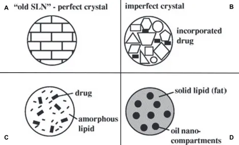
During storage, SLN triglycerides are subjected to a shift into the low energy and more ordered β modification. The formed high degree of order causes a reduction of imperfections in the crystal lattice and consequently, the drug expulsion (Fig. 3).
Figure 3. Mechanism of drug expulsion during storage of SLN dispersions; transition to highly ordered lipid crystal
[15]
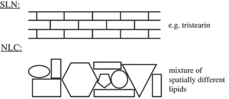
To obtain a sufficiently high drug load, the creation of a less ordered solid lipid matrix is necessary. In this case, the drug can dispose itself between the chains of the fatty acids or between the lipid layers and the imperfections. When SLN are formulated with mono acid highly purified glyceride, such as Tristearin, the drug loading is limited and very frequently drug expulsion occurs within hours or few days due to the formation of the perfect β modification [70]. The second generation of SLN, avoiding some disadvantages of classic SLN, is represented by Nanostructured Lipid Carriers (NLC), which are composed of a solid lipid matrix with a certain content of a liquid lipid phase [71].
Liquid lipids are able to better disperse lipophilic drugs than solid lipids. The liquid lipids form droplets within the solid lipid particles matrix, providing a high incorporation capacity and a control of drug release (Fig 4).
Figure 4. Perfect crystal in SLN comparable with a brick wall and structure with imperfections due to spacially very
The use of different lipid molecules (solid and liquid at room temperature) in the preparation of NLC allow the building of the matrix of the wall with different types of stones leaving enough imperfections to accommodate the drugs. In this way the problem of the formation of a perfect crystal, that seems a wall made of bricks, is overcome.
Especially, as schematized in figure 5, three types of NLC can be produced: 1. Imperfect type
2. Amorphous type 3. Multiple type
Figure 5. Different types of NLC compared to the ordered matrix of SLN (A); NLC types: imperfect type (B),
amorphous type (C), multiple type (D) [15]
The process of drug loading in SLN is due to the formation of lipid crystals. The formation of a perfect crystal causes drug expulsion. Thus, by avoiding crystallization and creating more imperfections in the crystal the drug loading is higher (Fig 5A).
NLC are solid but not crystalline particles. By using special mixtures of solid lipids and liquid lipids (e.g. isopropylmyristate, hydroxyloctacosanylhydroxylstearate) the obtained nanoparticles become solid after cooling but they don’t crystallize (Fig. 5C).
A B
From the production point of view, a too high concentration of drug in the molten lipidic phase could lead to an immediate drug expulsion during the cooling process or create a dilution in the cold water. Based on these supposes, the multiple type of NLC was developed (Fig. 5D).
In this last structure, the solid matrix of the lipid nanoparticle contains tiny liquid nano-compartments of oil, in which the drug solubility is higher. It follows an increase in the total drug loading capacity. The oil nano-compartments are all surrounded by solid matrix, allowing, also in this case, a prolonged drug release.
1.5.2 Monoolein aqueous dispersions (MAD)
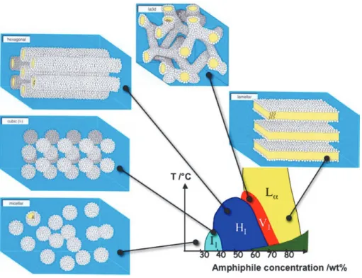
Surfactants, lipids and polymer molecules that have both polar and nonpolar components are called amphiphilic. The hydrophobic effect drives amphiphilic molecules into polar solvents to spontaneously self-assemble into a rich array to thermodynamically stable lyotropic liquid crystalline phases with characteristic lengths on the nanometer scale.
The content of water or other solvent molecules changes the self-assembled structures. A generic progression of phases, going from low to high amphiphile concentration, is: 1. Discontinuous cubic phase (micellar cubic phase)
2. Hexagonal phase (hexagonal columnar phase) (middle phase) 3. Lamellar phase
4. Bicontinuous cubic phase
5. Reverse hexagonal columnar phase
6. Inverse cubic phase (Inverse micellar phase)
At very low amphiphile concentration, the molecules will be dispersed randomly without any ordering. At slightly higher (but still low) concentration, amphiphilic molecules will spontaneously assemble into micelles or vesicles. This is done so as to 'hide' the hydrophobic tail of the amphiphile inside the micelle core, exposing a hydrophilic (water-soluble) surface to aqueous solution. These spherical objects do not order themselves in solution and are denoted by the symbol I1. This is a highly viscous, optically isotropic phase in which the micelles are arranges
on a cubic lattice.
At higher amphiphile concentrations the micelles fuse to form cylindrical aggregates of indefinite length, and these cylinders are arranged on a long-ranged hexagonal lattice. This lyotropic liquid crystalline phase is known as the 'hexagonal phase', or more specifically the 'normal topology' hexagonal phase and is generally denoted by the symbol HI.
At higher concentrations of amphiphile the 'lamellar phase' is formed. This phase is denoted by the symbol Lα and consists of amphiphilic molecules arranged in bilayer sheets separated by
layers of water. Each bilayer is a prototype of the arrangement of lipids in cell membranes. For most amphiphiles that consist of a single hydrocarbon chain, one or more phases having complex architectures are formed at concentrations that are intermediate between those required to form a hexagonal phase and those that lead to the formation of a lamellar phase. Often this intermediate phase is a bicontinuous cubic phase (Fig. 6).
Figure 6. Schematic showing the aggregation of amphiphiles into micelles and then into lyotropic liquid crystalline
The lamellar phase is the basis of the planar lipid bilayer of the vesicle structure, while the bicontinuous phase consists of a single continuous curved bilayer that forms a complex network with 3D cubic symmetry and continuous water channels. Cubic phase surface could assume different curvatures: Ia3d (gyroid surface, G), Pn3m (diamond surface, D), lm3m (primitive surface, P) [72] (Fig. 7).
Fig.7 Cubic phase surface curvatures. Individual lipids are shown as small circles with adjoined curved lines, representing the polar
headgroup and hydrophobic acyl chain respectively. The red and green regions represent water. [72]
Monoglycerides are polar lipids with poor water solubility that exhibit aqueous phase behaviour, reflecting their structural similarity to nonionic surfactants. The most studied, among aqueous monoglycerides systems, is that formed by monoolein, the unsaturated C18 monoglyceride.
As shown in figure 6, the formation of cubic phases is often localized in a narrow region between lamellar and hexagonal liquid crystalline phases. Differently, the monoolein-water system is the only that possesses a cubic phase region spanning large compositional and temperature ranges (Fig. 8).
Fig.8 Aqueous phase behaviour of the monoolein-water system, evidencing the existence of two cubic phases [73].
In the cubic phase, the minimal surface is formed by the self assembled bilayer that occurs as the hydrophobic or hydrophilic portions of the surfactants molecules line up to minimize their interaction with their opposite. The three structures, P-, D- and G- surfaces, are all bicontinuous (i.e., they divide space into two continuous but nonintersecting regions); in the case of cubic phases, two separate regions of hydrophilic material (water channels) are formed.
Cubic liquid crystals are transparent, isotropic viscous phases and physically stable in excess water [74-77]. Since cubic phases are characterized by high viscosity, they are difficult to administrate by parenteral routes. For this reason some researchers have developed the emulsification of cubic lipid phases in water resulting in the production of cubosomes that can be defined as nanoparticulate disperse systems characterized by high biocompatibility and bioadhesivity [78,79].
Dispersed particles of bicontinuous cubic phases were first observed during studies of fat digestion that simulated stomach contents by combining oil with lipase and bile salts [80].
One of the first published instances of the word “cubosomes” appears in a review written by Larsson [81] in which the author discussed about implications of bicontinuous cubic phases to
biological membranes. Cubosomes structures, such as bulk cubic phases, are critical elements of some biological processes and for example, occur naturally in bacterial cells [82].
The formation of cubosomes is possible in binary and ternary systems with a sufficiently large miscibility gap between the cubic phase and the solvent. Colloidal stabilization of cubosomes is good when Poloxamer 407 is used to provide steric stabilization against aggregation and coalescence, but other suitable polymers can be used as well [83].
Concerning their applications in pharmaceutical and biotechnological fields, cubosomes have been proposed as a delivery system able to provide both a solubilization benefit (increased drug payload) and a system for controlled or sustained release [84]. In particular, cubosomes show the ability to solubilize hydrophobic, hydrophilic and amphiphilic molecules (Fig. 9) and a high biodegradability by simple enzyme action [85].
Fig.9 Cubosome exhibiting its cavernous internal and cubic structure and its membrane composition with different drug-loading modalities.
Compared to liposomes or vesicles, cubosomes possess much higher bilayer area-to-particle volume ratios as well as higher viscous resistance to rupture [84]. Because of their properties, these versatile delivery systems can be administrable by different routes (such as orally,
parenterally, or percutaneously) [86].
The methods of preparation [87,88] and the inner structure [89,90] of cubosomes have been widely studied. Nevertheless, drug release from these systems has been poorly investigated [91].
1.6 AIMS AND ORGANIZATION OF THE THESIS
In recent years, the field of lipid nanostructures shows a growing interest from the pharmaceutical technology research groups world wide. In particular, Solid Lipid Nanoparticles (SLN), Nanostructured Lipid Carriers (NLC) and Monoolein Aqueous Dispersions (MAD) have been proposed as new carrier systems for many applications.
In this thesis different NLC, SLN or MAD formulations were developed as drug delivery systems specifically for antiparkinson or antimycotic drugs, in order to prolong their action and reduce the side effects. The thesis consists of seven main chapters.
The first chapter contains an introduction on nanoparticulate systems, focusing the attention on properties and the differences existing between non-lipid and lipid vectors.
The second and third chapters describe materials and methods used in this work respectively.
From chapter four to six, the applications of lipidic nanosystems as delivery vectors are analysed in order to improve the bioavailability and controlled release of incorporated drugs.
In chapter four, NLC and MAD formulations containing Bromocriptine are described and compared. In particular, characterization of morphology, size, inner structure and drug distribution of nanosystems have been made. Finally, in vitro and in vivo analyses have been performed.
In chapter five, four different new L-dopa derivatives are studied as model drugs for NLC and SLN formulations. After the inclusion in nanoparticulate systems, the derivatives have been analysed in dimensions, morphology, L-dopa release by time and in vitro release tests.
In chapter six, NLC and MAD are described as possible drug delivery systems for Clotrimazole, an antimycotic agent. After dimensional, morphology and stability studies, NLC and MAD have been viscosized in order to facilitate a mucosal application and delivery of the drug. The obtained gels have been analysed through rheological and calorimetric assays, then cytotoxic and anticandidal activities have been tested.
2. MATERIALS
The materials used in this work are only partially composed of individual chemical substances and their composition may differ according to the manufacturer. Therefore, the physicochemical properties and the producer details for all the materials used are given in the present section.
2.1 SOLID LIPIDS
Lipids that are solid at room temperature are saturated. The solid lipids used in this work are well tolerated, accepted for human use and they are characterized by in vivo biodegradability.
2.1.1 Tristearin
Tristearin is also known as glycerol tristearate, stearin, glyceryl tristearate, trioctadecanoin, hardened oil, stearin, tri-, spezialfett 118, stearic triglyceride.
His molecular formula is C57H110O6 and his molecular weight is 891.4797.
Tristearin is the primary fat in beef. It is a triglyceride, a molecule of glycerine has reacted with three molecules of the fatty acid stearic acid. It is a saturated fat, solid at room temperature. Tristearin was provided by Fluka (Buchs, Switzerland).
2.2 LIQUID LIPIDS (OILS)
Liquid lipids at room temperature are unsaturated. The oils used in this work are well tolerated and accepted for human use.
2.2.1 Miglyol
® 812
It is a liquid lipid at room temperature, having a density between 0.945 and 955 g/cm3. This oil
consists of medium chain triacylglycerols (C8-C10) (caprylic/capric triglycerides) and it is virtually colourless and of neutral odour and taste. It is used as skin oil and as dissolution medium for many substances. It is soluble in hexane, toluene, diethyl ether, ethyl acetate, acetone, isopropanol and ethanol 96%. It is miscible in all ratios with paraffin hydrocarbons and natural oils. The acid value is 0.1 mg KOH/g and the saponification value is between 325-345 mg KOH/g [92].
Miglyol 812 was purchased from Eigenmann & Veronelli (Rho, Milano, Italy).
2.2.2 Monoolein
Synonyms of monoolein are RYLO MG 19, Arlacel™ 186, D-L-alpha-monoolein, delta 9 cis monoolein, glycerol-1-monooleate, glycerol alpha-monooleate, glycerol monooleate, glyceryl monooleate, glyceryl cis-9-octadecenoate. His molecular formula is C21H40O4 and his molecular
weight is 356.54.
Monoolein is a monoacylglycerol with 9-cis-octadecenoic acid at the sn-1 position of glycerol. The mesophase propensities and structure of monoolein dispersed in water are of interest in a number of areas ranging from controlled uptake and release to cosmetic, food and pharmaceutical formulations.
RYLO MG 19 was purchased from Danisco Cultor (Grindsted, Denmark).
2.3 EMULSIFYING AGENTS
The International Union of Pure and Applied Chemistry (IUPAC) defines the properties of an emulsifying agent as a surfactant, which is positively adsorbed at interfaces and lowers the interfacial tension [93]. Surfactants have amphiphilic structures and are able to form micelles. Some polymers (i.e. poloxamers) can function in the same manner, if they have a sufficient
surface activity.
2.3.1 Pluronic F68
Lutrol F 68, (oxirane, methyl-, polymer with oxirane (75;30)) or poloxamer 188 is a water-soluble non-ionic polyoxyethylene-polyoxypropylene polymer. Its value in the HLB system (Hydrophile - Lipophile Balance) amounts to 29. The stabilizer and solution enhancer does not cause toxic reactions after parenteral, dermal or peroral administration [92]. Moreover, the transition time of gut is not influenced in GIT of rats [92]. Tenside micelles are not likely to occur in the concentration range of poloxamer used in this study.
Poloxamer 188 arranges only at higher concentrations and temperatures in form of micellar structures [94,95].
Lutrol F 68, was a purchased from BASF ChemTrade GmbH (Burgbernheim, Germany).
2.3.2 Pluronic F127
Pluronic F127 (PEO98-POP67-PEO98) or poloxamer 407, is a hydrophilic non-ionic surfactant of
the more general class of copolymers known as poloxamers. Poloxamer 407 is a triblock copolymer consisting of a central hydrophobic block of polypropylene glycol flanked by two hydrophilic blocks of polyethylene glycol. The approximate length of the two PEG blocks is 101 repeat units while the approximate length of the propylene-glycol block is 56 repeat units.
Poloxamer 407 shows thermo-reversible properties. Its fluid state at room temperature facilitates the administration while its gel state above sol-gel transition temperature promotes prolonged release of pharmacological agents. Pharmaceutical evaluation consists in determining the rheological behaviour (flow curve or oscillatory studies), sol-gel transition temperature, in vitro drug release using either synthetic or physiological membrane and (bio)adhesion characteristics. Poloxamer 407 formulations led to enhanced solubilisation of poorly water-soluble drugs and prolonged release profile for many galenic applications (e.g., oral, rectal, topical, ophthalmic, nasal and injectable preparations) but did not clearly show
relevant advantages when used alone. Combination with other excipients like poloxamer 188 or mucoadhesive polymers (i.e. xanthan gum, sodium alginate, methyl cellulose and others) promotes poloxamer 407 action by optimising sol-gel transition temperature or increasing bioadhesive properties. Inclusion of liposomes or micro(nano)particles in poloxamer 407 formulations offers interesting prospects, as well [96].
Pluronic F127 was obtained from BASF (Ludwigshafen, Germany).
2.3.3 Soybean phosphatidylcholine
Phosphatidylcholine (PC) is a phospholipid that incorporates choline as a head group. Phosphatidylcholine is a major constituent of cell membranes and is more commonly found in the exoplasmic or outer leaflet of a cell membrane. It is thought to be transported between membranes within the cell by phosphatidylcholine transfer protein (PCTP) [97].
Phosphatidylcholines are composed of a choline head group and glycerophosphoric acid with a variety of fatty acids, one being a saturated fatty acids and one being an unsaturated fatty acid. The phospholipids are such a major component of lecithin that in some contexts the terms are sometimes used as synonyms. However, lecithin extract consists of a mixture of phosphatidylcholine and other compounds.
In pharmaceutical applications it acts as a wetting and stabilizing agent, it helps in emulsifications and encapsulation and it is a good dispersing agent. It can be used in manufacture of intravenous fat infusions and for therapeutic use.
Phospholipon® 90G (P 90G) is a highly purified soybean lecithin that contains at least 90% phosphatidylcholine. P 90G was supplied by Rhône-Poulenc-Rorer (Germany).
2.3.4 Labrasol
Labrasol ® or caprylocaproyl macrogol-8 glycerides is a non-ionic water dispersible surfactant composed of well-characterised polyethylene glycol (PEG) esters, a small glyceride fraction and free PEG.
It is able to self-emulsify on contact with aqueous media forming a fine dispersion or to act as solubilizer and wetting agent. It is also used as bioavailability enhancer.
It is obtained from coconut oil and has very low toxicity with an LD50 of 22g/kg for rats. Labrasol® was purchased form Gattefossé (France).
2.4 WATER
The water used in all experiments was purified water by reverse osmosis and was obtained from a Milli Q Plus system (Millipore®, USA).
2.5 OTHER MATERIALS
FL-70 was used for SdFFF analyses. FL-70 is a detergent composed of water 88.8%, triethanolamine oleate 3.8%, sodium carbonate 2.7%, Alcohols, C12-14-secondary, ethoxylated 1.8%, Tetrasodium ethylenediaminetetraacetate 1.4%, Polyethylene glycol 0.9%, Sodium oleate 0.5%, Sodium bicarbonate 0.1%.
2.6 DRUGS
2.6.1 Bromocriptine
Figure 10. Chemical structure of Bromocriptine.
Bromocriptine (Fig. 10), an ergoline derivative, is a dopamine agonist that is used in the treatment of pituitary tumours, Parkinson’s disease (PD), hyperprolactinemia, neuroleptic malignant syndrome and type 2 diabetes.
Bromocriptine is a dopamine receptor agonist, which activates postsynaptic dopamine receptors. The dopaminergic neurons in the tubero-infundibular process modulate the secretion of prolactin from the anterior pituitary by secreting a prolactin inhibitory factor (thought to be dopamine); in the corpus striatum the dopaminergic neurons are involved in the control of motor function. Clinically, Bromocriptine significantly reduces plasma levels of prolactin in patients with physiologically elevated prolactin as well as in patients with hyperprolactinemia. The inhibition of physiological lactation as well as galactorrhea in pathological hyperprolactinemic states is obtained at dose levels that do not affect secretion of other tropic hormones from the anterior pituitary. Experiments have demonstrated that Bromocriptine induces long lasting stereotyped behaviour in rodents and turning behaviour in rats having unilateral lesions in the substantia nigra. These actions, characteristic of those produced by dopamine, are inhibited by dopamine
antagonists and suggest a direct action of Bromocriptine on striatal dopamine receptors.
Bromocriptine is a nonhormonal, nonestrogenic agent that inhibits the secretion of prolactin in humans, with little or no effect on other pituitary hormones, except in patients with acromegaly, where it lowers elevated blood levels of growth hormone in the majority of patients.
In about 75% of cases of amenorrhea and galactorrhea, Bromocriptine therapy suppresses the galactorrhea completely, or almost completely, and reinitiates normal ovulatory menstrual cycles.
Galactorrhea may take longer to control depending on the degree of stimulation of the mammary tissue prior to therapy. At least a 75% reduction in secretion is usually observed after 8-12 weeks. Some patients may fail to respond even after 12 months of therapy.
In many acromegalic patients, Bromocriptine produces a prompt and sustained reduction in circulating levels of serum growth hormone.
Bromocriptine produces its therapeutic effect in the treatment of Parkinson's disease, a clinical condition characterized by a progressive deficiency in dopamine synthesis in the substantia nigra, by directly stimulating the dopamine receptors in the corpus striatum.
The pharmacokinetics and metabolism of Bromocriptine in human subjects were studied with the help of radioactively labelled drug. Twenty-eight percent of an oral dose was absorbed from the gastrointestinal tract. The blood levels following a 2.5 mg dose were in the range of 2-3 ng equivalents/mL. Plasma levels were in the range of 4-6 ng equivalents/mL indicating that the red blood cells did not contain appreciable amounts of drug and/or metabolites. In vitro experiments showed that the drug was 90%-96% bound to serum albumin.
Bromocriptine was completely metabolized prior to excretion. The major route of excretion of absorbed drug was via the bile. Only 2.5%-5.5% of the dose was excreted in the urine. Almost all (84.6%) of the administered dose was excreted in the faeces in 120 hours.
Bromocriptine mesylate, 2-Bromo--ergocriptine methansulfonate salt (BC), amphetamine and 6-hydroxydopamine (6-OHDA) were purchased from Sigma Chemical Company (St Louis, MO, USA).
2.6.2 L-DOPA derivatives
L-Dopa (LD) is a direct precursor of dopamine (DA), which is deficient in the brains of patients suffering from Parkinson's disease (PD). Although LD is the treatment of choice, its metabolism generates a variety of free radicals that contribute to the progression of the disease, increasing the loss of nigrostriatal dopaminergic neurons [98]. In particular, it was demonstrated that systemic administration of LD in the rat increases the production of free radicals in the substantia nigra [99]; in addition, cell-death, both of the necrotic and the apoptotic types, was observed in neuronal and non-neuronal cell cultures treated with LD [100].
Experimental studies have also shown that LD alters cellular energy metabolism, probably by inducing oxidative damage of specific enzymes of the mitochondrial respiratory chain [101]. Miller et al. confirmed that catecholamines, including the neurotransmitter DA and its biochemical precursor LD, are subjected to autoxidation [102]. This process, which is accelerated in the presence of pro-oxidative transition metals, such as iron and copper, is enhanced by the presence of neuromelanin in dopaminergic neurons, due to its reported ability to accumulate iron and consequently acting through promotion of Fenton and Haber-Weiss reactions with production of potentially cytotoxic reactive oxygen species (ROS) [103].
These ROS have been hypothesized to play a role in the progressive and selective loss of nigrostriatal dopaminergic neurons that occurs in aging and in neurodegenerative disorders such as PD and Alzheimer disease [104]. This hypothesis suggests that inhibition of catecholamine autoxidation and the scavenging of ROS produced by such oxidation are important strategies for preventing or slowing down the progression of aging and aged-related neurodegenerative disorders. It is well known that low molecular weight natural free radical scavengers such as glutathione, vitamin E, carnosine and ascorbic acid have been extensively studied as useful neuroprotective agents [105,106].
However, the use of these natural antioxidants as therapeutic agents is limited, mainly due to the marginal efficiency of these scavengers to cross the blood-brain barrier. Conversely, antioxidant (e.g. ascorbate or R-tocopherol) interventions that do not affect iron accumulation
have shown only limited efficacy in lowering oxidant stress in the aging brain [107,108]. Thus new, nontoxic, therapeutic agents that improve both alterations in iron and antioxidant status may be needed.
Starting from these data, the synthetic study, performed by Professor Di Stefano group at the University of Chieti, was focused on providing molecular combinations obtained joining an antioxidant molecule with a therapeutic compound also able to generate a targeted antioxidant. These compounds could permit a targeted delivery of the antioxidant moiety directly to specific groups of cells, including neurons, where cellular stress is associated with pathology, including that associated with normal aging and neurodegenerative disorders [109]. With regard to PD, an analysis of the literature reveals that despite antioxidant therapy having been explored in a number of pathological conditions, the joining of an antioxidant molecule to a group capable of targeting a specific population of dying cells has not so far been considered.
In addition, another strategy adopted for the production of prodrug forms was the synthesis of dimeric derivatives, in which two identical structural molecules are linked together through a spacer and after administration are metabolized into their identical agents [110-113].
Derivative A (Der-A)
Der-A (Fig. 11) was synthesized through the interaction of 3,4-diacetyloxy-L-dopa methyl ester hydrochloride 3 with caffeic acid 4 [114-116]. Preliminary in vitro and in vivo studies evaluated chemical and enzymatic properties of this molecule and showed that Der-A is stable in aqueous solutions and improves the release of LD and dopamine in the brain [117].
Derivatives B (Der-B) and C (Der-C)
For the synthesis of Der-B and Der-C novel molecular combinations were proposed in which dopamine is linked to antioxidant and iron-chelating agents such as (R)-R-Lipoic acid.
These multifunctional codrugs, containing antioxidant molecules whose benefits have been demonstrated in several neurodegenerative disorders, could represent useful dopaminergic agents devoid of the pro-oxidant effects associated with the presence of the catecholic moiety [118].
The amides were synthesized by the classical methods through the interaction of (R)-R-lipoic acid with 2-(3,4-diacetoxy)-phenylethylamine (Fig. 12), and 2-(3,4- dihydroxy)-phenylethylamine (Fig. 13), respectively.
Figure 12. Chemical structure of Der-B.
Derivative D (Der-D)
Der-D (Fig. 14) was synthetized protecting all the three sensitive centres of LD (the carboxy function, the amino group and the catechol system) and following the classical methods through the interaction of 3,4-diacetyloxy-l-phenylalanine methyl ester hydrochloride with the respective dicarboxylic acid dichloride [119]. Der-D showed chemical stability in aqueous, acid and physiological solutions. Furthermore, in vitro studies showed a slow release of LD from Der-D in human and rat plasma.
Figure 14. Chemical structure of Der-D.
The physico-chemical characteristics of LD derivatives are reported in table I.
Table I. Physico-chemical characteristics of LD derivatives.
Molecular weight Melting point (°C) λ (nm) Colour
Der-A 429.202 140 254 Yellow ochre
Der-B 341.332 80 282 Grey
Der-C 397.399 60 220 Yellow phosphorescent
2.6.3 Clotrimazole
Clotrimazole (Fig. 15), an imidazole derivative, is an antifungal agent commonly used in the treatment of fungal infections (of both humans and other animals) such as vaginal yeast infections, oral thrush and ringworm.
It is commonly available as an over-the-counter substance in various dosage forms, such as a cream, and also (especially in the case of ear infection) as a combination medicine. It is also available as a troche or throat lozenge. Fungal infections can be slow to clear up, so the usual course for an anti-fungal agent is, in general, longer than the typical 3–7 days of an antibiotic. Additionally, Clotrimazole is used to treat the sickling of cells (related to sickle cell anaemia) by blocking ion channels in the RBC (red blood cell) membrane, keeping ions and water within the cell [120,121].
The primary mechanism of action of clotrimazole is against the division and growing of fungi [122]. Clotrimazole alters the permeability of the fungal cell wall and inhibits the activity of enzymes within the cell. Studies show that minimal concentrations of clotrimazole causes leakage of intracellular phosphorus compounds into the ambient medium along with the breakdown of cellular nucleic acids and an accelerated K+ efflux [122]. This leads eventually to
the cell's death. It does not appreciably spread through the user's body but remains at the point of application [123].
Potential for drug interactions with clotrimazole oral exists, as it is a potent, specific inhibitor of cytochrome P450 oxidase and may alter the metabolism of other drugs.
3. METHODS
3.1 NANOSYSTEMS PRODUCTION
3.1.1 NLC preparation
NLC were alternatively prepared by stirring, followed by homogenization or ultrasonication. Briefly, 1 g of lipidic mixture was melted at 70°C. The lipidic mixture concentration ranged 5% w/w (with respect to the total weight of dispersions) and was constituted of a mixture tristearin/tricaprin 2:1 w/w. The fused lipid phase was dispersed in 19 ml of an aqueous poloxamer 188 solution (2.5% w/w) at 13,500 rpm, 70°C for 1 min, using a high-speed stirrer
(Ultra Turrax T25, IKA-Werke GmbH & Co. KG, Staufen, Germany). The emulsion was
subjected to ultrasonication (MicrosonTM, Ultrasonic cell Disruptor) at 6.75 kHz for 15 min and
then cooled down to room temperature by placing it in a water bath at 22 °C. NLC dispersions were stored at room temperature.
In the case of BC-containing dispersions, 5 mg of the drug (0.025% w/w with respect to the total dispersions, 0.5% w/w with respect to the lipid phase) were added to the molten lipidic mixture and dissolved before adding to the aqueous solution.
LD derivatives-containing NLC were prepared by adding 5 mg of LD-derivate to the molten lipidic mixture and by adding it in the aqueous solution.
In the case of CLO-containing dispersions, 20 mg of the active were added to the lipid mixture and dissolved before addition to the aqueous solution.
3.1.2 SLN preparation
SLN were prepared by stirring and ultrasonication method, using the same parameters used for NLC production. Briefly, 0.8 g of tristearin and 0.005 g of soybean phosphatidylcholine were melted at 70°C. The fused lipid phase was added to 0.2 g of Labrasol and the obtained melted mixture was dispersed in 19 ml of an aqueous poloxamer 188 solution (2.5% w/w). The
emulsion was subjected to ultrasonication (MicrosonTM, Ultrasonic cell Disruptor) at 6.75 kHz for
15 min and then cooled down to room temperature by mean of a water bath. SLN dispersions were stored at room temperature.
Alternatively 2.5 (1x), 5 (2x) or 10 mg (4x) of Der-A were added to the molten mixture of
tristearin/soybean phosphatidylcholine/Labrasol, dissolved and added to the aqueous phase.
3.1.3 MAD preparation
Production of dispersions was based on the emulsification of monoglycerides/surfactant mixtures in water. In particular, the monoglyceride-based lipidic phase was composed of RYLO MG19.
Poloxamer 407 was used as surfactant in a concentration of 0.5% wt/wt with respect to the disperse phase. The concentration of the monoglyceride/surfactant mixture was 5% wt/wt with respect to the total weight of the dispersion. Typically, 2.25 g of RYLO MG19 plus 0.25 g poloxamer 407 were melted in a water bath (Haake FS bath, Enco sas, Karlsruhe, Germany). The obtained molten mixture was added dropwise into 47.5 mL of water at 70°C under mechanical stirring (Eurostar digital stirrer, IKA Labotechnik, Sardo, Torino, Italy) at 1500 rpm. The dispersion was maintained under stirring and was cooled to room temperature up to the solidification of lipid droplets (after 2 hours). Afterwards, the dispersion was heated to 60°C and subjected to homogenization at 13500 rpm (Ultra Turrax, Janke & Kunkel, Ika-Werk, Sardo, Italy) for 5 minutes. Then, the dispersion was cooled at room temperature and stored in glass vials at 25°C.
Twelve point five (12.5) mg of BC (0.55% w/w with respect to the monoolein, 0.025% w/w with respect to the dispersion) were added to the molten MO/poloxamer solution and dissolved before addition to the aqueous solution.
Conversely, In the case of CLO-containing MAD, 50 mg of the active were added to the molten MO/ poloxamer mixture and dissolved before addition to the aqueous solution.
3.2 NANOSYSTEMS CHARACTERIZATION
3.2.1 Water and dispersed phase loss after NLC and MAD production
The disperse phase that was lost on the paddle of the overhead mechanical stirrer was recovered and weighed. The dispersions were then weighed in order to evaluate the water evaporation due to high temperature and rapid stirring during production. The extent of water loss due to evaporation was calculated as shown in Equations 1 and 2, concerning MAD and NLC respectively:
water loss = W MO/P407/H20 - (Wdp + Wdisp) (1)
water loss = W Tris/Tric/P188 sol 2.5% - (Wdp + Wdisp) (2)
where W MO/P407/H2O is the weight of monoglyceride/poloxamer and water before dispersion; W Tris/Tric/P188 sol 2.5% is the weight of the lipidic mixture (tristearin/tricaprin 2:1 w/w) and the aqueous Poloxamer 188 solution, Wdp is the weight of disperse phase lost on the paddle; and Wdisp is the weight of dispersion after production.
MAD dispersions was then filtered through filter paper (Whatman®, 90mm Dia, England) in order to separate big monoglyceride-poloxamer aggregates. After filtration, both dispersion and filter were weighed. Finally, the filter was left to desiccate in an oven at 70°C for 12 hours and again weighed.
3.2.2 Particle density
In order to measure the particle density of MAD and NLC, eight aliquots of dispersion (1 ml each) were centrifuged 4 times at 20000 g for 20 min. The precipitates were collected and suspended in 1 ml of water. 200 µl of this suspension were poured on a glass watch previously weighted and left to desiccate, afterwards the glass watch was weighted and the particles weight was calculated by difference. The particles density was calculated through the “density = mass/volume” equation.
3.2.3 Photon correlation spectroscopy (PCS)
Photon correlation spectroscopy (PCS) is a technique used to determine the mean particle size diameter (mean PCS diameter) and the width of the particle size distribution expressed as polydispersity index (PI) [124-127]. The measurement using PCS is based on the light scattering phenomena in which the statistical intensity fluctuations of the scattered light from the particles in the measuring cell are measured. These fluctuations are due to the random movement of the particles in the dispersion medium because of the Brownian motion of the dispersion medium molecules (e.g. water). The measuring range of PCS is approximately from 3 nm to approximately 3 μm.
Usually a PCS device consists of a laser light which is focused to illuminate a small volume of the sample, which is a dilute suspension of particles. The light scattered from these particles is collected again by a lens and its intensity is measured by a photomultiplier. The diffusion rate of the particles depends on their size (at a known fluid viscosity and temperature). Hence, the size of these particles can be calculated from the rate of fluctuation of the scattered light intensity. The lower particle size limit for a measurement is determined by the scattering intensity and the experimental noise. When the suspended particles are small, they diffuse relatively fast, and the fluctuations in the scattered light are rapid. On the other hand, if the particles are large, they move slowly, and the fluctuations in the scattered light are slow. The detected intensity signals are used to calculate the auto-correlation function G(τ), from the decay of this correlation function the diffusion coefficient D of the particles is obtained. Once the diffusion coefficient is known, the equivalent diffusional spherical diameter can be calculated applying the Stokes-Einstein equation, which relates the diffusion coefficient D of a spherical particle to its diameter r:
3
where ρ is the viscosity of the surrounding medium, k is Boltzmann’s constant, T is the absolute temperature.
the detection of the scattered light at a certain angle (e.g. 90º or 173º) [125]. The photomultiplier (or photodiode in the new apparatus) signal is transferred to a correlator for calculation of the G(τ). This G(τ) is after that sent to a microprocessor for the calculation of D and the correlated mean particle size.
As it was previously mentioned, small particles diffuse faster than large ones, causing a stronger fluctuation in the scattering signal and a more rapid decaying G(τ). For a monodisperse particle population G(τ) is a single exponential, but if more than one size of particles is present the function is polyexponential. Deviation from a single exponential is used to calculate the PI, which is a measure of the width of the size distribution. The PI is 0.0 when a monodisperse particles population is measured. PI values of around 0.10-0.20 indicate a relatively narrow distribution, values of 0.5 and higher are obtained in case of very broad distributions.
Submicron particle size analysis was performed using a Zetasizer 3000 PCS (Malvern Instr., Malvern, England) equipped with a 5 mW helium neon laser with a wavelength output of 633 nm. The dispersant refractive index was 1.33 and the absorbance was 0.00. Glassware was cleaned of dust by washing with detergent and rinsing twice with sterile water. Measurements were made at 25° C at an angle of 90° with a run time of at least 180 sec. Samples were diluted with bidistilled water in a 1:10 v/v ratio. Data were analysed using the “CONTIN” method [128].
3.2.4 Sedimentation Field Flow Fractionation (SdFFF)
Sedimentation Field-Flow Fractionation (SdFFF) is a name describing a group of separation methods constituting a subclass of general field-flow fractionation (FFF).
SdFFF technique is ideally suited for the characterization of nanoparticles with highest resolution since it can separate nanoparticles by size and density in the range of 50 nm up to several µm. In case of very dense particles, such as gold sols, particles down to 10 nm can be analysed. Two identically sized particles can be still separated by Sd-FFF into two peaks, when the density is different.