Abstract. – Blow-out fractures usually in-volve the orbit in the floor or in the medial wall. Anyway, if the roof of the orbit is thin and direct compressive or buckling forces impact the or-bit the fracture can involve the upper roof. We describe the case of a blow-out fracture of the o r b i t a l ro o f w i t h e n o p h t a l mu s a n d c e r e -brospinal fluid leak from lacero-contusive sub-ciliar wound.
Key Words:
Orbital roof, Fracture, Blow out.
Introduction
Orbital fractures can occur in patients who have suffered blunt injury from traffic accidents, falls, violence or other orbital trauma1-6.
The term blow-out fracture was coined by Smith and Regan7 to describe an orbital floor
fracture caused by a very sudden increase in in-traorbital pressure without concomitant fracture of the orbital rim. Most blow-out fractures in-volve the orbital floor. Less often the medial or-bital wall is fractured, either alone or in conjunc-tion with the floor.8 Upward displacement
frtures involving the orbital roof occur usually ac-companied by extensive craniofacial fractures and involvement of the orbital rim. This type of fracture doesn’t share the hydraulic etiology of true isolated blow-out fractures. Anyway if the skull base is thin the fracture can share such eti-ology, as in the case reported.
Early diagnosis and treatment are very impor-tant since complications such as irreversible damage on the optic nerve and ascending infec-tions favored by liquoral fistula can occur. Or-bital computerized tomography (CT) with thin axial and coronal sections should be performed in an acute traumatized patient with a concurrent orbital trauma.
Reconstruction of the orbital roof is the key step of the surgical treatment and should be per-formed in every case.
European Review for Medical and Pharmacological Sciences
An orbital roof and anterior
skull base fracture: case report
P. GENNARO, V. MITRO, G. GABRIELE, F. GIOVANNETTI, A. FACCHINI
Department of Maxillofacial Surgery, Sapienza University of Rome, Rome, Italy
Corresponding Author: Arianna Facchini, MD; e-mail: [email protected] 117
We present the case of a fracture of the orbital roof with extent of the medial wall, the posterior wall until the frontal sinuses and the anterior skull base.
Case Report
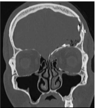
A male patient of 46 year old was admitted to Emergency Department of Policlinico Umberto I, Rome. The man had been subjected to a severe aggression. He referred intense pain and had a notable edema of the left hemiface as well as a lacero-contusive wound under the inferior eyelid. Ocular kinetic could not be evaluated. The CT showed a break in the continuation of the left or-bital roof, the posterior wall of the left frontal si-nus and the anterior skull base; the fracture re-sulted displaced and multifragmentary (Figure 1). Gassous bubbles in the frontal area were even present. The rectus superior and levator palpe-brae superioris muscles were attracted to the edge of the fracture and the periorbital fat showed edematous hemorrhagic imbibition.
At our Department we found several addition-al signs. There was enophtaddition-almus, mild ptosis with restricted eyelid elevation and important cerebrospinal fluid leakage. After consulting the ophthalmologist and the neurosurgeon the patient was brought to the operating theatre. The inter-vention was executed by the neurosurgeon and the maxillo-facial surgeon operating contextual-ly. A coronal incision was performed, the frontal bone was exposed, and a galeal-pericraneal flap was harvested. Then, an anterior craniotomy of the left frontal bone was executed. Next step con-sisted of the exposition of the fracture and of the reposition of the herniated orbital content (Figure 2). Therefore, the bone integrity was restored with a portion of splitted frontal bone (Figure 3). This portion was secured with a titanium plane plate (Figure 4). Then, a cranialization of the frontal sinuses with a cuff of temporal muscle and tissucol was performed (Figure 4). The bone fragments were covered by a galeal-pericraneal 2012; 16(4 Suppl): 117-120
Figure 1. Coronal CT scan showing the deplacement of
the orbital roof and periorbital edema.
Figure 2. Intraoperative image that evidences the
hernia-tion of the orbital content into the anterior skull base.
Figure 3. Frontal volet splitted and successively used to
re-cover the roof’s deficit.
Figure 4. The slipped volet recovers the orbital roof and is
stabilized by plane titanium plates.
P. Gennaro, V. Mitro, G. Gabriele, F. Giovannetti, A. Facchini
188910. Smith and Regan7 demonstrated that the
orbital content is necessary to produce a typical orbital floor blow out fracture. Indeed, the sud-den increase in intraorbital pressure causes the herniation of orbital content through the weakest part of the orbit.
Blow out fractures occur most often through the posterior orbital floor, medially to the infraor-bital groove8. Less frequently, the orbital plate of
the ethmoid is involved alone or in combination with the floor. Distribution of blow-out location results from a combination of bone’s thinness and it’s geometric relation to the orbital axis.
Orbital roof fractures usually are part of severe craniofacial traumas. Displacement of the orbital rim can cause linear extension into or comminu-tion of the orbirtal roof. In case of entrapment of the orbital soft tissues or persistent displacement of the globe, surgery is necessary to prevent per-manent functional and aesthetic defect11-13.
Mc Clurg and Swanson14 reported the first
case of a roof fracture causing diplopia as part of a more extensive skull fracture.
flap, which extended from the orbit roof to the upper-lateral portion of the ethmoidal cells. At this level a discontinuity was observed. Dural suspensions were executed and then the temporal bone was replaced with two plane plates. At two days from the intervention, the patient didn’t show cerebro-spinal fluid leak anymore. He was discharged from the Hospital on his fourth post-operative day without complications. The follow-ing clinical examinations showed a progressive functional and aesthetic good outcome.
Discussion
Orbital fractures can occur in patients who have suffered blunt injury from traffic accidents, falls, violence or other orbital trauma1-6.
The term ‘orbital blow-out fracture’ refers to the mechanism by which a blow to the eyeball is trans-posed to the orbital walls, causing them to fracture6.
This often involves injury to the orbital floor.4-9.
The first recorded description was by Lang in
Fractures of the orbital roof not associated with fractures of the orbital rim are unusual. Iso-lated orbital roof fractures can derive from pene-trating injuries15. Rarely, the orbital roof without
can be blown downward by distant skull frac-tures. This results from a sudden increase in
in-tracranial pressure transmitted through the anteri-or fossa and decompressed by a fracture of the orbital roof16.
The most appropriate timing for surgical repair after an orbital blow-out fracture is controver-sial1,4-6,10,11. The current accepted indications are:
internal orbital fracture with muscle incarcera-tion resulting in ocular motility restricincarcera-tion with diplopia, early enophthalmos (> 2 mm), and large orbital defects (> 50% of the floor or medi-al wmedi-all)5,13,17. Surgical intervention should be
per-formed within 2 weeks18,19.
For trapdoor fracture, early surgery within 2 to 4 days is more appropriate than a wait and watch period of 2 to 3 weeks4. The trapped
perimuscu-lar fat and connective tissue may set up a poten-tial compartment-type situation leading to tissue ischemia and the possibility of resultant muscular and perimuscular fibrosis4.
In 1974, Putterman et al20 recommended that
patients with pure blow-out fractures had to be kept under observation for 4 to 6 months at least to determine the rate of improvement. In 1991, de Man et al21 also suggested that a “wait-and
watch” policy was appropriate for blow-out frac-tures in adults.
Dutton et al17felt, however, that in some
situa-tions surgical repair should not be delayed, and therefore summarized the indications for early surgery (within one to two weeks). Furthermore, Kwon et al22reported that the recovery period in
children was shorter when surgical intervention was performed within 5 days after injury. Howev-er, surgery might be required within 2 weeks in adults. Bansagi and Meyer23also found that early
surgical intervention (< 2 weeks) resulted in a more complete return of ocular motility in a pe-diatric population, while Egbert et al9 suggested
surgery should be performed within 7 days for more rapid recovery. Egbert et al9 also found
surgery within one month of injury resulted in improvements of preoperative conduction deficit and diplopia in all his patients.
We think that when an orbital fracture is com-plicated by exophtalmus, enophtalmus, diplopia or liquoral fistula as in the previously described case, the intervention must be performed as soon as possible. We treated two days after the patient was injured an orbital roof fracture which ex-tended to the ethmoidal planum, the posterior wall of frontal sinus and the anterior skull base.
Isolated fractures of the orbital roof occur rarely and are often found when a thin anterior skull base is present. This thinness makes the
re-119
An orbital roof and anterior skull base fracture: case report
Figure 5. Coronal TC scan at two days after the
interven-tion: bone integrity restored.
Figure 6. Sagittal TC scan evidences the continuity
120
duction of the fragments difficult and unstable. For these types of fractures there exist several re-construction options. Among them are alloplastic materials and titanium meshes plates.
For our patient we chose to use autolog bone taken from the frontal volet because the fracture included ethmoidal planum and the possible communication with the nasal cavity raised the risk of ascending infections. Autolog bone tissue lowers the risk of infection and eliminates the risk of rejection. The volume of the orbital cavity was restored, the frontal sinuses cranialized and the cerebro-spinal fluid leakage interrupted. The functional and aesthetic outcome of the patient was satisfactory.
References
1) ROSADOP,DEVICENTEJC. Retrospective analysis of 314 orbital fractures. Oral Surg Oral Med Oral Pathol Oral Radiol Endod 2011 Apr 20.
2) RINNAC, ROCCHIG, VENTUCCIE, PAGNONIM. Iannetti G. Bilateral orbital roof fracture. J Craniofac 2009; 20: 737-734.
3) JOSEPH JM, GLAVAS IP. Orbital fractures: a review. Clin Ophthalmol 2011; 5: 95-100.
4) JORDAN DR, ALLENLH, WHITE J, HARVEY J, PASHBY R, ESMAELI B. Intervention within days for some orbital floor fractures: the white-eyed blowout. Ophthalmic Plast Reconstr Surg 1998; 14: 379-390.
5) HATTON MP, WATKINS LM, RUBIN PAD. Orbital frac-tures in children. Ophthalmic Plast Reconstr Surg 2001; 17: 174-179.
6) SUGAMATAA, YOSHIZAWA N. Clinical analysis of or-bital blowout fractures caused by a globe-to-wall contact mechanism. J Plast Surg Hand Surg 2010; 44: 278-281.
7) SMITHB, REGAN WF SR. Blowout fracture of the or-bit: mechanism and correction of the internal orbit fractures. Am J Ophtalmol 1957; 44: 733-739. 8) JONES DEP, EVANS JNG. “Blowout” fractures of the
orbit: an investigation into their anatomical basis. J Laryngol Otol 1967; 81: 1109-1120.
9) EGBERTJE, MAYK, KERSTENRC, KULWINDR. Pediatric orbital floor fracture: direct extraocular muscle in-volvement. Ophthalmology 2000; 107: 1875-1879. 10) LANGW. Traumatic enophthalmos with retention of perfect acuity of vision. Trans Ophthalmol Soc UK 1889; 9: 41-5.
11) STRANC MF, GUSTAVSON EH. Primary treatment of fractures of the orbital roof. Proc R Soc Med 1973; 66: 303-304
12) GERBINOG, ROCCIAF, BIANCHIFA, ZAVATTEROE. Sur-gical management of orbital trapdoor fracture in a pediatric population. J Oral Maxillofac Surg 2010; 68: 1310-1316.
13) CHENCT, CHEN YR. Update on orbital reconstruc-tion. Curr Opin Otolaryngol Head Neck Surg 2010; 18: 311-316.
14) MC CLURG FL, SWANSON PJ. An orbital roof fracture causing diplopia. Arch Otolaryngol 1976; 102: 497-98
15) GU Y O T L, LA R I N, BE N S O-LA Y O U N C, DE N I S D, CHOSSEGROS C, THIERY G. Orbital fractures in chil-dren. J Fr Ophtalmol 2011; 34: 265-274.
16) BRUCOLIM, GIARDAM, BENECHA. A peculiar orbital roof blow-in fracture. J Craniofac Surg 2011; 22: 358-360.
17) DUTTON JJ, MANSON PN, ILIFF N, PUTTERMAN AM. Management of blow-out fractures of the orbital floor. Surv Ophthamol 1990; 35: 279-298.
18) DULLEY B, FELLS P. Long-tern follow-up of orbital floor fractures with and without surgery. Mod Probl Ophthalmol 1975; 14: 467-470.
19) WILKINSRB, HAVINSWE. Current treatment of blowout fractures. Ophthalmology 1982; 89: 464-466. 20) PUTTERMAN AM, STEVEN T, URIST MJ. Non-surgical
management of blow-out fractures of the orbital floor. Am J Ophthalmol 1974; 77: 232-239. 21) DEMANK, WIJNGAARDE R, HESJ, DEJONG PT.
Influ-ence of age on the management of blow-out frac-tures of the orbital floor. Int J Oral Maxillofac Surg 1991; 20: 330-336
22) KWONJH, MOONJH, KWONMS, CHOJH. The differ-ences of blowout fracture of the inferior orbital wall between children and adults. Arch Otolaryn-gol Head Neck Surg 2005; 131: 723-727
23) BANSAGIZC, MEYERDR. Internal orbital fractures in the pediatric age group: characterization and manage-ment. Ophthalmology 2000; 107: 829-836.