doctor of philosophy in physics XXXI cycle
Giada Petringa
STUDY ON EFFICACY IMPROVEMENTS AND QUALITY OF THE PROTONTHERAPY
TREATMENTS
PhD Thesis
Tutor:
Prof. S. Romano Supervisors:
Dott. G.A.P. Cirrone Dott. G. Cuttone
Contents
1 Protontherapy: state of the art 11
1.1 The advent of protons in cancer therapy . . . 11
1.1.1 The rationale for using protons in radiotherapy . 13 1.2 Physical Properties . . . 13
1.3 Biophysical Propertie . . . 16
1.3.1 Stopping Power and Linear Energy Transfer . . . 16
1.3.2 Relative Biological Effectiveness . . . 17
1.4 Clinical implication . . . 18
1.5 New Ions for radiotherapy treatments . . . 19
1.6 New techniques for RBE enhancement . . . 22
1.6.1 High-Z nano-particles . . . 23
1.6.2 The Targeted Alpha Therapy . . . 24
1.6.3 The Binary Approaches driven by nuclear reactions 25 2 A new approach:the PBCT 31 2.1 Rationale for the enhancement protontherapy using the p-B reaction . . . 32
2.2 The p-B nuclear fusion reaction cross section . . . 33
2.3 Experimental proof of PBCT . . . 35
2.3.1 BSH enhances cell death . . . 35
2.3.2 Dependence of BSH-mediated cell killing enhance-ment upon proton energy . . . 36
2.4 Analytical estimation of RBE . . . 40
2.4.1 Evaluation of the alpha particles yield . . . 40
2.4.2 The LET and RBE calculation . . . 41
2.5 Imaging potentialities of PBCT . . . 46
2.5.1 Imaging by gamma prompt emission . . . 48
2.5.2 Experimental set-up . . . 51
2.5.3 Experimental results . . . 52
2.5.4 Gamma emission evaluation in a real treatment configuration . . . 56
3 The Monte Carlo approach 59 3.1 The method . . . 59
3.2 Simulations in medical physics . . . 60
3.3 The Geant4 simulation toolkit . . . 62
3.3.1 Geant4 use in medical physics . . . 64
4 Hadrontherapy: a MC application 67 4.1 Introduction . . . 68
4.2 Physics lists . . . 69
4.3 Primary Event Generator . . . 71
4.4 CATANA protontherapy facility of INFN-LNS . . . 71
4.4.1 Set-up of the beamline . . . 71
4.4.2 The scattering system . . . 72
4.4.3 Energy modulation system: modulator wheel . . . 74
4.5 ZD facility of LNS . . . 76
4.5.1 Set-up of the beamline . . . 76
4.5.2 The scattering system . . . 77
4.5.3 Energy modulation system . . . 79
4.6 Multidisciplinary beamline of INFN-TIFPA . . . 84
4.6.1 Set-up of the beamline . . . 84
CONTENTS 5
4.6.3 Energy modulation system . . . 89
5 LET in radiation therapy 91 5.1 Microdosimetry . . . 91
5.2 Rigorous definition of LET . . . 93
5.3 LET calculation via Monte Carlo approach . . . 94
5.3.1 Absorbed dose average LET (LD) . . . 95
5.3.2 Track average LET (LT) . . . 97
5.3.3 LET dependence on production cuts: a system-atic study . . . 99
5.3.4 The instability of the method 3 for the average dose LET calculation . . . 106
5.3.5 Validation against the ICRU data-base . . . 107
5.3.6 A study with light ions by using the Method 2 . . 108
6 RBE evaluation 113 6.1 The Relative biological effectiveness and the rationale of clinical use of 1.1 RBE value . . . 114
6.2 Modeling cellular radiation effect . . . 115
6.2.1 The Linear Quadratic Model . . . 116
6.2.2 The Local Effect Model . . . 117
6.2.3 The Microdosimetric Kinetic Model . . . 121
6.3 The Monte Carlo approach . . . 122
6.3.1 Survival fraction calculation: the LEM approach coupled to Geant4 . . . 123
6.3.2 Implementation of the parametrized L-Q model . 126 6.4 Study on S.F. prediction . . . 127
6.4.1 Simulated set-up . . . 128
6.4.2 Irradiation and experimental set-up . . . 128
6.4.3 The comparison with experimental data . . . 130
7 New Beam Profiler detect 141
7.1 The new developed beam profiling system . . . 142
7.1.1 System assembly . . . 142
7.1.2 Acquisition and Processing Software . . . 143
7.2 preliminary characterization . . . 144
7.2.1 Experimental Set-up . . . 144
7.2.2 Monte Carlo simulations . . . 147
7.2.3 Results . . . 148
7.2.4 Single spot characterization . . . 150
Introduction
The urgent need for radical radiotherapy research to achieve improved tumour control in the context of reducing the risk of normal tissue toxicity and late-occurring sequelae, has driven in recent decades the fast-growing development of cancer treatment by accelerated beams of charged particles (hadrontherapy). This appears to be particularly true for protontherapy, which has emerged as the most-rapidly expanding hadrontherapy approach, totalling over 170,000 patients treated thus far worldwide. The primary motivation for investigation into this area was based on the physical properties of charged particles, which can de-posit energy far more selectively than photons: thanks to the inverted depth-dose profile described by the Bragg curve.
However, one of the shortcomings of proton therapy resides in its lim-ited capability to locally control radioresistant cancers. Heavier parti-cles such as 12C ions can overcome such radioresistance because they
are densely ionizing. On the other hand, complications related to nu-clear fragmentation from the primary beam, along with a still partial understanding of the consequences of the exposure of normal cells to high-LET radiation, and also considering the complexity and high costs associated with a 12C treatment facility, fuelled research into exploring novel strategies with the aim to achieve alternative solutions for a lo-calized increase of proton damage efficiency (RBE).
The research in protontherapy is being configured today a new era 7
where new objectives in term of quality treatment improvement are reached and new methodologies to increase the radiobiological effect are investigated. This thesis includes both these aspects. The first part of the work is dedicated to a new proposed approach based on a nuclear reaction triggered by clinical protons. I report the first experimental proof of the idea theoretically proposed by Do-Kun Y. et al. in [158], based on the use of the p +11B → 3α nuclear fusion reaction to en-hance proton biological effectiveness exclusively in the tumour region through the generation of short-range high-LET alpha particles, thus being of potential clinical worth. The main results of this experiment are reported in Chapter 2. The remarkable observed effect has pushed on to starting a critical analytical study to reproduce the biological ef-fect. I performed a set of analytical calculations as well as Monte Carlo simulations ( Section 2.4 ) to reach this scope. Aroused particular in-terest also the study of prompt gammas emitted by the interaction of incident protons with boron target. This is a powerful technique to quantifying the absorbed boron by a tumor and monitoring online the treatments bringing out the RBE map on the treated region (Section 2.5). The work in this field has been presented in [116] [110] [112] [113] [122] leading different publications [39] [120] [119]. I won the Young In-vestigator Award of the European Radiation Research Society (ERRS) for the interesting studies on protontherapy enhancement field. The results reached on PBCT lead to a new INFN funded project called NEPTUNE (Nuclear process-driven Enhancement of Proton Therapy UNravEled). The main intent of this new project will be to consol-idated and explain the promising results on PBCT and investigate a new reaction p +19F → α +16O, which also generates secondary
parti-cles potentially able to lead a local enhancement of proton effectiveness. During its three years the NEPTUNE project will study different as-pects of these researches: from modelling (using analytical and Monte
CONTENTS 9 Carlo approaches), to microdosimetry and radiobiology.
As abovementioned another important item in the protontherapy re-search consist of investigations about the improvements in the treat-ments quality. In this thesis, Monte Carlo approach has been evaluated as a new promising strategy for the calculation of the main parame-ters of interest in medical physics and radiobiological applications. A systematic study to identify the best algorithm for the Linear Energy Transfer (LET) calculation has been performed (Chapter 5). This re-sulted in a new proposed algorithm and its application for the radiobi-ological effect estimation (Chapter 6). The LET is, in fact, the main physical quantity that describe the radiation quality and the mech-anism of released energy by ionizing particles. This is of particular importance for the prediction of radiobiological effects as a function of the radiation used in clinical environment. The work done has also included a new method for the Relative Biological Effectiveness (RBE) estimation (Chapter 6). The main results are reported in [123] [121] [115] [117].
A work on the proton therapy improvement cannot neglect a precise beam characterization as well as the optimisation of its transport pa-rameters. Accordingly, a specific task included in this thesis was focused on the improvement of the INFN (Istituto Nazionale di Fisica Nucle-are) multidisciplinary facilities: LNS (Laboratori Nazionali del Sud) and TIFPA (Trento Institute for Fundamentals Physics Applications). The main intent has been to increase the beam quality and the tech-niques for its monitoring. For these reasons the Monte Carlo simula-tions of the three beamline of interest have been optimized: CATANA (INFN-LNS); Zero Degree Experimental room (INFN-LNS) and the Multidisciplinary beamline of the TIFPA-INFN. Systematics studies to realize a scattering system for high energy proton and light ions beams (12C,4He,16O) as a well as a passive system for the beam energy
modu-lation (Chapter 4) have been performed. In the end, a new detector for the online dose profiler acquisition has been developed and character-ized for its future installation in the abovementioned facilities (Chapter 7). The work carried out on the beam quality improvement has been realized in strictly connection with the MOVE-IT (MOdelling and Ver-ification for Ion beam Treatment planning), INFN project. The main aim of Move-it (2017-2019) was, in fact, to achieve a synergy among dif-ferent expertise (i.e. detectors development, algorithms to optimize the treatment planning systems, radiobiological experience with in-vivo and in-vitro experiments) to further refine and optimize the current treat-ment planning systems for proton beams. These studies have resulted in [114] [111] [146] [26].
Chapter 1
Protontherapy: state of the art
1.1
The advent of protons in cancer therapy
Cancer therapy is a multi-modality approach including surgery, chemother-apy, radiotherapy and immunotherapy. Conventional radiotherapy is typically administered using external energetic photon (5-20 MeV) or electron beams. The first medical application of ionizing radiation, using x-rays, occurred in 1895 [128] [127]. In the following decades, ra-diation therapy became one of the main treatment options in oncology [58] [79]. Over the past years, in fact, many improvements have been made in order to optimize the treatments. Technical advances have been aimed mainly to reduce the dose to the healthy tissue while main-taining prescribed doses to the tumor target [93] [90]. Computerized treatment planning and advanced imaging techniques have influenced the beam delivery precision [125] [84]. The advantages of proton radi-ation therapy, compared with conventional photon radiradi-ation therapy, were outlined for the first time by Wilson in 1964 [153]. He proposed the idea of utilizing the finite range of the Bragg peak of proton beams for treating targets deep within healthy tissue and was the first that de-scribe the potential of proton beams for medical purposes. Nowadays, proton therapy represents an established alternative to photon
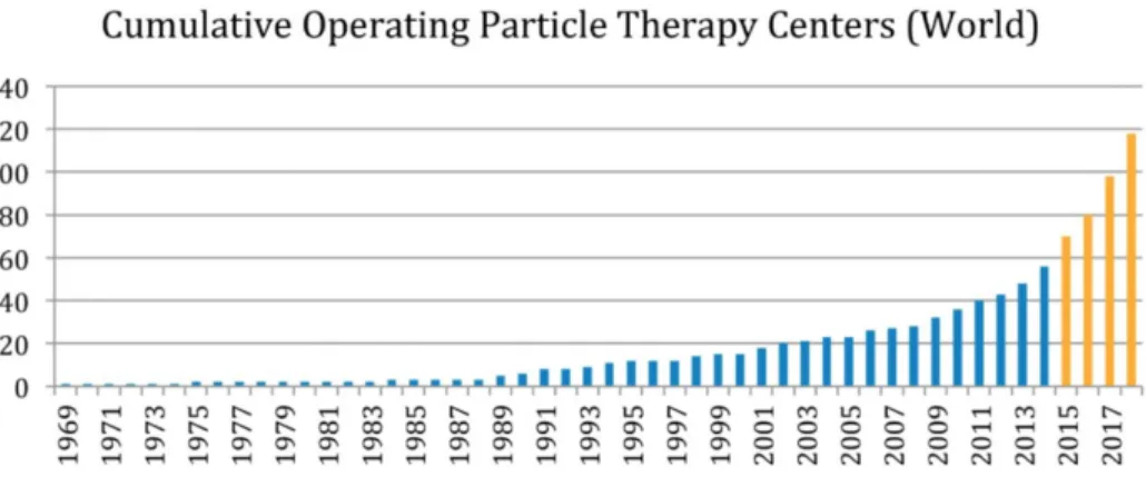
therapy for the treatment of specific types of cancer. According to the latest updates of the Particle Therapy Co-Operative Group (PTCOG) [61], more than 80 hadrontherapy centers are currently in operation and this number is going to increase in the next years, as reported in Figure 1.1.
Figure 1.1: Number of worldwide hadron therapy centers as a function of the years. Last four years trend in yellow
Besides the advantages offered by the peculiar inverted depth-dose profile, protons also show an enhanced biological effectiveness in cell killing. This is related to the increased Linear Energy Transfer (LET) compared to X-rays when protons are close to the Bragg peak [46]. High-LET value are associated with a localized energy deposition re-sulting in the induction of enhanced, unrepairable biological damage. Consequently, charged particles are often defined as densely ionizing radiation, in contrast to photons being considered sparsely ionizing ra-diation.
1.2. PHYSICAL PROPERTIES 13
1.1.1
The rationale for using protons in
radiother-apy
Since the beginning of clinical radiation therapy, one of the main goal of radiotherapy was to restrict the irradiated volume to the site and shape of the target volume [40] [45]. The strongest argument to consider protons for radiation therapy is the fact that, thanks to their sharp dose profile, entering in the matter (Bragg Peak),they are suited to irradiate a tumor at any depth of the body with a minimum dose to the surrounding healthy tissues.
1.2
Physical Properties
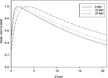
Photons of short wavelengths (X- or γ rays) interact to the medium througth stochastic events, such as inelastic or Compton scattering, photoelectric processes and pair production [133]. Due to the statistical nature of these absorption processes as well as the strong deflection during the interactions with medium atoms, a photon beam reveals an initial build-up, followed by a region of exponentially decreasing dose (see Figure 1.2).
In classical mechanics, the transfer of kinetic energy is inversely pro-portional to the square of the velocity (dE/dx ∼ 1/ν2). Due to their
low mass, accelerated electrons reach rapidly high velocities close to the speed of light (at energies > 1 MeV). As the electron velocity ap-proaches the speed of light c, the energy loss per unit length become independent of the energy (dE/dx ∼ 1/c2). As a consequence,
relativis-tic electrons deposit a constant energy dose per unit length. In water with its density similar to tissue, this dose corresponds to approx 2 MeV/cm [91]. The low mass renders electrons subject to strong lateral scattering. Bremsstrahlung photons which are produced by stopping process of the electrons in the target nuclei, cause the low intensity tail
Figure 1.2: Depth dose distributions of incident photons in water with different energy
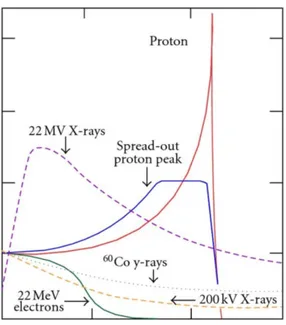
at the end of the depth-dose curve. For protons and all heavier ions, the absorption curve in matter shows a slow initial increase with pene-tration depth and a rise and fall towards the end of the particle’s range [75]. Because of their much higher mass ions the lateral scattering is significantly less than electrons, as shown in Figure 1.3
Accelerated atomic nuclei of the therapeutically relevant kinetic en-ergy interact predominantly via Coulomb forces with the target elec-trons of the traversed matter. This leads to excitation and ionization of atoms along the track of the travelling particle. Quantitatively, the en-ergy loss per unit path length, also called Stopping Power is described by the Beth formula [18] [21]:
dE
dx ∼ Kn0(Zef f)
2/β2[ln((2m
ec2β2/I(1 − β2)) − β)] (1.1)
where K is a constant n0 the electron density of the target material,
1.2. PHYSICAL PROPERTIES 15
Figure 1.3: Bragg peak and Spread-Out Bragg Peak (SOBP) for a proton beam in comparison with photon and electron dose distributions
projectile in units of speed of light (β = ν/c). I the mean ionization energy of the target atoms, and me the rest mass of the electron. For
low particle velocity (ν << c and β << 1) the Bethe formula can be reduced to:
dE
dx ∼ Kn0(Zef f)
2/ν2[ln((2m
ec2/I)] (1.2)
Under these conditions, the stopping power varies mainly with Z2 ef f/ν2.
With decreasing speed, dE/dx should increase. Most ion particles travel the same distance in a monoenergetic beam. But not all are involved in the same number of collisions. Their range is, therefore, somewhat dif-ferent. This phenomenon is called straggling. In tissues, the difference is of the order of 1% of the mean range for protons [59]. For heavier ions, range straggling varies approximately inversely to the square-root of the particle mass. The transverse spread of an infinitely narrow
proton beam amounts to approximately 5% of its initial range. Just as for range straggling, the angular deflection from the incident beam direction by multiple scattering decrease with increasing charge and mass.
1.3
Biophysical Propertie
1.3.1
Stopping Power and Linear Energy Transfer
The linear energy transfer or LET is a measure for the energy deposited by an ionizing particle travelling through matter [159]. It’s close to the stopping power concept but with some conceptual discrepancies: while the stopping power can be seen as a material property (depending on electron density) describing the energy absorbed by matter, LET ex-presses the loss of particle energy [83]. If all secondary electron energies are considered, LET, numerically is equal to the Stopping Power. The LET, generally expressed in keV/µm is the major parameter to discern qualitatively the biological effect of different kinds of radiations. The LET is widely used to characterized ion-induced damage even if has been shown that is not a good parameter to describe the full spectrum of biological radiation effects. The limitations of LET become partic-ularly evident when ions of different atomic number are compared. In particular for LET values greater than 100 keV/µm, different biological responses can be observed for particles with the same LET, as shown in Figure 1.4.
This is due to the different mechanisms of energy depositions. Ions of the same velocity produce tracks of secondary electrons with the same kinetic energy but the dose density within the track, i.e. the number of secondary electrons produced, is different [82]. Today the LET is one of the most studied parameters in the field of hadrontherapy. It’s strictly linked to the biological damage and some aspects of the
1.3. BIOPHYSICAL PROPERTIE 17
Figure 1.4: Relative Biological effectiveness (RBE) of ion beams as a function of Linear Energy Transfer (LET) with the same cell line. energy deposition in a real mixed radiation field are investigated by using micro/nano dosimeter detectors and new algorithms. All of these aspects will be discussed in detail in the Chapter 5.
1.3.2
Relative Biological Effectiveness
Prior to the clinical use of accelerated ions, their biological effects were studied in several experimental test. In the 1950s, the Uppsala cy-clotron was intensively used to study such biological effects [89]. The main aim of these experiments was to determine the ion biological effec-tiveness in comparison to the effect of a reference radiation (mostly 250 kV x-rays), i.e. the so-called Relative Biological Effectiveness (or RBE) [5]. Early studies revealed an RBE close to one for protons meaning that the proton dose required to produce a given effect was comparable to the equivalent dose from photons. More refined studies however, indicated that low-energy (< 1MeV) and very high-energy (>1 GeV)
protons can reach an RBE-value of 2 or more, depending on radiobi-ological endpoint studied. An elevated biradiobi-ological effectiveness in the Bragg peak region has clearly been demonstrated for ions heavier than helium [60].
1.4
Clinical implication
Today proton therapy is a well-established treatment option for many tumor types and sites. Advantages when using protons in favour of pho-tons have been shown in terms of Tumor Control Probability (TCP) and/or Normal Tissue Complication Probability (NTCP). Various dosi-metric studies clearly demonstrate superior normal tissue sparing with protons. It is well recognized that protons are extremely valuable to treat tumors close to critical structures [140] [77]. In the pediatric pa-tient population, the impact of the decreased total absorbed energy in the patient with protons is most significant. One example is the treat-ment of medulloblastoma, a malignant tumor in the medulla and that extends in the cerebellum [77] [28] [151]. Treatment with photon ra-diation therapy invariably causes a significant dose to heart, lung and abdominal tissue as well as the organ at risk in the cranium, something that can be largely avoided using protons as shown in the Figure 1.5.
These facts have boosted proton therapy in particular for pediatric patients. Although the dose distributions achievable with protons are superior to those achievable with photons, it is debatable whether the advantages of proton therapy are clinically significant for all treatment sites. Nowadays there is an ongoing discussion about the clinical trials that show a significant advantage in outcome by using protons.
1.5. NEW IONS FOR RADIOTHERAPY TREATMENTS 19
Figure 1.5: Dose distributions for a proton (left) and photon (right) craniospinal plan prescribed to 23.4 Gy (relative biological equivalents) are illustrated for comparison. The proton craniospinal plan provides considerable sparing of normal tissues anterior to the spinal canal and delivers a significantly reduced total integral dose to the patient. Num-ber of worldwide hadron therapy centers as a function of the years. In yellow is reported the trend registered in the last four years
1.5
New Ions for radiotherapy treatments
Protons are the charged particles most largely used in clinical prac-tice and for which the largest clinical experience has been acquired. Remarkably, this is somehow in contrast with the improved physical properties offered by heavier ions [142]. This contradiction is due to the fact that the biological effectiveness of other ions is still affected by large uncertainties, which do not fulfil clinical constraints [144]. An intense discussion has once again arisen in the literature in recent years concern-ing the possibility of optimizconcern-ing the clinical potential of charged particle therapy by extending the spectrum of therapeutic ions beyond protons and carbon ions [138] [85]. Currently, innovative treatment-planning
allowing a simultaneous optimization of different particle beams are investigated as well as new techniques to characterize the ion beams from the point of view of the physical and biological characteristics with a level of accuracy high enough to allow their clinical use [138]. Up to now, many studies were directed to improve the description of helium and oxygen beams for clinical use on the basis of experimental data and transport code (Monte Carlo and analytical). As well-known, the strong advantage offered by the physics of charged particles is the increasing LET in the Bragg peak, which allows high RBE to be ob-tained corresponding to the tumor. However, the LET variation along the depth-dose profile of different ions deserves an accurate discussion. It has been shown that the portion of the depth-dose curve where high-LET is released is strongly influenced by the particle type. From this point of view, while light particles like proton and Helium produce high-LET only on the distal part of the Bragg curve, in the case of carbon ions the high-LET region and therefore in healthy tissues, as shown in Figure 1.6. Thus, intermediate ions from lithium to beryllium might represent a valid compromise [144].
The very high LET radiations are interesting for its effectiveness in killing hypoxic cells that are normally radioresistant. The Oxygen Enhancement Ratio (OER), defined as the ratio of doses under hypoxia to normoxic conditions needed to obtain the same biological effect, be-comes close to one only at LET values that are accessible for ions heavier than carbon [73] [126]. From this point of view, Oxygen and Neon ions might be a promising tool. However, due to their increased charge, such heavy ions release a comparably high LET along the peak, thus increasing the risk of normal tissue damage. This statement has sug-gested to combining light and heavy ion modalities inside a treatment plan, i.e. by forwarding high-LET 16O ions only to the hypoxic parts of the target, while covering the rest with lighter particles (p,4He). In
1.5. NEW IONS FOR RADIOTHERAPY TREATMENTS 21
Figure 1.6: Profiles of dose-averaged linear energy transfer for the ir-radiation of an extended target of 2.5 X 2.5 X 2.5 cm3 centered at 8 cm depth in water, with a field optimized on a uniform physical dose (2Gy) [144]
the current version of GSI (Helmholtz Centre for Heavy Ion Research) in-house treatment planning system TRiP98 (TReatment planning for Particles, 1998 edition) the possibility to activate several ion modalities inside a single plan has been recently inserted [1] [84]. Figure 1.7 repre-sents the first tests of plans optimized for a uniform target survival level of 10%. In that case, three cases were analysed: single-ion optimiza-tions with opposite4He or16O fields, and quadruple-field optimization
with two pairs of opposite fields (16O + 16O and 4He +4He) [138].
There are ongoing studies of16O beams, investigating also the
radio-biological properties of ions, but the complete description of the beam still needs dedicated research, accounting for all the different effects [144]. This is the reason because there was a particular emphasis in new methodologies to optimize the uniformity of ions beam (especially
Figure 1.7: Survival distribution for single-ion optimizations with oppo-site 4He or 16O fields, and quadruple-field optimization with two pairs of opposite fields (16O + 16O and 4He + 4He) [138].
the study and results obtained in the design and realisation of a new scattering system and passive modulation system for future in-vitro and in-vivo studies by using light ions beams.
1.6
New techniques for biological
effective-ness enhancement
In the last decades, research efforts in the field of conventional radio-therapy have reduced the dosimetric gap between photons and protons in terms of tumour conformation [33] [84] [86] [132] [134]. Nevertheless, significant accomplishments have marked advances in proton therapy over the same period. Although it can be envisaged that technological developments will continue to improve the dose profiling achievable with photon therapy, the latter cannot be expected to fully match the dosi-metric advantage offered by protons since the integral dose difference cannot be overcome. Several radiobiological uncertainties, concerning
1.6. NEW TECHNIQUES FOR RBE ENHANCEMENT 23 the late effects of sub-lethally damaged healthy cells and cell signalling-mediated modifications of the tumour microenvironment, persist that, coupled with economical issues, hamper12C-based hadrontherapy wider
adoption [98]. In the last years, therefore, many strategies have been designed with the aim of increasing the biological effectiveness of light hadron beams. Possible methodologies investigated chemical radiosen-sitizing agents or, more recently, electromagnetic-driven enhancement of local dose through high-Z metallic nano-particles [68]. All these ap-proaches additionally attempt to exploit energy-dependent mechanisms in order to differentiate conveniently between healthy tissue (entrance channel) and tumor (target).
1.6.1
High-Z nano-particles
Radiation therapy is a critical treatment approach in the radiotherapy field. A sufficient damage to DNA by using ionizing particles can arrest cell growth and prevent metastasis [68]. But the primary drawback of the conventional radiotherapy with X-rays are the collateral damages: there is a little distinction in absorption between healthy and malignant tissues, and thus doses must be limited in order to mitigate unwanted damage to the tumor surroundings. Recently, it has been demonstrated that heavy elements can be radiosensitizers: drugs contain high Z ions can enhance the effects of ionizing radiation through the so-called high Z effect, or what has come to be known as Auger therapy. Heavy elements have significantly higher photoelectric cross-sections than soft tissue for sub-MeV energies, approximated for X-ray energies by the equation:
σpe ∝
Zn
e3 (1.3)
where σpe is the cross-section, E=hν is the photon energy, Z is the
the value of E. When ionized by X-ray or γ ray energy, mid- to high-Z elements (roughly Br and up) can produce a cascade of low-energy Auger electrons that can locally enhance the effective radiation dose. Dense inorganic nano-particles can also provide radiation dose enhance-ment that depends upon the composition and size of the particles, up-take of particles into cells, and the energy of the applied radiation. In the last years, Au nano-particles have been under investigation as possible agents for selective amplification of radiation dose in tumors. The promising in-vitro experiments performed by using gold Au nano-particles was followed by several studies examining their mechanism action as well as attempting to optimize the concentration, size, and the energy and dose of applied X-rays [56]. Alternatives to Gold are understudied i.e. Bismuth (Bi, Z = 83) and Platinum (Pt, Z = 78). Dose enhancement is also predicted to increase with decreasing nano-particle size because the smaller nano-nano-particles accumulate closer to the nucleus, where they can cause the greatest damage. However, the radiotherapy approach based on high-Z nano-particles is still discussed by the scientific community because appear complicated to find the correct relation between the deposited local dose and the observed bio-logical effect. Research efforts are underway to increase the efficiency of nano-particle-based treatments and improve the physical and chemical description.
1.6.2
The Targeted Alpha Therapy
At today, the radioimmunotherapy is considered a good candidate to treatment the radioresistant cancer cells. Monoclonal Antibodies (MAbs) that recognize tumor-associated antigens are conjugated to alpha or beta-emitting radionuclides that can provide selective systemic radio-therapy to primary and metastatic tumor sites. A number of targeted agents for radioimmunotherapy have been successfully constructed and
1.6. NEW TECHNIQUES FOR RBE ENHANCEMENT 25 labelled with alpha, beta and Auger emitters [43]. The different phys-ical properties of the radiations confer some advantages and disadvan-tages in various clinical situations, as schematically illustrated in Figure 1.8. Alpha particles have the advantage of high specificity thanks to the densely ionizing track and short path length (comparable to cell diam-eters). Beta particles can traverse several millimeters in soft tissue but the ionization is quite sparse. Auger particles have a high ionization probability but energy is deposited in the nanometer scale, being com-parable to the size of DNA, so the radioisotope should be used within the cell nucleus for effect.
The Targeted Alpha (α)-particle Therapy (TAT) is a methodology where α-emitting nuclides are conjugated to a carrier, normally an an-tibody [9]. Generally, an alpha particle with an energy from 4 to 9 MeV can deposit about 100 keV/µm within a few cell diameters (40–90 µm), causing direct DNA double-strand breaks, which lead to cancer cell apoptosis. Today the TAT is potent enough to eradicate dissem-inated cancer cells or cancer stem cells that are minimally susceptible to chemo- or radio-resistance. The relative biological effect of alpha particles is from 3 to 7, which means that for the same absorbed dose, the acute biological effects of alpha particles are 3 to 7 times greater than the damage caused by external beam photons or beta radiation. This methodology is then ideally suited to liquid cancers or micro-metastases. However, the regression of metastatic melanoma lesions after systemic TAT in phase I clinical trial has extended the applica-tion to solid tumors also [10].
1.6.3
The Binary Approaches driven by nuclear
re-actions
As already mentioned, the advent of hadrontherapy has allowed the study and development of new non-invasive therapeutic modalities. In
Figure 1.8: Alpha,beta and Auger radiation: the cells were dyedfluo-rescent green,the nucleus was dyed blue and yellow dots represent the ionization probability. (A) Short path length of alpha particles (a few cell diameters) and densely ionization along the track compared with (B) the longer path length of beta particles and (C) very short path length of Auger particles. The figure A and B were adapted from [43].
this framework, it has been widely investigated the so-called binary-systems. A binary system consists of two separate components that are combined to achieve a therapeutic effect. Briefly summarized, it’s a two-step procedure: firstly, the patient is injected with a
tumor-1.6. NEW TECHNIQUES FOR RBE ENHANCEMENT 27 localizing drug containing a non-radioactive isotope that has a cross-section of many times greater than the other elements present in tissues such as hydrogen, oxygen, and nitrogen. In the second step, the pa-tient is radiated with an incident ionizing radiation (such as neutron or protons). Beside lose they energy penetrating tissues, the incident par-ticles may also inelastically interact with the injected isotopes, which a subsequent emission of charged particles that can selectively kill tumor cells. The Boron Neutron Capture Therapy (or BNCT) is one of the most famous binary approach already applied in clinical practice. Re-cently, a new promising method called Proton Boron Capture Therapy (or PBCT) has been proposed. PBCT will be discussed extensively in the Chapter 2 of this thesis work.
The Boron Neutron Capture Therapy
Boron Neutron Capture Therapy is based on the reaction of low energy neutrons with10B producing two high-LET particles: a 7Li ion and an
α particle. The cross section of the 10B(n, α)7Li reaction is 3837 barn
at neutron thermal energies (Q-value = 2.790 MeV). With a probability of 94%, the reaction gives rise to the Li ion in an excited state, which returns to its fundamental level with a γ emission of 478 keV. In the rest of the cases, the reaction takes place at the fundamental level [74]. A scheme of the reaction channels is reported in Figure 1.9.
The principle of the therapy is that tumoural cells can be loaded with 10B and subsequently irradiated with thermal neutrons. When
the reaction takes place inside a cell, the two ionizing particles can cross the nucleus and cause non-repairable damages to the DNA. The potential efficacy of BNCT lies in its selectivity: using, in fact, proper borated compounds the tumoural cells can be loaded with higher boron concentration compared to the normal cells. This fact was experimen-tally observed since two drugs were synthesized and used in BNCT
Figure 1.9: Scheme of reaction channels involved into BNCT
research. These are the Boronophenylalanine (BPA) and the Sodium-Dodecaborane (BSH), which are still used in the BNCT research pro-grams for their ability to concentrate the boron atoms preferably in tumoural cells [29] [147]. When irradiating the tissue with neutrons, the total absorbed dose is made up of different components. Some of these dose components are a non-selective background radiation that cannot be avoided, as they originate from the interaction with tissue elements. The selective dose delivery is based on the boron concen-tration ratio between the tumour and the healthy tissue, that must be as high as possible. Today critical issues that must be addressed in-clude the need for more selective and effective boron delivery agents, the development of methods to provide quantitative estimates of tumor boron content before treatment, and a need for randomized clinical tri-als with an unequivocal demonstration of therapeutic efficacy [97]. The best survival data from the clinical trials performed for the treatment of glioblastoma multiforme are comparable with those obtained by current standard therapy.
1.6. NEW TECHNIQUES FOR RBE ENHANCEMENT 29 The Proton Boron Capture Therapy
Recently, a method to induce an enhancement of the biological efficacy of proton therapy and called PBCT, has been suggested [158]. It is based on the use of Boron atoms that, if localized inside a tumour mass, promote the occurrence of specific nuclear reactions and consequently the production of high-LET radiation. Specifically, this approach leads to the possibility to exploit the 11B(p, α)8Be nuclear fusion reaction
channel where three α particles, with an average energy around 4 MeV, are emitted [17]. The11B(p, α)8Be reaction proceeds predominantly by
sequential decay through the ground or the first excited state of 8Be,
that immediately decays into two secondary α particles. These parti-cles have a range (around 30 µm in water) sufficient to release most of their energy in the cell nucleus, and high-LET values (77 keV/µm), able to severely damage the DNA and produce an enhancement of the pro-ton beam biological efficacy [120]. The first experimental in-vitro test has been performed at Laboratori Nazionali del Sud (LNS) of Istituto Nazionale di Fisica Nucleare (INFN) in Catania (I). The experimental campaign has suggested that the achievable enhanced effect is due to the damage of high-LET particles inside the irradiated cells [39]. At to-day, other experimental campaigns with different cell lines are ongoing as well as a systematic Monte Carlo and analytical study to describe the observed effect. This is one of the main topic of this thesis work and widely reported and discussed in the Chapter 2.
Chapter 2
A new approach for the
radiobiological enhancement of
clinical proton beams: the
Proton Boron Capture Therapy
In the last years, many strategies have been designed to increase the biological effectiveness of proton beams. In the framework of this thesis the use of p-11B nuclear reaction, triggered by clinical protons
gener-ating short-range high-LET alpha particles inside the tumors, thereby allowing a highly localized DNA-damaging action has been investigated [39]. In this Chapter is reported the first experimental results with appearing evident the radiobiological effect due to the boron atoms in-jection in a biological sample. The first Monte Carlo and analytical calculations performed to reproduce the biological effect as well as a critical study conducted to investigate the potentiality of Boron atoms as a potential imaging technique is described.
2.1
Rationale for the enhancement
proton-therapy using the p-B reaction
Charged particle inverted dose-depth profile represents the physical pil-lar of protontherapy[106]. On the other hand, there is no obvious ra-diobiological advantage in the use of protons since their LET in the clinical energy range (a few keV/µm, at mid-Spread-Out Bragg Peak, SOBP) is too low to achieve a cell killing effect significantly greater than in conventional radiotherapy [82] [81]. This currently prevents protontherapy from being useful against intrinsically radioresistant can-cers. A well-known relationship links physical radiation quality (LET) and its biological effectiveness (RBE), based on the notion that cellu-lar lethality increases with the degree of DNA damage clustering, i.e. complexity, which reflects the nano-scale mode of radiation action [108] [107]. Therapeutic12C ion beams show an LET at mid-SOBP of about 50 keV/µm, conferring these particles a greater RBE for tumour cell killing, which is the radiobiological justification for their use against radioresistant cancers [131]. However,the non-negligible dose deposi-tion beyond the SOBP due to nuclear fragmentadeposi-tion and economical issues encumber this form of hadrontherapy. In this context, strategies combining greater RBE at cell tumour inactivation while maintaining reasonably low-dose levels in healthy tissues are desirable. Historically, the first approach to predict a tumour-confined increase of radiobio-logically effective dose by irradiation with a primary beam is BNCT, which exploits the10B(n, α)7Li reaction [74] [29] . It requires: a)
neu-trons, whose availability and dosimetry are not trivial; b) selectivity in boron uptake by tumour cells only [78] [147]. Another binary approach has been proposed that exploits the11B(p, α)8Be reaction, whose cross section resonates at 675 keV [17]. In protontherapy such energies are those of protons as they slow down across the tumour region. The
2.2. THE P-B NUCLEAR FUSION REACTION CROSS SECTION33 latter eliminates the requirement for selective boron uptake by cancer cell as alpha particles will be not generated, in principle, in healthy tis-sues where incident proton energy is too distant from that of the cross section maximum; together with the growing number of protontherapy centres, this elegantly bypasses the main drawbacks of BNCT.
2.2
The p-B nuclear fusion reaction cross
section
The proton-boron nuclear reaction considered is usually formalized as
11B(p, α)8Be. It has a positive Q-value (8.7 MeV) and is often referred
to as proton-boron fusion reaction since the incident proton is com-pletely absorbed by the 11B nucleus [17]. This reaction has gathered
interest since the 1930s because of the process ability to produce copi-ous numbers of alpha particles in an exothermic reaction. According to the literature, the p-B nuclear fusion reaction shows three resonant energies (0.162 MeV, 0.675 MeV and 2.64 MeV) and can be described as a two-step reaction. A proton interacting with a11B nucleus induces the formation of a 12C∗ compound nucleus formed in the 2− or 3− ex-cited state. If the 12C∗ nucleus is formed in its 2− state, it will decay
to the first 2+ state of8Be emitting one alpha-particle with l = 3 [139].
If the 12C∗ nucleus is formed in its 3− state, then the primary alpha
particles can be emitted either with l = 1 from the decay to the first 2+ 8Be excitated state, or with from the decay to the 0+ 8Be ground
state. In either case, the remaining8Be (2+or 0+) nucleus immediately
decays into two secondary alpha particles with l′ = 2. Alpha particles emitted in the first stage present a well-defined energy distribution and are commonly referred as α0 and α1 if the 8Be 2+ or the 0+ states
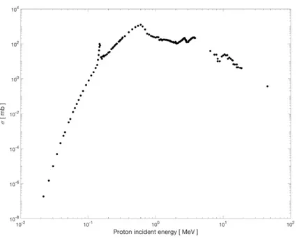
are populated, respectively [92]. In the Figure 2.1 is reported the total cross section for α1 emission, the most probably reaction channel.
Figure 2.1: Experimental total cross section of p +11B → 3α fusion reaction for the most probable α1 channel decay
Few authors report that a very unlikely fourth channel, character-ized by a maximum cross section of 10 µb in the 2.0 - 2.7 MeV energy range, can be also populated. In this case, the12C∗ directly breaks into three α particles skipping the intermediate 8Be stage, resulting in a continuous energy distribution. The emitted α particles exhibit a wide energy spectrum with a predominant energy around 4 MeV as shown in Figure 2.2.
Such a reaction has been considered very attractive for the genera-tion of fusion energy without producing neutron-induced radioactivity [158] [50]. As said above, the p-B fusion reaction is expected to play a strategic role in medical applications improving the effectiveness of protontherapy. Besides the advantage of using a neutron-free nuclear fusion reaction, the relevance of this method stems from the fact that the fusion reaction cross section becomes significantly high at relatively
2.3. EXPERIMENTAL PROOF OF PBCT 35
Figure 2.2: Spectrum of alpha particles emitted as a function of incident proton energy
low incident proton energy, i.e. around the Bragg peak region. Actu-ally, in conventional protontherapy, the proton beam is typically slowed down inside the tumour thickness (the Bragg peak region). Thus, most of the beam energy (i.e. dose released) is delivered to the tumour cells. Assuming that a given concentration of11B nuclei is present
preferen-tially, but not exclusively, in the tumour volume, fusion reaction events can be triggered by the incoming slow protons generating a relevant yield of highly DNA-damaging alpha particles localized in the tumour region.
2.3
Experimental proof of PBCT
2.3.1
BSH enhances cell death following proton
ir-radiation
To test whether the 11B(p, α)2α reaction results in an enhancement of cell killing by therapeutic proton beam irradiation, cells from the human prostate cancer line DU145 were irradiated with graded doses
at the middle position of the clinical Spread-Out Bragg Peak of the INFN-LNS protontherapy ocular facility. Irradiations were performed in the presence of two concentrations of BSH. As a control, DU145 prostate cancer cells grown and irradiated without BSH were used. The considered BSH concentrations were equivalent to 40 ppm (parts per million) and 80 ppm of 11B. These values were chosen in order to maximize the 11B content in the cells on the basis of what is done for the 10B-enriched BSH for BNCT [147].
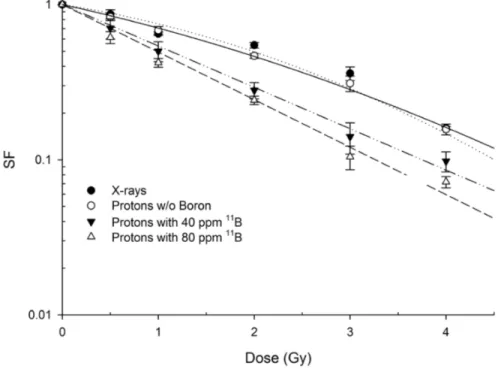
Boron treatment enhanced proton biological effectiveness resulting in a significant increase of mortality of DU145 cells. Cells that were irradiated after pre-treatment with, and in the presence of, boron-containing BSH exhibited a greater radiosensitivity in comparison with cells exposed to radiation alone [96]: BSH-treated cells yielded a much steeper clonogenic dose-response curve than that obtained for cells grown and irradiated in BSH-free medium (Figure 2.3).
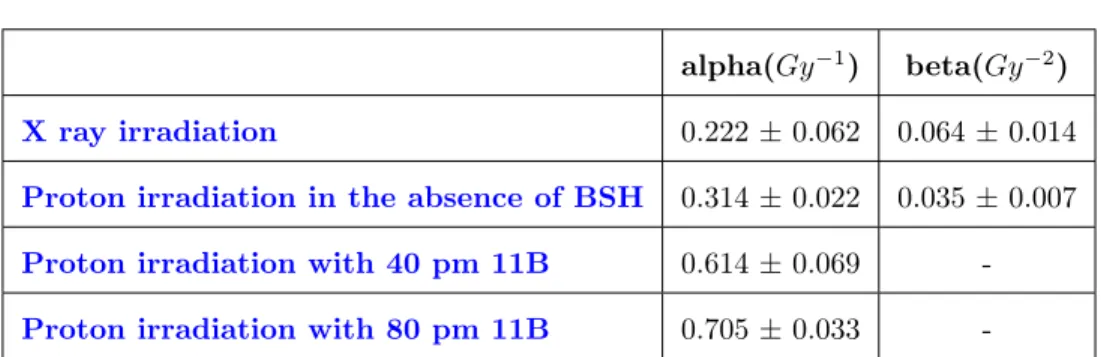
The clonogenic Survival Fraction (SF) following irradiation with protons alone was best fitted to a linear-quadratic function of dose, i.e. SF = exp (-α*D-β ∗ D2), with data from proton irradiation in the presence of BSH exhibiting a purely exponential behaviour as a function of dose. Least-square fitting parameters are reported in Table 2.1.
A slight yet not statistically significant effect due to boron con-centration was observed. Based upon the measured survival dose-responses, a calculated Dose Modifying Factor (or DMF) of 1.46 ± 0.12 was determined at the 10 survival level (DMF10).
2.3.2
Dependence of BSH-mediated cell killing
en-hancement upon proton energy
The pB nuclear fusion reaction critically depends on the incident pro-ton energy; hence, its radiobiological effectiveness can be expected to vary along the clinical proton SOBP. To verify this hypothesis, the
in-2.3. EXPERIMENTAL PROOF OF PBCT 37
Figure 2.3: Clonogenic dose response curves of DU145 irradiated with therapeutic protons in the presence or absence of BSH at mid-SOBP. Data are weighted mean values plus standard error from four indepen-dent experiments in the case of proton irradiation in the absence of BSH (open circles) and in the presence of the compound at highest concen-tration used (80 ppm, open triangles) Two experiments were performed with cells irradiated in the presence of 40 ppm of11B. X ray-irradiation
survival data are also shown for comparison.
duction of cell killing in the presence of the boron compound at the concentration of 80 ppm 11B (Figure 2.3), was investigated irradiating
the cancer DU145 cell line at the beam entrance (P1 position), at the SOBP distal end (P3 position), and at the middle of the SOBP (P2) as above reported (Figure 2.4).
The panel in Figure 2.5 shows the clonogenic survival dose-response curves derived from the three positions along the SOBP, in the absence
alpha(Gy−1) beta(Gy−2) X ray irradiation 0.222 ± 0.062 0.064 ± 0.014 Proton irradiation in the absence of BSH 0.314 ± 0.022 0.035 ± 0.007 Proton irradiation with 40 pm 11B 0.614 ± 0.069 -Proton irradiation with 80 pm 11B 0.705 ± 0.033
-Table 2.1: Cell killing dose-response fitting parameters. Calculated values for α and β parameters are obtained from the fitting of experi-mental data by linear-quadratic model for radiation-induced cell killing, are reported. Statistically equivalent to zero β values were found for proton irradiation in presence of BSH.
Figure 2.4: Cell irradiation along the proton SOBP. Measured dose and calculated LET profile for cellular irradiation at different positions along the clinical proton SOBP at INFN-LNS. Shown are the three depths along the SOBP at which cells were irradiated and the corre-sponding calculated LET values (open squares). Dose profiles as ob-tained by direct measurement by Markus chamber and by Monte Carlo simulations
or presence of BSH.
2.3. EXPERIMENTAL PROOF OF PBCT 39
Figure 2.5: Clonogenic survival along the proton SOBP. Data shown here refer to dose-response curves obtained at positions P1, P2 and P3 as indicated in Figure 2.4along the clinical proton SOBP.
clinical SOBP, proton irradiation alone resulted in a progressive increase in cell killing from P1 to P3. Interestingly, data clearly show no effect of BSH at the beam entrance. A DMF of about 1.4 was confirmed at 10% cell survival at mid-SOBP: here, fitting parameters were α = (0.309 ± 0.022) Gy−1 and β = (0.040 ± 0.006) Gy−2 for proton irradiation
without BSH and α = (0.653 ± 0.018) Gy−1 in the presence of 80 ppm 11B. At the distal end of the SOBP, however, BSH appeared to be even more effective with a recorded DMF of 1.75 ± 0.13: at this position, cell killing was best described by a pure exponential for both protons alone and protons in the presence of BSH, with values for the α parameter of (0.541 ± 0.027) Gy−1 and (0.952 ± 0.053) Gy−1. These experimental results and particularly the lack of a measurable effect due to the presence of 11B at beam entrance where the incident
proton energy is the highest, confirm that the enhancement of biological effectiveness is caused by the occurrence of p-B nuclear fusion events, which have a higher probability (i.e. higher cross section) at relatively low energy (MeV level) of the incoming protons, i.e. towards the end of their range.
2.4
An analytical estimation of the
radiobi-ological effect
Monte Carlo simulations coupled to analytical calculations were per-formed to estimate the number of alpha particles generated along the SOBP from the 11B(p, α)2α reaction and quantify the emission neces-sary to produce the observed radiobiological effect. The experimental total cross section was directly applied to proton energy spectra ex-tracted along the Bragg Peak. The estimated alpha particles yield was then applied to quantify the total dose-average LET and estimate the radiobiological observed effect. A detailed description of the applica-tion used for the Monte Carlo calculaapplica-tions as well as the algorithm adopted for LET computing and reproducing the radiobiological effect are reported in Chapter 4, 5 and 6 respectively.
2.4.1
Evaluation of the alpha particles yield
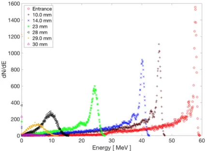
Hadrontherapy is a publicly available Geant4 application and was used to reproduce the whole beamline and the cells irradiation conditions during the radiobiological experiments. A standard water phantom for clinical absolute dosimetry, divided into slices of 100 µm, was also simulated: here particles fluence, energy spectra, dose and LET distri-bution were recorded. The energy spectra of the incident proton beam calculated along the SOBP are reported in Figure 2.6
The alpha particles produced were estimated applying experimental cross section (Figure 2.1) to the simulated energy spectra (2.6). Specif-ically, alpha particle yields were calculated assuming a11B layer of 100
µm and a density of 8 ∗ 10−5 g/cm3 corresponding to 80 ppm of 11B, that is the concentration used in the experiments. A number of histo-ries of the order of 109 were simulated to deliver a total dose of 2 Gy at the middle of the SOBP, according to the experimental results. The
2.4. ANALYTICAL ESTIMATION OF RBE 41
Figure 2.6: Energy spectra of the incident proton beam calculated at several depths along SOBP
results are reported in Figure 2.7 where the SOBP (in arbitrary units) is plotted along with primary proton fluence and the corresponding generated alpha particles. The dose related to each components, the number of incident protons and the emitted alpha particles are shown in Figure2.8.
2.4.2
The LET and RBE calculation
The estimated alpha energy spectrum has been then used to calculate the radiobiological quantities of interest like Linear Energy Transfer and the biological damage expressed in term of DMF. In Figure 2.9 the av-erage dose-LET along the SOBP calculated in two different conditions is reported:
• Condition 1: when only primary incident protons are considered; • Condition 2: when the contribution of the generated alpha
parti-Figure 2.7: Number of alphas particles generated in the p-B reac-tion. Fluence of primary protons and total alphas from the reaction
11B(p, α)2α produced in each slice. The experimental dose distribution
is also shown in arbitrary units.
cles is added to the primary protons.
LETs for the Condition 1 and 2 were estimated with Equations 2.1 and 2.2, respectively: LETp = dE1p· dE1p dxp1 + dE p 2 · dEp2 dxp2 + ... + dE p N · dENp dxpN dE1p+ dE2p+ ... + dENp (2.1) LETp+LETα = dE1p·dE1p dxp1 + ... + dE p N · dENp dxpN + dE α 1 · dEα 1 dxα 1 + ... + dE α N · dEαN dxα N dE1p+ ... + dENp + dEα 1 + ... + dENα (2.2) In both cases, at middle of SOBP (in water), average dose-LET resulted to be 5.69 keV/µm. Appear then evident that to obtain an
2.4. ANALYTICAL ESTIMATION OF RBE 43
Figure 2.8: Dose distribution as a function of the depth for incident protons and generated alpha particles, supposed that 2 Gy are released at the MID-SOBP, according to the experimental data
evaluable increment in the LET value is necessary to consider some different order of magnitude in the estimated alpha particles yield. The expected effect of the11B(p, α)2α reaction seems to be negligible, being the dose and LET due to this reaction channel orders of magnitude lower than the contribution due to the primary protons. When, at the same depth, the presence of 105 alphas is considered applying Formula
2.2, the augmented averaged LET due to the high-LET alpha particles (Condition 2) resulted 12.40 keV/µm. In Table 2.2 is reported the necessary LET value for the experimental SOBP (Figure 2.2) to obtain the experimental dose modified factor at the positions P1, P2 and P3. The DMF estimation reported in the Table 2.2 has been performed using the parametrisation and the MKM model described in Chapter 6. The Surviving Fraction curve, experimentally measured without boron, has been chosen as reference to demonstrate the enhancement effect.
Figure 2.9: Estimated increment of LET-dose when alphas from the p-B reaction are taken into account. Averaged LET-dose calculated with Equation 2.1 when only primary protons are considered (blue triangles) and when the contribute of the generated alphas is considered (red crosses) applying Equation 2.2. The experimental dose distribution is reported in arbitrary units.
Position Depth in Proton LET Total LET Dose Modifying
water [mm] [KeV/µm] [KeV/µm] Factor (DMF)
Entrance 5 1.96 3.66 1.17± 0.37
Mid-SOBP 21 4.24 9.53 1.36± 0.44
Distal 29.8 20.62 33.92 2.04 ± 0.66
Table 2.2: DMF estimation (last column) at three different depths along the SOBP; Proton dose averaged LET and Total dose averaged LET due to the contribute of alpha particles are also reported.
2.4. ANALYTICAL ESTIMATION OF RBE 45 The estimation of theoretical DMF calculated at the same experimen-tal positions along the Bragg curve has shown a maximum deviation of the order of 15% in comparison to the experimental results obtained with the clonogenic analysis (Section 2.3). To further substantiate the reasonableness of the analytical calculation, has been applied the MKM model considering the supposed incremented total dose averaged LET at the experimental MID-SOBP position. In Figure 2.10 is reported the Survival Fraction curve calculated with and without boron concen-tration as well as the experimental data.
Figure 2.10: The Experimental survival fraction with and without boron injection (yellow and blue triangles, respectively) as a well as the estimated damage calculated by applying MKM model ad parametrized approach described in Chapter 6
In conclusion, radiobiological data suggest that the 11B(p, α)2α re-action is a good candidate to justify the observed increase in the bi-ological effectiveness of the clinical proton beam. However desirable, it’s not simple provide analytical computation able to explain the
bio-logical results, for instance by correlating the biobio-logical effect with the total number of α-particles that can be expected to be generated under our experimental conditions. The current knowledge of biological radi-ation action has nonetheless established that, the biological effects of low-energy high-LET radiations cannot be interpreted solely on the ba-sis of macroscopic concepts like the absorbed dose or the average LET distributions. This is due to the intrinsically inhomogeneous nature of energy deposition events along radiation tracks, which becomes more significant with their increasing ionization density. Therefore, micro-and nano-dosimetric approaches must be taken into account to anal-yse the effects arising at cellular level. In addition, the role of extra-targeted phenomena, such as the bystander effect whereby cells that have not been traversed directly by radiation tracks may express cyto-genetic damage, is still largely undetermined in such scenario thereby contributing to the overall uncertainty between the physical dose dis-tribution at the micro or nano-dosimetric level and at the cellular one.
2.5
Imaging potentialities of PBCT : A
study of gamma-ray emission by proton
beam interaction with injected Boron
atoms for future medical imaging
ap-plications
The presence of Boron inside the neoplastic cells can also be exploited to spatially map the biological distribution of the Boron and, hence, the tumour regions where the RBE enhancement occurs [120] [119]. Char-acteristic gamma prompt emission from p-B interaction can in fact be detected, thus allowing the determination of the spatial distribution of their emission point. This possibility was discussed by D.K. Yoon et
2.5. IMAGING POTENTIALITIES OF PBCT 47 al. in [158] who proposed the use of 11B isotope. In this thesis has
been to investigate the potentiality of this technique. A critical analy-sis revealed that the vision described in [158] is quite optimistic for two reasons. First, the prompt-gammas of 0.718 MeV are emitted from the
11B(p,p’)11B inelastic scattering channel (and not the fusion channel,
as they erroneously state), and they are the same as generated by the interaction of protons with the main constituents of the human body (12C and 16O). Moreover, the maximum concentration of B nuclei that can be reached in a tumor, conferring minimal cell toxicity, is in the order of 10−5 g/cm3 [147], five orders of magnitude less than the value proposed in [158]. With such small densities, prompt gamma rays emit-ted at high incident proton energies are completely drowned out by the gamma particles generated by the interaction of the proton with the biological tissue.
The purpose of this study has been then to investigate the configuration discussed in [158], determining its real feasibility and establishing its limitations. Furthermore, a complementary approach was found. The gamma prompt spectra obtained with proton irradiation on11B and10B have been measured in a clinical configuration during an experimental campaign carried out at LNS-INFN. The same set-up has been simu-lated using the Talys [80] nuclear reaction code and outputs compared with the experimental results. This procedure enabled the selection of useful prompt-gamma lines and simultaneous validation of the code. Once validated, Talys was adopted to extend the study, considering different materials and to make additional considerations for the future improvement of this technique. The comparison has shown that the emission from the Boron atoms is not sufficient to produce a detectable signal at least at high energies. In this framework, a new approach based on using natural Copper directly bonded to Boron was proposed. The characteristic gamma prompts from Copper are characterized by
energies in a region far from that present in the background. This will permit their detection avoiding the unwanted interference coming from human body elements.
2.5.1
Imaging by gamma prompt emission
When a sample is irradiated with energetic protons, several nuclear re-actions, such as (p, p’γ), (p, γ) and (p, α) with gamma prompts are emitted in a temporal scale ranging in the 10−19to 10−9 s. These occur also during a typical proton-therapy treatment, where many secondary prompt gamma rays are induced from the proton-tissue interaction. In particular, discrete gamma ray emission can be generated from the ex-cited states of 16O and 12C that are the most abundant human tissue constituents. In the last decade, many experimental studies [137, 125] and simulations [149, 124] have shown that gamma prompt detection can be adopted as online dose monitoring and beam range estimation [87], as well. In this investigation, four characteristic gamma peaks (0.429 MeV, 0.719 MeV, 1.022 MeV and 1.435 MeV), coming from the p-11B and p-10B targets, were investigated in order to evaluate their
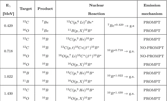
po-tential use in imaging applications. These peaks are generated by both B isotopes, even with different mechanisms (prompt and non-prompt), and from different reaction channels. Table 2.3 summarizes the reaction channels generating the investigated gamma lines. The maximum total production cross sections per channel and the corresponding emission mechanisms, are also reported.
According to literature [63, 92, 32], the gamma-prompts emitted from p-11B and p-10B interactions have the same energy as the ones generated by the interaction of protons with 16O and 12C (see Table 2.4).
2.5. IMAGING POTENTIALITIES OF PBCT 49 Eγ T ar get Pro duct Nuclear Emissio Maxim um gamma emission Nuclide pro duction Reaction Mec hanism cross section[m b] cross section [m b] 0.429 10 B 7B e 10 B ( p, α ) 7B e ∗ 7B e ∗ 0 . 429 → g.s. PR OMPT 27.8 at 19 Me V 56.8 at 19 Me V 11 B 7B e 11 B ( p, nα ) 7B e ∗ PR OMPT 17.18 at 19 Me V 287.7 at 19 Me V 0.718 10 B 10 B 10 B ( p, p ′) 10 B ∗ 10 B ∗ 0 . 718 → g.s. PR OMPT 163.5 at 11 Me V 253.8 at 11 Me V 10 B 10 B 10 B ( p, n ) 10 C ∗ ( β + ) 10 B ∗ NO-PR OM P T -134.4 at 12 Me V 11 B 10 B 10 B ( p, 2 n ) 10 C ∗ ( β + ) 10 B ∗ NO-PR OM P T -2.39 at 30 Me V 11 B 10 B 11 B ( p, d ) 10 B ∗ PR OMPT 82.09 at 22 Me V 126.9 at 22 Me V 1.022 10 B 10 B 10 B ( p, p ′) 10 B ∗ 10 B ∗ 0 . 718 → g.s. PR OMPT 38.5 at 11 Me V 253.8 at 11 Me V 11 B 10 B 11 B ( p, d ) 10 B ∗ PR OMPT 20.72 at 22 Me V 26.9 at 22 Me V 1.430 10 B 10 B 10 B ( p, p ′) 10 B ∗ 10 10 B ∗ 2 . 15 → 10 B ∗ 0 . 718 PR OMPT 10.26 at 11 Me V 253.8 at 11 Me V 11 B 10 B 11 B ( p, d ) 10 B ∗ PR OMPT 6.61 at 22 Me V 126.9 at 22 Me V T able 2.3: Summary of the reaction channels and corresp ondi ng characteristic gamma emission, o ccurring in the p-10 B and p-11 B in teractions. F or eac h p eak, the considered target and corresp onding resid ual n ucleus are rep orted (columns tw o and three); In columns four and fiv e the reaction channels are describ ed and the de-excitation pro ducing the gamma (column fiv e); Finally , the emission mec hanism (column six) and the maxim um gamma pro duction total cross sections (column sev en) a re rep orted as w ell as th e n uclide pro duction cross sections (column eigh t).
Eγ
Target Product Nuclear Emission
[MeV] Reaction mechanism
0.429 12C 7Be 12C(p,6Li)7Be∗ 7Be∗0.429→ g.s PROMPT 16O 7Be 11B(p, X)10B∗ PROMPT 0.718 12C 10B 12C(p,3He)10B∗ 10B∗0.718→ g.s. PROMPT 12C 10B 12C(p, t)10C∗(β+)10B∗ NO-PROMPT 16O 10B 16O(p,7Li)10C∗(β+)10B∗ NO-PROMPT 16O 10B 16O(p, X)10B∗ PROMPT 1.022 10B 10B 12C(p,3He)10B∗ 10B∗1.022→ g.s. PROMPT 11B 10B 16O(p, X)10B∗ PROMPT 1.430 12C 10B 12C(p,3He)10B∗ 10B∗1.430→ g.s. PROMPT 16O 10B 16O(p, X)10B∗ PROMPT
Table 2.4: Summary of reaction channels occurring in the p-12C and
p-16O interactions generating the gamma lines of interest. For each peak
the target and corresponding residual is reported (columns two and three); The nuclear reaction and emission mechanism is also included (columns four and five). Column six indicates the emission mechanism.
2.5. IMAGING POTENTIALITIES OF PBCT 51
2.5.2
Experimental set-up
Experimental runs have been performed at the zero-degree experimen-tal hall of INFN-LNS where 62 MeV proton beams, accelerated by the LNS superconducting cyclotron, are available. Gamma spectra were measured using a cylindrical, high-purity germanium detector (HPGe) GEM series model 70220-S, connected to a signal amplifier and an analog-to-digital converter MCA Amptek model 8000-A. The detector has a nominal energy resolution of 1.84 keV (at 1.33 MeV, as from60Co)
and a relative efficiency of 60.08%. Its sensitive volume has dimensions of 69.2 mm and 81.8 mm in diameter and height, respectively. Energy calibration was performed using a 152Eu source. The experimental
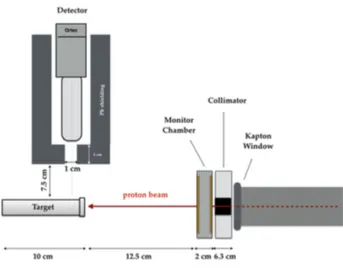
set-up is shown in Figure 2.11. Targets were positioned at 15 cm from the beam exit point corresponding with the Kapton window, making the interface between vacuum and air. The HPGe detector front surface was 7.5 cm from the cylindrical target, with its axis perpendicular to the beam.
Figure 2.11: Schematic representation of the experimental setup used for the gamma spectra measurement
The 62 MeV proton beam exits in air, traversing a 50 µm Kapton. It is then intercepted by a PMMA (PolyMethylMethacrylate) collimator fixing the beam circular spot size at 10 mm in diameter. Just beyond the collimator, a transmission ionization chamber is adopted for the on-line monitoring of the total released dose. It is composed by two Kapton/Gold foils 25 µm in thickness, separated by 9 mm of air [44]. An electric field of 800V between the two foils ensures the complete charge collection at the adopted rate. The role of the transmission chamber was essential to normalize the number of detected gamma rays with respect to the total incident proton flux. Finally, the beam reaches the Boron sample and it is completely stopped in it with a range of 3.41 ± 0.14 cm. The detector was shielded with a 5 cm thick lead wall to minimize the environmental gamma radiation. The Boron targets, produced at the INFN-LNS chemical workshop, consisted of two Mylar cylinders, 10 cm in length and with an external and internal diameter of 31 mm and 15 mm, respectively. One of the two targets is shown in Figure 2.12. They were filled alternatively with pressed
11B and10B powders, and in both cases the resultant density was 1.15
g/cm3. The bases of the Mylar cylinders were plugged with two 25 µm kapton foils to avoid Boron powder dispersion and at the same time to minimize the proton beam energy loss.
2.5.3
Experimental results
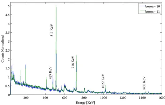
The two gamma spectra obtained with the irradiation of 10B and 11B are shown in Figure 2.13, both normalized to the same beam flux of 1.69*107 particles. They were acquired in a fixed time interval of 500 seconds. Proton beam current was kept at the value of about 15 pA at sample surface, in order to reduce pile up effects. Spectra were then normalized taking into account the total beam flux.
2.5. IMAGING POTENTIALITIES OF PBCT 53
Figure 2.12: Right: picture of one of the irradiated Boron targets; the mylar cylinder has an internal diameter of 15 mm and its thickness is chosen to minimise gamma attenuation. It contains the pressed Boron powder; Two kapton foils on its base prevent Boron powder leakage. Left: the target sketch with the mylar cylinder, the Boron insert and the kapton window.
annihilation after electron-positron pair production in the target mate-rial and in the detector itself. Moreover 0.429 MeV, 0.718 MeV, 1.022 MeV and 1.435 MeV peaks are present in agreement with the reac-tions reported in Table 2.3. Experimental gamma spectra have been compared against Talys analytical simulations performed with the out-gamdis modality [62]. Such a modality corresponds to an option in Talys which permits the gamma emission yield calculation taking into account all possible nuclear transitions. The real experimental concen-tration of Boron nuclei on the target was considered. Furthermore, the experimental detection solid angle and the total production cross sec-tions were taken into account in the total gamma yield calculation. The main aim of this study being the comparative evaluation of the different gamma contribution from11B and 10B isotopes irradiated with clinical proton beams, it was compared the relative difference between the ana-lytical (Talys) and experimental data. Table 2.5 reports the percentage
Figure 2.13: Normalized gamma rays spectra from p-10B (blue) and
p-11B (green). The energies of the four studied peaks are indicated together with the annihilation peak.
difference of each gamma yield line produced from p-11B and p-10B
interactions. The total count N of each considered gamma line has been evaluated for both targets, as the integral under the correspon-dent peaks. The counts percentage difference obtained as the difference between 11B and 10B counts divided by 11B yield in the experimental
case (∆exp) has then been compared with the corresponding one (∆ana)
calculated with the Talys analytical simulations. The results of these comparisons have been reported in Table 2.5 for three different target positions, corresponding to different proton beam energy intervals.
The differences reported in Table 2.5 between the calculations and experimental observations are all within the acceptable precision re-quired for it (estimated to be the order of 40%) and making us confident