Maurizio Caocci, IFNα-MEDIATED SUPPRESSION OF JCV IS mTOR PATHWAY DEPENDENT, tesi di dottorato in Life Sciences and Biotechnologies, Università degli studi di Sassari
Università degli studi di Sassari
Biomedical Science department
PhD Course in Life Sciences and Biotechnologies
IFNα-MEDIATED SUPPRESSION OF JCV IS mTOR PATHWAY DEPENDENT
Thesis coordinator: Prof. Caterina Serra
Department of Biomedical Science University of Sassari
Thesis advisor: Dr. Kamel Khalili
Director of Center for Neurovirology Lewis Katz School of Medicine, Temple University
Thesis co-advisor: Ph.D student Prof. Antonina Dolei Maurizio Caocci Department of Biomedical Science
University of Sassari
Maurizio Caocci, IFNα-MEDIATED SUPPRESSION OF JCV IS mTOR PATHWAY DEPENDENT, tesi di dottorato in Life Sciences and Biotechnologies, Università degli studi di Sassari
Università degli Studi di Sassari Corso di Dottorato di ricerca in Life Sciences and Biotechnologies
La presente tesi è stata prodotta durante la frequenza del corso di dottorato in Life Sciences and Biotechnologies dell’Università degli Studi di Sassari, a.a. 2017/2018- XXX ciclo, con il sostegno di una borsa di studio cofinanziata con le risorse del P.O.R. SARDEGNA F.S.E. 2007-2013 - Obiettivo competitività regionale e occupazione, Asse IV Capitale umano, Linea di Attività l.3.1 “Finanziamento di corsi di dottorato finalizzati alla formazione di capitale umano altamente specializzato, in particolare per i settori dell’ICT, delle nanotecnologie e delle biotecnologie, dell'energia e dello sviluppo sostenibile, dell'agroalimentare e dei materiali tradizionali”.
La tesi è stata prodotta, altresì, grazie al contributo della Fondazione di Sardegna.
Maurizio Caocci, IFNα-MEDIATED SUPPRESSION OF JCV IS mTOR PATHWAY DEPENDENT, tesi di dottorato in Life Sciences and Biotechnologies, Università degli studi di Sassari
TABLE OF CONTENTS
1. INTRODUCTION ... 1.1 Human Polyomaviruses……... 1.2 Human Polyomavirus JC... 1.2.1 JCV genome and structure……... 1.2.2 Non- Coding Control Region (NCCR)... 1.2.3.1 The large T-Antigen... 1.2.3.2 The small t-antigen and T’…………... 1.2.4 Late Proteins... 1.2.4.1 The VP1 protein... 1.2.4.2 VP2 and VP3 proteins………... 1.2.5 Genotyping classification of JCV……... 1.2.6 JCV life cycle…………... 1.2.7 JCV oncogenic potential……... 1.3 CNS diseases associated with JCV infection: Progressive Multifocal Leukoencephalopathy (PML) ………... 1.4 The big IFNs family…... 1.4.1. Type I IFNs... 1.4.2 Type I IFN receptors signaling... 2. AIM OF THE THESIS ... 3. MATERIAL AND METHODS... 4. RESULTS... 5. DISCUSSION AND CONCLUSIONS...
Maurizio Caocci, IFNα-MEDIATED SUPPRESSION OF JCV IS mTOR PATHWAY DEPENDENT, tesi di dottorato in Life Sciences and Biotechnologies, Università degli studi di Sassari
Maurizio Caocci, IFNα-MEDIATED SUPPRESSION OF JCV IS mTOR PATHWAY DEPENDENT, tesi di dottorato in Life Sciences and Biotechnologies, Università degli studi di Sassari
1. INTRODUCTION
1.1 Human Polyomaviruses
JCV belongs to the Polyomaviridae family, comprising non-enveloped tumor viruses with icosahedral capsids containing small, circular, double-stranded DNA genomes [White et al., 2005]. The Polyomaviridae family encloses a large number of members that are able to infect various animal species including rodents, rabbits, birds and primates, including human beings [Imperiale, 2001; Delbue et al., 2012]; in particular, to date, have 13 polyomaviruses infecting humans been discovered [White et al., 2013; Feltkamp et al., 2013; Moens et al ,2017]. The JC human polyomavirus was isolated in 1971 and in the same year the human polyomavirus BK (BKV) was discovered [Gardner et al., 1971; Padgett et al., 1971]. The transmission route is not fully understood. In humans, the first site of infection seems to be the respiratory tract, and specifically the tonsils, where the DNA of both JC and BK viruses has been isolated [Kato et al., 2004]. Subsequently, they can spread, by replication in lymphoid cells and through the circulatory system. BKV and JCV can reach other organs, such as the kidney, while JCV can reach the central nerve system (CNS), where these viruses cause persistent and latent infection [Wei et al., 2000; Pietropaolo et al., 2003; Zhong et al., 2007].
In the immunocompetent host, the primary infection is subclinical, but severe conditions of immunosuppression (caused, for example, by the administration of immunosuppressive therapies, congenital and acquired immunodeficiency, as well as kidney and kidney transplantation, pregnancy, diabetes, and other chronic diseases) stimulate viral reactivation, characterized by the release of viral particles in the urine [Doerries, 2006; Ferrante et al., 1997; Thomas et al., 2007; Zhong et al., 2007]. BKV and JCV are widespread throughout the human population, with greater than 90% of adults exhibiting JCV-specific antibodies. Antibodies persist significantly throughout lifespan and, in particular, different serological studies have highlighted an earlier and faster acquisition of antibodies against BKV rather than JCV [Kim et al., 2001; Frisque et al., 2006; Knowles, 2006; Lundstig and Dillner, 2006). The human BK and JC polyomaviruses share 75% genomic homology sequence and they are morphologically similar to murine oncogenic polyomavirus (PyV). They share a common ancestor with the monkey polyomavirus (SV40), whose oncogenic activity has been associated with human mesothelioma [Frisque et al., 1984; Shah, 2007].
Maurizio Caocci, IFNα-MEDIATED SUPPRESSION OF JCV IS mTOR PATHWAY DEPENDENT, tesi di dottorato in Life Sciences and Biotechnologies, Università degli studi di Sassari
Recently, several studies have led to the identification of new members of the Polyomaviridae family [White et al., 2013]. In 2007 a molecular screening study that tested for the viruses in nasopharyngeal drawings of children with respiratory tract infections led to the identification of a new human polyomavirus, called Karolinska Institute Polyomavirus (KIPyV). It is phylogenetically related to other human polyomaviruses at the early genomic region level, but has a low level of homologous sequences with the Late genomic region (<30% of identified amino acids) [Allander et al., 2007].
In the same year another group of researchers identified a fourth member of the Polyomaviridae family, named Washington University Polyomavirus (WUPyV), through the analysis of genomic sequences found in bronchial lavage of patients with severe respiratory inflammation. Following the sequencing of the entire viral genome (5.229 bp), a homology has emerged between the structural characteristics of the WUPyV genome and those of the viruses of the family of Polyomaviridae. In addition, preliminary data showed similarities between WUPyV and KIPyV, both in terms of genomic structure and phylogenetic relationships, and about their tropism [Gaynor et al., 2007; Dalianis et al., 2009]. These data suggest how these newly discovered polyomaviruses can be classified within a subgroup of the Polyomaviridae family, with its own biological and pathogenetic characteristics [Gaynor et al., 2007].
In 2008, Feng and colleagues performed a study on Merkel Carcinoma Cells (MCC), a rare but aggressive skin cancer, to test the role of the viral infections in this cancer. In the samples analyzed, they found the presence of transcripts of the TAg protein of a polyomavirus never described, called then Merkel Cell Polyomavirus (MCPyV) [Feng et al., 2008]. Then analyzing MCPy sequences, it was found a truncated form of TAg, that contains Rb protein binding domain (Shuda et al., 2008).
In 2010 Schowalter and colleagues isolated human polyomaviruses 6 and 7 in skin samples. They found that these viruses are released chronically from the epidermis by virions [Schowalter et al., 2010]. Also, in 2010 van der Meijden and co-workers discovered a new human polyomavirus associated with Trichodysplasia Spinulosa in a immunocompromised patient [Van der Meijden et al., 2010]. In 2011, the Human Polyomavirus 9 was isolated in a patient who had kidney transplantation and immunosuppressant drug treatments [Scuda et al., 2011]. The Malawi Polyomavirus was isolated in 2012, both in fecal samples taken from a
Maurizio Caocci, IFNα-MEDIATED SUPPRESSION OF JCV IS mTOR PATHWAY DEPENDENT, tesi di dottorato in Life Sciences and Biotechnologies, Università degli studi di Sassari
healthy baby in Malawi, and in diarrheal stool specimens taken from an American child from St. Louis [Siebrasse et al., 2012]. In 2013 a human polyomavirus was isolated, STI Polyomavirus, from the fecal microbiota of a healthy Malawense child [Lim et al., 2013]. The most recently human polyomaviruses discovered are the human polyomavirus 12 (HPyV12), isolated from the gastrointestinal tract [Korup et al., 2013] and the New Jersey polyomavirus (NJPyV), [Mishra et al., 2014].
1.2 Human JC Polyomavirus
The John Cunningham virus (JCV) is a member of the Polyomaviridae family, genus Orthopolyomavirus, with double stranded circular DNA, first isolated in 1971 from the brain of a patient suffering of progressive multifocal leukoencephalopathy (PML) [Padgett et al., 1971]. It has been found that JCV is the etiologic agent of PML. It was first described by Astrom and colleagues in 1958 [Astrom et al., 1958]. Therefore, the JC virus must be considered a neurotropic virus with both neuropathogenic and oncogenic properties. This is further supported by the etymology of the name of the family, in that polyoma derives from the Greek poli = many and oma = tumors. JC virus is associated with many tumors, both of glial (including gliomas and medulloblastomas) and non-glial origin. It has been found in many non-neuronal tissues, such as tumor biopsies of the gastro-intestinal (G.I.) tract and particularly in rectal colon cancer [White and Khalili, 2011].
The transmission of JCV through the population is not completely clear. It seems that the virus can be spread through the respiratory tract as well as through a fecal-oral route, since viral genomic sequences were found in the human gastrointestinal tract and in urban waste water [Boland et al., 2005]. The mechanism of viral entry in the host cell has not been entirely clarified, but it is known that cellular receptors for JCV are glycoproteins, containing one sialic acid residue bound in α-(2,6) position. However, it is also known that the virus can use serotoninergic receptor 5-HT2α [Brew et al., 2010]. The strong JCV tropism for human glial cells, for kidney cells and (with less efficiency compared to the previous ones) for B lymphocytes is due to these molecules. The restricted tropism for the CNS is confirmed by in vitro studies and laboratory animals [Raj and Khalili, 1995; Khalili et al., 2001]. JC virus is ubiquitous: specific antibodies have been found in 70% of the adult population and primary infection occurs predominantly during childhood. The infection does not necessarily correlate with a pathological state, occurring in most cases asymptomatically (primary viremia).
Maurizio Caocci, IFNα-MEDIATED SUPPRESSION OF JCV IS mTOR PATHWAY DEPENDENT, tesi di dottorato in Life Sciences and Biotechnologies, Università degli studi di Sassari
Subsequently JCV establishes a latent infection in the kidney, where it can be reactivated, due to immunological changes in the host. Reactivation is characterized by viral excretion in urines and virus migration, through the circulatory system, to the CNS, where oligodendrocytes are infected [Caldarelli-Stefano et al., 1999; Perez-Liz et al., 2008; Delbue et al., 2013].
1.2.1 JCV genome and structure
JC virus has an icosahedral capsid, sized 45 nm, constituted by 72 pentamers, with a molecular weight of 3.2x10⁶ Daltons [Stehle and Harrison, 1997; Diotti et al., 2013]. The capsid encloses the genome of the virus: a circular double-stranded DNA filament, of about 5.3 Kb, with a content in C+G very much similar to that of mammalian DNA (40-42%). The viral genome is associated with cell histones (H1, H2A, H2B, H3 and H4) in which form a complex, "viral mini-cromosome", that is structurally indistinguishable from the chromatin of host cell [Cole, 1996; Ahsan and Shah, 2006; Delbue et al., 2013].
The viral DNA can be functionally divided into three regions: an early region, a late region and a complex non-coding control region (NCCR). The early region encodes for the non-structural Large T (TAg) Antigen and small Antigen (tAg) proteins, that play a role in the replication, transcription, and expression of the viral genome. The late region encodes for the VP1, VP2, VP3 and VPx proteins: the most abundant protein is VP1 (39 kDa), with 360 molecules per capsid, followed by VP2 (37 kDa) and VP3 (24 kDa), contributing to the structure with 30-60 molecules per capsid. The VP1 protein, which is exposed on the surface of the capsid, is the one that determines the receptor specificity [Ferenczy et al., 2012]. The VPx protein is commonly called agnoprotein; it is about 71 aa long and facilitates the transport of VP1 to the core, thereby favoring the formation of virions [Carswell and Alwine, 1986]. Between the early region and the late region, there is the NCCR, ideally divided into ―boxes‖, from which early genes are transcribed counter-clockwise, and the late genes in the clockwise direction, on the complementary strand [Khalili and White, 2006] (Fig.1).
Maurizio Caocci, IFNα-MEDIATED SUPPRESSION OF JCV IS mTOR PATHWAY DEPENDENT, tesi di dottorato in Life Sciences and Biotechnologies, Università degli studi di Sassari
Figure 1. JCV genome map (Boland, 2005)
1.2.2 Non-Coding Control Region (NCCR)
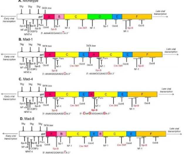
The NCCR contains the origin of replication (ori), the TATA box to which RNA polymerase II binds during transcription, the binding sites for T-Ag and cell transcription factors, bidirectional promoters and enhancers (cis-acting nucleotide sequences, that exemplify their function increase up to 200 times the transcription rate of the genes that they control). Unlike the coding regions that are well preserved, the NCCR region is hypervariable and contains determinants for neurotropism [Bellizzi et al., 2013]. The archetype NCCR (CY) is divided into 6 regions, called Box A (36pb), B (23pb), C (55pb), D (66pb), E (18pb) and F (69pb). Each box contains binding sites for cell transcription factors involved in viral transcription [Yogo et al., 1990]. These binding sites can undergo deletions or duplications, generating new viral variants, with different tropism and pathogenicity compared to the archetype strain. The archetype sequence is found in the kidney and urine of healthy individuals. It has never been associated with PML and the virus with this sequence is not infectious in cell culture models (Fig. 2) [Yogo and Sugimoto, 2001; Pietropaolo et al., 2003; Mischitelli et al., 2005].
Maurizio Caocci, IFNα-MEDIATED SUPPRESSION OF JCV IS mTOR PATHWAY DEPENDENT, tesi di dottorato in Life Sciences and Biotechnologies, Università degli studi di Sassari
Figure 2. Structure of JCV NCCR (Bellizzi, 2013)
The NCCR prototype has been isolated from tissues of patients with PML, and derives by a rearrangement of the archetype sequence. The original prototype is the Mad-1, which contains a NCCR consisting of A-C-E sequence of 98-pb repeated in tandem (A-C-E-A-C-E-F), with the consequent duplication of the TATA box, and specific binding sites for particular factors of cell transcription, including NF-1 and Spi-B. The TATA boxes contained in the sequences of 98 pb repeated in tandem are ultimately determinant for the transcription of both early and late viral genes [Frisque, 1983; Ferenczy et al., 2012].
In addition, in Mad-1, the loss of the B and D boxes determines the formation of binding sites for cell transcription factors essential for the expression of viral genes, including YB-1 / Purα and NF-1, and Oct-6 duplication. It has been observed that the T → G transition in the binding site for Spi-B results in the increase of the binding affinity for this important transcription factor [Marshall and Major, 2010; Marshall et al., 2012]. The Mad-4 variant represents a variant of the
Maurizio Caocci, IFNα-MEDIATED SUPPRESSION OF JCV IS mTOR PATHWAY DEPENDENT, tesi di dottorato in Life Sciences and Biotechnologies, Università degli studi di Sassari
Mad-1 prototype. It is identical to Mad-1, except for the presence of 19 bp alongside the second TATA box in the proximal position [Padgett et al., 1977]. Finally, the Mad-8 variant is the NCCR variant found most commonly in PML patients. It has a structure similar to the Mad-1 prototype, with a large deletion/insertion but also with different minor insertions and point mutations [Martin et al. 1985].
With the discovery of new rearranged NCCR sequences, the JCV variants were divided into two large groups: Class I viruses, characterized by a 98-bp tandem repeats NCCR (Mad-1); and Class II viruses, containing strains showing variations in the NCCR of class I viruses [Jensen and Major, 2001; White et al., 2009]. A further classification, instead, organizes the viral sequences of the NCCR in four variants:
- Type I variants, which do not contain insertions in the ACE organization of the NCCR and are then subdivided into IS type variants (with only one ACE sequence ) and IR variants (with repetitions of the ACE sequence) such as Mad-1 and Mad-4 (Fig. 1.7A) [Padgett et al., 1976; Frisque et al., 1984; Jensen and Major, 2001];
- Type II variants, which show 23pb and 66pb insertions, corresponding respectively to box B and box D, in the ACE sequence of 98 pb, and are subdivided into IIS (or archetype-like) and IIR containing insertion and deletions in respect to the IIS sequence, as in the Mad-8 strain [Yogo et al., 1990; Ferenczy et al., 2012].
1.2.3 Early proteins
The early proteins are involved in replication, gene regulation, and viral transformation. They are the large T-Ag protein, the small t-Ag protein, and the T prime proteins (T'135, T'36 and T'165). These multifunctional proteins are encoded by five different genes, from a single precursor of mRNA, that is alternatively spliced. They play a role in regulating the virus cycle and cell transformation.
1.2.3.1 The large T-antigen
The T-antigen is a protein of 688 amino acids, and regulates the transition of the transcription from early to late proteins, as well as the replication of the viral genome [Diotti et al., 2013]. In particular, T-Ag modulates the cell signaling pathways, in order to push the cells to the S phase,
Maurizio Caocci, IFNα-MEDIATED SUPPRESSION OF JCV IS mTOR PATHWAY DEPENDENT, tesi di dottorato in Life Sciences and Biotechnologies, Università degli studi di Sassari
and to activate the replication and the host transcription apparatus (DNA polymerase α and transcription factors), in order to replicate and transcribe viral DNA. Crucial event for the progression of the cell cycle, is the interaction of large T-Ag with Rb and p53 oncosuppressive proteins [Orba et al., 2010; Diotti et al., 2013]. The interaction with p53 prevents the apoptotic process, that is induced when there is a wrong cell activation of the S phase, and promotes viral replication in cells blocked in the G2 phase [Del Valle et al., 2001; Del Valle and Khalili, 2010]. Thus, this interaction confers to T-Ag its oncogenic properties, and is considered to be the main factor involved in cellular transformation and in tumorigenesis.
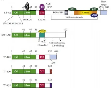
The oncogenic potential of JC polyomavirus has been extensively demonstrated in vitro in non-permissive cells, in which the viral replication cycle is not completed, but it induces the cell to transform [Khalili et al., 2001, White and Khalili, 2005]. In vivo studies conducted on transgenic mice showed how, in the onset of neuroblastoma of the adrenal gland, the replication of the entire viral genome was not required, but only the production of the T-Ag protein (Small et al., 1986; Franks et al., 1996). These experiments have also highlighted how the T-Ag interacts with several cell oncosuppressors, including p53, altering the course of normal regulatory processes of the cell cycle, by promoting the acquisition of the transformed phenotype [Del Valle et al., 2001; Khalili et al., 2001; Khalili et al., 2003a; Khalili et al., 2003b; Caracciolo et al., 2006]. Structurally, the T-Ag can be subdivided into different functional domains (Fig. 1.8) [Delbue et al., 2012] that, starting from the N-terminal are:
- DNaJ domain, which binds to HSc70 cellular factor and to αpolymerase;
- The LXCXE motif, which is specifically linked to members of the Rb family, inactivating the functionality;
- NLS domain, necessary for nuclear protein localization; - The helicase domain;
Maurizio Caocci, IFNα-MEDIATED SUPPRESSION OF JCV IS mTOR PATHWAY DEPENDENT, tesi di dottorato in Life Sciences and Biotechnologies, Università degli studi di Sassari
Figure 3. T-Ag structure (Del Bue, 2012)
1.2.3.2 The small t-antigen and the T '
The role of t-Ag in JCV life cycle is not completely clarified; however it has been shown that the t-Ag, similarly to T-Ag, is involved in the induction of the cellular S phase. In fact, it interacts and inactivates the regulating factors belonging to the Rb family and phosphatase-protein A (PP2A) [Bollag et al., 2010]. This binding prevents the dephosphorylation of the Agnoprotein, promoting viral replication. It is known that the reduction of the levels of t-Ag or PP2A protein results in a decrease in replication of JCV[Sariyer et al., 2008].
T '(first) proteins were discovered in 1995 by Trowbridge and Frisque. They were thought to be degradation products of the T-Ag, since they present a sequence homology equal to 132 amino acids [Trowbridge and Frisque, 1995]. Afterwards, it was understood that the T 'proteins are generated by an alternative splicing of the T-Ag. In fact, they have the same J and LXCXE (Cr2) domains in the N-terminal portion, but they differ at the C-terminal sequence, for their degree of phosphorylation, that affects the binding of T's with members of the Rb family (pRb, p107 and p130) and with the Hsp70 chaperone protein. In particular, the interaction between the T' proteins and the Hsp70 chaperon, and the inactivation of the pRb, p107 and p130 proteins results in the release of a cell transcription factor belonging to the E2F family. This factor promotes the switching of the cell cycle from the G1 to S phases [Prins and Frisque, 2001; Sami Saribas et al., 2014]. (Figure 4)
Maurizio Caocci, IFNα-MEDIATED SUPPRESSION OF JCV IS mTOR PATHWAY DEPENDENT, tesi di dottorato in Life Sciences and Biotechnologies, Università degli studi di Sassari
Figure 4. Graphic rapresentation of JCV early proteins (Sami Saribas, 2014)
1.2.4 Late proteins 1.2.4.1 The VP1 protein
The VP1 protein is the main viral capsid protein. In fact, for each pentamer of VP1, there is a single molecule of VP2 or VP3 [Sthele and Harrison, 1997]. In addition, the VP1 protein contains epitopes recognized by the immune system, and is implicated in the recognition of host cell receptors, hence allowing the infection of the host cell [Weissert, 2011]. The sequence analysis of the region encoding VP1 was used to define the different viral genotypes.
The sequence analysis of the region encoding the VP1 protein allowed also to detect characteristic mutations present in some isolates from patients with PML and never highlighted in healthy subjects. In particular, the mutations in the positions L55, K60, N265, S267 and S269 are related to the development of PML, and are localized at the binding site for the receptor of which they alter the specificity [Sunyaev et al., 2009; Gorelik et al., 2011]. Although these mutations are PML-specific, they were not found in all the isolates (52% of the positive samples), suggesting a cofactor role in the onset of the disease. Other specific polymorphisms, located in the amino acid positions 74, 75, 117 and 128 of viral VP1s seem to correlate with a more favorable PML prognosis [Delbue et al., 2009].
Maurizio Caocci, IFNα-MEDIATED SUPPRESSION OF JCV IS mTOR PATHWAY DEPENDENT, tesi di dottorato in Life Sciences and Biotechnologies, Università degli studi di Sassari
1.2.4.2 VP2 and VP3 proteins
The VP2 and VP3 late proteins promote the translocation of the virus to the nucleus and its capsid disassembly [Geiger et al., 2011; Inoue and Tsai, 2011]. They also participate in the capsid assembling, making the innermost layer [Sami Saribas et al., 2014]. It has been shown how the link between VP2 and VP3 proteins, chaperone Hsp70 and T-Ag leads to the accumulation of pre-virions within the nucleus of infected cells. Specifically, Hsp70 is associated with VP2 and VP3 late proteins, and it interacts with T-Ag, in the cytoplasm or in the nucleus. This interaction induces a conformational change of T-Ag, that increases its affinity to bind the origin of viral replication, hence beginning the DNA synthesis. The newly synthesized DNA is immediately caught by the VP2 and VP3 capsidic proteins, that start the virion assembly process. Subsequently, the nucleocapsids are covered by the VP1 protein, becoming mature virions [Sami Saribas et al., 2014].
1.2.4.3 VPx or Agnoprotein
Agnoprotein (or VPx) is a late protein, active during the last phase of JCV infection. Although its function has not been well understood, it has been hypothesized that it could play the role of a viroporine, promoting the release of the virus from cells [Suzuki et al., 2010]. The Agnoprotein also interacts with T-Ag, in order to reduce the levels of viral replication, by promoting the transcription of late genes and the assembly of mature virions. Some Authors have shown how Agnoprotein is involved in tumorigenesis. In fact, in the infection of non-permissive cells, it acts in synergy with T-Ag, altering the regulation of metabolic cellular processes, by promoting the acquisition of the transformed phenotype. In particular, it contributes to mutations at the genomic DNA level, blocking the activity of the proteins involved in the repair mechanisms of damaged DNA [Khalili et al., 2005].
1.2.5 The genotypic classification of JCV
The classification of JC virus in genotypes and subtypes is based on the sequence analysis of the region encoding the VP1 protein. This classification was introduced by Agostini in 1996, and is based on the analysis of point mutations in a 215 bp gene fragment of VP1, comprised between
Maurizio Caocci, IFNα-MEDIATED SUPPRESSION OF JCV IS mTOR PATHWAY DEPENDENT, tesi di dottorato in Life Sciences and Biotechnologies, Università degli studi di Sassari
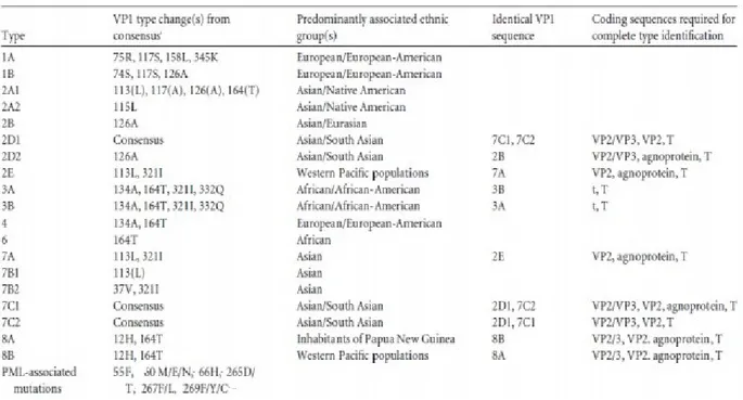
the 1710-1924 nucleotides [Agostini et al., 1996; Hansjurgen et al., 2001). This classification uses a combination of numbers followed by letters to describe genotypes and subtypes [Chang et al., 1996]. Eight major genotypes have now been identified (the genotype 5 is a subgroup of genotype 3), classified on the basis of some polymorphic sites in the VP1 region [Ferenczy et al., 2012] (Table 1).
Table 1. JCV genotypes and subtybe: classification based on VP1 gene differences. (Ferenczy,
2012)
Different viral subtypes related to different geographic areas of the planet have been identified, developed as a result of migratory flows, that led to geographic isolation of some human populations. JCV has also turned out to be a great marker to study human migratory flows, by allowing researchers to trace new migratory paths and confirm those already known [Jobe et al., 2001; Sugimoto et al., 2002; Kmieciak et al., 2008; Hirsch et al., 2013].
Genotypes 1, 4 and their subtypes are more common in Europe and in the United States of America (USA) [Sugimoto et al., 2002]. The 2 and 7 genotypes, and the related subtypes, have been found in Asian populations and in Native Americans. The genotype 3 was isolated in a patient coming from Tanzania, and seems to be prevalent in Africa, along with genotype 6. The genotype 8 and its subtypes are peculiar of people of West Pacific. In Italy, genotype 1 is the prevalent in the healthy population (57.8%), followed by genotype 4 (27.8%), while genotype 2 is rarely found (13.4%) and the genotype 3 is almost completely absent (1%) [Pagani et al., 2003]. Several authors have found with high frequency the presence of genotype 2 (subtype 2B)
Maurizio Caocci, IFNα-MEDIATED SUPPRESSION OF JCV IS mTOR PATHWAY DEPENDENT, tesi di dottorato in Life Sciences and Biotechnologies, Università degli studi di Sassari
in cerebral tissue and spinal fluids of patients with PML, suggesting the existence of a biological difference in the neuropathogenic potential of the various genotypes [Agostini et al., 1997; Ferrante et al., 2001; Mischitelli et al., 2005].
1.2.6 JCV life cycle
JCV life cycle begins with the binding of the VP1 protein to specific receptor glycoproteins on the surface of the target cell, containing sialic acid and negatively charged [Austin and Fehlings, 2008]. Generally, the sialic acid is in position α- (2,6) terminal, and is capable of binding to a LSTc pentasaccharide or a serotoninergic 5HT2A receptor [Elphick et al., 2004 Dugan et al., 2005; Neu et al., 2010]. In the phase of adsorption to the cell, also the VP2 and VP3 proteins play a key role, facilitating the binding of the VP1 protein to the cellular receptor (Cole, 1996; Pho et al., 2000]. Like all DNA viruses, JCV enters the cytoplasm, through a process of clatrine-mediated endocytosis [Pho et al., 2000; Querbes et al., 2004]. Subsequently, the virion is translocated within the endoplasmic reticulum (RE), from which, by a degradation process called ERAD (Endoplasmic-Reticulum-Associated protein degradation), is released into the cytoplasm, to move towards the nucleus. The entrance into the nucleus core involves the interaction with nuclear porous complexes, as shown in Figure. [Ferenczy et al., 2012].
Once into the nucleus, the virion is subjected to uncoating, and the viral minicromosome is transcribed from the cellular RNA polymerase II, to produce the early mRNAs, T-Ag and t-Ag, that can be detected 12 to 15 hours after infection. The regulation of viral transcription depends not only on the availability of host proteins, but also on the type of sequence of the NCCR present in the viral genome. In fact, the more this sequence will present binding sites for specific transcriptional cell factors, the more the entire replication process will proceed, and the number of virions produced will be faster and greater [Ferenczy et al., 2012]. Replication of the viral genome in vivo can be observed only after 3-5 days from infection, and it goes on for several weeks, by using the cellular DNA polymerase and thymidine-kinase enzymes (Feigenbaum et al., 1987; Khalili et al., 1987). Especially T-Ag determines the beginning of viral DNA synthesis, by binding to the ORI and, from this site, replication of viral DNA proceeds bi-directionally. Viral DNA replication is controlled by species-specific factors, which determine also the virus tropism. In fact, only few cells have the proteins necessary to complete the viral
Maurizio Caocci, IFNα-MEDIATED SUPPRESSION OF JCV IS mTOR PATHWAY DEPENDENT, tesi di dottorato in Life Sciences and Biotechnologies, Università degli studi di Sassari
life cycle; among them the most important one is the NF-1 protein, that can modulate in vivo JCV replication, depending on the isoform and the cell type involved [Muller and Mermod; 2000; Monaco et al., 2001; Major, 2010].
The replication and assembly of the JCV capsid occur in the areas of the nucleus characterized by the accumulation of specific proteins, such as Daxx and SP100, called "Nuclear domain 10 bodies" (ND10 bodies). During the replication process, the archetype NCCR rearranges specific sequences of the control region.
For example, the sequence of the Mad-1 prototype is characterized by two linking sites for the Pura, YB-1, LCP-1 and GF-1 factors, four sites for Oct-6 / tst-1 / SCIP and NF-1 factors and six for Spi-B factor hematopoietic transcription [Tamura et al., 1988; Amemiya et al., 1992; Tada and Khalili, 1992; Wegner et al., 1993; Shivakumar and Das, 1994; Chen and Khalili, 1995; Chen et al., 1997; Ferenczy et al., 2012]
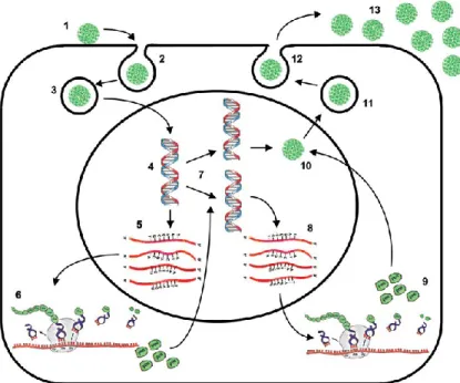
The NCCR of the Mad-4 variant is characterized by one deletion of 19 bp. This leads to the elimination of the TATA box placed in the terminal position, but it does not affect the replication capabilities of the virus, as sequences were found in subjects with PML [Martin et al., 1985; Mandl and Frisque, 1986; Cubitt et al., 2001; Gosert et al., 2010; Reid et al., 2011]. Concurrently with the replication of the viral genome, the transcription and translation of late mRNAs begin; these mRNAs encode for the capsid proteins and the Agnoprotein. The activation of late genes requires that the T-Ag interacts with some components of the cell transcriptional apparatus, such as TATA Binding Protein (TBP), TATA box-associated factors (TAFs) and some transcriptional factors, including Sp1 [Kim et al., 2000]. The expression of the structural viral proteins occurs from two mRNA molecules: the 16S and the 19S ones. From the first mRNA, the VP1 capsid protein is produced, and from the second mRNA the VP2, VP3 and Agnoprotein viral proteins are translated. Then, the structural proteins are transported into the nucleus, where the virion assemblies are visible at 24/48h. Mature virions are released from the host cell by cell lysis, even if by electron microscopy has also been observed the secretion of virions through an intact cell membrane. Which of these two mechanisms is the one preferred by the virus to escape from the host cell is still under study [Ferenczy et al., 2012]. (Figure 5)
Maurizio Caocci, IFNα-MEDIATED SUPPRESSION OF JCV IS mTOR PATHWAY DEPENDENT, tesi di dottorato in Life Sciences and Biotechnologies, Università degli studi di Sassari
Figure 5. Schematic rapresentation of JCV life cycle. The following steps are indicated: 1—
Binding of JC virions to cell surface receptor, 2—endocytosis, 3—nuclear import, 4 uncoating, 5—transcription of the early region, 6—translation of the early proteins, T-antigen and t antigen, 7—viral DNA replication, 8—transcription of the late region, 9—translation of Agnoprotein and the capsid proteins VP1, VP2 and VP3, 10—virionassembly, 11—nuclear export, 12—virion release from the cell, 13—infectious viral particles.
Among DNA viruses, the JC virus is the one with the longest replication cycle. Cell lysis occurs 24-48 hours since the beginning of the life cycle, and often a large part of progeny remains associated with cellular debris.
1.2.7 JCV Oncogenic Potential
When the expression of early genes is followed by the replication of viral DNA and from the expression of late genes, the result is a productive infection. The most striking example is the infection of oligodendrocytes, which are permissive to JCV infection and are destroyed during the infectious process, leading in vivo to the appearance of PML.
However, other scenarios are also possible. In non-permissive cells, only the expression of early genes is observed, since these cells are not capable to support neither the viral replication nor the expression of late genes. In this situation, the expression of T-Ag leads to the inactivation of
Maurizio Caocci, IFNα-MEDIATED SUPPRESSION OF JCV IS mTOR PATHWAY DEPENDENT, tesi di dottorato in Life Sciences and Biotechnologies, Università degli studi di Sassari
oncosuppressors, to the de-regulation of the signaling pathways and to genomic instability, which can contribute to cellular transformation [Khalili et al., 2003a]. The oncogenic potential of the JC polyomavirus has been extensively demonstrated in vitro in non-permissive cells, in which the viral replication cycle is not completed, leading to neoplastic transformation. The factors contributing to the permissiveness of a cell type can be, for example, the cell development status or the differentiation state. This can be observed in the same oligodendrocytes, that allow the progression of lytic infection only if fully mature, but, when they are in an undifferentiated form, they lead to the expression of the T-Ag, with consequent development of an oligodendroglioma [Khalili et al., 2003a]. Some authors suggested a possible involvement of the virus in the onset and/or in the development of brain tumors, B cell lymphomas and colorectal cancer, after the detection of the DNA, the mRNA for the T antigen and the same T antigen protein in tumors [Shadan et al., 2002; Khalili et al., 2003a; Khalili et al., 2003b; Hory et al., 2005; Enam et al., 2006; Lin et al., 2008].
Other authors have demonstrated the correlation between some CNS tumors (lymphomas, gliomas, oligodendrogliomas) and development or progression of PML [Caldarelli-Stefano et al., 2000; Del Valle et al., 2001; Gallia et al., 2001; Croul et al., 2003]. The presence of JC DNA only, by itself is not indicative of a causative effect in the onset of tumors, but viral genes have to be expressed. In case of the T-Ag protein, as widely discussed, the understanding of its oncogenic properties could be a starting point to develop therapeutic drugs, capable of blocking the JC virus-associated tumor progression [Khalili et al., 2003a].
1.3 CNS diseases associated with JCV infection: Progressive Multifocal Leukoencephalopathy (PML)
PML is a rare brain demyelinating pathology, characterized by the infection of glial cells, affecting predominantly adults and only occasionally children. Patients have neuropsychological deficits at the time of the onset of the disease. Common symptoms are: motor deficits, altered state of consciousness, visual disturbances and ataxia. However, there are also atypical manifestations,which include cerebellar syndrome, meningitis, and meningoencephalitis, progressive myoclonic ataxia, muscular degeneration associated with extrapyramidal signs.
Maurizio Caocci, IFNα-MEDIATED SUPPRESSION OF JCV IS mTOR PATHWAY DEPENDENT, tesi di dottorato in Life Sciences and Biotechnologies, Università degli studi di Sassari
The PML is a progressive disease, and leads to death within few months, when the patient is in permanent immunodeficiency conditions. The average survival varies with respect to the patient’s status: in HIV-negative individuals, it lasts few months only; in HIV-positive patients and under treatment with antiretroviral therapies, the survival increases, even though serious CNS complications can occur. The neuropathological description of PML was reported for the first time in 1958, by Astrom and colleagues, following the analysis of brain tissues from two patients with chronic lymphocytic leukemia and from a patient with Hodgkin's lymphoma [Astrom et al. 1958]. The etiology of this disease was not known, until Zu Rhein and Chou, in 1965, observed the presence of viral particles in the brain lesions typical of PML [Zu Rhein and Chou, 1965]. Viral particles were then isolated in fetal human brain cultures in 1971, by Padgett and colleagues [Padgett et al., 1971]. The virus was named JC (JCV), after the name of the first patient (John Cunningham) the virus was isolated from, using brain tissue for culture and virus isolation [Padgett et al., 1971]. At the beginning, PML was thought to be a rare complication of hematologic tumors or systemic autoimmune disorders; however, PML incidence was found increased by 50 times, with the spread of the HIV pandemic, from the mid Eighties onward. Nowadays, PML is the cause of death in 3-5% of AIDS patients [Major, 2010].
The onset of the disease can also be observed following organ or stem cells transplantation, and, recently, new cases of PML are associated with the use of therapeutic monoclonal antibodies (mAbs) in patients with various diseases, such as multiple sclerosis, Chron’s disease, Lupus, non-Hodgkin lymphoma and autoimmune hematologic diseases.
The incidence of PML in patients under immunotherapy depends on the drug used. For example, the risk of PML for patients under therapy with rituximab, a humanized monoclonal antibody, directed against the CD-20 antigen, was estimated to be 1/4000 when used in patients affected by Lupus, and 1/25000 when given to subjects with rheumatoid arthritis [Clifford et al., 2011]. Very high incidence (1/500) was observed in patients with psoriasis treated with efalizumab, a humanized antibody directed against an adhesion molecule present on the T lymphocytes. For this reason, the drug was withdrawn from the market [Molloy and Calabrese, 2009]. It is likely that many of these therapies lead to the onset of PML subsequently to the weakening of immuno-surveillance. It has been shown that the lymphopenia of the CD4+ and CD8+ T lymphocytes caused by the use of natalizumab, efalizumab and rituximab is one of the factors of primary risk to the occurrence of the disease [Uleri et al., 2017; Arru et al., 2014].
Maurizio Caocci, IFNα-MEDIATED SUPPRESSION OF JCV IS mTOR PATHWAY DEPENDENT, tesi di dottorato in Life Sciences and Biotechnologies, Università degli studi di Sassari
PML pathogenesis in patients treated with natalizumab is complex, and still is it to be defined if the disease is caused by JCV reactivation directly in the CNS or at peripheral level.
On the other hand, several drugs, such as natalizumab and rituximab, cause depletion of mature B cells in the periphery, with the consequent mobilization of immature B cell cells and bone marrow potential spread of latent virus in the brain [Ferenczy et al., 2012]. Currently, there is no effective therapy able to counteract PML pathology.
1.4 The big family of IFNs
The importance of interferon as an antiviral agent has been discovered during viral interference studies. [Isaacs and Lindenmann, 1957, Nagano and Kojima, 1958]. In 1957, two researchers, Isaacs and Lindenmann, studying the effect of heat-inactivated influenza virus on infected chicken cells, discovered a factor that protected cells from both homologous and heterologous viruses [Isaacs and Lindenmann, 1957]. This observation, along with the work done by Nagano and Kojima in 1958 [Nagano and Kojima, 1958], has paved the way for subsequent studies that have define the interferon system in a detailed way [Samuel, 2001]. After the first studies leading to the discovery of Type I [Isaacs and Lindenmann, 1957] and Type II Interferon in 1965 [Wheelock, 1965], more recently in 2003 two different studies have led to the discovery of interferon-like cytokynes known as IFNλ1, λ2 , and λ3 or interleukin-29 (IL-29), IL-28A, and IL-28B respectively [Kotenko, 2003, Sheppard, 2003] and form IFNs of type III. Interferons are cytokines of fundamental importance in viral defense, each with a well-identified and specific role. In particular, type I interferons induce an antimicrobial states in infected cells preventing the spread of infective agents to neighboring cells, particularly viruses. They modulate the innate immune response thereby promoting the delivery of antigen by competent cells and enhancing the action of NK cells, narrowing the pro-inflammatory pathways and producing cytokines. Another function is to stimulate the adaptive immune system by provoking the response of T cells and B and consequently the immune memory [Ivashkiv, 2014]. They also induce antiproliferative activity: recent studies have led to the observation of autophagy phenotypes in tumor cells. This new type I IFN function could explain the positive feedback circuit for its production [Schmeisser,2014]. Type I IFNs are protective in acute viral infections but may have protective or deleterious roles in bacterial infections and autoimmune diseases. While IFNβ is produced from the majority of cells, IFNα is essentially produced by
Maurizio Caocci, IFNα-MEDIATED SUPPRESSION OF JCV IS mTOR PATHWAY DEPENDENT, tesi di dottorato in Life Sciences and Biotechnologies, Università degli studi di Sassari
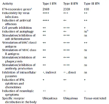
hematopoietic cells, such as plasmacytoid dendritic cells [Ivashkiv, 2014]. IFNγ is mainly produced by T lymphocytes, natural killer cells, and antigen-presenting cells (APCs) as monocytes, macrophages and dendritic cells. IFNγ secretion from natural killer cells (NK) and APC appears to be important in the early stages of infection, while secretion by T cells appears to be important in adaptive immune response. (Table 2)
Table 2. Biological activities of Interferons.
The aforementioned functions are the result of interactions of different family members of interferons which act within a complex system with agonistic and antagonistic effects. Many viruses have developed a system to escape the complex interferon system.
Based on their structure, three distinct IFN families (I-III) are recognized. Differences are based on specific cellular receptors used, cells involved and specific biological functions. Type I IFNs encode 13 partially homologous IFNα subtypes in humans, a single IFNβ and other monochromatic products (IFNε, IFNτ, IFNκ, IFNω, IFNδ and IFNδ) to date not yet well-defined [Trinchieri, 2010, Gryne 2017]. Type II IFNs consist only of IFNγ, produced by T cells and NK cells [Schoenborn, 2007]. Type III IFNs are IFNλ1, IFNλ2 and IFNλ3, and the newly discovered IFNλ4 [O'Brien, 2014], which have a spectrum of action similar to that of type 1
Maurizio Caocci, IFNα-MEDIATED SUPPRESSION OF JCV IS mTOR PATHWAY DEPENDENT, tesi di dottorato in Life Sciences and Biotechnologies, Università degli studi di Sassari
IFNs, but exerted only on a limited number of cell types, since their receptors are present only on the surface of epithelial cells [Witte, 2010].
1.4.1 Type I IFNs
IFNα and IFNβ are the best defined and widely expressed IFNs. Both participate in the antiviral and antimicrobial responses, by limiting the spread of infection to nearby cells [Yan, 2012] and modulating the innate immune response, thereby promoting the delivery of antigen by competent cells and enhancing the action of NK cells. Another function is to stimulate the adaptive immune system by provoking the response of the T cells and B and consequently the immune memory [Trinchieri, 2010 and Ivashkiv, 2014]. Importantly, because they can act in different pathways, inducting different genes, the same IFN can lead to opposite effect, depending on dosages and on timing. (Swetly, 1974)
1.4.1.2 Type I IFN production and signaling pathway
The majority of body cells can produce both IFNα and IFNβ in response to stimulation of different patterns receptors (PRRs) mainly for microbial products [Capobianchi, 2015, McNab, 2015]. After the binding of IFNs to their receptors, a pathway is activated and leads to the downstream activation of key molecules, such as IRF family of transcription factors, that activate the transcription of genes encoding IFNα and β. The pathway that induces the production of IFNα and IFNβ can be divided into different waves of gene transcription. First, the IFNβ gene transcription relies on IRF3, this set the transcription of IRF7. IRF7 in a positive feedback leads to the induction of a second wave of gene transcription, including IFNα-encoding genes [Honda, 2006] and NFκB can be required as a cofactor [Ivashkiv 2014, Honda 2006]. IFNα and IFNβ bind a heterodimeric transmembrane receptor, the IFNα receptor (IFNαR) which is composed of IFNαR1 and IFNαR2 subunits, leading to signaling pathways and induction of Interferon-Stimulated Genes [De Weerd,2012]. In the typical IFN-induced signaling pathway, after the binding of IFNs to IFNαR, it is shown the activation of the receptor-associated protein tyrosine kinases Janus kinase 1 (JAK1) and tyrosine kinase 2 (TYK2). The TYK2 phosphorylates the latent cytoplasmic transcription factors signal transducer and activator of transcription 1 (STAT1) and STAT2 [Stark, 2012],
Tyrosine-Maurizio Caocci, IFNα-MEDIATED SUPPRESSION OF JCV IS mTOR PATHWAY DEPENDENT, tesi di dottorato in Life Sciences and Biotechnologies, Università degli studi di Sassari
phosphorylated STAT1 and STAT2 dimerize and translocate to the nucleus, where they assemble with IRF9, to form a trimolecular complex, called IFN-stimulated gene factor 3 (ISGF3). ISGF3 binds to its cognate DNA sequences, which are known as IFN-stimulated response elements (ISREs), thereby activating directly the transcription of thousands of Interferon-Stimulated Genes (ISGs). The phosphoinositide 3-kinase (PI3K)-mammalian target of rapamycin (mTOR) pathway, NF-κB and MAPK pathways can also be activated downstream of IFNαR. This variety in signaling pathways could explain the broad effects of IFNα and β. It allows the transcription of genes important for viral restriction and also a large number of genes [Ivashkiv, 2014] that encode cytokines and chemokines, antibacterial effectors, pro-apoptotic and anti-apoptotic molecules, and molecules involved in several metabolic processes [Rauch, 2013].
Recent studies have highlighted new possible ways of regulating type I IFNs production: in physiological conditions, the commensal microbiota maintain the production of type I IFNs at basal levels [Abt MC, 2012]. Thus, immune cells can respond rapidly to baseline type I IFNs stimulation. Small amounts of IFNα and β also maintain STAT1 and IRF9 [Gough, 2012] basal expression levels, through an autocrine loop. In this context, and in conjunction with IFNαR activation, immune system cells may respond rapidly to microbial or viral stimuli. Type I IFNs production may be induced also by host factors, such as the TNFα cytokine, which follow the IRF1 signaling pathway, rather than that of IRF3 and IRF7 [Yarilina, 2008], and the macrophage colony stimulating factor (MCSF) [Ivashkiv, 2014].
Type I IFN effects: Cell Resistance and Immune Response
Interferon-stimulated genes, following activation by IFNα and β, play a primary role in limiting viral replication [Yan, 2012]. The ISG gene products can act by containing multiple steps of viral replication: inhibiting viral transcription, translation and replication, degrading viral nucleic acids by altering cellular lipid metabolism [MacMicking, 2012]. The importance of the interferon system is underlined by the fact that many viruses have developed different systems to interfere with their production [Capobianchi, 2015]. These systems include: inhibition of IFN induction, or by inhibition of their signaling pathways, activation of IFN regulatory factors (IRFs), STAT or NF-кB. Another recently studied feature is the action of type I IFNs in regulating immune cell functions, such as myeloid cells, B cells, T cells, and NK cells. IFNα
Maurizio Caocci, IFNα-MEDIATED SUPPRESSION OF JCV IS mTOR PATHWAY DEPENDENT, tesi di dottorato in Life Sciences and Biotechnologies, Università degli studi di Sassari
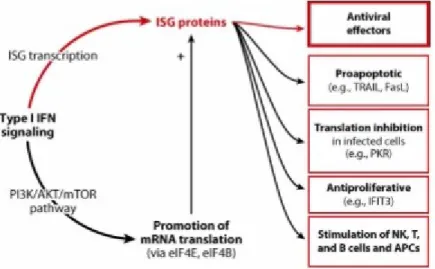
influences, thus increasing immune responses, solving viral infections more effectively and improving memory generation [McNab, 2015]. A summary of type I IFN-mediated function is depicted in Figure 6.
Figure 6. Interferon Stimulated Genes (ISG) and effects of IFNs (Fensterl, 2015)
In this context, IFNα has an activating effect on immature Myeloid Dendritic Cells (mDCs), increasing cell surface expression of MHC Class I and II molecules and co-stimulation molecules, such as CD80 and CD86, associated with greater ability to stimulate T cells [Ito, 2001, Montoya, 2002]. The mDCs are also stimulated by IFNα to present the antigen during viral infections [Spadaro, 2012] and allow their migration to the lymph nodes through the upregulation of chemochine receptors [Parlato, 2011]. IFNα regulates mDC and pDC in vivo by inducing downregulation of anti-apoptotic molecules, upregulation of pro-apoptotic molecules and caspase activation [Swiecki, 2011]. IFNα and β together with IL-12 increases the cytolytic activity of NK and CD8 + T cells and the production of IFNγ in vitro and in vivo, promotes Th1 polarization of CD4 + T cells, as well as long-term survival and memory T cell term. IFNα / β also promote the production of immunoglobulins by favoring the differentiation of B cells into immunoglobulin secreting plasma cells [Swiecki, 2011].
In summary, the interferons are potent immunomodulators, with specific immunomodulatory patterns, which may vary among different types and subtypes. In general, in the short term the interferons promote the activity of already differentiated cells, while, with prolonged exposure of cells to interferon, or with higher IFN concentrations, the antigrowth effect of interferons prevails; thereferore, they may reduce the replacement of exhausted cells with their precursors, thus causing inhibition of their immune functions [Capobianchi et al., 2015].
Maurizio Caocci, IFNα-MEDIATED SUPPRESSION OF JCV IS mTOR PATHWAY DEPENDENT, tesi di dottorato in Life Sciences and Biotechnologies, Università degli studi di Sassari
2. AIM OF THE THESIS
Infection by JCV is very common and is widespread worldwide; it is associated with the severe demyelinating disease progressive multifocal leukoencephalopathy (PML) and no therapies are still available.
Assetta et al (2016) found that JCV infection of primary human renal epithelial cells induced interferon production and activated interferon-stimulated gene expression. In the latter study, it was also noted that phosphorylated STAT1 and IFN regulatory factor-9 (IRF9) translocated to the nucleus in JCV-infected cells and that blocking the IFNAR and neutralization of IFN-α and IFN-β partially relieved inhibition of JCV infection (Assetta et al, 2016).
Our hypothesis comes from previews studies which reported that interferons negatively regulate JCV (Co et al, 2007; Assetta et al, 2016). Co et al (2007) reported that JCV replication was significantly inhibited by IFN in primary human fetal glial cells and neutralizing anti-IFN antibody rescued the inhibitory effect of IFN.
The aim of this thesis is to study the effect of IFN-α on JCV replication understanding the role of signal transducer and activator of transcription (STAT) family, STAT1 and STAT2, and other downstream factors involved in IFNs pathways. The understanding of the pathway involved could help in the development of treatments or combined treatments to prevent PML spread.
Maurizio Caocci, IFNα-MEDIATED SUPPRESSION OF JCV IS mTOR PATHWAY DEPENDENT, tesi di dottorato in Life Sciences and Biotechnologies, Università degli studi di Sassari
3.MATERIAL AND METHODS Cell culture
The TC620 human oligodendroglioma cell line was cultured at 37°C, 5% CO2 in DMEM + 10% FBS + 1% penicillin/streptomycin. PHFA (human fetal primary astrocytes) cells were obtained in six-well plates from the Comprehensive Neuro-AIDS Center (CNAC) tissue culture core at Temple University Lewis Katz School of Medicine and cultured in astrocyte growth media (Dulbecco’s modified Eagle's medium (DMEM): F12 medium supplemented with 10% fetal bovine serum, 10% GlutaMAX, insulin and gentamicin (10 μg/ml) at 37°C under 5% CO2 atmosphere.
JCV Infection
PHFA cells were plated in 100mm dishes and infected with JCV Mad-1 strain at moi = 1 and treated with IFN-α or/and IFN-β, and/not LY294002 for 14 days and harvested together with uninfected control cultures. Cells were transfected with luciferase reporter plasmid for the JCV early and JCV late promoters, harvested and analyzed after 48 hours. In parallel, the growth media supernatants of the cells was also collected to measure viral load by Q-PCR.
Luciferase reporter assay
TC620 cells were plated in 6 well plates and transfected with the reporter construct JCVE-LUC contains the JCV promoter from the Mad-1 strain linked to the luciferase gene in the early and late orientation and empty DNA vector was used as a control to normalize. After 48 h, cells were harvested with reporter lysis buffer for the luciferase reporter system provided by the kit (Promega, USA) and analyzed. Activities were normalized to control without expression plasmid.
Reagents
LY294002, a specific cell permeable phosphatidylinositol 3-kinase inhibitor, was used on PHFA cells at the concentration of 1nMol/L. IFN-α and IFN-β were used at a concentration of 100 UL/ml.
Maurizio Caocci, IFNα-MEDIATED SUPPRESSION OF JCV IS mTOR PATHWAY DEPENDENT, tesi di dottorato in Life Sciences and Biotechnologies, Università degli studi di Sassari
Cell fractionation
PHFA were treated with or without 100 U/ml IFN-α for 10 or 20 min and harvested for cell fractions using the NE-PER nuclear and cytoplasmic extraction kit according to the Manufacturer’s instructions (Thermo Scientific Pierce, Rockford IL; Cat. # PI-78835).
Western blots
Cells were harvested using trypsin, pelletted and incubated with TNN buffer for 30 minutes. After centrifugation at 12000 RPM the supernatant was collected and the concentration measured. 50 μg of whole cell extract proteins were resolved by 10% polyacrylamide SDS gel and transferred to nitrocellulose membrane. Blots were incubated with specific primary antibody (1/1000 dilution) and secondary antibody (1/10000 dilution) and visualized with an Odyssey CLx Imaging System (LI-COR, Inc., Lincoln, NE) using LI-COR Odyssey software. Band intensities were quantified using the ImageQuant software (Molecular Dynamics, GE Healthcare Bio-Sciences, Pittsburgh PA) and intensities normalized to α-tubulin or GAPDH or LAMIN a/c.
Antibodies
Primary antibodies: anti-PI3K/Phospho-PI3K (diluition 1/1000, Cell Signaling Technologies, Massachussets, USA) was used to check phosphatidylinositide 3-kinases and its phosphorilated form. Protein Kinase B and its phosphorilated form was checked using Anti-AKT/Phospho-AKT (diluition 1/1000, Cell Signaling Technologies, Massachussets, USA). To check STAT-1 and its phosphorilated form were used anti-STAT-1/Phospho-STAT-1 (1/1000, Stanta Cruz, Texas,USA). STAT-2/Phospho-STAT-2, STAT3,STAT5 (diluition 1/1000, Cell Signaling Technologies, Massachussets, USA). Expression of VP1 was checked using Anti-VP1 (1/500, Calbiochem, Massachussets,USA). Lamin A/C was used as a nuclear/cytoplasm extraction loading control (diluition 1/1000, Cell Signaling Technologies, Massachussets, USA). α-tubulin (1/10000, sigma, Missouri,USA) and GAPDH (1/1000, Santa Cruz, Texas,USA) were used as loading controls. Secondary antibodies: IRDye® 680RD Goat Anti-Mouse Li-COR dyes and IRDye® 680RD Goat Anti-Rabbit Li-COR dyes.
Maurizio Caocci, IFNα-MEDIATED SUPPRESSION OF JCV IS mTOR PATHWAY DEPENDENT, tesi di dottorato in Life Sciences and Biotechnologies, Università degli studi di Sassari
Immunocytochemistry (ICC)
PHFA cells were serum-starved overnight with 0.5% BSA, pre-treated with Rapamycin for three hours and then either untreated or treated with 100 U/ml IFN-α for 20 min. Cells were fixed in 4% paraformaldehyde in PBS for 10 min, washed, permeabilized for 5 min with 0.1% TritonX-100, blocked for 30 min with 5% normal goat serum and incubated 3 h at 37°C with rabbit anti-STAT-1 and anti-PhosphoSTAT-1 at a 1:100 dilution in PBS. Cells were then washed, incubated for 2 h with secondary FITC-conjugated goat anti-rabbit secondary antibody at a 1:250 dilution, washed, mounted with DAPI-containing mounting medium (VECTASHIELD, Vector Laboratories Inc. Burlingame, CA) and viewed by fluorescence microscopy.
Maurizio Caocci, IFNα-MEDIATED SUPPRESSION OF JCV IS mTOR PATHWAY DEPENDENT, tesi di dottorato in Life Sciences and Biotechnologies, Università degli studi di Sassari
4. RESULTS
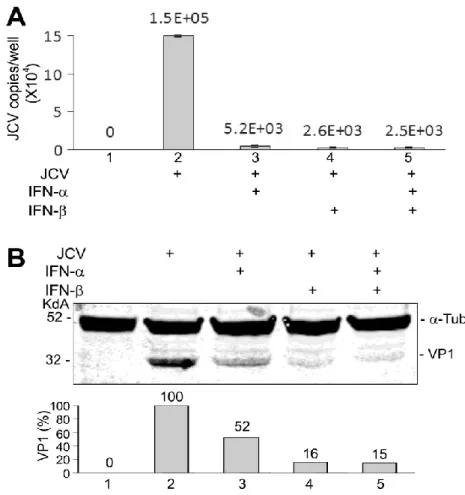
IFN-α and IFN-β inhibits JCV replication in Primary Human Fetal Astrocytes (PHFA). Firstly, we examined the effect of IFN- α and IFN-β on the replication of JCV in primary human fetal astrocytes (PHFA). Treatment of PHFA with IFN-α and IFN-β or both caused a drastic reduction in JCV replication of almost 100-fold as measured by the viral content in the culture supernatant (Fig. 1A). Similarly, IFN-α and IFN-β or both caused a reduction in the level of expression of the viral capsid VP1 (Fig. 1B). We conclude that both IFN-α and IFN-β inhibit JCV replication.
Figure 1. Inhibition of JCV replication in PHFA by IFN-α and IFN-β. PHFA were infected with the Mad-1 strain of JCV at moi = 1 and treated with IFN-α or/and IFN-β (100 U/ml), as indicated, for 14 days and harvested. A. Culture supernatants were assayed for virus using qPCR. Each viral load was measured in duplicate and presented as a histogram with the
Maurizio Caocci, IFNα-MEDIATED SUPPRESSION OF JCV IS mTOR PATHWAY DEPENDENT, tesi di dottorato in Life Sciences and Biotechnologies, Università degli studi di Sassari
standard deviation shown as an error bar. B. Fifty micrograms of total cell extract were electrophoresed on a 10% polyacrylamide SDS gel and analyzed by Western blot for VP1. Alpha-tubulin (α-Tub) was the loading control. The intensity of the VP1 band in each lane was quantified using the ImageQuant software (Molecular Dynamics, GE Healthcare Bio-Sciences, Pittsburgh PA) and these are shown in the lower part of the panel.
IFN-α inhibits JCV early and JCV late transcription.
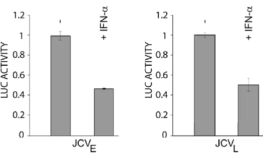
Next, we tested the effect of IFN-α and IFN-β on JCV transcription in TC620 cells using luciferase reporter constructs for the JCV early and late promoters. As shown in figure 2 upon IFNα treatment the luciferase levels were dramatically reduced to basal level for JCV early meanwhile JCV late was 50% reduced. We concluded that IFN-α inhibits both JCV early and JCV late transcription.
Figure 2. Effect of IFN-α on JCV early and JCV late transcription. TC620 cells were transfected with luciferase reporter plasmid for the JCV early and JCV late promoters. After 24h, cells were treated with or without 100 U/ml IFN-α. After 24 h, cells were harvested and
Maurizio Caocci, IFNα-MEDIATED SUPPRESSION OF JCV IS mTOR PATHWAY DEPENDENT, tesi di dottorato in Life Sciences and Biotechnologies, Università degli studi di Sassari
assayed for luciferase activity as described in material and methods. Activities were normalized to values without IFN-α. The bar represents one standard deviation.
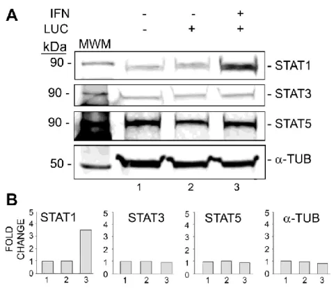
IFN-α induces expression of STAT1 but not STAT3 or STAT5.
In order to examine the effect of IFN-α on expression of STAT proteins, PHFA cells were treated with or without IFN-α and analyzed by Western blot for STAT1, STAT3 and STAT5 (Fig. 3). The level of STAT1 was increased over 3-fold whereas STAT3 and STAT5 were unchanged. Thus IFN-α treatment leads to the induction of STAT1.
Figure 3. Effect of IFN-α on STAT1, STAT3 and STAT5 expression. TC620 cells were transfected with luciferase reporter plasmid for the JCV early promoter and treated with or without 100 U/ml IFN-α. After 48 h, cells were harvested and fifty micrograms of total cell extract were electrophoresed on a 10% polyacrylamide SDS gel and analyzed by Western blot for STAT1, STAT3 and STAT5. Alpha-tubulin (α-Tub) was the loading control. The intensity
Maurizio Caocci, IFNα-MEDIATED SUPPRESSION OF JCV IS mTOR PATHWAY DEPENDENT, tesi di dottorato in Life Sciences and Biotechnologies, Università degli studi di Sassari
of the bands in each lane was quantified using the ImageQuant software (Molecular Dynamics, GE Healthcare Bio-Sciences, Pittsburgh PA) and are shown in the lower part of the panel.
IFN-α causes STAT1 subcellular redistribution to the nucleus.
To investigate the subcellular partition of STAT1 between the nucleus and cytoplasm, TC620 cells were treated with IFN-α, harvested and nuclear/cytoplasmic proteins extracted. The level of nuclear STAT1 was increased about 2-fold after both 10 and 20 minutes IFN-α treatment indicating that IFN-α causes translocation of STAT1 to the nucleus.
Figure 4. Effect of IFN-α on STAT1 subcellular distribution. PHFA were treated with or without 100 U/ml IFN-α for 10 or 20 min and cytoplasmic and nuclear fractions prepared as described in Materials and Methods. The distribution of STAT1 in these fractions was measured by Western blot. The purity of fractions and equal loading was assessed by Western blot for α-tubulin and lamin A/C. The nuclear and cytoplasmic STAT1 bands were quantified by densitometry and are shown a histogram of the percentage of STAT1 present in the nucleus.
Maurizio Caocci, IFNα-MEDIATED SUPPRESSION OF JCV IS mTOR PATHWAY DEPENDENT, tesi di dottorato in Life Sciences and Biotechnologies, Università degli studi di Sassari
IFN-α causes phosphorylation of STAT1 but not STAT2.
IFN-α activates the JAK/STAT pathway resulting in the phosphorylation of STAT proteins (Larner et al, 1993; Darnell et al, 1994). We examined the phosphorylation of STAT1 and STAT2 in response to IFN-α in PHFA using Western blot analysis with phosphospecific antibodies. As shown in Figure 5, STAT1 was robustly phosphorylated following IFN-α treatment for 24 h but STAT2 phosphorylation was unchanged.
Figure 5. Effect of IFN-α on pSTAT1/STAT1 and pSTAT2/STAT2. PHFA were treated with IFN-α (100 U/ml) for 24 h, cells were harvested and 50 µg of total cell extract were electrophoresed on a 10% polyacrylamide SDS gel, electrophoresed and analyzed by Western blot for pSTAT1/STAT1 and pSTAT2/STAT2. Alpha-tubulin (α-Tub) was the loading control.
Maurizio Caocci, IFNα-MEDIATED SUPPRESSION OF JCV IS mTOR PATHWAY DEPENDENT, tesi di dottorato in Life Sciences and Biotechnologies, Università degli studi di Sassari
As well as the JAK/STAT pathway, many additional JAK-dependent signal transduction cascades are activated by interferons, including the phosphatidylinositoI 3-kinase (PI3K) pathway (Platanias and Fish, 1999). PI3K is strongly and specifically inhibited by LY294002 (Vlahos et al, 1994). In the next experiment, we explored the effect of LY294002 on JCV replication in PHFA (Fig. 6). LY294002 enhanced JCV replication as measured by the production of viral capsid protein VP1 (Fig. 6A, compare lanes 2 and 3) and the level of virus in the culture supernatant, which increased almost 8-fold (Fig. 6B).
Figure 6. Effect of the PI3K inhibitor LY294002 on JCV replication. PHFA were infected with the Mad-1 strain of JCV at moi = 1 and treated with or without LY294002 (1 nM), as indicated, for 14 days and harvested. A. Fifty micrograms of total cell extract were electrophoresed on a 10% polyacrylamide SDS gel and analyzed by Western blot for VP1, pAKT/AKT, pPI3K/PI3K. Alpha-tubulin (α-TUB) was the loading control. B. Culture supernatants were assayed for virus
Maurizio Caocci, IFNα-MEDIATED SUPPRESSION OF JCV IS mTOR PATHWAY DEPENDENT, tesi di dottorato in Life Sciences and Biotechnologies, Università degli studi di Sassari
using qPCR. Each viral load was measured in duplicate and presented as a histogram with the standard deviation shown as an error bar.
Rapamycin alters STAT1 translocation to the nucleus
Rapamycin binds and inhibits mammalian target of rapamycin (mTOR), a signaling complex downstream of PI3K/AKT and mTOR complexes regulate IFN-induced AKT phosphorylation and are involved in IFN-dependent signaling (Kaur et al, 2012). As shown in the first two panels (control and rapamycin only treated) both STAT1 and pSTAT1 proteins are spread into the cytoplasm and nucleus without a specific accumulation site but,upon interferon treatment the majority of STAT1 and pSTAT1 are accumulated around the outside nucleus surface. Thus IFN-α treatment leads to the induction of mTOR pathway.
Figure 7. Effect of rapamycin on the redistribution of STAT1 induced by IFN-α. PHFA were plated and treated without or with IFN-α in the presence or absence of rapamycin, and then immunocytochemistry was performed for (A) STAT1 or (B) pSTAT1.