U N I V ER SI T À P O LI T E C N I C A ? DELLE M A R C H E FA CO LTÀ D I M E D I C I N A E C H I R U R G I A
Ph.D, Thesis
IdmtìfuxLtixm ofa new Siamar^for ceHac cRsease andother
gCuten-reCated (tisorders
DOTTORATO DI RICERCA- XXXI° CICLO
Curriculum Scienze Biomediche
Pediatria Generale e Specialistica
Tutor
Prof. Carlo Catassi
Anno accadem ico 2015-2018
Dottorando
Dott. AnM K. Verma
2
3
Dedicated to the loving memory of Prof. Elio Tonutti….
….Gone but Never Forgotten
Safe in the arms of Jesus4
Acknowledgment
Firstly, I would like to express my sincere gratitude to my Ph.D. supervisor Professor
Carlo Catassi for his continuous support of my Ph.D. study and related research, for
his patience, motivation, and immense knowledge. His guidance helped me all the time of research and writing of this thesis. I could not have imagined having a better advisor and mentor for my Ph.D. study.
My very special gratitude goes to Dott. ssa Elena Lionetti and Dott. ssa Simona Gatti for their advice at every step. I appreciate the help of Dott. ssa Tiziana Galeazzi for her incessant guidance in the laboratory. I am thankful to Dott. ssa Giada Del Baldo, Dott.
ssa Roberta Annibali, Dott. ssa Elisa Franceschini, Dott. ssa Matilde Rossi and Dott. ssa Vera dominijanni for their support from clinical side.
Along with them, I appreciate the help of my fellow labmates Dott. ssa Lucia Zampini,
Dott. ssa Lucia Padella, Dott. ssa Rita Lucia Marchesiello, Dott. ssa Chiara Monachesi, Dott. Alessio Coreanni, Dott. ssa Azzurra Pignotti, and Dott. ssa Ilaria Giretti for their support. I feel obliged to my lab mate and a good friend Dott. ssa Chiara Monachesi for her unceasing assistance. I am indeed very thankful for each
one of them for their support, they have made my Ph.D. years extremely relaxed.
Apart from my scientific team, I am thankful to Emanuela Maria Mariani (Secretary,
Department of Pediatrics, Università Politecnica delle Marche, Ancona, Italy) for
taking care of my Ph.D. administrative work and documentation since 2015.
I would like to express my sincere gratitude to Professor Govind Makharia (India) for his enormous guidance and support. I always remember his valuable lessons that gave me the courage to face challenges.
I would like to thank Dr. Giovanni Maria Maggiore, a vibrant scientist, and a nice friend, for allowing me to learn HLA-DQ typing techniques in his laboratory (Palermo, Italy), I also recognize his immense support during my learning about HLA.
I would like to offer special thanks to Professor Elio Tonutti (late), who is now safe in the arms of Jesus, without his help and guidance this journey was not possible. I am
5
very thankful to his whole research team Dott. Ssa Martina Fabris and Dott. ssa
Desre Fontana for their help and support during my learning days at University
Hospital, Udine (Italy).
I express my gratefulness towards, Professor Alessio Fasano, to me, he is one of the best supervisor a best friend. I am thankful to Prof. Fasano, for providing me the opportunity to join his research team at Massachusetts General Hospital, Boston,
MA (USA) as an external research scholar, and gave access to the laboratory and
research facilities. From his research team, I am grateful to Dr. Gloria Serena, for letting me learn advanced techniques during my visit to Boston (USA). I am thankful to
Dr. Anna Sapone for her support and cannot explain my gratitude in words for Susan Marie Flaherty for her support during my stay in Boston (USA).
I am grateful to my friends Dr. Pushpanjali Dasauni and Dr. Alka Sing for their scientific and moral support during the whole Ph.D. years. I appreciate the help and support of Alka Singh that she did for me during my study period.
I would like to express my sincere respect and love to Dott. ssa Eleonora Bove, a true well-wisher, and wife of Prof. Carlo Catassi, for her love, for her care, for her attention and for letting me feel I am living with my family.
I am very grateful to Dr. Suman lal for her endless emotional support. Her valuable advice always encourages me to become a decent person.
Above all, I would like to thank Anu Verma a fantastic engineer and my wife for her love and constant support, for all those late nights and early morning and for keeping me sane over the past few months. Thank you for understanding me best and for being my best friend. I owe you everything.
Last but not least, I would like to thank my family, my parents especially my brother
(Ajay K Verma) and sister (Indu Verma) for supporting me spiritually throughout the
learning years.
I am very much thankful to Università Politecnica delle Marche for proving me chance to be a part of this great institution and making arrangement for my Ph.D. study.
In the end, I would again like to pay my best regard to my professor (Carlo Catassi) for his tremendous help, without his support this thesis was not possible. He is a true leader and true inspiration.
6
Table of content
Introduction ... 6
Chapter 1. Gluten-related disorders: An introduction ...15
Celiac Disease ...19
Dermatitis herpetiformis ...19
Gluten Ataxia ...20
Wheat Allergy ...22
Gluten sensitivity (GS)/Non Celiac Gluten Sensitivity (NCGS)...24
Chapter 2. Celiac disease ...27
Chapter 3.History of CD diagnostic Biomarkers ... 41
Chapter 4. Need of novel CD biomarkers for adherence to GFD ... 50
Chapter 5. Development of my Ph.D.: A journey from Master’s to Doctorate
...
52Project 1. Biomarkers for the adherence to Gluten Free Diet ...57
Project 2. Per day gluten exposure determination in celiac disease patients on gluten free diet ...73
Project 3. Gluten contamination in naturally or labeled gluten-free products marketed in Italy ..81
Project 4. Contribution of oral hygiene and cosmetics on contamination of gluten-free diet ...86
Project 5. Validation of a novel single-drop rapid HLA-DQ2/-DQ8 typing...90
Project 6. Verification HLA-DQ2 and HLA-DQ8 alleles distribution in native South Indian Population ...95
Project 7. Re-exploring the iceberg of celiac disease in children ... 106
Project 8. The increasing prevalence of celiac disease: what is the role of an improved diagnostic accuracy?... 108
Project 9. Establishment of IgA EMA biopsy method ... 111
Project 10. Non-immunological biomarkers for assessment of villous abnormalities in patients with celiac disease ... 115
Project 11. Gluten sensitivity in patients with cerebellar ataxia: A prospective cohort study .... 118
7
Introduction
Cereal crops (Wheat, barley, rye, oat, corn) are the foremost portion of human food.
Among those, wheat is the most prevalent cereal grain and worldwide staple food. Due
to the visco-elastic property of wheat flour dough and due to the major storage protein
i.e. ‘gluten’, it is used extensively in the food industry primarily to prepare bread and pasta.1 Gluten is a ubiquitous material presents in almost every diet in apparent or its
concealed form. Apart from food industry gluten is used widely in cosmetics, bakeries,
toiletries, beverages drugs and numerous industries.2–4
Consumption of gluten in the form of wheat and closely related cereals gives birth to
several wheat related disorders collectively termed as “Gluten-Related disorders
(GRD)” that include Celiac disease (CD), Dermatitis herpetiformis (DH), Gluten
Ataxia (GA), Wheat Allergy (WA), and Non-Celiac Gluten Sensitivity (NCGS).5 GRD
gradually emerged as an epidemiologically relevant phenomenon with a global
prevalence of around 5%.5,6,7
For GRD, the only treatment available so far is the complete avoidance of gluten and
adherence to a strict gluten-free diet (GFD). Despite the well-recognized efficacy of the
GFD, complete adherence to GFD is challenging to achieve, numerous studies have
reported up to 50% incomplete compliance in celiac patients.8,9 It has been calculated
that gluten-free products with <20 mg/kg (or parts per million = ppm) of gluten level are
safe over a wide range of daily consumption.10 This maximum tolerable amount of
gluten contamination (<20 ppm) is recommended by Codex Alimentarius, US Food and
Drug Administration (FDA) and European Food Safety Authorities (EFSA).11–13
Among different GRD, a considerable amount of work has been done towards CD. Its
8
documented that help making a diagnose of CD. For other GRD so far, there is no
specific and reliable biomarker discovered. Available serological tests work well for
screening and diagnosis purposes. After the diagnosis of disease, for the follow-up
purpose, serological markers are not very reliable as autoantibody titers do not match
with the histological findings or with the symptoms of CD.14,15 In patients following a
GFD, elevated antibodies levels take about 6-24 months to reduce in blood.16 This is a
matter of concern that the requirement of adherence to GFD vary in each GRD, while a
strict and lifelong GFD is the only option for CD, NCGS requires repeated gluten
challenges. A strict adherence to the GFD is certainly necessary to improve the
symptoms, despite this, there are no clear guidelines to monitor the adherence to the
diet moreover, there are no validated biomarkers to assess the compliance. 17
This Ph.D. work has principally focused on the finding and validation of suitable
non-invasive biomarkers for diagnosis as well for the assessment of the adherence to GFD.
To fulfill this goal, I with my research team in the supervision of Professor Carlo
Catassi have extensively investigated the efficacy of different potential
blood-dependent non-invasive biomarkers for the diagnosis (Rapid HLA DQ typing method,
EMA Biopsy) as well as for the adherence to GFD [Alkylresorcinol (AR), GlutenImmunogenic Peptides (GIP urine test), Intestinal Fatty Acid Binding Protein (i-FABP)]. Additionally, the efficacy of blood-independent biomarkers has also
been investigated, as in the quantitative analysis of gluten in food products through
different antibodies that specifically designed to investigate the gluten content in food
products (e.g. R5 and G-12 ab). I have discussed the details of each biomarker in the
9
10
Awards
• First award for oral presentation in Società Italiana di Gastroenterologia Epatologia e Nutrizione Pediatrica (SIGENP) Congress Rome, Italy. 2017
• “Young Investigator Travel Grant”, World Congress of Pediatric Gastroenterology,
Hepatology and Nutrition (WCPGHAN), Canada. 2016
• “Young Researcher Award”, Società Italiana di Gastroenterologia Epatologia e
Nutrizione Pediatrica (SIGENP) award, Italy. 2016
• Full Ph.D. fellowship, Università Politecnica delle Marche, for Ph.D. in Biomedical Sciences, Italy, 2015
Professional Membership
• Italian Society of pediatric gastroenterology, hepatology, and Nutrition (SIGENP) • International Society for the study of Celiac Disease (ISSCD)
Reviewer
• BMC Health Services Research
• Journal of Gastroenterology and Hepatology Open (JGH Open)
•
Annals of pediatricsExternal Reviewer
• Nutrients • Food • Safety • SustainabilityEditorial board member
• Journal of public health and nutrition • Nutrition and public health
11
Featured Publications: S.
No. Title Authorship status Year Journal
Impact factor 1. Gluten contamination in naturally or labeled gluten-free products marketed in Italy [Project 3] First & corresponding Published 2017 Nutrients 4.2 2. Comparison of diagnostic performance of the IgA anti-tTG test vs IgA anti-native gliadin
antibodies test in detection of celiac disease in the general population [Project 8] First & corresponding Published 2018 CGH 7.9 3. Validation of a novel single-drop rapid HLA-DQ2/-DQ8 typing method to identify subjects susceptible to celiac disease [Project 5]
First Published 2018 JGH Open
Newly launched journal 4. Contribution of oral hygiene and cosmetics on contamination of gluten-free diet: do celiac customers
need to worry about? [Project 4] First & corresponding Published 2019 JPGN 2.8 5.
Celiac disease in the
year 2020: still
increasing, largely
undetected
[Project 7]
Fourth
12
Status of the research studies (projects) performed during Ph.D. duration (2015-2018)
Project no. Project title Status Duration Publication status
1.
Biomarkers for the adherence to Gluten Ongoing 2018 -
2. Per day gluten exposure determination in celiac disease patients on
gluten-free diet Ongoing 2018 -
3. Gluten contamination in naturally or labeled gluten-free products
marketed in Italy Completed 2016-2017 Published
4. Contribution of oral hygiene and cosmetics on contamination of
gluten-free diet Completed 2018 Published
5. Validation of a novel single-drop rapid HLA-DQ2/-DQ8 typing method to
identify subjects susceptible to celiac disease Completed 2017-2018 Published
6. Verification of HLA-DQ2 and HLA-DQ8 allele distribution in native south
Indian population Ongoing 2018 -
7.
Re-exploring the iceberg of celiac disease in children Completed 2015-2017 Under review
8. The increasing prevalence of celiac disease: what is the role of an
improved diagnostic accuracy? Completed 2017 Published
9.
Establishment of IgA EMA biopsy method Completed 2018 -
10. Non-immunological biomarkers for assessment of villous abnormalities in
patients with celiac disease Completed 2015-2018 Under review
11. Gluten sensitivity in patients with cerebellar ataxia: A prospective cohort
13
Abbreviations:
AGA Anti Gliadin antibodies
Anti tTG ab anti Tissue transglutaminase antibodies
AOAC Association of Official Analytical Chemists
AR Alkylresorcinol
ATI Amylase-Trypsin inhibitors
AU arbitrary units
BAT Basophil activation test
BP Base pair
CD Celiac disease
DGP De-amidated gluten peptide
DH Dermatitis herpetiformis
EFSA European Food Safety Authorities
ELISA Enzyme-Linked ImmunoSorbent Assay
EMA Endomysial antibodies
ESPGHAN European Society of Pediatric Gastroenterology Hepatology
EU European Union
FA Food allergy
FDA Food and Drug Administration
FDR First degree relatives
GA Gluten Ataxia
GFD Gluten-free diet
GIP Gluten immunogenic peptides
GRD Gluten-related disorders
14
HLA Human Leukocytic Antigen
HMW-GS High Molecular Weight Glutenin Subunits
HPLC Hi-performance liquid chromatography
HSCT Hematopoietic stem cell transplantation
IEL Intraepithelial lymphocytes
i-FABP Intestinal Fatty Acid Binding proteins
IFN Interferon IgA Immunoglobulin A IgE Immunoglobulin E IgG Immunoglobulin G IHC Immunohistochemistry IL Interleukin
IUIS International Union of Immunogenic Societies
LMW-GS Low Molecular Weight Glutenin Subunits
MA Molecular-based allergy
mAb Monoclonal antibody
Mg Milligram
ml Mililiter
MW Molecular weight
NCE Non-celiac enteropathies
NCGS Non-celiac gluten sensitivity
NPV Negative predictive value
pH Power of hydrogen
ppm Parts per million
PPV Positive predictive value
PWG Poramin working group
RT Real-time
15
SPT Skin prick test
TG6 tissue transglutaminase 6
TGAse Tissue transglutaminases
Th T helper cells
UK United Kingdom
ULN Upper limits of normal
USA United State of America
WA Wheat Allergy
WDEIA wheat dependent exercise-induced anaphylaxis
WHO World health organization
16
Chapter 1
Gluten-related disorders: An introduction
Wheat (Triticum aestivum) is a cereal grain which is the world’s most favored staple food.18 In 2016, global wheat production was 749 million tonnes.19 Cultivation of wheat
was started about 10,000 years ago during the Neolithic period in the fertile crescent of
the middle east. The wheat kernel contains 8%–15% of protein, from which 10%–15% is
albumin/globulin and 85%–90% is gluten (Figure 1).20
Gluten is a complex mixture of hundreds of related but distinct proteins, including some
toxic proteins fractions that are mainly gliadin and glutenin.21 Alcohol soluble gliadins
are further classified in their primary structures into α-, γ- and ώ-gliadins, poorly alcohol soluble glutenin proteins can be divided into high molecular weight glutenin
subunits (HMW-GS), low molecular weight glutenin subunits (LMW-GS) [Figure 1].22 The gliadins have high proline and glutamine content, known as prolamins,
characterized by high levels of glutamine (38%) and proline residues (20%). humans
inherently lack endopeptidases (an enzyme) that cleave bonds between proline and
glutamines. The incomplete digestion of gliadin by digestive tract enzymes leads to the
generation of many bigger peptides that eventually causes dietary diseases.20 In the last
decades, multiple disorders have been reported due to the ingestion of gluten. There is
a general agreement for the term “Gluten-Related Disorders” that is an umbrella-term
to be used collectively for all such disorders with 5% worldwide prevalence. 5,23,24 GRD
is the umbrella-term to be used for describing all conditions related to ingestion of
17
WA, and NCGS.5 Each disorder has discussed in separate sections. Classification of
GRD is shown in Figure 2.
Figure 1: Breakdown of wheat components.
HMW-GS: High Molecular weight glutenin subunit; LMW-GS: Low molecular weight glutenin subunit
Starch (60-70 %) Protein (8-15 %) Moisture (10-15%) Lipid (1-2 %) Ash (4 %) Albumin/ Globulin Gluten (85-90 %) Gliadin (50 %) Glutenin (50 %) α γ ω HMW-GS LMW-GS
Wheat Kernel
18
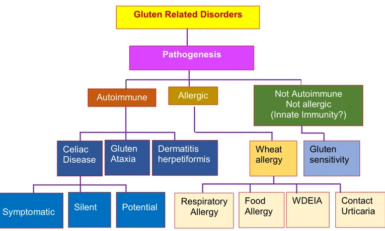
Figure 2: Proposed new nomenclature and classification of gluten-related disorders.
Gluten Related Disorders
Pathogenesis Autoimmune Allergic Celiac Disease Not Autoimmune Not allergic (Innate Immunity?) Gluten Ataxia Dermatitis herpetiformis Wheat allergy Gluten sensitivity
Symptomatic Silent Potential
Respiratory Allergy Food Allergy WDEIA Contact Urticaria
19
Celiac Disease
Celiac disease (CD) is an autoimmune condition characterized by permanent
intolerance to dietary gluten, a protein complex found in wheat, rye, and barley,
occurring in genetically predisposed individuals i.e individuals display HLA-DQ2 and/or
HLA-DQ8 alleles. At least 1% of the general population all over the world is affected by
CD. The hallmarks of the active CD are the presence of serum autoantibodies (e.g. IgA
anti-transglutaminase; anti tTG ab and anti-endomysial antibodies; EMA) and a small
intestinal enteropathy characterized (in typical cases) by villous atrophy, crypt
hypertrophy and increased number of intraepithelial lymphocytes (IELs). Treatment of
CD is based on the lifelong exclusion of gluten-containing food from the diet.GFD
determines the gradual disappearance of symptoms and serum autoantibodies, and the
normalization of the intestinal histological architecture. CD is explained further in
chapter 2.
Dermatitis herpetiformis
The term dermatitis herpetiformis (DH), was introduced by Louis Adolphus During in
1884, is a chronic skin inflammatory condition due to the indolence of gluten that results
in skin rashes, blisters filled with watery fluid. Elbows and upper forearms are the most
affected body parts (almost 90% DH patients), other common sites are knees,
shoulders, sacrum, face, scalp, neck and trunk. About 1 in every 10,000 in the UK and
USA and slightly higher in Europe, 4-6 in every 10,000 in Finland and Sweden are
20
DH is often reported from Asia and Africa. The age of onset of DH is about 15-40
years, DH is more common in males than in females. About 5 % of first degree relatives
of DH patients develop DH. Approximately 90%-95% of DH patients express
HLA-DQ2/-DQ8 haplotypes.25,26 DH is a manifestation of CD, the exact causal mechanism is
unknown to date. It is not understood very well, why only some patients with CD
develop DH. In DH, IgA is present in skin and inflammatory cells, EMA and anti tTG ab
occur in serum. Epidermal transglutaminase-3 (TG3) is an effective serological test for
DH. Suspected individuals are advised to undergo for a skin biopsy to check the IgA
uninvolved skin and could be considered for duodenal biopsy test, about 60-75%
patients with DH show villous abnormalities, patients with normal endoscopy also show
a minor change in the mucosa e.g. increased IELs. Diagnosis of DH is mostly
dependent on serology and skin biopsy, a duodenal biopsy is not strictly advised to DH
patients.27 Once the diagnosis of DH is confirmed, a gluten elimination from the diet is
recommended to the patients even they show a normal duodenal mucosa. A lifelong
strict GFD heals the complications and it also helps reduce other associated intestinal
conditions.28
Gluten Ataxia
Ataxia is a general term that defines the symptoms and signs resulting from cerebral
dysfunction. Major symptoms of incoordination of limbs, gain instability slurred speech.29
Gluten ataxia (GA) is an extra-intestinal, autoimmune disorder caused by the ingestion
of gluten in genetically susceptible individuals and characterized by the damage to the
cerebellum resulting in ataxia. GA is probably one of the commonest cause of idiopathic
21
So far, the exact prevalence of GA is not known. However, in the last two decades,
multiple key studies have found a high prevalence of native anti-gliadin antibody (AGA)
in GA patients. 31–33
Elimination of gluten from the diet has shown a satisfactory improvement in GA
symptoms.29 About 95% of patients with GA have Human Leukocytic Antigen (HLA)
-DQ2 or -DQ8 positive. However, 10%-15% of AGA prevalence has been reported in the
general population. AGA test also remained a first line serological test for CD several
years before. This evidence suggest that AGA is, however, not a specific serological
test for GA. Nevertheless, in the last decades, significant deposition of transglutaminase
antibodies has been found around the brain vessels (mainly in the cerebellum) of GA
patients. Antibodies against tissue transglutaminase 6 (tTG6), a primarily
brain-expressed transglutaminase, have been found in patients with GA.34,35 At present, TG6
ab test in association with the AGA test is used as a serological test. However, in an
interesting study, Hadjivassiliou and co-workers found that 73% of patients with
idiopathic sporadic ataxia positive for AGA, were also positive for TG6 antibodies.35 This
observation suggested a correlation between AGA and TG6 positivity but does not
clarify whether TG6 antibodies are more sensitive or specific for GA than AGA.35,36
For making a diagnosis for GA serological tests including AGA (IgG and IgA), anti-tTG2
antibodies and, if available, IgG and IgA anti-tTG6 antibodies are considered as a
screening test. Considering the level of these abs, patients should undergo a duodenal
biopsy. However, irrespective to the presence of an enteropathy, patients positive for
any of these antibodies with no alternative cause for their ataxia should be offered a
22
usually takes 6-12 months. Stabilization or even improvement of the ataxia after one
year would be a strong indicator that the patient suffers from GA.32,36
Wheat Allergy
Wheat allergy (WA) is a specific wheat dependent allergic condition where ingested
wheat causes an IgE mediated immediate allergy reaction in both adult and children.
This involves urticaria, angioedema, bronchial obstruction, nausea, and abdominal pain.
Depending on the route of the allergen and immunologic mechanism WA could be
classified in multiple types e.g., occupational asthma (backer’s asthma) and rhinitis, food allergy (FA) affecting the skin, the gastrointestinal tract or respiratory tract, wheat
dependent exercise-induced anaphylaxis (WDEIA) and contact urticarial. According to
the US Food and Drug Administration (FDA), the overall prevalence of WA
(self-reported as well as clinically diagnosed) is 0.4%.37 WA shows a greater prevalence in
children than adults.37,38 However, in adults most common condition is WDEIA where
ingested food and physical exercise together display the most common symptoms e.g.,
diarrhea and bloating. Among multiples WA conditions, backer’s asthma and rhinitis are the most ancient and well-recognized allergic response due to the inhalation of wheat
flour.39,40 About 10%-15% of Baker's and pastry factory workers are affected by this
condition.41 Several allergenic proteins have been recognized for WA. Recently, 21
well-classified wheat allergens listed in the updated database of WHO/IUIS allergen
nomenclature.42 Skin prick test (SPT), in-vitro specific Immunoglobulin E (sIgE) assay
and functional tests (a bronchial challenge test in baker’s asthma and a double-blind placebo-controlled food challenge or an open oral food challenge in FA) are considered
23
these tests have lower sensitivity and their positive predictive value (PPV) is limited to
75%.39 Reagents, used in commercially available SPE kits are not highly purified they
are only a mixture of water and salt soluble wheat that lacks allergens from insoluble
gliadin fraction and required a significant improvement in the kit performance.39,40 On
the other hand, sIgE assay is about 70-75% more sensitive that SPE test but is about
60% less specific that SPE test due to the cross-reactivity with pollens43.
Molecular-based allergy (MA) diagnostics could be a promising method for the
diagnosis of WA. In recent time, wheat flour extracts (omega-5 gliadin) based
ImmunoCAP™ assay, alpha-amylase/trypsin inhibitors (ATIs) based ISAC™ assay is
reasonably satisfactory markers.40 However, with approximately 20% missed cases,
sIgE to omega-5 gliadin assay is highly reliable and now widely used to identify the
patients with WDEIA44 Recently introduced flow cytometry-assisted basophil activation
test (BAT) seems to be promising in vitro functional test for the diagnosis of
immediate-type allergy. In clinical practice, the use of BAT is increasing. However, it is an
24
Gluten sensitivity (GS)/Non Celiac Gluten Sensitivity (NCGS)
Gluten sensitivity (GS) or more simply Non-Celiac Gluten Sensitivity (NCGS) was
introduced during the 1980s but strong attention was given in the last two decades
when a large number of individuals erupted with NCGS.47 So far, insufficient knowledge
is poised about the specific diagnostic criteria, biomarker, treatment and management
of NCGS. However, to date, four international scientific expert meetings have been
called to make a consensus about NCGS. A panel of experts discussed the definition,
diagnostic algorithm, advances, and current trends on NCGS. First, two expert meetings
were organized in London, 2011 and Munich, 2012 respectively.5,39 The third expert
panel meeting was held in Salerno (Italy) 2014 to make criteria of how the diagnosis of
NCGS should be confirmed, this is called “The Salerno Expert’s Criteria”.48 Latest meeting (4th) has been held in Bolzano (Italy) in 2018. According to Salerno criteria,
“Non-Celiac Gluten Sensitivity is a non-autoimmune, non-allergic, intestinal and extra-intestinal symptoms due to the ingestion of gluten-containing food in subjects that are
not affected by either CD or WA”.48
Classical symptoms of NCGS include abdominal pain, bloating, bowel habit
abnormalities (diarrhea/constipation) and systemic manifestations such as foggy mind,
headache, fatigue, joint, and muscle pain. In NCGS, celiac-specific antibodies may
remain absent, the villous structure remains normal with a variable status of HLA-DQ
alleles (50% positivity) and variable status of native (first-generation) anti-gliadin
25
Till date, the definite prevalence of NCGS is unknown but it is frequently reported.
However, a study done in the USA estimate a high prevalence of NCGS i.e. 6%.39 Until
now, there is no specific biomarker designated for the NCGS, most specific biomarkers
for CD (IgA anti tTG ab and EMA) do not provide a reliable result. Nevertheless, the IgG
class of anti-native gliadin antibody (IgG AGA) test shows a somewhat satisfactory
result and a recommended sero-test for suspected individuals.31,5 The recommended for
NCGS is the complete exclusion of gluten for the diet (i.e. GFD) and a gluten challenge.
However, before advising a GFD, a suspected individual should undergo complete
clinical and laboratory evaluation while on a normal diet (before GFD) to exclude the
possibility of having CD or WA. After 6 weeks of normal diet (gluten-containing diet) the
suspected individual is kept on 6 weeks of complete GFD. After this period, seven days
of gluten challenge is given to the patient followed by a one-week washout period of
strict GFD and by the crossover to the second one-week challenge. During all this
duration patient’s condition is monitored. Depending on the examination final diagnosis
is made.5
Although so far, there is no certain biomarker for the diagnosis for NCGS, three
gluten-related disorders (WA, CD, and NCGS) can be discriminated based on their combined
clinical, biological, genetic and histological data following the algorithm given by Sapone
26
Figure 3: Proposed algorithm for the differential diagnosis of gluten-related disorders, including celiac disease, gluten sensitivity, and wheat allergy
EGD with Biopsies Suspected GS WA diagnosis confirmed WA ruled out
• tTG IgA +/- EMA + total IgA • Deamidated AGA IgA • AGA YES YES Potential CD CD diagnosis confirmed GS ruled out consider other diagnoses GS diagnosis confirmed YES YES Tests + Challenge + tTG and/or dAGA + Gluten Challenge + NO NO NO Biopsy positive NO
History and Physical Exam-Initial Evaluation- Consider Differential Diagnosis
Wheat Allergy (WA) Celiac Disease (CD)
Gluten Sensitivity (GS)
• Specific skin prick tests • Wheat specific serum IgE • Gluten challenge
27
Chapter 2
Celiac disease
Introduction
Among the wide range of adverse reactions caused by the gluten, CD is the
longest-studied systemic, autoimmune disorder with the best-known pathology.
CD is a heritable, inflammatory condition of the small intestine, takes place due to
ingestion of immune-dominant dietary peptide (gliadin in wheat, hordein in barley and
secalin in rye) in the predisposed individuals who possess specific Human Leukocytic
antigen (HLA) -DQ2 and/or HLA-DQ8 alleles. Ingestion of these peptides eventually
causes villous atrophy, crypt hyperplasia, malabsorption, and extra-intestinal
manifestations. Lifelong Gluten-free diet (GFD) is currently the only accepted therapy
for CD that leads to normalization of the histological lesion.
Epidemiology
CD once thought to be an uncommon disease but now At least 1% of the world
population is affected with CD.49–52 While a large number of subjects are expected to
have CD, the majority of them still remain undiagnosed.53 In the year 1996, our group
conducted one large CD screening study in Italian school children of 6-15 years of age
reported prevalence of silent CD as 0.5%.54 Pioneer CD population screening studies
based on IgA native AGA as the first level test (conducted between 1992 and 2003)
reported a prevalence of CD ranging from 0.25 to 1.13%, with an overall pooled
anti-28
tTG have described a prevalence rate ranging from 0.2 to 2.4%, with a pooled
prevalence rate of 0.9% (Table 1). These data suggest that CD prevalence has
increased in recent years, a hypothesis that is also supported by longitudinal studies
performed in North America and Europe.55,56 Initially, it was exclusively thought as the
disease of western countries but now it is considered to be relatively common
throughout the world.51,57–59 In the United States, the CD is believed to affect 0.5%-1.0%
of the general population.60 In Northern African populations (Morocco, Algeria, Tunisia,
Libya, and Egypt) the prevalence of CD was 0.28%-5.6% in the general population.61–63
In Australia and New Zealand, the overall prevalence is 1:82 (1.2%).64 There is a large
variation in prevalence of CD between countries, from the lowest record incidence of
0.2% in Germany to as high as 2-3% in Finland and Sweden.51
In Asian countries which shows both serological and biopsy prevalence of CD 1.6% and
0.5% respectively.65 Among Asian countries, in India CD has been explored
respectfully. Some elegant CD screening studies have been conducted in past years
showed overall biopsy-proven CD prevalence in India is more or less 1% (I was among
the authors in two of three studies).65–67 Some possible reasons for increasing CD
prevalence could be, due to advance diagnosis or increased awareness. Instead of the
availability of several highly sensitive and specific non-invasive serological markers (e.g.
anti tTG ab), intestinal biopsy is still considered as the gold standard for diagnosis of
29
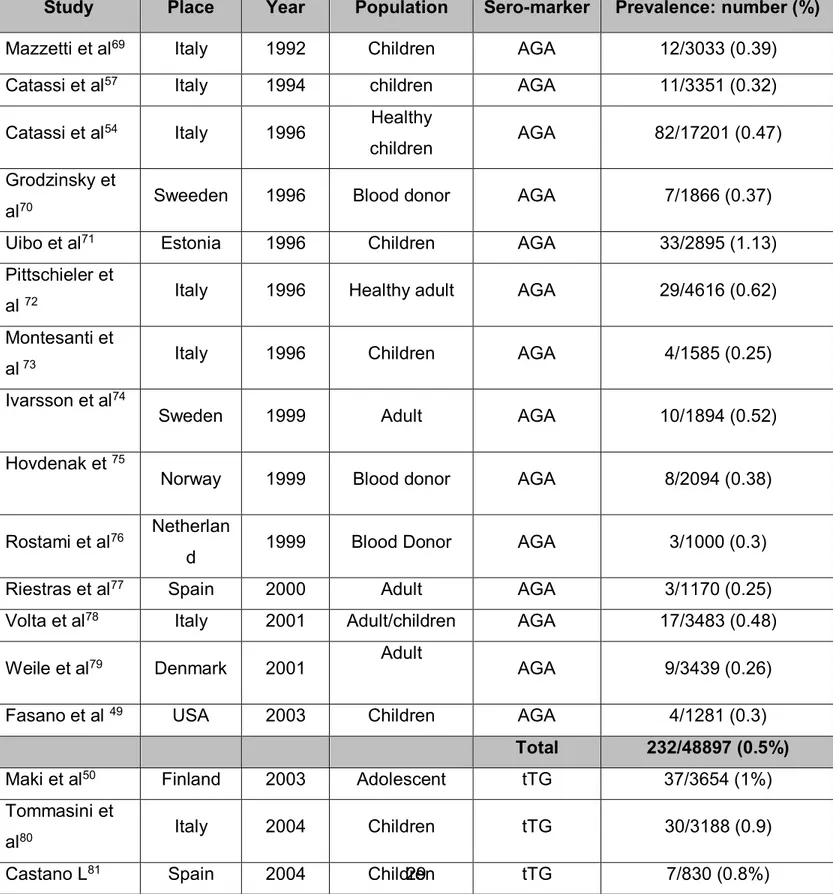
Table 1: Summary of CD prevalence studies based on AGA and tTG as a first level screening tool in different countries.
Study Place Year Population Sero-marker Prevalence: number (%)
Mazzetti et al69 Italy 1992 Children AGA 12/3033 (0.39)
Catassi et al57 Italy 1994 children AGA 11/3351 (0.32)
Catassi et al54 Italy 1996 Healthy
children AGA 82/17201 (0.47)
Grodzinsky et
al70 Sweeden 1996 Blood donor AGA 7/1866 (0.37)
Uibo et al71 Estonia 1996 Children AGA 33/2895 (1.13)
Pittschieler et
al 72 Italy 1996 Healthy adult AGA 29/4616 (0.62)
Montesanti et
al 73 Italy 1996 Children AGA 4/1585 (0.25)
Ivarsson et al74
Sweden 1999 Adult AGA 10/1894 (0.52)
Hovdenak et 75
Norway 1999 Blood donor AGA 8/2094 (0.38)
Rostami et al76 Netherlan
d 1999 Blood Donor AGA 3/1000 (0.3)
Riestras et al77 Spain 2000 Adult AGA 3/1170 (0.25)
Volta et al78 Italy 2001 Adult/children AGA 17/3483 (0.48)
Weile et al79 Denmark 2001 Adult AGA 9/3439 (0.26)
Fasano et al 49 USA 2003 Children AGA 4/1281 (0.3)
Total 232/48897 (0.5%)
Maki et al50 Finland 2003 Adolescent tTG 37/3654 (1%)
Tommasini et
al80 Italy 2004 Children tTG 30/3188 (0.9)
30
Clinical features
CD is a lifelong disease (once a celiac, always a celiac) where individuals may present
GI symptoms, extraintestinal symptoms, no sign or symptoms at all and nutritional
deficiencies as well. The clinical manifestations of CD are classical (signs and
symptoms of malabsorption such as diarrhea, steatorrhea, weight loss & growth failure)
or non-classical and symptomatic (with evident GI and/or extra-intestinal symptoms) or
asymptomatic.88,89 There are several classical and non-classical symptoms of CD
summarized in Table 2.
Menardo et al82 Italy 2006 Adult tTG 10/1002 (1%)
Akbari et al 83 Iran 2006 Adult tTG 27/2799 (1%)
Vilppula et al84 Finland 2008 Adult tTG 60/2815 (2.1%)
Mustalahti et al51 Finland 2010 Adult tTG 113/4846 (2.4%) Germany 8/3038 (0.2%) Italy 32/4781(0.7%) Marine et al85 Spain 2011 Children tTG 11/780 (1.4%) Adult 10/3450 (0.3%)
Dalgic et al86 Turkey 2011 Children tTG 95/20190 (0.5%)
Alarida et al 87 Libya 2011 Children tTG 19/2920 (0.7%)
31
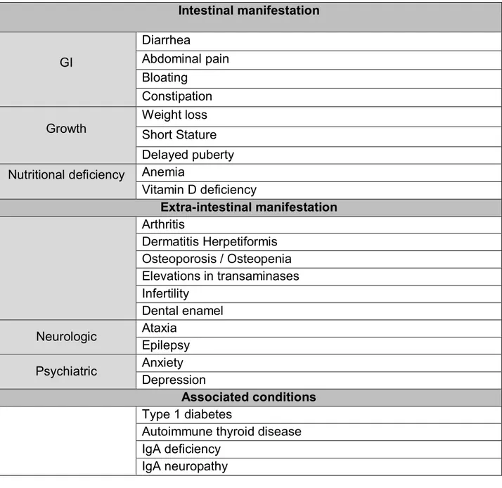
Table 2: Clinical features of celiac disease
Classification of CD
Significant improvement in the diagnostic methods in CD has changed the concept of
understanding the presentation of CD. It is not mandatory that an individual suffering
from CD will always preset the classical symptoms of CD (diarrhea, growth failure).90 A
Intestinal manifestation GI Diarrhea Abdominal pain Bloating Constipation Growth Weight loss Short Stature Delayed puberty
Nutritional deficiency Anemia
Vitamin D deficiency Extra-intestinal manifestation Arthritis Dermatitis Herpetiformis Osteoporosis / Osteopenia Elevations in transaminases Infertility Dental enamel Neurologic Ataxia Epilepsy Psychiatric Anxiety Depression Associated conditions Type 1 diabetes
Autoimmune thyroid disease IgA deficiency
32
large number of adult CD individuals present atypical symptoms of CD. Based on the
presentation, CD has been classified in the multiple forms that are described in Table 3
Table 3: Classification of celiac disease
Types Features
Classical
Associated with features of intestinal malabsorption, gluten-induced villous atrophy and other classic histological features, the presence of GI symptoms (diarrhea, steatorrhea, abdominal distension, iron-deficiency anemia, and weight loss or growth failure). Children with classical CD present with chronic diarrhea, vomiting, abdominal distension and failure to thrive, muscle wasting and poor appetite and show signs of emotional distress and lethargy.
Non-classical
Associated with the atypical or extraintestinal manifestations, such as unexplained iron deficiency anemia, short stature, osteoporosis,
arthritis, delayed puberty, infertility, peripheral neuropathy,
hypertransaminasemia, dermatitis herpetiformis and dental enamel defects at the time of diagnosis.
Atypical
Patients generally have little or no GI symptoms but come to medication because of other reasons such as iron deficiency, osteoporosis, short stature, or infertility, generally, have fully developed gluten-induced villous atrophy. Since these patients are “asymptomatic” from the GI perspective, a large number remain undiagnosed.
Asymptomatic
Associated with no symptoms or commonly associated with CD, even on detailed questioning and presence of villous abnormalities, these patients are diagnosed only through the testing of populations enrolled in screening program or through family studies
Potential It is characterized by the presence of CD-specific autoantibodies in
the blood of patients without histological abnormalities in small intestinal biopsies or may develop intestinal damage later. These
33
patients are at increased risk of developing CD as indicated by positive CD serology.
Silent
It is characterized by asymptomatic patients having gluten-induced villous atrophy. They are discovered after serologic screening or perhaps during endoscopy and biopsy for another reason. These patients are clinically silent or associated with atypical features of CD such as iron deficiency or osteoporosis.
Latent
It is associated with the previous diagnosis of CD that responded to a GFD and retains normal mucosal histology or manifests only an increase in IELs. It also represents patients with normal intestinal mucosa, on a gluten-containing diet who will subsequently develop CD.
Refractory
It represents patients with true CD (i.e. not a misdiagnosis) who do not or no longer respond to a GFD. Some of these patients develop complications such as ulcerative jejunoileitis or enteropathy-associated T-cell lymphoma.
Pathogenesis
The principal toxic component of wheat gluten belong to a family of closely related
proline and glutamine-rich proteins called gliadins.91 In-vitro and in-vivo studies (rats
and human) have confirmed that CD is triggered by the presence of a 33-mer peptides
(LQLQPFPQPQLPYPQPQLPYPQPQLPYPQPQPF) that develops due to the partial
digestion of gliadins because the gliadin remain stable toward breakdown by all gastric,
pancreatic, and intestinal brush border membrane endoproteases.15,39,40 This peptide
was identified as the primary initiator of the inflammatory response to gluten in celiac
34
smaller peptides but because of the lack of the proly-endopeptidase among gastric,
pancreatic and brush border enzyme relatively large gluten peptide that is rich in proline
and glutamine remain after initial digestion.92 High proline content renders these
proteins resistant to complete proteolytic digestion in the human intestine. Proline-rich
fragments of gluten that are resistant to processing by luminal enzymes survive
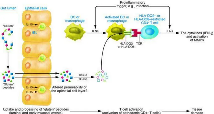
digestion and transported across the mucosal epithelium as polypeptides (Figure 4).
Tissue transglutaminase-2 (tTG), a calcium-dependent autoantigen is a ubiquitous
enzyme which is released in the intestinal mucosa during tissue injury, has a role in
tissue repair and cross-linking proteins by forming iso-peptides bond between
glutaminase and lysine residues.93 tTG also has a high avidity from gluten peptides and
under certain conditions (e.g. low pH) and in the absence of lysine residue, can
deamidate glutamine, which converts neutral glutamine to negatively charged glutamic
acid. These negatively charged glutamic acid residues manifest an increased binding
affinity for the disease-relevant HLA-DQ2/-DQ8 molecules. Once bound to HLA-DQ2/8
molecule, the gluten peptides HLA-DQ complex can activate CD4 T-helper 1 (Th1) cells
in the mucosa of the small intestine that recognize these complex.94 The gluten-reactive
CD4 T-cells produce interferon (IFN-γ) on activation. IFN-γ is also produced by T-cells
in the epithelium. Interleukin (IL-15), produced by either mononuclear cells in the lamina
propria or by enterocytes, stimulates T-cells to migrate to the epithelium and facilitate
the killing of enterocytes by upregulated expression of mic by enterocytes and NKG2D
by intraepithelial T-cells. IL-15 production is stimulated by gluten.95,96 Gluten can also
induce production of the intestinal peptide zonulin, which acts on tight junctions and
35
Figure 4: Pathogenesis of Celiac disease
Diagnosis and management of celiac disease
The diagnosis of CD is established by the European Society for Pediatric
Gastroenterology Hepatology and Nutrition (ESPGHAN) criteria (designed especially for
pediatric subjects) is based on the combination of clinical manifestations, positive
CD-specific serological tests and histopathological evaluation of duodenal biopsies.
Following different tests are used for the CD diagnosis.
Serology: Serological tests are considered as the first line investigation for the
screening of CD. Several antibodies such as anti tTG ab, EMA and DGP have been
used to evaluate the patients susceptible to CD.42 All these antibodies are based on
IgG-36
based tests are used for screening of IgA deficient patients to avoid the possibility of
false negative results. Of all the serological tests, due to its high sensitivity and
specificity, IgA anti-tTG ab is widely used sero marker for the screening of CD. The
specificity of anti-tTG ab is >90% and the sensitivity is in the range of 90-96%.97
Nowadays simple non-invasive serological markers have a major role in the diagnostic
algorithm, particularly in children, due to their high sensitivity and specificity.98
HLA-DQ typing: HLA-DQ genes (HLA-DQ2/-DQ8) play a primary role in the
development of CD. Clinically, HLA-DQ genotyping does not provide a final diagnosis of
the CD but indicates the necessary predisposition required for the development of
CD.99,100 Due to its high negative predictive value (NPV), the absence of HLA-DQ
predisposing alleles makes the diagnosis of the CD unlikely. Different combinations of
HLA-DQ CD predisposing alleles also determines the level of risk of CD development.
For instance, individuals presenting a double dose of the HLA-DQ B1*02 variant remain
at high risk of developing CD in comparison to those expressing a single dose of the
DQB1*02 allele.99,100 A single HLA determination in such subjects may set them free
from future surveillance and unnecessary follow-up in the clinic (repetitive serology,
duodenal endoscopy/biopsy).101 However, HLA typing is not sufficient for the diagnosis
of CD because of its modest sensitivity (HLA-DQ2, 70%-99.8%; HLA-DQ8, 1.6%-38%)
and specificity (HLA-DQ2, 69%-77%; HLA-DQ8, 77%-85%). HLA-DQ genotyping may
be useful in the diagnosis of CD in those individuals who are having negative serology,
but histological findings are suggestive of CD.102,103
Endoscopy: Endoscopic features such as scalloping of mucosal folds, loss of circular
37
suggestive of villous atrophy in CD. However, these endoscopic features are neither
sensitive nor specific enough for the diagnosis of CD as these endoscopic changes
have been reported in other small bowel disorders such as tropical sprue, Crohn’s disease, HIV enteropathy etc. Hence, endoscopy merely provides a means to obtain
biopsies for histopathological evaluation.104
Histopathological analysis: Although the diagnosis of CD can be suspected on clinical
or laboratory grounds, or because of serological tests. Histology of the proximal
duodenum is still the gold standard for the diagnosis.68 The histopathology of small
intestinal biopsy is characterized by typical architectural abnormalities that include
partial to total villous atrophy, crypt lengthening with an increase in the crypt to villi ratio,
structural abnormalities in epithelial cells and increase in IELs. The histopathological
changes of CD are classified using modified Marsh (mMarsh) classification (Table 4).105
Table 4: mMarsh Classification of histological findings in CD (Oberhuber)
Marsh Type IEL / 100 enterocytes – duodenum Crypt hyperplasia Villi
0 <30 Normal Normal
1 >30 Normal Normal
2 >30 Increased Normal
3a >30 Increased Mild atrophy
3b >30 Increased Marked atrophy
3c >30 Increased Complete atrophy
According to the latest ESPGHAN criteria 2012.106 The following guideline should be
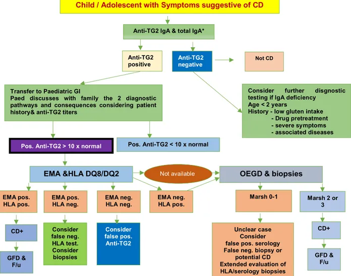
used while making the diagnosis of child/adolescent with symptoms of CD (Figure 5)
38
Figure 5: Symptomatic patient. CD=celiac disease; EMA=endomysial antibodies;
F/u=follow-up; GFD=gluten-free diet; GI=gastroenterologist; HLA=human leukocyte
antigen; IgA=immunoglobulinA; IgG=immunoglobulinG;
OEGD=oesophagogastroduodenoscopy; TG2=tranglutaminase 2 EMA pos.
HLA neg.
Marsh 2 or 3 Consider further disgnostic testing if IgA deficiency
Age < 2 years
History - low gluten intake - Drug pretreatment - severe symptoms - associated diseases EMA pos. HLA pos. Unclear case Consider false pos. serology False neg. biopsy or
potential CD Extended evaluation of HLA/serology biopsies
Marsh 0-1
Child / Adolescent with Symptoms suggestive of CD
Anti-TG2 IgA & total IgA*
Transfer to Paediatric GI
Paed discusses with family the 2 diagnostic pathways and consequences considering patient history& anti-TG2 titers
Anti-TG2 positive
Anti-TG2
negative Not CD
Pos. Anti-TG2 < 10 x normal
EMA &HLA DQ8/DQ2 OEGD & biopsies
EMA neg. HLA neg. EMA neg. HLA pos. Consider false neg. HLA test. Consider biopsies Consider false pos. Anti-TG2 CD+ CD+ Not available GFD & F/u GFD & F/u
39
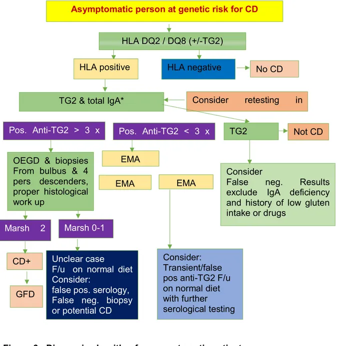
Figure 6: Diagnosis algorithm for asymptomatic patient Marsh 2
Pos. Anti-TG2 > 3 x
Consider retesting in
EMA
Unclear case
F/u on normal diet Consider:
false pos. serology, False neg. biopsy or potential CD
Marsh 0-1
Asymptomatic person at genetic risk for CD
HLA DQ2 / DQ8 (+/-TG2)
TG2 & total IgA*
HLA positive HLA negative No CD
Pos. Anti-TG2 < 3 x
EMA
Consider
False neg. Results
exclude IgA deficiency and history of low gluten intake or drugs
EMA
Consider: Transient/false pos anti-TG2 F/u on normal diet with further serological testing CD+ GFD TG2 Not CD
OEGD & biopsies From bulbus & 4 pers descenders, proper histological work up
40
The latest ESPGHAN guideline precisely recommends avoiding the histological
assessment in children and adolescents with signs or symptoms suggestive of CD with
high anti tTG ab titers with levels >10 times Upper Limits of Normal (ULN), supported by
a positive EMA test also with positive HLA-DQ2 and/or HLA-DQ8 alleles.106
Treatment
Currently, the only effective treatment for CD is a strict lifelong GFD which results in
clinical, serological and histological improvement.28 Unfortunately, the complete
avoidance of gluten intake is extremely difficult due to hidden gluten contamination. CD
patients are highly sensitive to the toxic effect of gluten. Even protracted ingestion of
traces of gluten (10–50 mg daily) may damage the integrity of the small intestinal mucosa. By combining toxicity data with the observed food intake, it has been
calculated that gluten-free products with <20 mg/kg (or parts per million = ppm) of
gluten contamination are safe over a wide range of daily consumption. Codex
Alimentarius regulation, also endorses that gluten-free food should contain < 20 ppm of
gluten in total.11 Effective therapy for CD is strict adherence to GFD which often restricts
patient’s social activities; affects the quality of life and causes nutritional deficiencies. Therefore, there is a need for alternative or adjunctive treatments of CD. Several
treatment strategies have been under investigation. Some of these are enzyme therapy,
inhibition of intestinal permeability, tTG inhibitors, HLA-DQ blockers and cytokine
41
Chapter 3
History of CD diagnostic Biomarkers
Introduction of wheat in the human diet arose the possibility of development of
syndromes including CD. Evidence of CD were present from 1st and 2nd centuries AD
but the major breakthrough was contributed by Dr. Wim Dicke in 1950, in his Ph.D.
thesis, showed that exclusion of wheat from the diet on children led to dramatic
improvement in the symptoms of CD.109 In the late 19th century, abnormalities of the
lining of the small intestine in autopsy were described. Finally in 1954 abnormal
mucosal lining was demonstrated by Paulley.110 Early study of HLA (preliminary HLA B)
suggested a relationship with CD considered as potential a genetic biomarker for CD.
During ’60s, the diagnosis of CD was based on clinical symptoms, stool characteristics,
and the effects of a GFD upon symptoms and the histology of intestinal lesions.
Circulating antibodies to gluten were reported in 1958 and till 1961 with other food
components.111,112 This finding was not highly specific for CD and was discoverable
even in non-celiac individuals. In 1971, antibodies to reticulin was reported but due to its
compromised specificity it was not accepted as a specific test for the CD.113 During, the
1970s and the beginning of the 1990s, the discovery of autoantibodies such as reticulin
and anti-endomysial antibodies in the serum of patients with CD laid the groundwork for
today’s understanding of CD.114–116 As the understanding had been developed towards
the pathogenesis of CD with the revelation of technology, several biomarkers were
developed to diagnose CD. Most crucial biomarkers that were developed for this
purpose are described below and detail of some leading biomarkers for the CD is given
42
1. Native Anti-Gliadin Antibody (AGA): In 1958, Berger described alcohol soluble
native antigliadin antibody (AGA).117During 70’s clinical use of AGA was started. IgA
class of AGA was the first serological marker to be used for CD diagnosis and
screening.118 Although the test detected antibodies to gliadin both IgG and IgA class
but the AGA test had shown a better accuracy through IgA class. During that time,
AGA method was the only marker present for the diagnosis of CD.119 However, these
antibodies clearly lacked specificity. IgA AGA appeared to offer fair to good
performance in children, with a sensitivity and specificity > 80% in most of them. In
fact, the old ESPGHAN guidelines, in addition to the serological tests, required three
biopsies for CD diagnosis.120 Due to its less specificity AGA test was not accepted as
a sole sero-test. However, during ’90s some leading CD epidemiology studies were
conducted AGA as the primary diagnostic marker.52,55,65,66,69 Even though the
guidelines have discouraged to use, however, this test had inappropriately, never
been abandoned. It has been observed that IgG class AGA, but also IgA, are present
in patients with autism.121–125 Moreover, it has recently been observed that about half
of patients suffering from NCGS do have high-IgG AGA and this remains, to date, the
only laboratory marker to diagnose NCGS.39
2. Anti Endomysial Antibody Test (EMA): Discovery of anti-endomysium antibody
test (EMA) has started a new era in the diagnosis of CD with remarkable specificity
and sensitivity over 90%. IgA class of EMA test provided support of specificity with
AGA, so, for several years, tests for AGA plus EMA were the laboratory approach
suggested and most widely used. The endomysium is the perivascular connective
43
antigen in endomysium is tissue transglutaminase-2.126 Tissue transglutaminase is a
ubiquitous calcium-dependent enzyme that crosslinks proteins. When it reacts with
gliadin, neoepitopes are formed. It is thought that the immunological response to
these neoepitopes may initiate the mucosal damage in CD. The commercially
available tests for EMA detect IgA class auto-antibody directed against the
endomysium in monkey esophagus by indirect immunofluorescence.115 More recent
work using human umbilical cord tissue as a substrate has shown improved
sensitivity and correlation with villous atrophy and has overcome the ethical issue of
using samples from endangered species.127,128 The technique of indirect
immunofluorescence for IgA EMA is both subjective and more labor intensive than
the ELISA tests which are used for IgA and IgG AGA. However, it has been
consistently demonstrated that EMA has superior sensitivity and specificity than
assays for AGA and anti-reticulin antibodies. The IgA EMA test is as easy as an
ELISA test. Nevertheless, it requires experienced pathology to read the EMA slide
so, the prediction of EMA is observation dependent. To be 100% sure diagnosis with
EMA test, a small bowel biopsy is necessary.129 Nevertheless, First generation IgA
AGA together with the combination of EMA, was a popular choice for the clinicians.
During ’80-’90s, several CD population screening studies were performed in different
countries using the native IgA-AGA test as the first line serological marker. 52,57,69,71
3. Anti-Tissue Transglutaminase antibody (anti tTG ab): Following the discovery
that calcium-dependent tTG is the major auto-antigen responsible for EMA positivity
in the late ’90s by the group of Dieterich who identified the tTG or Type 2
44
developed and this gradually replaced the AGA testing in first-line CD screening,
due to higher sensitivity and specificity. Earlier, the commercial immunoassay kits
were based on guinea pig or human extractive enzyme, based on this method in
pediatric patients, the sensitivity of these tests was reported between 89% and 96%
with a specificity higher than 92%.130–133 The sensitivity in adults of the IgA anti-tTG
assay, using human recombinant tTG, ranges between 95% to 100% and the
specificity between 97% to 100%.134,135 Later the commercial ELISA kits were
developed with recombinant human enzymes. Subsequently showed a higher
diagnostic accuracy and became almost a standard.132,134 The sensitivity of IgA
anti-tTG assays, using human recombinant anti-tTG, in adults ranges between 95% to 100%
and the specificity between 97% to 100%134,135 with the highly reliable performance
anti tTG ab was the most reliable test for CD so far. With the discovery of anti tTG
ab further discovery of diagnostic tools for CD probably got a halt. This enzyme
plays a significant biological role, catalyzing the bond between glutamine and lysine
in different proteins. It is important in the processes of tissue repair and it is also
involved in the removal of cell debris after cell death and apoptosis.136 When
excessive gluten penetrates the mucosa, an immune response with antibody
formation is triggered. This response results in mucosal damage and the subsequent
release and activation of transglutaminase. Gluten, being rich in glutamine, may also
be the target of the enzyme, which can bind it to other proteins including
transglutaminase itself. In the “Guidelines for the diagnosis and treatment of celiac disease” produced by the North American Society for Pediatric Gastroenterology, Hepatology and Nutrition and the latest ESPGHAN criteria, the IgA
anti-45
transglutaminase test is recommended for CD screening, rather than the EMA
test.106,137 During the last 20 years, many screening CD studies have been
performed worldwide, using the IgA anti-tTG test as the first-line test.49,50,66,80,82,90 A
high concordance rate was reported between EMA and tTG test. These studies
invariably reported higher CD prevalence compared to older AGA-based studies.
However, it remains unclear whether this higher frequency of CD reflected a true
increase in the prevalence of the disease or is related at least in part to the higher
sensitivity of the IgA anti-tTG-based diagnostic algorithm.106,138 It has already been
emphasized that IgA anti-tTG autoantibody levels may reflect the degree of mucosal
damage present,139–142 although there are reports of high levels of IgA anti-tTG in
patients without CD.143 Different groups of researchers have attempted to determine
whether high-IgA anti-tTG levels can justify avoiding biopsies, especially in pediatric
patients.
4. De-amidated gliadin peptides (DGP)/Second generation AGA: The deamidated
gliadin peptides (DGP) in patients with CD bind to the circulating antibodies with
higher specificity than the native peptides (AGA),144 and in particular to residues
containing the PEQ tripeptide, as the core epitope in celiac patients.145 Later it was
found that this DGP ab had a higher diagnostic accuracy than the AGA test, both in
terms of sensitivity and of specificity.146 Subsequent studies performed with
commercial kits confirmed the accuracy of the data reported in early studies. Data on
the sensitivity of IgA antibodies (DGP-A) in adults ranged from 83.6% to 98.3% with
specificity between 90.3% to 99.1%.147–149 The sensitivity observed for the IgG class
46
early studies clearly highlighted the high specificity of the anti-DGP-G, considerably
better than the classic IgG AGA. A study showed that the anti- DGP, particularly of the
IgG class, maintaining a high specificity, were positive in the majority of patients
negative for anti-tTG, both children and patients with selective IgA deficiency.150
These data were confirmed in further studies.151–153 Considering the results of
published studies, it is indisputable that the anti-DGP, especially of the IgG class,
have a diagnostic performance higher than AGA and add value in the diagnosis of CD.
Nevertheless, there is still not widespread agreement on their use. Some authors
affirm that anti-DGP should be used mainly in children under 2 years of age,154
whereas other authors regard them as a marker of diagnostic accuracy comparable to,
47
Table 5: Major diagnostic biomarker for CD their sensitivity, specificity, pros, and cons
S.
No. Name
Sensitivity (range)
Specificity
(range) Pros Cons Use
1. EMA IgA >90% (77.9–100) 98% (90–100) The most specific test.
• More time consuming to perform, • More expensive. • Observer dependent To confirm tTG IgA positive patients 2. Anti-tTG IgA >95% (67–100) >95% (92–100) The most sensitive test Recommende d for first level screening test
Lack of standardization Best test for the initial
screening of the patients
3. Anti-tTG IgG >70% (54.7–100) >90% (80–100) Often positive in IgA deficient patients
The variable diagnostic accuracy of commercial kits
Useful in IgA deficient patients
4. Anti-DPG IgG >90% (80.1–98.6) >90% (90.3–100) Often positive in antitTG IgA negative children
Less accurate with respect
to anti-tTG IgA Recommended in children
and in IgA deficient patients 5. Anti-DPG IgA >90% (80.7–98.3) >90% (86.3–99.1) Often positive in children
Less accurate with respect to anti-tTG IgA and
anti-DPG IgG Useful in children
6. Anti-actin IgA >50% (25.7–80) >85% (85–100) Highly correlated to mucosal atrophy
Lack of specificity, the IFA method is highly observer
and substrate dependent Useful to evaluate mucosal