Review Article
Genetic Engineering of Dystroglycan in Animal Models of
Muscular Dystrophy
Francesca Sciandra,
1Maria Giulia Bigotti,
2Bruno Giardina,
3Manuela Bozzi,
3and Andrea Brancaccio
1,21Istituto di Chimica del Riconoscimento Molecolare, CNR c/o Istituto di Biochimica e Biochimica Clinica,
Universit`a Cattolica del Sacro Cuore, 00168 Roma, Italy
2School of Biochemistry, Bristol University, Bristol B58 1TD, UK
3Istituto di Biochimica e Biochimica Clinica, Universit`a Cattolica del Sacro Cuore, Roma, Italy
Correspondence should be addressed to Francesca Sciandra; [email protected] and Andrea Brancaccio; [email protected]
Received 3 October 2014; Accepted 11 March 2015 Academic Editor: Gouri Shankar Pandey
Copyright © 2015 Francesca Sciandra et al. This is an open access article distributed under the Creative Commons Attribution License, which permits unrestricted use, distribution, and reproduction in any medium, provided the original work is properly cited.
In skeletal muscle, dystroglycan (DG) is the central component of the dystrophin-glycoprotein complex (DGC), a multimeric protein complex that ensures a strong mechanical link between the extracellular matrix and the cytoskeleton. Several muscular dystrophies arise from mutations hitting most of the components of the DGC. Mutations within the DG gene (DAG1) have been recently associated with two forms of muscular dystrophy, one displaying a milder and one a more severe phenotype. This review focuses specifically on the animal (murine and others) model systems that have been developed with the aim of directly engineering
DAG1 in order to study the DG function in skeletal muscle as well as in other tissues. In the last years, conditional animal models
overcoming the embryonic lethality of the DG knock-out in mouse have been generated and helped clarifying the crucial role of DG in skeletal muscle, while an increasing number of studies on knock-in mice are aimed at understanding the contribution of single amino acids to the stability of DG and to the possible development of muscular dystrophy.
1. Introduction
The extracellular matrix receptor dystroglycan (DG) is highly expressed in skeletal muscle and in several developing and adult tissues, typically in cell types that adjoin basement membranes, such as epithelial and neural tissues [1–3].
DG is composed of two subunits,𝛼- and 𝛽-DG, deriving from a posttranslational cleavage of a single mRNA species encoded by a single gene (DAG1) [4].𝛼-DG is an extracellular protein characterized by an extensive and heterogeneous gly-cosylation mainly concentrated within an elongated central mucin-like region which separates two globular domains, the N- and C-terminal domains [5]. 𝛼-DG binds with high affinities to the LG domains-containing extracellular proteins, such as laminin-𝛼2, perlecan, and agrin, and in turn interacts noncovalently with the𝛽-subunit, a transmembrane protein [6]. The cytosolic domain of𝛽-DG is anchored to
actin through the interaction with dystrophin [7–9], and 𝛽-DG also constitutes a scaffold for proteins involved in signal transduction such as Gbr2 and ERK [10,11].
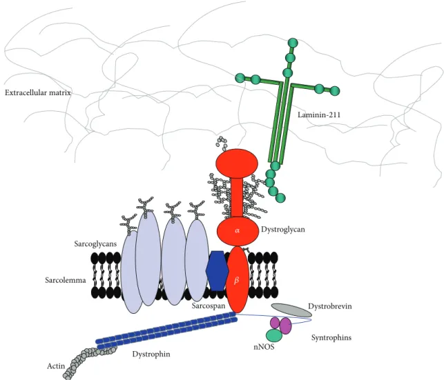
In skeletal muscle, DG is the central component of the dystrophin-glycoprotein complex (DGC), a multisubunit protein complex which links the actin cytoskeleton to the extracellular matrix [12] (Figure 1). Other members of the DGC include transmembrane proteins such as sarcoglycans and sarcospan and multiple cytoplasmic proteins, including dystrobrevin and syntrophins.
The role of the DGC in muscle is to provide mechanical reinforcement to the sarcolemma and to maintain membrane integrity during cycles of contraction and relaxation. In fact, mutations in any components of the DGC cause distinct forms of muscular dystrophy [13]. In humans, mutations in dystrophin lead to Duchenne and Becker muscular dystrophy [14], mutations in sarcoglycans cause limb-girdle muscular Volume 2015, Article ID 635792, 10 pages
Actin Dystrophin Sarcospan Laminin-211 Sarcolemma Dystroglycan Syntrophins nNOS Dystrobrevin Extracellular matrix Sarcoglycans 𝛼 𝛽
Figure 1: Schematic representation of the dystrophin-glycoprotein complex (DGC) in skeletal muscle. This multiprotein complex anchors the extracellular matrix (ECM) to actin and other components of the cytoskeleton. O-Mannosylated𝛼-DG is a central component of this complex and serves as binding partner for a number of ECM proteins containing LG domains, such as laminin-211.𝛽-DG is a transmembrane protein and binds the actin cytoskeleton via the direct interaction with dystrophin. Other intracellular molecules being a part of, or associated with, DGC are dystrobrevin, syntrophins, and neural nitric oxide synthase (nNOS).
dystrophy [15–19], and mutations in laminin-𝛼2 cause
con-genital muscular dystrophy [20]. Recently, mutations in DAG1 have been reported in three patients, affecting DG function by impairing glycosylation of𝛼-DG or by presum-ably disrupting the𝛼/𝛽-DG binding interface [21–23].
Moreover, several mutations in 12 proteins involved in the O-mannosyl-glycosylation pathway of𝛼-DG have been identified so far which lead to a variety of clinical symptoms, including severe muscular dystrophy and abnormal central nervous system development and function. These diseases are defined as “secondary dystroglycanopathies” (for recent reviews see [24,25]). The defective O-mannosyl glycosylation of𝛼-DG impairs its multiple interactions with its extracel-lular partners, eventually destabilizing the link between the cytoskeleton and the extracellular matrix. Secondary loss of 𝛼- and 𝛽-DG at the muscle membrane also occurs in
Duchenne and Becker muscular dystrophies [26] and in some forms of limb-girdle muscular dystrophy [18].
The recovery of DG glycosylation state via transgenic overexpression of LARGE, a putative enzyme involved in the first steps of the posttranslational processing of𝛼-DG, has been proposed as a therapeutic strategy for muscular dystrophy [27], although with conflicting outcomes [28–30]. The recently emerging data on patients affected by primary and secondary dystroglycanopathies reinforce the notion that a correct expression and modification of DG are crucial for muscle fibres stability and function. A relevant amount of genetic engineering work has been carried out so far on the DAG1 gene in several laboratories. This review will be focused on the animal models generated to understand the function of DG in skeletal muscle, as well as in other tissues, and to better understand its involvement in neuromuscular disorders (Tables1and2).
Table 1: DG mouse models characterized by a muscle and/or central nervous system phenotype.
Mouse model Muscular
dystrophy CNS involvement NMJs
Chimaeric mice [34] Progressive — Disorganized and disrupted
MCK-Cre/DG-null [38] Mild — Normal
GFAP-Cre/DG-null [45] — Neuronal migration errors, brain
malformation —
MORE-DG-null [38,39] Severe
Neuronal migration errors, brain malformations, and ocular defects (WWS
phenotype)
— Nestin-Cre/DG-null [47] and
Crx-Cre/DG-null [50] — Abnormal retinal physiology —
DGT192M/DGT192M[21] Mild Some neurological impairments Compromised
DGY890F/Y890F[56] Normal Normal Normal
DGY890F/Y890F/mdx [56] Ameliorated — Ameliorated
DGWToverexpression [40] Normal Normal 25% smaller than normal
but only 1% are aberrant
DGWToverexpression/mdx [40] Not ameliorated — Not ameliorated
DGS654Aoverexpression [63] Mild — Compromised
DGΔ𝛽cyt/Δ𝛽cyt[47] — Mild effects in the retina —
—: not analysed.
Table 2: Mouse models in which DG was targeted in tissues other than skeletal muscle and brain and additional DG animal models with muscle and central nervous system defects.
Animal model Phenotype
Kidney specific DG knock-out mouse (podocin-Cre/DG-null,
Pax2-Cre/DG-null, Pax3-Cre/DG-null, HoxB7-Cre/DG-null) [64] Normal Schwann cells specific DG knock-out mouse (P0-Cre/DG-null)
[66] Severe neurological dysfunctions
DG knock-out in Caenorhabditis elegans [68] Defects in gonad and vulval epithelium and in motoneurons RNAi knock-out of DG in Drosophila melanogaster [73,74] Muscle degeneration and neuronal defects
Inhibition of DG translation via morpholino antisense in zebrafish
[78] Muscle defects
Zebrafish patchytail [79] Dystrophic muscles, ocular and central nervous system defects
Zebrafish dag1hu3072[81] Muscular dystrophy
Inhibition of DG translation via morpholino antisense in Xenopus
laevis [82–86]
Defects in the somitogenesis, epidermal differentiation, the retinal and renal developing
Overexpression of DG in Xenopus laevis embryos [86] Aberrant neuromuscular junctions
2. From Knock-Out Mice to
the Different Strategies to Circumvent
Embryonic Lethality
In 1997 the DG knock-out mouse was generated and analyzed in Kevin Campbell’s Laboratory [31]. The targeting vector was designed to replace a portion of the DAG1 second coding exon with the neo-cassette following homologous recombination. DAG1-null allele resulted in a deletion in the exon including the 3splice acceptor site and a large portion of the coding sequence of𝛼-DG. Animals that were heterozygous for the targeted allele appeared healthy and bred normally. Inter-estingly, DG transcripts in skeletal muscle of heterozygous
mice were only 10–20% lower than those in wild-type mice, suggesting a compensatory increase in the expression level of the untargeted allele. Accordingly, DG protein levels in skeletal muscle were also comparable between wild-type and heterozygous animals. However, the DG knock-out was lethal for homozygous mice embryos that died at the embryonic day 6.5 because of the disorganization of Reichert’s membrane, one of the first specialized extraembryonic basement mem-branes. The absence of laminin receptor precluded the assem-bly of laminin in a network and the distribution of laminin and collagen-IV appeared patchy, suggesting a crucial role of DG in the organization of the basement membranes [31]. This conclusion was further confirmed by the molecular analyses
of the embryoid bodies derived from homozygous DAG1-null ES cells in which an ordered basement membrane failed to form [32].
As DG is involved in the development of basement membranes, it certainly is fundamental for normal human development, and the failure to identify null mutations in DAG1 linked to muscular dystrophies in humans is probably due to early embryonic lethality of such mutations. Inter-estingly, Frost et al. described a patient affected by a mild myopathy with central nervous system involvement who was heterozygous for a DNA deletion which included also the DG gene [33]. In this patient, only 50–60% of native DG is produced and correctly glycosylated thus showing a much lower degree of compensation compared to the heterozygous DG-null mouse. Although other genes present in the same deleted region could account for the phenotype, this case report suggests the possibility that the heterozygosis for DG-null mutations (haploinsufficiency) could produce patholog-ical consequences in humans. A substantial genetic screening effort, carried out on an enlarged number of patients, would be necessary for the identification of additional cases that may be related to the haploinsufficiency of DG.
To circumvent the embryonic lethality of the DG knock-out mouse, highly chimaeric mice, generated with ES cells targeted for both DAG1 alleles, were generated [34]. In chimaeric mice deficient in DG rescued from the embryonic lethality, skeletal muscle differentiated normally but they developed a progressive muscular dystrophy reminiscent in many respects of that of mice with double mutations in dystrophin and utrophin [35]. Significant differences in fibre size, central nuclei, and connective tissue infiltration charac-terized the skeletal muscle histology of DG-null chimaeric mice that die at 13 months [34]. DG plays a crucial role also in stabilizing acetylcholine receptors [36] and consequently in chimaeric mice the neuromuscular junctions (NMJs) were grossly disorganized and disrupted [34]. In the most severely affected mice, the heart appeared dilated and with an extensive connective tissue hyperplasia. At the sarcolemma of DG-null chimaeric mice the entire DGC complex was disassembled, with dystrophin and sarcoglycans absent in many fibres. However, laminin-𝛼2, perlecan, and agrin were expressed at wild-type levels and the basement membrane appeared organized in an ordered network. It is likely that, in differentiated skeletal muscle, the expression of integrins or other extracellular matrix receptors exert an important compensatory effect in supporting the skeletal muscle differ-entiation and basement membrane assembly [37]. However, the DG-null chimaeric mice pointed out the central role of DG in the maintenance of the DGC and muscle integrity.
An additional step forward in understanding the func-tional role of DG in skeletal muscle came from the condi-tional inactivation of skeletal muscle DG using the Cre-loxP system under the muscle creatine kinase (MCK) promoter [38]. The MCK-Cre/DG-null mice were viable and born with the expected frequency; they developed muscular dystrophy around 4–6 weeks of age but the phenotype became milder with advanced age. As a matter of fact, satellite cells, which had not been targeted by the Cre recombinase, supported the muscle regeneration and formation of novel fibres expressing
DG and the other components of DGC. Moreover, in old mice, muscle fibres appeared hypertrophic and larger, as compared with controls and with the other mouse models of muscular dystrophy. Like in chimaeric DG-null mice, also in MCK-Cre/DG-null mice, laminin-𝛼2 was expressed and the basement membrane was correctly assembled. However, while in chimaeric DG-null mice the NMJs were disrupted, in MCK-DG-null mice they were preserved.
In MORE-DG-null mice [38], the inactivation of DAG1 was driven by Cre recombinase under the control of the Mox 2 promoter enabling the targeting of DG in all tissues of the embryo, while DG was still expressed in extraembryonic membranes to circumvent embryonic lethality. MORE-DG-null mice were significantly smaller than control littermates, a majority of the mice died within 48 h after birth, and the remaining mice typically failed to survive the fourth postnatal week. In addition, MORE-DG-null mice exhibited profound muscle weakness and muscular dystrophy was present at birth, reminiscent of a secondary dystroglycanopa-thy phenotype (see next paragraph) [39]. Consistent with the results obtained with MCK-Cre/DG-null mice, MORE-DG-null mice displayed severe impaired regeneration capacity since satellite cells were also targeted by Cre recombinase under the control of the Mox 2 promoter [38].
The phenotype observed in chimaeric and conditional knock-out mice demonstrated the importance of DG for the stability of the DGC and for the structural integrity of the sarcolemma. However, the overexpression of DG in trans-genic mice onto an mdx background did not inhibit muscular dystrophy; on the contrary, it exacerbated the phenotype by decreasing the utrophin and sarcoglycans expression at the sarcolemma [40].
3. Conditional DG Knock-Out in the Brain
Recapitulates the Outcome of Secondary
Dystroglycanopathies
Fukuyama congenital muscular dystrophy (FCMD), muscle-eye-brain disease (MEB), and Walker-Warburg syndrome (WWS) are congenital muscular dystrophies (CMDs) with associated developmental brain defects [41–43]. The genes that are mutated in these disorders are those of the enzymes involved in the O-mannosyl glycosylation of𝛼-DG, in par-ticular O-mannosyl transferase 1 (POMT1), protein-O-mannosyl transferase 2 (POMT2), protein-O-linked man-nose beta 1,2-N-acetylglucosaminyltransferase (POMGnT1), and fukutin, an enzyme indirectly implicated in a pathway to further modify the phosphorylated O-linked mannose located in the mucin-like domain of𝛼-DG [44].
The conditional DG knock-out mouse in the brain was produced, using the Cre-LoxP methodology, in order to anal-yse the function of DG in the central nervous system and to demonstrate the role of DG in the brain malformations seen in CMDs [45]. Brain-selective expression of Cre recombinase was accomplished using a human glial fibrillary acid protein (GFAP) promoter expressed as early as embryonic day 13.5. GFAP-Cre/DG-null mice followed the expected Mendelian distribution and were fertile.
In the GFAP-Cre/DG-null mice cerebral cortex, DG was not expressed in the astrocytes abutting the brain surface (glia limitans) and cerebral microvessels, in radial glia and in a subset of neurons that are the progeny of radial glia [45,46]. Also the localization of dystrophin isoforms was impaired in these cells. The basal lamina of the glia limitans that plays a critical role for normal cortical development was severely disrupted. The results were a number of brain structural developmental defects similar to those seen in MEB, WWS, and FCMD patients. The abnormalities of the glia limitans permitted the overextended migration of neurons in the developing brain, which is the most important diagnostic feature of cobblestone lissencephaly observed in the most severe cases of secondary dystroglycanopathy. Such mice also lacked the usual fissure between the brain’s hemispheres, a characteristic of WWS, and suffered from an overabundance of glia.
Despite the large similarities with CMDs, GFAP-Cre/DG-null mice did not recapitulate the most severe characteristics observed in the brain of patients affected by WWS. On the contrary, the earlier suppression of DG expression in MORE-DG-null mice (in which Cre recombinase under the control of the Mox 2 promoter operates at E7.5) was sufficient to cause malformations that broadly resembled the clinical spectrum of WWS, including hydrocephalus and ocular malformations with structural defects of both the anterior and posterior chambers of the eye [39].
The DAG1 gene was targeted to generate other mouse models for the detailed analysis of the role of DG in the central nervous system and in the visual function. For example, the following two novel DG mouse models were created and analyzed by Satz and colleagues [47]: (1) the conditional knock-out nestin-CRE/DG-null mouse, in which Cre recombinase was under the control of the rat nestin enhancer expressed in neuroepithelial precursor cells and in the retina as early as embryonic day 9.5 (further analysed in [48, 49]) and (2) a knock-in mouse expressing a trun-cated DG lacking the entire𝛽-DG cytodomain (in which a premature stop codon was inserted after Lys778 resulting in the presence of only 4 amino acids in the cytodomain of 𝛽-DG). Surprisingly, according to the authors, a reasonable number of knock-in mice were obtained [47]. It remains unclear whether these mice would display some pathologic effect in their skeletal muscle.
Moreover, Omori and colleagues developed a retinal photoreceptor-specific DG conditional knock-out (Crx-Cre/DG-null) mice, in order to analyze the role of DG localized at the presynaptic elements of photoreceptor cells [50]. The results showed the crucial role of presynaptic DG for both the formation of proper photoreceptor ribbon synaptic structures and normal retinal electrophysiology.
4. DG Knocking-In: A Powerful Tool to Dissect
the Role Played by Specific Amino Acids
4.1. DG𝑇190𝑀/DG𝑇190𝑀, the First Case of Primary Dystrogly-canopathy. The first case of a homozygous missense mutation in the DAG1 gene was described in a 16-year-old patient [21]originally described as affected by a mild form of limb-girdle muscular dystrophy associated with mental retarda-tion but normal brain imaging (recently classified as limb-girdle muscular dystrophy 2P [51]). Immunofluorescence and immunoblot of muscle biopsies showed that 𝛼-DG was hypoglycosylated and had a reduced affinity toward laminin. The mutation (T192M), located within the RNA binding protein-like domain of the𝛼-DG N-terminus [52], is supposed to reduce, via a mechanism that needs to be fully elucidated yet, the binding between 𝛼-DG and the glycosyltransferase LARGE, an interaction that is essential for the posttranslational modification of𝛼-DG and for the DG’s laminin-binding activity [53]. Recently, a mild form of muscular dystrophy characterized by hypoglycosylated𝛼-DG was associated with the compound heterozygous missense mutations (V74I and D111N) with both mutated sites located within the N-terminal domain of𝛼-DG and a pathological molecular mechanism similar to the one described for the T192M mutation was hypothesized [22].
A knock-in mouse was generated introducing the muta-tion T192M (that in mouse corresponds to T190M) by homol-ogous recombination [21]. Heterozygous mice were normal, while homozygous knock-in mice presented abnormalities consistent with those observed in the patient. Analysis of muscle biopsies revealed hallmarks of muscular dystrophy, such as centrally nucleated fibres, and a hypoglycosylated 𝛼-DG with a decreased laminin-binding activity. The T190M mutation interfered also with the organization of the NMJ. Although no structural abnormality was evident in the brains of the knock-in mice, these mice had abnormal hind limb clasping, a phenotype common to mouse models featur-ing neurologic impairment. Interestfeatur-ingly, in the heart, the mutated𝛼-DG still bound to laminin and no obvious signs of any pathological abnormality were observed.
Recently, a novel homozygous mutation in DAG1 has been identified in a Libyan family with two siblings affected by a dystroglycanopathy resembling a MEB-like condition [23]. The mutation (C669F) hits an amino acid already identified as an important site for the interaction between𝛼- and 𝛽-DG and for the overall stability of the complex, but the knock-in mouse model has not been generated yet [54,55].
4.2. DG𝑌890𝐹/𝑌890𝐹/mdx. Although not directly linked to a specific disease, a knock-in mouse hitting a tyrosine within the cytodomain of𝛽-DG represented a tool for dissecting the role of the phosphorylation of DG in the skeletal muscle and for testing novel therapeutic ideas [56].
In fact, previous studies suggested that tyrosine phospho-rylation of DG at the site Y892 may be an important mech-anism for modulating the association of DG with its cellular binding partners dystrophin and utrophin and may also work as a signal for proteasome degradation of DG [57–59].
The knock-in mouse DGY890F/Y890Fwas generated using homologous recombination in ES cells [56]. Both heterozy-gous and homozyheterozy-gous DGY890F/Y890Fmice appeared normal and healthy. Skeletal muscle analysis of knock-in mice did not reveal any differences or abnormalities compared to the wild-type mice.
In order to assess whether the inhibition of tyrosine phosphorylation in DG had any beneficial effect on dys-trophic skeletal muscle, DGY890F/Y890F/mdx mice were further generated [56]. Interestingly, the expression of Y890F mutant DG in an mdx background significantly improved the muscle phenotype, reducing the number of centrally nucleated fibres and the levels of creatine kinase. Moreover, also an improve-ment in resistance to eccentric contraction-induced injury was observed in DGY890F/Y890F/mdx mice. Changing a single phosphorylation site in DG reinforced the DGC sarcolemma localization preventing the proteasome degradation of DG. In fact, the inhibition of tyrosine phosphorylation of DG in mdx mice was sufficient to restore the sarcolemma localization of DG,𝛼-sarcoglycan, and sarcospan also in the absence of dystrophin, while utrophin was confined at the NMJ [56]. Moreover, an increase in the plectin expression/localization at the sarcolemma was also observed. Plectin is a cytolinker protein that binds 𝛽-DG at different sites, thus providing a stabilizing link between DG and the cytoskeleton [60]. It was already known that treatment with proteasomal inhibitors improves the muscle pathophysiology in some mouse models, such as mdx mice [61] or𝑑𝑦3𝐾mice [62], and in this context the DGY890F/Y890F/mdx mouse highlights novel targets for therapeutic intervention.
5. Mice Overexpressing DG
The overexpression of multiple and randomly integrated copies of the coding sequence of DG was obtained by microinjection of a pBS-HSAvpA cDNA construct into fer-tilized CB6 oocytes. Mice overexpressing wild-type DG were normal compared to control [63]. Interestingly, transgenic lines overexpressing DG mutated in the cleavage site S654 were also created in order to understand the role of the posttranslational cleavage resulting in the production of the two interacting subunits [63]. In the transgenic DGS654Amice only the uncleaved DG precursor was correctly expressed, while the expression of endogenous and processed DG was inhibited. In DGS654A mice, most muscles were dystrophic with increased levels of central nuclei. The lack of the DG cleavage and the presence of muscular dystrophy corre-late with altered glycosylation of 𝛼-DG. In addition, the expression of dystrophin and𝛼-sarcoglycan decreased, while utrophin and laminin-𝛼5 were upregulated, probably as a secondary effect of muscle regeneration. Aberrant NMJs were observed in DGS654A mice, although also DGWTmice occasionally showed fragmented NMJ [63].
6. Conditional DG Knock-Out Mice in Other
Tissues Compared to Skeletal Muscle
DG is also highly expressed in epithelia and in the periph-eral nervous system (Table 2). To study the role of DG in the kidney, different conditional knock-out mice were created to selectively delete DG from podocytes, ureteric bud, metanephric mesenchyme derivatives, and all renal epithelial cells using the Cre-lox system under the control
of podocin, HoxB7, Pax-3, and Pax-2 promoters, respectively [64]. Surprisingly, DG deletion from kidney resulted in no aberrant phenotypes. Kidney formation and function proceeded normally in the absence of DG and the only detectable abnormality was a mild increase of the glomerular basement membrane thickness. This observation was further confirmed in chimaeric mice generated with fukutin-null embryonic stem cells expressing a hypoglycosylated𝛼-DG, in which minor glomerular structure abnormalities were found without functional renal defects [65]. These results suggest that DG and its correct glycosylation may be important in the maintenance of podocyte architecture and extracellular matrix assembly; however, the presence of integrin 𝛼3𝛽1 as an additional laminin receptor and basement membrane organizer in podocytes may preserve the structure and functionality of the kidney [65]. Accordingly, there are no reported renal dysfunctions in human patients with impaired DG glycosylation.
To analyse the role of DG in peripheral nerves, DG was disrupted selectively in Schwann cells using the P0 protein promoter and Cre-loxP technology [66]. The loss of DG caused severe neurological dysfunctions, including a slow nerve conduction. In P0-Cre/DG-null mice the myelin sheaths around the nerves were structurally abnor-mal and extended throughout the internodal segments. The DG-interacting proteins, sarcospan, sarcoglycan, and 𝛼-dystrobrevin, were lost from the membrane and laminin was not deposited around the nerves. These findings point to the crucial role of DG in myelin integrity and in node of Ranvier structure and function [67].
7. Other Animal Models to Study
the DG Functions in Muscle and Central
Nervous System
DG and most of the members of DGC are highly conserved in vertebrates, including fish, and invertebrates. Therefore, DG offers a wide range of possible animal models aside from mouse to understand the role of DG in the pathogenesis of muscular dystrophies.
7.1. Caenorhabditis elegans. Interestingly, in Caenorhabditis elegans, DG is expressed in epithelial and neuronal tissues but not in muscle. Indeed, a deletion, cg121, that removes most of the coding and some of the 3untranslated region of DGN-1 gene, resulted in viable but sterile animals, with neuronal defects and normal muscles [68]. DG1 contains the N-terminal immunoglobulin-like domain of vertebrate𝛼-DG and a shorter mucin-like region, while the𝛼/𝛽 proteolytic cleavage region and the residues involved in binding WW and SH3 domain-containing proteins such as dystrophin are all missing [68].
7.2. Drosophila melanogaster. All known components of the DGC are present in the fruit fly [69]. Several Drosophila DG isoforms are generated via alternative splicing [70,71]. Only one of these contains the full mucin-like domain, characterized by significant levels of glycosylation, while DG isoforms that lack the mucin-like domain are required to
maintain polarity in the follicular epithelium, suggesting that the isoforms can have different functional roles in Drosophila [71]. Moreover, Drosophila DG is not cleaved in𝛼- and 𝛽-subunits but it is expressed as a single polypeptide [72].
Interestingly, muscle-specific RNAi-mediated knock-down of DG, as well as of dystrophin isoforms, led to age-dependent, progressive climbing deficits, severe muscle degeneration in adult flies, and defects in neurons migration and eye development [73]. Haines and colleagues, analysing larvae carrying mutant alleles of DG, also established that DG is required in Drosophila larval muscles to maintain integrity [74].
The similar defects observed in both flies and humans make Drosophila an attractive model for further studies on clarifying the role of the DGC. In particular, using the RNAi knockdown mutants of DG, many genes had been identified as possible regulatory genes of DG and dystrophin, such as genes involved in muscle function and components of Notch, TGF-𝛽, and EGFR signalling pathways [75]. Recently, it was also shown that in Drosophila the expression level of DG may be buffered in a homeostatic fashion via a mechanism mediated by the miR-310s complex which acts directly on the alternative DG 3-UTR [76]. Deficiencies in the miR-310s complex resulted in cobblestone brain, a phenotype reminiscent of human lissencephaly type II [76]. This evidence represents a seminal result, paving the way for identifying similar regulation mechanisms also in higher vertebrates and mammals.
7.3. Danio rerio. In recent years, Danio rerio (zebrafish) has emerged as a powerful genetic tool to study muscle diseases [77]. Disruption of DG translation using an anti-sense morpholino oligonucleotide (MO) approach, led to the destabilization of the embryos muscle, with loss of sarcomere organisation and necrosis of the developing muscle [78]. The NMJ and central nervous system appeared normal. The lack of the DG protein impaired also the localization of dys-trophin. Interestingly, in an ENU (N-ethyl-N-nitrosourea) mutagenesis screen aimed at identifying genes responsible for skeletal muscle disorders, a DG homozygous mutant, patchy-tail, was found to show impaired locomotion behaviour, dystrophic muscles and ocular and central nervous system defects [79]. The point mutation resulted in a missense amino acid change of valine to aspartic acid (V567D) within the Ig-like domain in the C-terminal region of a-DG, leading to a destabilization and degradation of the protein [79,80]. The absence of DG led to a reduced expression of dystrophin and laminin-a2. The sarcolemma appeared grossly disorganized and detached from the extracellular matrix. NMJs were normal in patchytail fish, but severe abnormalities of brain and eyes were observed and the embryos did not survive more than 10 days post fertilization. This case represents so far the first case in which a single point mutation is able to induce the complete depletion of DG from tissues, highlighting the importance that even single aminoacids may have for the stability and/or folding pathway of the DG precursor [56,80]. An additional loss of function mutation was identified in a zebrafish affected by muscular dystrophy (dag1hu3072) due to a nonsense mutation (R398>Stop) within
the mucin-like region of a-DG, causing the complete loss of DG [81]. The absence of DG led to the dislocation of dystrophin and to muscle fibre detachment, followed by disruption of sarcolemma integrity.
7.4. Xenopus laevis. The possible DG functions in different tissues of Xenopus laevis were analysed using the morpholino knock-out approach. It was shown that, during Xenopus development, DG is important for the somitogenesis and that the interaction between DG and the extracellular matrix is indispensable for the alignment of the myoblasts in the somites [82]. The loss of DG influenced the epidermal differentiation in the retinal and renal development [83–85]. Moreover, the injection of rabbit DG RNA into Xenopus embryos produced an overexpression of DG that altered the acetylcholine receptors aggregation and the NMJ structure [86].
8. Perspectives
Starting from the 1997 knock-out mouse [31], during the last 17 years, an impressive amount of work has been carried out already on DG at the genetic level. It is likely to expect in the next few years a further increase both in the number of patients/families identified who carry mutations specifically within the DAG1 gene and in the number of animal genetic models that will be generated and analyzed. In particular, it could be particularly interesting to focus on the 3region of DAG1, corresponding to the𝛼/𝛽-DG interface, the genetic clinical screenings carried out on still unassigned cases of myopathy presenting with symptoms that may suggest the presence of a dystroglycanopathy. In this specific and very small region (that could be therefore analyzed inexpensively) in fact, mutations have been found to grossly affect the stability and the maturation of the complex, in zebrafish as well as in human patients [23,79,81].
Via such comparative studies, a full circle will be com-pleted in the elucidation of the function(s) of DG in muscle and nonmuscle tissues. Moreover, the genetic data on human patients and the generation of novel animal models will cer-tainly boost the research on potential therapeutic approaches for human muscular dystrophies.
Conflict of Interests
The authors declare no conflict of interests.
Acknowledgment
Maria Giulia Bigotti is the recipient of a Research Career Re-entry Fellowship from the Wellcome Trust, which is hereby gratefully acknowledged.
References
[1] M. Durbeej, M. D. Henry, M. Ferletta, K. P. Campbell, and P. Ekblom, “Distribution of dystroglycan in normal adult mouse tissues,” Journal of Histochemistry and Cytochemistry, vol. 46, no. 4, pp. 449–457, 1998.
[2] M. Durbeej and K. P. Campbell, “Biochemical characterization of the epithelial dystroglycan complex,” The Journal of Biological
Chemistry, vol. 274, no. 37, pp. 26609–26616, 1999.
[3] K. Matsumura, H. Yamada, T. Shimizu, and K. P. Camp-bell, “Differential expression of dystrophin, utrophin and dystrophin-associated proteins in peripheral nerve,” FEBS
Let-ters, vol. 334, no. 3, pp. 281–285, 1993.
[4] O. Ibraghimov-Beskrovnaya, J. M. Ervasti, C. J. Leveille, C. A. Slaughter, S. W. Sernett, and K. P. Campbell, “Primary structure of dystrophin-associated glycoproteins linking dystrophin to the extracellular matrix,” Nature, vol. 355, no. 6362, pp. 696–702, 1992.
[5] A. Brancaccio, T. Schulthess, M. Gesemann, and J. Engel, “Electron microscopic evidence for a mucin-like region in chick muscle𝛼-dystroglycan,” FEBS Letters, vol. 368, no. 1, pp. 139– 142, 1995.
[6] F. Sciandra, M. Bozzi, M. G. Bigotti, and A. Brancaccio, “The multiple affinities of𝛼-dystroglycan,” Current Protein & Peptide
Science, vol. 14, no. 7, pp. 626–634, 2013.
[7] A. Suzuki, M. Yoshida, K. Hayashi, Y. Mizuno, Y. Hagiwara, and E. Ozawa, “Molecular organization at the glycoprotein-complex-binding site of dystrophin. Three dystrophin-associated proteins bind directly to the carboxy-terminal portion of dystrophin,” European Journal of Biochemistry, vol. 220, no. 2, pp. 283–292, 1994.
[8] D. Jung, B. Yang, J. Meyer, J. S. Chamberlain, and K. P. Campbell, “Identification and characterization of the dystrophin anchor-ing site on𝛽-dystroglycan,” The Journal of Biological Chemistry, vol. 270, no. 45, pp. 27305–27310, 1995.
[9] G. Rosa, M. Ceccarini, M. Cavaldesi, M. Zini, and T. C. Petrucci, “Localization of the dystrophin binding site at the carboxyl ter-minus of𝛽-dystroglycan,” Biochemical and Biophysical Research
Communications, vol. 223, no. 2, pp. 272–277, 1996.
[10] M. Cavaldesi, G. Macchia, S. Barca, P. Defilippi, G. Tarone, and T. C. Petrucci, “Association of the dystroglycan complex isolated from bovine brain synaptosomes with proteins involved in signal transduction,” Journal of Neurochemistry, vol. 72, no. 4, pp. 1648–1655, 1999.
[11] H. J. Spence, A. S. Dhillon, M. James, and S. J. Winder, “Dystroglycan, a scaffold for the ERK-MAP kinase cascade,” The
EMBO Reports, vol. 5, no. 5, pp. 484–489, 2004.
[12] J. M. Ervasti, K. Ohlendieck, S. D. Kahl, M. G. Gaver, and K. P. Campbell, “Deficiency of a glycoprotein component of the dystrophin complex in dystrophic muscle,” Nature, vol. 345, no. 6273, pp. 315–319, 1990.
[13] R. D. Cohn and K. P. Campbell, “Molecular basis of muscular dystrophies,” Muscle & Nerve, vol. 23, no. 10, pp. 1456–1471, 2000.
[14] E. P. Hoffman, R. H. Brown Jr., and L. M. Kunkel, “Dystrophin: the protein product of the duchenne muscular dystrophy locus,”
Cell, vol. 51, no. 6, pp. 919–928, 1987.
[15] C. G. Bonnemann, R. Modi, S. Noguchi et al., “𝛽-sarcoglycan (A3b) mutations cause autosomal recessive muscular dystrophy with loss of the sarcoglycan complex,” Nature Genetics, vol. 11, no. 3, pp. 266–273, 1995.
[16] V. Nigro, E. De Sa Moreira, G. Piluso et al., “Autosomal recessive limb-girdle muscular dystrophy, LGMD2F, is caused by a mutation in the𝛿-sarcoglycan gene,” Nature Genetics, vol. 14, no. 2, pp. 195–198, 1996.
[17] S. Noguchi, E. M. McNally, K. Ben Othmane et al., “Mutations in the dystrophin-associated protein gamma-sarcoglycan in
chromosome 13 muscular dystrophy,” Science, vol. 270, no. 5237, pp. 819–822, 1995.
[18] S. L. Roberds, F. Leturcq, V. Allemand et al., “Missense muta-tions in the adhalin gene linked to autosomal recessive muscular dystrophy,” Cell, vol. 78, no. 4, pp. 625–633, 1994.
[19] L. E. Lim, F. Duclos, O. Broux et al., “𝛽-Sarcoglycan: charac-terization and role in limb-girdle muscular dystrophy linked to 4q12,” Nature Genetics, vol. 11, no. 3, pp. 257–265, 1995. [20] A. Helbling-Leclerc, X. Zhang, H. Topaloglu et al., “Mutations
in the laminin𝛼2-chain gene (LAMA2) cause merosin-deficient congenital muscular dystrophy,” Nature Genetics, vol. 11, no. 2, pp. 216–218, 1995.
[21] Y. Hara, B. Balci-Hayta, T. Yoshida-Moriguchi et al., “A glycan mutation associated with limb-girdle muscular dystro-phy,” The New England Journal of Medicine, vol. 364, no. 10, pp. 939–946, 2011.
[22] M. Dong, S. Noguchi, Y. Endo et al., “DAG1 mutations associ-ated with asymptomatic hyperCKemia and hypoglycosylation of𝛼-dystroglycan,” Neurology, vol. 84, no. 3, pp. 273–279, 2015. [23] T. Geis, K. Marquard, T. R¨odl et al., “Homozygous dystroglycan mutation associated with a novel muscle-eye-brain disease-like phenotype with multicystic leucodystrophy,” Neurogenetics, vol. 14, no. 3-4, pp. 205–213, 2013.
[24] D. Live, L. Wells, and G.-J. Boons, “Dissecting the molecular basis of the role of the O-mannosylation pathway in disease: 𝛼-dystroglycan and forms of muscular dystrophy,” ChemBioChem, vol. 14, no. 18, pp. 2392–2402, 2013.
[25] L. Wells, “The O-mannosylation pathway: glycosyltransferases and proteins implicated in congenital muscular dystrophy,” The
Journal of Biological Chemistry, vol. 288, no. 10, pp. 6930–6935,
2013.
[26] C. di Blasi, L. Morandi, R. Barresi, F. Blasevich, F. Cornelio, and M. Mora, “Dystrophin-associated protein abnormalities in dystrophin-deficient muscle fibers from symptomatic and asymptomatic Duchenne/Becker muscular dystrophy carriers,”
Acta Neuropathologica, vol. 92, no. 4, pp. 369–377, 1996.
[27] R. Barresi, D. E. Michele, M. Kanagawa et al., “LARGE can functionally bypass 𝛼-dystroglycan glycosylation defects in distinct congenital muscular dystrophies,” Nature Medicine, vol. 10, no. 7, pp. 696–703, 2004.
[28] F. Saito, M. Kanagawa, M. Ikeda et al., “Overexpression of LARGE suppresses muscle regeneration via down-regulation of insulin-like growth factor 1 and aggravates muscular dystrophy in mice,” Human Molecular Genetics, vol. 23, no. 17, pp. 4543– 4558, 2014.
[29] C. H. Vannoy, L. Xu, E. Keramaris, P. Lu, X. Xiao, and Q. L. Lu, “Adeno-associated virus-mediated overexpression of LARGE rescues𝛼-dystroglycan function in dystrophic mice with mutations in the fukutin-related protein,” Human Gene
Therapy Methods, vol. 25, no. 3, pp. 187–196, 2014.
[30] C. Whitmore, M. Fernandez-Fuente, H. Booler et al., “The transgenic expression of LARGE exacerbates the muscle pheno-type of dystroglycanopathy mice,” Human Molecular Genetics, vol. 23, no. 7, pp. 1842–1855, 2014.
[31] R. A. Williamson, M. D. Henry, K. J. Daniels et al., “Dystro-glycan is essential for early embryonic development: disruption of Reichert’s membrane in Dag1-null mice,” Human Molecular
Genetics, vol. 6, no. 6, pp. 831–841, 1997.
[32] M. D. Henry and K. P. Campbell, “A role for dystroglycan in basement membrane assembly,” Cell, vol. 95, no. 6, pp. 859–870, 1998.
[33] A. R. Frost, S. V. B¨ohm, R. N. Sewduth et al., “Heterozygous deletion of a 2-Mb region including the dystroglycan gene in a patient with mild myopathy, facial hypotonia, oral-motor dyspraxia and white matter abnormalities,” European Journal of
Human Genetics, vol. 18, no. 7, pp. 852–855, 2010.
[34] P. D. Cˆot´e, H. Moukhles, M. Lindenbaum, and S. Carbonetto, “Chimaeric mice deficient in dystroglycans develop muscular dystrophy and have disrupted myoneural synapses,” Nature
Genetics, vol. 23, no. 3, pp. 338–342, 1999.
[35] A. E. Deconinck, J. A. Rafael, J. A. Skinner et al., “Utrophin-dystrophin-deficient mice as a model for Duchenne muscular dystrophy,” Cell, vol. 90, no. 4, pp. 717–727, 1997.
[36] F. Montanaro, S. H. Gee, C. Jacobson, M. H. Lindenbaum, S. C. Froehner, and S. Carbonetto, “Laminin and𝛼-dystroglycan mediate acetylcholine receptor aggregation via a MuSK-independent pathway,” Journal of Neuroscience, vol. 18, no. 4, pp. 1250–1260, 1998.
[37] U. Mayer, “Integrins: redundant or important players in skeletal muscle?” The Journal of Biological Chemistry, vol. 278, no. 17, pp. 14587–14590, 2003.
[38] R. D. Cohn, M. D. Henry, D. E. Michele et al., “Disruption of DAG1 in differentiated skeletal muscle reveals a role for dystroglycan in muscle regeneration,” Cell, vol. 110, no. 5, pp. 639–648, 2002.
[39] J. S. Satz, R. Barresi, M. Durbeej et al., “Brain and eye malforma-tions resembling Walker-Warburg syndrome are recapitulated in mice by dystroglycan deletion in the epiblast,” The Journal of
Neuroscience, vol. 28, no. 42, pp. 10567–10575, 2008.
[40] K. Hoyte, V. Jayasinha, B. Xia, and P. T. Martin, “Transgenic overexpression of dystroglycan does not inhibit muscular dys-trophy in mdx mice,” The American Journal of Pathology, vol. 164, no. 2, pp. 711–718, 2004.
[41] Y. Fukuyama, M. Osawa, and H. Suzuki, “Congenital pro-gressive muscular dystrophy of the Fukuyama type—clinical, genetic and pathological considerations,” Brain & Development, vol. 3, no. 1, pp. 1–29, 1981.
[42] M. Haltia, I. Leivo, H. Somer et al., “Muscle-eye-brain disease: a neuropathological study,” Annals of Neurology, vol. 41, no. 2, pp. 173–180, 1997.
[43] B. Cormand, H. Pihko, M. Bay´es et al., “Clinical and genetic distinction between Walker-Warburg syndrome and muscle-eye-brain disease,” Neurology, vol. 56, no. 8, pp. 1059–1069, 2001. [44] C. Godfrey, E. Clement, R. Mein et al., “Refining genotype-phenotype correlations in muscular dystrophies with defective glycosylation of dystroglycan,” Brain, vol. 130, no. 10, pp. 2725– 2735, 2007.
[45] S. A. Moore, F. Saito, J. Chen et al., “Deletion of brain dystro-glycan recapitulates aspects of congenital muscular dystrophy,”
Nature, vol. 418, no. 6896, pp. 422–425, 2002.
[46] S. Noell, K. Wolburg-Buchholz, A. F. Mack et al., “Evidence for a role of dystroglycan regulating the membrane architecture of astroglial endfeet,” European Journal of Neuroscience, vol. 33, no. 12, pp. 2179–2186, 2011.
[47] J. S. Satz, A. R. Philp, H. Nguyen et al., “Visual impairment in the absence of dystroglycan,” Journal of Neuroscience, vol. 29, no. 42, pp. 13136–13146, 2009.
[48] J. S. Satz, A. P. Ostendorf, S. Hou et al., “Distinct functions of glial and neuronal dystroglycan in the developing and adult mouse brain,” The Journal of Neuroscience, vol. 30, no. 43, pp. 14560–14572, 2010.
[49] T. D. Myshrall, S. A. Moore, A. P. Ostendorf et al., “Dystroglycan on radial glia end feet is required for pial basement membrane integrity and columnar organization of the developing cerebral cortex,” Journal of Neuropathology and Experimental Neurology, vol. 71, no. 12, pp. 1047–1063, 2012.
[50] Y. Omori, F. Araki, T. Chaya et al., “Presynaptic dystroglycan-pikachurin complex regulates the proper synaptic connection between retinal photoreceptor and bipolar cells,” The Journal of
Neuroscience, vol. 32, no. 18, pp. 6126–6137, 2012.
[51] V. Nigro and M. Savarese, “Genetic basis of limb-girdle muscu-lar dystrophies: the 2014 update,” Acta Myologica, vol. 33, no. 1, pp. 1–12, 2014.
[52] D. Bozic, F. Sciandra, D. Lamba, and A. Brancaccio, “The structure of the N-terminal region of murine skeletal muscle 𝛼-dystroglycan discloses a modular architecture,” Journal of
Biological Chemistry, vol. 279, no. 43, pp. 44812–44816, 2004.
[53] M. Kanagawa, F. Saito, S. Kunz et al., “Molecular recognition by LARGE is essential for expression of functional dystroglycan,”
Cell, vol. 117, no. 7, pp. 953–964, 2004.
[54] N. Watanabe, T. Sasaoka, S. Noguchi, I. Nishino, and T. Tanaka, “Cys669-Cys713 disulfide bridge formation is a key to dystroglycan cleavage and subunit association,” Genes to Cells, vol. 12, no. 1, pp. 75–88, 2007.
[55] F. Sciandra, M. Bozzi, S. Morlacchi, A. Galtieri, B. Giardina, and A. Brancaccio, “Mutagenesis at the𝛼-𝛽 Interface impairs the cleavage of the dystroglycan precursor,” FEBS Journal, vol. 276, no. 17, pp. 4933–4945, 2009.
[56] G. Miller, C. J. Moore, R. Terry et al., “Preventing phosphory-lation of dystroglycan ameliorates the dystrophic phenotype in mdx mouse,” Human Molecular Genetics, vol. 21, no. 20, Article ID dds293, pp. 4508–4520, 2012.
[57] M. James, A. Nuttall, J. L. Ilsley et al., “Adhesion-dependent tyrosine phosphorylation of𝛽-dystroglycan regulates its inter-action with utrophin,” Journal of Cell Science, vol. 113, no. 10, pp. 1717–1726, 2000.
[58] J. L. Ilsley, M. Sudol, and S. J. Winder, “The interaction of dystrophin with 𝛽-dystroglycan is regulated by tyrosine phosphorylation,” Cellular Signalling, vol. 13, no. 9, pp. 625–632, 2001.
[59] F. Sotgia, G. Bonuccelli, M. Bedford et al., “Localization of phospho-𝛽-dystroglycan (pY892) to an intracellular vesicular compartment in cultured cells and skeletal muscle fibers in vivo,” Biochemistry, vol. 42, no. 23, pp. 7110–7123, 2003. [60] G. A. Rezniczek, P. Konieczny, B. Nikolic et al., “Plectin 1f
scaffolding at the sarcolemma of dystrophic (mdx) muscle fibers through multiple interactions with𝛽-dystroglycan,” The Journal
of Cell Biology, vol. 176, no. 7, pp. 965–977, 2007.
[61] G. Bonuccelli, F. Sotgia, F. Capozza, E. Gazzerro, C. Minetti, and M. P. Lisanti, “Localized treatment with a novel FDA-approved proteasome inhibitor blocks the degradation of dystrophin and dystrophin-associated proteins in mdx mice,” Cell Cycle, vol. 6, no. 10, pp. 1242–1248, 2007.
[62] V. Carmignac, R. Qu´er´e, and M. Durbeej, “Proteasome inhibi-tion improves the muscle of laminin𝛼2 chain-deficient mice,”
Human Molecular Genetics, vol. 20, no. 3, pp. 541–552, 2011.
[63] V. Jayasinha, H. H. Nguyen, B. Xia, A. Kammesheidt, K. Hoyte, and P. T. Martin, “Inhibition of dystroglycan cleavage causes muscular dystrophy in transgenic mice,” Neuromuscular
Disorders, vol. 13, no. 5, pp. 365–375, 2003.
[64] G. Jarad, J. W. Pippin, S. J. Shankland, J. A. Kreidberg, and J. H. Miner, “Dystroglycan does not contribute significantly to
kidney development or function, in health or after injury,” The
American Journal of Physiology—Renal Physiology, vol. 300, no.
3, pp. F811–F820, 2011.
[65] K. Kojima, H. Nosaka, Y. Kishimoto et al., “Defective glyco-sylation of𝛼-dystroglycan contributes to podocyte flattening,”
Kidney International, vol. 79, no. 3, pp. 311–316, 2011.
[66] F. Saito, S. A. Moore, R. Barresi et al., “Unique role of dystro-glycan in peripheral nerve myelination, nodal structure, and sodium channel stabilization,” Neuron, vol. 38, no. 5, pp. 747– 758, 2003.
[67] S. Occhi, D. Zambroni, U. del Carro et al., “Both laminin and Schwann cell dystroglycan are necessary for proper clustering of sodium channels at nodes of Ranvier,” The Journal of
Neuroscience, vol. 25, no. 41, pp. 9418–9427, 2005.
[68] R. P. Johnson, S. H. Kang, and J. M. Kramer, “C. elegans dystroglycan DGN-1 functions in epithelia and neurons, but not muscle, and independently of dystrophin,” Development, vol. 133, no. 10, pp. 1911–1921, 2006.
[69] T. E. Lloyd and J. P. Taylor, “Flightless flies: Drosophila models of neuromuscular disease,” Annals of the New York Academy of
Sciences, vol. 1184, pp. e1–e20, 2010.
[70] M. J. Greener and R. G. Roberts, “Conservation of components of the dystrophin complex in Drosophila,” FEBS Letters, vol. 482, no. 1-2, pp. 13–18, 2000.
[71] M. Schneider, A. A. Khalil, J. Poulton et al., “Perlecan and dystroglycan act at the basal side of the Drosophila follicular epithelium to maintain epithelial organization,” Development, vol. 133, no. 19, pp. 3805–3815, 2006.
[72] W.-M. Deng, M. Schneider, R. Frock et al., “Dystroglycan is required for polarizing the epithelial cells and the oocyte in
Drosophila,” Development, vol. 130, no. 1, pp. 173–184, 2003.
[73] H. R. Shcherbata, A. S. Yatsenko, L. Patterson et al., “Dissecting muscle and neuronal disorders in a Drosophila model of muscular dystrophy,” The EMBO Journal, vol. 26, no. 2, pp. 481– 493, 2007.
[74] N. Haines, S. Seabrooke, and B. A. Stewart, “Dystroglycan and protein O-mannosyltransferases 1 and 2 are required to maintain integrity of Drosophila larval muscles,” Molecular
Biology of the Cell, vol. 18, no. 12, pp. 4721–4730, 2007.
[75] M. M. Kucherenko, M. Pantoja, A. S. Yatsenko et al., “Genetic modifier screens reveal new components that interact with the
Drosophila dystroglycan-dystrophin complex,” PLoS ONE, vol.
3, no. 6, Article ID e2418, 2008.
[76] A. S. Yatsenko, A. K. Marrone, and H. R. Shcherbata, “miRNA-based buffering of the cobblestone-lissencephaly-associated extracellular matrix receptor dystroglycan via its alternative 3 -UTR,” Nature Communications, vol. 5, article 4906, 2014. [77] L. S. Steffen, J. R. Guyon, E. D. Vogel et al., “Zebrafish orthologs
of human muscular dystrophy genes,” BMC Genomics, vol. 8, article 79, 2007.
[78] M. J. Parsons, I. Campos, E. M. A. Hirst, and D. L. Stemple, “Removal of dystroglycan causes severe muscular dystrophy in
Zebrafish embryos,” Development, vol. 129, no. 14, pp. 3505–3512,
2002.
[79] V. Gupta, G. Kawahara, S. R. Gundry et al., “The zebrafish
dag1 mutant: a novel genetic model for dystroglycanopathies,” Human Molecular Genetics, vol. 20, no. 9, Article ID ddr047, pp.
1712–1725, 2011.
[80] D. Pirolli, F. Sciandra, M. Bozzi et al., “Insights from molecular dynamics simulations: structural basis for the V567D mutation-induced instability of zebrafish𝛼-dystroglycan and comparison
with the murine model,” PLoS ONE, vol. 9, no. 7, Article ID e103866, 2014.
[81] Y.-Y. Lin, R. J. White, S. Torelli, S. Cirak, F. Muntoni, and D. L. Stemple, “Zebrafish fukutin family proteins link the unfolded protein response with dystroglycanopathies,” Human Molecular
Genetics, vol. 20, no. 9, pp. 1763–1775, 2011.
[82] M. Hidalgo, C. Sirour, V. Bello, N. Moreau, M. Beaudry, and T. Darrib`ere, “In vivo analyzes of dystroglycan function during somitogenesis in Xenopus laevis,” Developmental Dynamics, vol. 238, no. 6, pp. 1332–1345, 2009.
[83] C. Sirour, M. Hidalgo, V. Bello, N. Buisson, T. Darrib`ere, and N. Moreau, “Dystroglycan is involved in skin morphogenesis downstream of the Notch signaling pathway,” Molecular Biology
of the Cell, vol. 22, no. 16, pp. 2957–2969, 2011.
[84] A. Lunardi, F. Cremisi, and L. Dente, “Dystroglycan is required for proper retinal layering,” Developmental Biology, vol. 290, no. 2, pp. 411–420, 2006.
[85] V. Bello, C. Sirour, N. Moreau, E. Denker, and T. Darrib`ere, “A function for dystroglycan in pronephros development in
Xenopus laevis,” Developmental Biology, vol. 317, no. 1, pp. 106–
120, 2008.
[86] R. D. Heathcote, J. M. Ekman, K. P. Campbell, and E. W. Godfrey, “Dystroglycan overexpression in vivo alters acetyl-choline receptor aggregation at the neuromuscular junction,”
Submit your manuscripts at
http://www.hindawi.com
Hindawi Publishing Corporation
http://www.hindawi.com Volume 2014
Anatomy
Research International
Peptides
Hindawi Publishing Corporation
http://www.hindawi.com Volume 2014
Hindawi Publishing Corporation http://www.hindawi.com
International Journal of
Volume 2014
Zoology
Hindawi Publishing Corporation
http://www.hindawi.com Volume 2014
Molecular Biology International
Genomics
International Journal of Hindawi Publishing Corporation
http://www.hindawi.com Volume 2014
The Scientific
World Journal
Hindawi Publishing Corporation
http://www.hindawi.com Volume 2014
Hindawi Publishing Corporation
http://www.hindawi.com Volume 2014
Bioinformatics
Advances inMarine Biology
Journal of Hindawi Publishing Corporationhttp://www.hindawi.com Volume 2014
Hindawi Publishing Corporation
http://www.hindawi.com Volume 2014
Signal Transduction
Journal ofHindawi Publishing Corporation
http://www.hindawi.com Volume 2014
BioMed
Research International
Evolutionary Biology International Journal of
Hindawi Publishing Corporation
http://www.hindawi.com Volume 2014
Hindawi Publishing Corporation
http://www.hindawi.com Volume 2014 Biochemistry Research International
Archaea
Hindawi Publishing Corporation
http://www.hindawi.com Volume 2014
Hindawi Publishing Corporation
http://www.hindawi.com Volume 2014
Genetics
Research International
Hindawi Publishing Corporation
http://www.hindawi.com Volume 2014 Advances in
Virology
Hindawi Publishing Corporation http://www.hindawi.com
Nucleic Acids
Journal ofVolume 2014
Stem Cells
International
Hindawi Publishing Corporation
http://www.hindawi.com Volume 2014
Hindawi Publishing Corporation
http://www.hindawi.com Volume 2014
Enzyme
Research
Hindawi Publishing Corporation
http://www.hindawi.com Volume 2014
International Journal of