DEPARTMENT OF BIOMEDICAL SCIENCES
PhD COURSE IN LIFE SCIENCES ANDBIOTECHNOLOGIES UNIVERSITY OF SASSARI
Coordinator: Prof. Sechi Leonardo Antonio
Genomic Landscape of Local Prostate Cancer in Sardinia Population by
Tiansheng Zeng
Supervisors: Prof. Leonardo.A.Sechi
Department of Biomedical Sciences University of Sassari Prof. David J. Kelvin
Division of Immunology, International Institute of Infection and Immunity Shantou University Medical College
Index
Abstract ... 1
ChapterⅠ.Introduction ... 2
1. Incidence, mortality of prostate cancer worldwide ... 2
2. Risk factors of prostate cancer ... 3
2.1 Race and ethnicity ... 3
2.2 PSA Screening ... 4
2.3Environmental factors ... 5
2.4 Age ... 7
2.5Infectious diseases ... 7
2.6 Family history ... 9
3. Heritability of prostate cancer ... 10
3.1Loci with low penetration explain 19% of the hereditary of prostate cancer ... 10
3.2 Rare mutations on susceptible genes explain 15% of the hereditary of prostate cancer ... 11
3.2.1 HOXB13 ... 12
3.2.2 BRCA1/BRCA2 ... 13
3.2.3 DNA mismatch repair genes ... 15
3.2.4 Other prostate cancer germline risk variants ... 15
5. Molecular evidence for racial disparities of prostate cancer ... 18
6. Oncogenesis and development of tumors ... 19
7. Prostate cancer diagnosis ... 22
7.1 PSA screening ... 22
7.2 Biopsy ... 23
7.3 Risk stratification ... 24
7.4 Development of prostate cancer diagnosis and risk stratification ... 25
7.4.1 PSA derived ... 25 7.4.1.1PHI index ... 26 7.4.1.2 The 4Kscore ... 27 7.4.2 Urine-derived biomarkers ... 28 7.4.2.1 SelectMDx ... 28 7.4.2.2 ExoDx ... 28 7.4.3 Genic Tests... 29
8. Treatment of prostate cancer ... 30
8.1 Active monitoring for very-low risk prostate cancer ... 30
8.2 Activemonitoringfor low risk prostate cancer ... 31
8.3 Treatment for patients with low risk cancer ... 31
8.4 Treatment for locally advanced prostate cancer ... 31
8.5CRPC treatment ... 32
8.5.2 PAPR inhibitor ... 33
8.5.3 Siupleucel-T ... 33
8.5.4 Chemotherapy ... 33
8.5.5 Ra223mCRPC ... 34
9. Translate genetics to biology and therapeutics ... 34
9.1 AR signaling ... 36 9.1.1 GnRH ... 36 9.1.2 AR ... 37 9.1.2.1 AR-LBD ... 37 9.1.2.2 AR-nonLBD ... 38 9.1.3 AR-binding protein ... 39 9.1.4 CYP17A1 ... 40 9.1.5 5alpha reductase ... 40
9.2 DNA repair Defects ... 42
9.3 PIK3-AKT pathway ... 43
9.4 ETS gene rearrangements ... 45
9.5 TP53 ... 47
9.6 WNT signaling ... 48
9.7 The RAS-RAF-MEK signaling... 49
10. Objectives ... 50
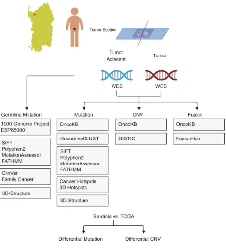
1. Patients and Samples... 52
2. DNA extraction from FFPET tissue ... 53
3. Whole Exome Sequencing Library Preparation ... 53
4. Sequence data Quality control. ... 54
5. Read mapping and processing. ... 54
6. Somatic SNP and INDEL calling and annotation ... 55
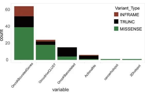
7. Identification of mutation drivers ... 55
8. Germline SNVs and INDELs calling and candidate germline risk identification ... 56
9. Fusion calling, filtering, ORF prediction and visualization ... 56
10. Copy number variation calling, filtering and driver copy number variation identification ... 57
11. Integrative analysis of SNP, INDEL, fusion and copy number variation 58 12. Differential gene expression and genomic alterations in prostate cancer. 58 13. PCR-based ERG fusion detection ... 58
Chapter Ⅲ. Results: Genomic landscape of Localized Prostate Cancer in a Sardinian cohort... 60
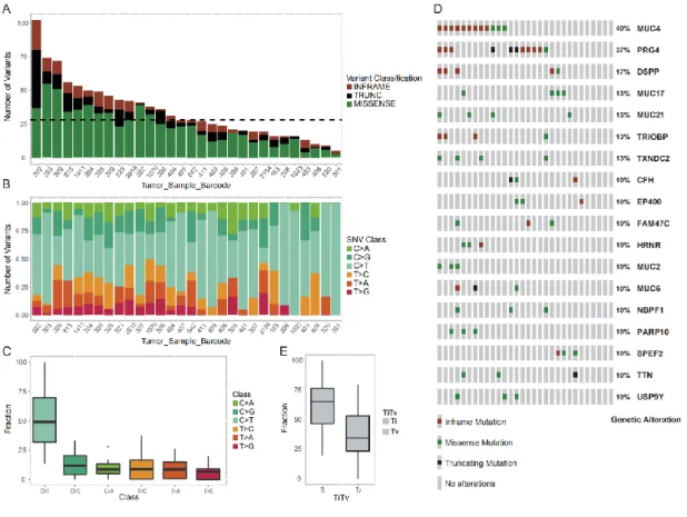
1. General summary of clinical and sequencing parameters... 60
3. Somatic copy number variation in Sardinia prostate cancer ... 71
4. Comparison of gene mutations between prostate cancer in the TCGAdatabase and Sardinia cohort... 78
5. Novel BTBD7-SLC2A5 fusions and ETS family status in prostate cancer of Sardinia ... 85
6. Germline risk mutations in the patients ... 88
ChapterⅤ. Discussion ... 93
Conclusion and prospect ... 109
A brief summary of the Chinese prostate cancer study ... 110
Data and material accession numbers... 113
Abbreviations ... 113
Acknowledgment ... 115
Abstract
Race and ethnicity are risk factors for prostate cancer. In the United States, African American men have the highest rate of mortality followed by Caucasians, and Asian Americans. The effects of race and ethnicity on prostate cancer are also reflected in different frequencies of ETS family fusion in different groups. ETS family fusions is the most common alteration in prostate cancer of Caucasian men at a frequency of ~50%, however, they are lower in African Americans and Chinese at 20-30%. Most of the genomic prostate cancer studies are focused on cohorts of European ancestry, leaving minority groups underrepresentation. Furthermore, in racial mixing, the ethnic contribution to risk is unclear. Sardinia population is an isolated Mediterranean population, and a purported refuge population of Neolithic ancestry with much lower incidence of prostate cancer than that in mainland Europe. Here, we conducted a genomic prostate cancer genomic study on a Sardinia cohort diagnosed with local prostate cancer. We identified a novel germline risk mutation ARSD-G320D occurring in 53 percent of the patients, somatic UGT family amplifications which occurred in 20% the patients, a novel in-frame fusion BTBD7-SLC2A5 occurred in 12 % of the patients. In addition, we pointed out that IRF8 deletion at 16q24.2 is a candidate driver in prostate cancer and patients with IRF8 deletion have worse prognosis. Our data revealed similarities and disparities in genomic alterations of prostate cancer
on Chinese prostate cancer cohort and have seen greater molecular disparities from TCGA cohort than in the Sardinian prostate cancer cohort. In Chinese cohort we have identified 37 genes significantly mutated and 20 of them have not implicated in prostate cancer in Caucasian and reveals a set of genomic markers that may inform the ethnic disparities.
ChapterⅠ.Introduction
1. Incidence, mortality of prostate cancer worldwide
Prostate cancer is the most common and fifth fatal cancer in men worldwide[1]. In 2012, an estimated of 1.1 million people were diagnosed with prostate cancer, and 307,000 deaths[2]. The incidence and mortality of prostate cancer vary greatly in the world [2-4].
The highest incidence of prostate cancer is observed in Oceania (111.6/100,000), followed by North America (97.2/100,000), Western Europe (94.9/100,000), Nordic Europe (85.0/100,000) and the Caribbean (79.8/100,000), while the incidence is much lower in Southeast Asia (11.2/100,000), North Africa (10.6/100,000), East Asia (10.5/100,000) and South-Central Asia (4/100,000) [2].
The highest prostate mortality is observed in the Caribbean (29.3/100,000), followed by South Africa and Central Africa (24.4/100,000, 24.2/100,000). As the incidence in Caribbean as high as 79.8/100,000, the ratio of mortality/incidence is 37%, much lower than that in South and Central Africa which is up to 90% [2, 4]. Although both the incidence and mortality in Southeast Asia, Central and South Asia are low, the ratio of mortality/incidence is as high as 64%[4]. Nevertheless, almost 70% of the newly diagnosed cases with prostate cancer in the world are in more developed regions such as North America, Oceania and Northern and Western Europe, the ratio of mortality/incidence in those area is only between 10-18% [2, 4] and the prostate cancer mortality rate has been decreasing over time[5, 6].
2. Risk factors of prostate cancer
2.1 Race and ethnicity
The incidence and mortality of prostate cancer vary greatly by geographic regions, strikingly, in the United States, the incidence and mortality of prostate cancer varies considerably by race and ethnicity [7, 8]. The prostate cancer incidence of the North America Africans is up to 208.7 per 100,000 and the mortality is up to 47.2 per 100,000, while prostate cancer incidence in the Asian American, Native Hawaiians and Pacific Islander (AANHIP) is only 67.8/100000 which is almost one third of North America Africans, and one half of Non-Hispanic Whites (123/100000) [8]. The
incidence, the mortality, and the ratio of mortality to incidence is lowest in AANHIP rather than North America Africans and Non-Hispanic Whites [8]. Even though, disparities in the diagnosis, treatment, and survival of prostate cancer patients of different races are often attributed to socio-economic status and access to healthcare [9, 10], after adjusting for those effects, racial disparities in prostate cancer incidence and mortality rates in United State remain significant [11]. In the United States, a white person has a 16% lifetime risk of prostate cancer and a 2.5% chance of death from prostate cancer, while black people are 70% more likely to develop prostate cancer and 40% more likely to die [12].
2.2 PSA Screening
In 1970 Wang and Valenzuela found that PSA was a highly sensitive marker of prostate cancer [13]. A longitudinal research project in Baltimore, revealed the relationship between serum PSA and prostate cancer [14]. In the late 1980s, PSA screening for prostate cancer diagnosis was adopted in the United States and subsequently in Europe, thus making the incidence of prostate cancer in the United States and Europe increase rapidly in the 1990s [15, 16]. However, due to the low specificity of PSA screening for high-grade, clinically significant disease, a considerable proportion of people were subjected to unnecessary prostate biopsy or diagnosed with indolent cancer resulting in overtreatment [17]. In Europe and the
United States, an estimated 23-42% of prostate cancer cases were over-diagnosed due to PSA screening (2002) [15]. Based on a large clinical study in the United States, USPSTF (US Preventive Services Task Force) recommended against PSA-based screening for prostate cancer for men of any age in 2012 [18], led to 18% relative decreasing in PSA screening rates for men aged over 50 between 2010 and 2013 [19]. However, among men greater than 75 years old recorded in the SEER database, the proportion of men presenting with metastatic disease increased from 2011 (7.8%) to 2013 (12.0%). A study conducted by ERSPC found that for every 1,000 men screened for PSA, three patients were prevented from metastasis and one was prevented from cancer-specific death. Based on these observations, in 2017, the USPSTF revised its recommendations and suggested that the decision to perform a PSA test for men aged 55 to 69 should be individualized by consulting their doctors [20]. Currently PSA screening rates in the United States and Europe are higher than that in other regions in the world. The latest data show that the screening rate for prostate cancer is 30.7% for American whites, 28.1% for American blacks and 25% for Asian Americans [21].
2.3Environmental factors
The incidence of prostate cancer in East Asia (~10/100,000) is much lower than that in North American Asians (~67/100,000) indicating that environment may be important risk factors for is prostate cancer. With the growth of economic and the
adoption of Western lifestyles during the last decades, the incidence of prostate cancer in East Asia has increased significantly. Singapore, Japan and Taiwan have experienced a sharp increase in the incidence of prostate cancer, from a low level (~5/100,000) to 30/100,000, 30/100,000 and 40/100,000 respectively [22-24]. The incidence of prostate cancer in China increased from 5/100,000 (2000) to 10/100,000 (2011) in ten years, and it is still growing. Current evidences have shown that obesity, high-fat diet, smoking and sunshine exposure may be risk factors for prostate cancer. Epidemiological studies on whether obesity increases the incidence of prostate cancer are very inconsistent, instead obesity increases the risk of prostate cancer progression [25, 26]. Animal model studies have shown that high-fat diets may induce prostate cancer progression through affecting growth factor signaling, lipid accumulation, inflammation and endocrine regulation. In addition, epidemiological evidence reveals that smoking is associated with the prognosis of prostate cancer[27], but its molecular mechanism is not well understood. There are studies indicating the short-day in high latitudes regions resulting high incidence of prostate cancer due to the insufficient sunshine exposure affecting vitamin D synthesis[28]. Nevertheless, low fat, vegetables, tomatoes, olive oil rich diets and exercise are protective for prostate cancer[29, 30].
2.4 Age
Age has a great impact on prostate cancer incidence and mortality. The risk of prostate cancer begins to increase in men with age over 40 years old and increases sharply when over 50 years old [31]. Over two-thirds of prostate cancer cases are older than 65 years old [31, 32]. However, there is evidence that the older you are, the more likely you are to develop prostate cancer, and it has been hypothesized that if men do not die of other causes, prostate cancer is inevitable. Men under 35 have almost no risk of prostate cancer. Therefore, the lower incidence of prostate cancer in Africans than that in North America Africans is largely due to the lower life expectancy of African males. With the aging of the global population, we need to pay attention to the increasing burden of prostate cancer.
2.5Infectious diseases
Over the years, several studies have focused on the association between prostate cancer and viral infections including human papillomavirus (HPV), herpesviruses including cytomegalovirus (CMV), human herpes simplex virus type 2 (HSV2), human herpesvirus type 8, (HHV8) and Epstein-Barr virus (EBV), polyomavirus BKV and xenotropic murine leukemia virus-related virus[33]. But a systematic review in 2013 indicated that there was insufficient epidemiological evidence showing that a single infectious pathogen was associated with prostate cancer, which
may due to the limited sample sizes in the study, resulting in the impossibility to assess non-persistent infection, and on the other hand, prostate cancer may be associated by multiple infectious pathogens [34]. A recent analysis of 5000 prostate cancer patients and 6000 healthy people has found that HPV16 infection increased the risk of prostate cancer [35].
Due to the development of second-generation sequencing technology, the doctrine of sterility in urine has been overthrown [36, 37]. Urinary microbial homeostasis and pathogenic microorganisms may cause prostate cancer inflammation and thus promote tumorigenesis [38]. Several bacteria have been proven to cause bacterial inflammation of the prostate, including E.coli. and other species of Enterobacteriaceae [39]. Propionibacterium acnes has also been demonstrated to cause prostate cancer inflammation and promote tumorigenesis [40-43]. Evidence also shows that some sexually transmitted pathogens such as Chlamydia trachomatis, Neisseria gonorrhoeae, or Trichomonas vaginalis are associated with prostate cancer [44-47]. Although it is hard to find a direct link between a single pathogen and the occurrence and development of prostate cancer, the relationship between various infections promoting inflammation of prostate cancer and the occurrence of prostate cancer has been gradually elucidated.
2.6 Family history
The genetic risk of familial prostate cancer was first proposed in the 1950s. Johns et al. carried out statistical analysis of 13 case-control studies, showing that men with a family history of prostate cancer had a 2.5-fold increased risk for prostate cancer. In a subsequent large number of studies, it was confirmed that men with a family history of prostate cancer had an increased risk of prostate cancer, especially with the presence of early onset, a relative, or multiple cases in the family. In 2015, Albright et al. conducted a retrospective analysis of 443 men with complete family history and assessed the relative risk of PC [48]. All these men had complete ancestral genealogy data. It was found that the risk of prostate cancer was increased by 2.46 if there was only one male immediate relative suffered from prostate cancer, while the risk reached by 7.65 when at least four male immediate male relatives had been diagnosed with prostate cancer [48]. In cases who have at least one immediate relative was diagnosed with prostate cancer before the age of 50, the risk was 5.54 [48]. Current studies have shown that the risk of prostate cancer in men is also increased due to rare germline risk variants such as BRCA1/BRCA2 or DNA mismatch, and the presence of breast cancer, ovarian cancer, rectal cancer and Lynch syndrome in families [49]. It is generally believed that the history of prostate cancer, breast cancer, ovarian cancer, rectal cancer and Lynch syndrome in relatives within three generations increased the risk of prostate cancer in men [50].
3. Heritability of prostate cancer
Early genetic quantitative studies of identical and fraternal twins have showed that the heritability of prostate cancer is 42% - 58%, higher than that of any other malignant tumors [51-53]. Linkage analysis based on pedigree prostate cancer has identified several chromosomal loci related to prostate cancer genetics [54-58]. In 1996, Jeffery et al analyzed 66 families at high risk for prostate cancer and identified for the first time the susceptibility locus of chromosome 1q24-25, which was named Hereditary prostate cancer 1(HPC1) [54]. Through this method, several chromosomes with different prostate susceptibility loci were also identified, including chromosomes 2, 3, 5, 6, 8, 10, 11, 13, 15, 17, 19, 20 and 22 [59, 60]. However, the chromosome regions identified by linkage analysis are too large to accurately identify the gene variant affecting the disease. Subsequent genome-wide association analysis (GWAS) and genome-wide exome sequencing further revealed susceptible variants in these regions, such as rs1006908 on chromosome 8q24 [61] and HOXB13 (G84E) on chromosome 17q21.[62]
3.1Loci with low penetration explain 19% of the hereditary of prostate cancer
Similar to other complex genetic diseases, the inheritance of prostate cancer is affected by variants with high frequency but low penetration and low frequency but high penetration in the populations [63, 64]. Genome-wide association analysis can
identify SNPs with high frequency but low risk. In 2016, Gudmundsson et al. reported the first genome-wide association analysis of prostate cancer, which confirmed that chromosome 8q24 carries prostate cancer susceptibility genes [65]. With the increase of GWAS research sample size and the development of chip imputing, the efficacy of GWAS in identifying prostate cancer susceptible SNPs has been greatly optimized, and more susceptible sites have been identified [66-69]. Scyumacher et al. recently have identified 63 new prostate cancer sites by analyzing data from 140,000 men[70]. Todate, 167 prostate cancer susceptibility loci have been identified by genome-wide association analysis [70]. These loci with high frequency and low penetration explain about 19% of the familial risk of prostate cancer [70]. However, most of the GWAS associated sites are located in the non-coding regions, and the molecular mechanisms of their effects on prostate cancer is still unresolved [71]. One hypothesis is that variants in these loci are associated coding regions affect prostate cancer and the other one is that these loci affect the transcriptional regulation region of coding genes.
3.2 Rare mutations on susceptible genes explain 15% of the hereditary of prostate cancer
In recent years, the effect of variants with low frequency and high penetration on the heredity of prostate cancer has become clear, such as HOXB13, BRCA1/BRCA2, DNA mismatch and DNA damage repair pathway [72]. These variants account
forabout 5% of the family risk of prostate cancer [73, 74]. In the following we will introduce the mechanism and clinical manifestations of the population frequency of these gene mutations on prostate cancer.
3.2.1 HOXB13
In 2012, Ewing et al. scanned 200 genes in 17-21-17q22 of 96 patients in different prostate cancer family clusters, and found that 18 patients had HOXB13 G84E rare mutations [62]. Furthermore, they verified the mutation in 5083 unrelated prostate cancer cases of European descent and 1401 control subjects. The mutation rate was approximately twenty-fold higher in the prostate cancer cases (1.4% or 72 in 5083) than in the controls (0.07% or1 in 1401)[62]. Odds ratio of HOXB13 G84E for the development of prostate cancer was 5.1 among men with positive family history and early onset and 1.7 among men with no family history and late onset [62]. Subsequently, in a large international family risk study of prostate cancer, 5% of prostate cancer families had the HOXB13 G84E variant [75], with the highest variant frequency around 20% in Finland, followed by Sweden around 8.2% [76]. Evidence suggests that HOXB13 G84E appeared most prevalent in the Nordic population, with a moderate penetration rate [77]. Different HOXB13 mutations have also been detected in prostate cancer cases in other racial or ethnic groups, including in African [62] (G216C and R229G), Asians (G135E) [78] and Portuguese (A128D,F240L)[79],
but the frequency and impact of these variants on the risk of prostate cancer remains to be further confirmed.
The HOXB13 gene encodes for a homeobox related transcription factor regulating a gene expression cascade which is critical for prostate development [80]. Other than that, HOXB13 protein has been shown to interact with Androgen Receptor signaling [81, 82], but its role in prostate tumorigenicity remains unclear and needs to be further investigation.
3.2.2 BRCA1/BRCA2
The relationship between BRCA1/BRCA2 and hereditary breast and ovarian cancer first revealed that BRCA1/BRCA2 mutations increase the risk of breast and ovarian cancer[83]. Subsequent studies have found that rare mutations in BRCA1/BRCA2 also increase the risk of prostate cancer [84]. Many studies have shown that BRCA1 mutations increase the risk of prostate cancer between 1.07 and 3.75 (odds ratio), while BRCA2 mutations increase the risk of prostate cancer more significantly between 4.65 and 8.6 (odds ratio). In a BCLC study involving 173 BRCA2 mutated families, the overall risk associated with prostate cancer in BRCA2 was 4.65, but in men who developed the disease before age 65, the risk rose to 7.33. In the study of prognostic analysis of patients with BRCA1/BRCA2 mutation, PSA-free survival and
overall five-year survival were significantly reduced in patients with BRCA2 mutations. Strikingly, in a recent genomic study that including 150 metastatic castration resistance prostate cancers (mCRPC), 8% of the patients had germline variants of BRCA2. BRCA1 and BRCA2 occur in about 0.87% and 5% of familial prostate cancers. Both BRCA1/BRCA2 mutations increases the risk of prostate cancer and BRCA2 mutations are associated with early onset of disease and prognosis. Due to relatively low numbers of cases carrying BRCA1/BRCA2 and the variability of specific mutations, the differences in mutation frequencies among different races is still unknown.
BRCA1/BRCA2 is a tumor suppressor gene encoding proteins that repair damaged double-stranded DNA by high-fidelity replication using the undamaged sister chromatid as a template [85]. BRCA1 has a broad range of functions, including recruiting effector factors to double strand break sites, regulating end resection of DSBs, activating G1/S, S-phase, and G2/M checkpoints, and mediating non-homologous end joining, and single-stranded DNA annealing repair pathways [85, 86]. The BRCA2 protein mainly recruits RAD51 and HR60 to DSB at the beginning of homologous recombination repair [87]. Deleterious mutations in BRAC1 and BRCA2 impair BRCA1/BRCA2 function and lead to instability of DNA, thus promoting carcinogenesis [88].
3.2.3 DNA mismatch repair genes
DNA mismatch repair gene mutations was first identified as the causative gene of Lynch syndrome[89]. Researchers subsequently observed that Lynch syndrome patients had a higher incidence of cancer than that of the normal population, especially in rectal cancer. In 2004, Harakdsdottir et al. analyzed the SEER database and observed 11 out of 188 men with Lynch syndrome developed prostate cancer (relative risk 4.87). In 2009, Grindedal et at. confirmed that DNA MMR mutations increased the risk of prostate cancer by analyzing 106 men with DNA MMR mutations in the Norwegian Cancer Registry [90]. Ryan et al. assessed the relative risk of prostate cancer associated with DNA MMR mutation to be 3.36 [91]. Then Rosty et al. estimated the relative risk of prostate cancer associated with MSH2 (5.8), MLH1(1.1), and MSH6(1.3)respectively[92]. Inactivation of MMR proteins, result in a high rate of microsatellite instability (MSI) in their tumors [93, 94].
3.2.4 Other prostate cancer germline risk variants
Other germline mutations are also associated with the risk of prostate cancer. In a study investigating germline risk mutations in metastatic prostate cancer, researchers found that 1.87% of metastatic tumors had CHEK2 germline mutations, 1.6% of ATM, 0.43% of PALB2 and 0.43% of RAD51D germline mutations [95]. A Polish study has found that NBN657del5 was present in 9% of pedigree prostate cancer
patients, and 2% of sporadic prostate cancer patients, while the mutation frequency in the control group was only 0.6%. In addition, in the recent TCGA pan-cancer study for screening germline risk variants, it suggested that germline variants on BRIP1, DKC1, EPCAM, GALNT3, MTAP, PMS2, POT1, RAD51C, RECQL, SEPRINA1, TSC1, TSC2, UROD and some other genes might also affect prostate cancer.
4. Somatic Genomic alterations in prostate cancer
Normal cells need to acquire multiple abilities to transform into tumor, including sustained growth signals, desensitization of growth inhibition signals, resistance to death, unlimited replication and proliferation, sustained angiogenesis, tissue infiltration and metastasis, avoidance of immune surveillance, and abnormal energy metabolism. Inflammation and genomic instability generate somatic mutations that expedites the acquisition of tumor promoting abilities. Before innovation of next generation sequencing technology, researchers identified a number of somatic mutation events in prostate cancer through fluorescent in-situ hybridization and comparative genomic hybridization, including loss of NKX3-1 [96, 97] and PTEN[98], amplification of MYC [99, 100] and AR [101]. Subsequent advances in gene expression profiling technologies led to the discovery of ETS family transcript factor over expression[102], and the discovery of recurrent TMPRSS22-ETS gene fusions in prostate cancer [103]. With the development of next generation sequencing
and bioinformatic analysis technologies comprehensive examination of prostate cancer genomes became an efficient tool for identification of cancer related mutations. In early prostate cancer genomic studies using next-generation sequencing, a large number of novel prostate cancer genomic alterations were identified [104-111]. However, due to the limitation of the sample sizes and early generation analytical methods, these studies failed to uncover the full spectrum of the complex heterogeneity of prostate cancer genomes. In 2015, the Cancer Genome Atlas (TCGA)published a multi-omic genomic study which included 333 cases of primary prostate cancer [112]. Researchers found that 74% of prostate cancers could fall into seven molecular subtypes: 1) ERG fusion (46%), 2) ETV1 fusion (8%), 3) ETV4 fusion (4%), 4) FL1 fusion (1%), 5) SPOP mutation (11%), 6) FOXA1 mutation (3%) and 7) IDH1 mutation (1%) [112]. Besides these reported mutations, TP53, PTEN, PIK3CA, RB1 and other gene alterations were also altered. In 2018, Joshua Armenia et al. combined genomic data from 1013 prostate cancer samples and identified that 97 genes were significantly mutated in prostate cancer, 70 of which had not been reported before, such as the ubiquitin ligase CUL3 and the transcription factor SPEN [113]. In addition, they defined a new molecular prostate cancer subtype that has mutations on epigenetic related genes [113]. Other related studies have also revealed that local indolent prostate cancer had lower numbers of molecular mutations [114], instead metastatic prostate cancer had higher mutation burden rates [115].
The unveiling of prostate cancer genome provided insights into prostate cancer biology and have enabled the identification of novel drug targets, diagnosis and risk stratification biomarkers for this disease. The molecular biology, diagnosis, risk stratification and therapeutic development of prostate cancer are presented in Chapter 1.7,1.8and 1.9.
5. Molecular evidence for racial disparities of prostate cancer
In recent years, the molecular basis for racial disparities of prostate cancer has accumulated. Early GWAS studies revealed the 8q24 locus [58, 61, 116] and some other prostate cancer risk alleles [117, 118] were associated with African Americans. GWAS studies based on men from different populations, such as Latino [119], South Asian [120], Japanese [68, 121], and Chinese [122, 123] ancestries have further identified potential race-specific prostate cancer risk alleles. Later whole exome sequencing revealed that the rare mutation HOXB13 G84E occurred more frequently in the family prostate cancer of the Nordic populations [62, 76, 77, 124] and the rare variation in Tet2 is associated with clinically relevant prostate carcinoma in African Americans [125]. Most recently, prostate cancer genomic studies revealed ETS family fusions was the most common alteration in prostate cancer of Caucasian men at a frequency of ~50%, however, they are much lower in African American and Chinese [103, 126, 127]. Similar to ETS fusion, PTEN loss is more frequently found in
prostate cancer of Caucasian men than other ethnicities [128]. There are also other race-specific prostate cancer genomic alterations such as LSAMP loss [129], ERF loss-of-function mutations [130, 131] and CDC27-OAT [132] fusion in prostate cancer of African Americans. However, most of the genomic prostate cancer studies are focused on cohorts of European ancestry, leaving minority groups under represented [133]. Furthermore,in racial mixing, the ethnic contribution to riskis not fully understood.
6. Oncogenesis and development of tumors
The size of the prostate is slightly larger than the walnut. It is located at the bottom of the pelvic cavity, under the neck of the bladder, on the urethra; behind the pubis, before the rectum, and surrounds the junction of the bladder mouth and the urethra, with a seminal vesicle gland attached, and urethra and the vas deferens wrapped [134].
(Figure 1.1) The main function of the prostate is to secrete and store prostatic fluid.
Prostatic fluid can be mixed with sperm to form semen which contains sperm and semen. About 10% to 30% of the semen is made from the prostate [134, 135]. The prostate also contains smooth muscle tissue, which helps ejaculation[135]. As prostate is located upstream of most male genital organs, including the vas deferens, epididymis and testis, the prostate is also considered the first line of defense for the male reproductive system against foreign antigens or pathogens from the bladder and
lower urethra [136]. The prostate is divided into transitional zone, central zone and peripheral zone. It consisted of 30 to 50 glands, with each one consisting of three basic types of cells, basal cells, luminal cells and neuroendocrine cells [137]. Most prostate cancers originate from luminal cells in the peripheral area of prostate cancer [138, 139]. The normal epidermal cells of the prostate develop into prostatic intraepithelial neoplasia, then local prostate cancer, then locally advanced prostate cancer, and eventually developed into metastatic prostate cancer [140-144]. With increasing age, inflammation prostatic atrophy is common [145, 146]. Inflammation atrophy has a larger impact area, especially in the peripheral areas with high incidence of prostate cancer [147]. Inflammation can be caused by infection and disruption of the epithelial barrier [33, 148]. Evidence suggests that inflammation-induced oxidative stress or reactive oxygen species (ROS) cause mutations in cell genes, leading to the transformation of normal cells to prostatic intraepithelial neoplasia[149-151]. In 2016, Mani, R.S. et.al found that inflammatory cytokine signals such as tumor necrosis factor (TNF) signals in epithelial cells result in DNA breaks which eventually lead to the fusion of TMPRSS2-ERG [152]. Other studies indicate bacterial infection leads to low expression of NKX3-1 in prostate epidermal cells [149]. Proliferative luminal epithelial cells of intermediate phenotype which abnormally express gene such as CDKN1B, GSTA1, COX2, MYC, PSA, AR, NKX3-1, MET, GSTP1 are enriched in prostatic proliferative inflammatory atrophy (PIA) [148, 153]. These studies suggested that inflammation can transform normal
cells to prostatic intraepithelial neoplasia. Besides, environmental toxins may cause DNA damage in normal epidermal cells of the prostate by triggering oxidative stress, which may lead to the carcinogenesis of epidermal cells, such asheterocyclic amines ingested from burnt diets through blood circulation to the prostate or by the urine reflux into the prostate also trigger oxidative stress [154-156].
Figure1.1 The size of the prostate is slightly larger than the walnut. It is located at
the bottom of the pelvic cavity, under the neck of the bladder, on the urethra; behind the pubis, before the rectum, and surrounds the junction of the bladder mouth and the urethra, with a seminal vesicle gland attached, and urethra and the vas deferens wrapped.
Most prostate cancers are indolent, and only a small proportion of prostate cancers progress aggressively to metastatic tumors. Metastasis prostate cancer undergo
Epithelial to mesenchymal transition (EMT) to migrate to the adjacent lymph nodes and then to the lungs, liver or bone[157, 158]. Recent studies have suggested that circulating tumor cells [159, 160] and exosomes[161] may play an important role in tumor metastasis. Despite the efforts to reveal the mechanism of prostate cancer metastasis and to develop drugs for the treatment of metastatic prostate cancer, metastatic prostate cancer has not yet been cured so far.
7. Prostate cancer diagnosis
Current clinical diagnosis of prostate cancer is based on the framework established in the 1990s. It mainly includes three main indicators: serum PSA level, rectal digital examination and tissue biopsy. Patients with serum PSA greater than 4.0 or rectal finger positive need to be further confirmed by biopsy [162, 163].
7.1 PSA screening
In 1970s, Wang and Valenzuela found that PSA was a highly sensitive biomarker of prostate cancer[13]. Then, in a longitudinal study in Baltimore, the relationship between PSA in serum and prostate was further elucidated [14]. In the late 1990s, PSA was used to screen prostate cancer because of its high sensitivity to detect prostate cancer, replacing the previously used prostatic acid phosphatase (PAP)[164].
PSA is a glycoprotein and composed of 237 amino acids, secreted by prostate epithelial cells. Normally, PSA is not released into the blood, because of natural blood-epithelial barrier between the prostatic duct system and the peripheral circulation system, thus maintaining low concentration in the blood. The invading and migration of prostate cancer cells disrupt blood-epithelial barrier, resulting in increasing of PSA concentration in the blood. Unfortunately, blood PSA elevation is also observed in benign prostatic hyperplasia (BPH) and prostatitis [165]. High levels cannot distinguish cancer from inflammation especially in patients with serum PSA levels <10 ng/ml[166]. Instead of only serum PSA level, the ratio of free PSA to total PSA, PSA density, PSA ROS curve have been shown to increase the specificity of PSA, but with limited effect [167].
7.2 Biopsy
The mode of prostate biopsies has evolved considerably over the years. Open or finger-guided transperineal biopsy with low accuracy was the standard method for biopsy sampling in the early 1920s. This method is very invasive, leading to high incidence of urinary incontinence and ED, and patients need to spend a long time in hospital to recover. The emergence of transrectal ultrasound (TRUS) guided prostate biopsy greatly improves the accuracy of biopsy and reduces the side effects of tissue biopsy [168]. In 1998, Levine and colleagues found that 12-core prostate biopsy
increased the detection rate of prostate cancer by 30% compared with 6 cores[169]. In 2003, the guideline for prostate cancer biopsy was revised to 12 needle biopsy and enhanced sampling of the anterior and lateral regions [170]. Nevertheless, needle biopsy has a significant chance of missing the prostate cancer. Thirty percent of patients with negative biopsy were identified as prostate cancer positive by repeat biopsy [169, 170]. In addition, transrectal ultrasound guided prostate cancer puncture also increases the risk of bacterial infection of the prostate [164].
7.3 Risk stratification
The risk stratification system for prostate cancer was originally derived from the D'Amico classification [171] of low-risk, medium-risk, high-risk disease or the the Epstein criteria [172] for low-risk (clinically unimportant) cancer mainly based on Gleason score of tissue puncture biopsy, serum PSA level and clinical classification. However, the inaccuracy of the risk stratification resulted in a high proportion of prostate cancers were inaccurately assessed, which lead to over-treatment or inadequate treatment.
7.4 Development of prostate cancer diagnosis and risk stratification 7.4.1 PSA derived
Fortunately, the understanding of the biology of PSA secreting and maturation helps to the identification of greater specificity of PSA isoform biomarkers. In particular, the detection level of the [-2] pro-PSA ('p2PSA') in serum can improve the diagnostic specificity of prostate cancer compared with free PSA or total PSA. PSA is initially translated as inactive pre-pro PSA, which carries a 17-amino acid signal peptide. During the secretion of PSA, pre-pro PSA is cleaved to pro-PSA by removing the signal peptide. Mature PSA, pro-PSA still requires cleavage by human kallikrein 2 (hK2) to remove the Pro precursor peptide of 7 amino acids at N terminal. In this step, alterative splicing isoforms have been found including [-1], [-2], [-4], [-5] and [-7] pro-PSA [173].
Figure1.2 Molecular forms of PSA. The arrows with dashed line mean the forms of
PSA that go from the cell to the blood. PSA: prostate specific antigen, BPSA: benign PSA, iPSA: intact PSA, PSA-ACT: Alpha 1-antichymotrypsin-PSA, PSA-API: alpha1-trypsin inhibitor PSA, PSA-A2M: alpha 2macroglobulin, hK-2: human kallicrein 2, hk-4: human kallicrein 4.
7.4.1.1PHI index
In 2012, the Prostate health index (PHI)was approved by the U.S. Food and Drug Administration (FDA) for aiding doctors to determine whether a patient with PSA level 4-10ng/ml requires biopsy[174]. Prostate health index (PHI) is a diagnostic index for prostate cancer that combines serum total PSA, free PSA and [-2] pro-PSA
levels. It is calculated by the formula ([-2] pro-PSA/free PSA) * tPSA. Patients with high serum level of total PSA and [-2] pro-PSA, and with low level of free PSA have higher PHI scores and are more likely to suffer from prostate cancer [174]. Fossati and colleagues evaluated PHI index in a total of 2034 patients with PSA at 2.5-10 ng/ml. In the ROC analysis, the AUC (Area under the curve) of PHI was 0.77, while the AUC of -2] pro-PSA was 0.76, % fPSA was 0.68, PSA was only 0.5[175].
7.4.1.2 The 4Kscore
The 4Kscore combines four prostate-specific biomarkers including total PSA, free PSA, intact PSA, and human kallikrein 2 [hK2] with clinical information to provide men with an accurate and personalized measure of their risk for aggressive prostate cancer [176, 177]. The 4Kscore Test was evaluated in multiple cohorts in Europe and subsequently validated in a US multicenter prospective study. Overall, the 4Kscore Test reduced prostate biopsy rates by 94% in men and there is evidence that the 4Kscore can predict the likelihood of cancer spreading to other parts of the body in the next 20 years [178-180].
7.4.2 Urine-derived biomarkers 7.4.2.1 SelectMDx
SelectMDx is a urine-based test based on prostate cancer genomic biomarkers HOXC6 and DLX1, which together have a 76% sensitivity (reliability) for detecting a prostate cancer that is aggressive enough (Gleason score of 7 or greater) to require treatment [181]. When additional risk factors (namely age, PSA, PSA density, family history and rectal examination) are added to the calculation, the accuracy of the test (negative predictive value) for excluding prostate cancer rises to 98%, with an expected total reduction of biopsies of 42%. So clearly there is great potential to avoid unnecessary prostate biopsies. A multi-centre scientific study made in 2016 and published in European Urology confirms the accuracy of the test[181]. while a 2018 study in the Journal of Urology has concluded that „routine use of the SelectMDx urinary biomarker panel to guide biopsy decision making improved health outcomes and lowered costs in American men at risk for prostate cancer [182]. This strategy may optimize the value of prostate cancer risk assessment in an era of increasing financial accountability.
7.4.2.2 ExoDx
ExoDx Prostate(IntelliScore) is a clinically validated, non-digital rectal exam (DRE) urine-based liquid biopsy test that predicts the presence of high-grade (Gleason score
≥7) prostate cancer for men 50 years of age and older with a PSA 2 – 10 mg/mL presenting for an initial biopsy. A “rule out” test, ExoDx Prostate(IntelliScore) is designed to more accurately predict whether a patient presenting for an initial biopsy does not have high-grade prostate cancer and, thus, could potentially avoid an initial biopsy and, instead, they continue to be monitored [183]. ExoDx Prostateanalyzes the urine for three biomarkers on exoRNA that are expressed in men with high-grade prostate cancer. Using a proprietary algorithm that combines the relative weighted expression of the three-gene signature, the test assigns an individual risk score for patients ranging from 0 to 100. A score >15.6 is associated with an increased likelihood of high-grade prostate cancer on a subsequent biopsy [184].
Additional molecular markers of prostate cancer based on different urine are being studied, such as MI-Prostate Score based on urinary secretory microRNA, Progensa and Protarix based on urinary proteomics[185].
7.4.3 Genic Tests
In 2017, the Philadelphia Consensus recommended that prostate cancer patients with a family history of HBOC and prostate cancer, as well as metastatic prostate cancer, need to undergo genetic screening for germline risk variants on genes includingBRCA1/2, HOXB13, and DNA mismatch repair genes [50]. Risk variants in
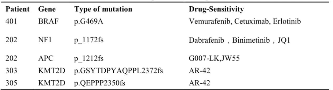
these genes not only increase the susceptibility to prostate cancer, but are also partly associated with the early onset and prognosis of prostate cancer or treatment selection [50].The risk genes and evidence discussed in the Philadelphia Consensus are shown in Table 3.
8. Treatment of prostate cancer
Alterative treatment selection for prostate cancer patients are mainly based on risk classification. In recent years, with the available of new targeted therapies and immunotherapy, genic testing become a supplemental option to guide the treatment selection [49].
8.1 Active monitoring for very-low risk prostate cancer
Recent studies have shown that very low-risk prostate cancer tends to remain indolent. The probability of their progression to metastatic prostate cancer or cause death is very low. To avoid side effects caused by treatment, in 2017, the American Urological Association announced that it is best for men who are diagnosed with very low-risk prostate cancer to actively monitor disease progression rather than receive treatment.
8.2 Activemonitoringfor low risk prostate cancer
Men who are diagnosed with low-risk prostate cancer should also be given priority in actively monitoring disease progression as well.Active monitoring is usually performed on patients with low-risk prostate cancer undergoing serum PSA monitoring, repeated prostate biopsy and MRI. Datahave shown that patients with low-risk prostate cancer under active monitoring have a probability of dying from prostate cancer less than 1% within10 years.[186]
8.3 Treatment for patients with low risk cancer
Because some lesions affect the daily life of patients, for patients with low-risk local prostate cancer, radical prostatectomy, external beam radiotherapy and brachytherapy are often used clinically. Othertreatments, such as cryotherapy, high-intensity focal ultrasound and photodynamic therapy, are also used [186].
8.4 Treatment for locally advanced prostate cancer
For patients with locally advanced or high-risk prostate cancer, recent studies have shown that radical prostatectomy plus ADT or radiotherapy plus ADT can significantly reduce the 10-year mortality risk compared with ADT alone. However, 10% to 20% of patients havebiochemical recurrence with elevated serum PSA in 2~3
years after ADT treatment and develop into castration resistant prostate cancer (CRPC) followed by metastasis to bone, lung and other sites (mCRPC). The median survival time of patients diagnosed with castration resistance prostate cancer is 15 to 36 months[187].
8.5CRPC treatment
8.5.1 Second Generation Androgen Receptor Antagonists
Although many patients develop resistance after 2~3 years of ADT treatment, mCRPC is AR signaling dependant. In 2012, the second generation AR antagonist Enzalutamide [188]was approved by FDA in the United States for the treatment of mCRPC. In clinical trials, Enzalutamide significantly improved overall survival (OS) and progression-free survival (PFS) in patients with prostate cancer after chemotherapy.
In 2012, Abiraterone, an inhibitor of CYP17A1, was also approved by FDA to treat mCRPC. CPY17A1 is an important enzyme in androgen synthesis pathway. It can effectively inhibit androgen synthesis by inhibiting CYP17A1. Clinical data showed that abiraterone combined with low-dose synthetic glucocorticoids effectively prolonged progression-free survival of the patients (5.6 vs 3.6 months, P < 0.001)[189].
8.5.2 PAPR inhibitor
A recently completed phaseclinical trial showed that PARP inhibitor (Lynparza/a/olaparib) significantly improved disease-free progression survival in mCRPC patients with BRCA1/2, and ATM mutations, compared with Enzaluramide and Abiraterone [190].
8.5.3 Siupleucel-T
Sipuleucel-T is an autologous dendritic cell vaccine targeting Prostatic acid phosphatase (PAP). In 2010, the FDA approved sipuleucel-T as the first and only immunotherapy for mCRPC[191]. In clinical trials, the 36-month survival rate in the Sipuleucel-T group was 31.7% while 23.0% in the placebo group[192].
8.5.4 Chemotherapy
In 2004, TAX327 reported that chemotherapy drug docetaxel improves outcomes in mCRPC. Tannock et al. demonstrated in a randomized trial of 1,000 men with mCRPC that docetaxel chemotherapy improved patient survival by nearly 3 months, with 45% of patients having a 50% reduction in PSA [193]. In 2015, the taxanes
represented by docetaxel in combination with prednisone have been used as first-line treatments for mCRPC patients [194].
8.5.5 Ra223mCRPC
Ra223 was approved by FAD in 2013 for the treatment of mCRPC with bone metastases. Clinical trial shows that Ra223 can increase the median survival time of mCRPC patients with bone metastases by 4.5 months[195].
9. Translate genetics to biology and therapeutics
The treatment of mCRPC has made great progress, but due to the heterogeneity of tumors and drug resistance during treatment, the fact is that mCRPC still cannot be cured. In recent years, a large number of new prostate cancer driver genes (potential targets) have been identified in prostate cancer genomic studies, and the breakthroughs in immune cell therapy and immune checkpoint therapy in other tumors have encouraged researchers to move forward.
In 1941, Charles Huggins first reported the beneficial effect of androgen ablation on metastatic prostate cancer. This discovery greatly inspired researchers to develop androgen deprivation therapy (ADT) for prostate cancer. ADT therapy is still a very important therapy for prostate cancer. Although 10% to 20% of patients have
biochemical recurrence with elevated serum PSA in 2~3 years after ADT treatment and progresses to CRPC which is associated with a poor prognosis.
Recent advances in prostate cancer genetics and genomics have provided considerable insights into prostate cancer biology and have identified a considerable number of cancer drivers which can be exploited as novel drug targets.
By 2018, More than 97 somatic drivers, 20 susceptibility genes, 167 germline risk alleles, in prostate cancer have been identified. To unveil the targetability of them, Wedge et al. conducted computational chemogenomic analysis of prostate cancer drivers and identified 11 targets of approved drugs, 7 targets of investigational drugs, and 62 targets with compounds that may be active and should be considered candidates for future clinical trials[196].
The following summarizes the most promising targets for prostate cancer discovery and highlight key signaling pathways as potential sources of targets including Androgen receptor signaling, the PIK3-AKT signaling, the WNT signaling, the DNA repair defects, the MAPK signaling.
9.1 AR signaling 9.1.1 GnRH
Gonadotropin-Releasing Hormone (GnRH), also known as Luteinising-hormone releasing hormone (LHRH), plays a crucial role in anti-androgen therapy [197]. Natural GnRH receptor agonists are secreted by hypothalamus and activate the receptors in the pituitary, increasing the release of LH (luteinizing hormone[198]) and ACTH (adreno-cortico-tropic-hormone) from the pituitary[199]. LH and ACTH promote corresponding target organs respectively to increase androgen secretion. In 1971, the chemical structure of GnRH in pigs was obtained [200]. Then a series of active analogue agonists were developed.
The persistent activation of GnRH receptor by GnRH analogue agonists depletes the pituitary of LH and ACTH, and the ultimate result is that androgen levels continue to drop to a very low level. In a phase III trial for patients with locally advanced prostate cancer, the 5-year clinical disease-free survival rate was 40% (95% CI 32% - 48%) in the radiotherapy group compared to 74% (95% CI 67% - 81%, P < 0.001) in theGnRH analogues and radiotherapy combined therapy group [201]. Therefore, GnRH analogues can significantly improve the outcome of locally advanced prostate cancer patients.
The persistent activation of GnRH receptor in the early stage causes a temporary rise in testosterone levels and promotes the progression of the disease. To resolve the adverse effects of GnRH receptor agonists, the development of GnRH receptor antagonists has been investigated. Antagonists inactivate the GnRH receptor by competitive binding with receptors. In 1997, Abarelix, a potent GnRH receptor antagonist, was successfully developed. Subsequently, several GnRH receptor antagonists, such as Degarelix, and Relugolix were introduced into the market [202, 203].
9.1.2 AR
9.1.2.1 AR-LBD
In 1990, the first generation of non-steroidal androgen receptor antagonists, flutamide [204] was approved and rapidly used as an important drug in the treatment of advanced prostate cancer, followed by Nelutamide and bicalutamide [205]. Flutamide must be absorbed in the gastrointestinal tract and metabolized in the liver to be activated leading to hepatotoxicity [206]. In addition, Drug resistance to the first generation non-steroidal androgen receptor antagonists caused by T877A[207],W741C[208] and F876L[209] mutations in the ligand binding domain of AR is usually observed within 1 year after first administration. In 2012, the second-generationnon-steroidal androgen receptor antagonist Enzalutamide was
approved by FDA. Enzalutamide is able to overcome the resistance caused by W741C mutation and has 8-fold greater affinity for AR than the first generation of non-steroidal androgen receptor antagonist[188]. Apalutamide with the same mother-ring chemical structure as Enzalutamide has been approved to treat CRPC in 2018 [210]. There was no obvious hepatotoxicity for the two drugs but seizures or rash and hypothyroidism are common side effects [188, 211]. Drug resistance is usually observed about 2-3 year later after administration because of AR F867L mutations, over-expression of AR-V7, AR co-activator, or activation of glucocorticoid receptor signaling [212].
A new non-steroidal androgen receptor antagonist,Darolutamide which is able to overcome the resistance caused by AR mutations including F867L, W741L and T877A has shown stronger antitumor activity and stronger AR affinity than Enzalutamide [213, 214]. It should be approved by the FDA to treat the mCRPC in the near future.
9.1.2.2 AR-nonLBD
Androgen receptor (AR) is a steroid hormone receptor in the nucleus, which contains a central DNA binding domain (DBD), ligand binding domain (LBD), and hinge domain and N-terminal domain(NTD). Mutations in AR-LDB domains and the
expression of AR-V7 without AR-LDB domain can lead to resistance to Enzalutamide and other second generation of AR antagonists [212, 215]. To overcome the resistance, drugs that bind AR non-LBD domain become a new strategy. Niclosamide has long been used as an anthelmintic, but it have been found that it can promote the degradation of AR-V7and effectively inhibit the growth of tumors [216]. Other strategies for degradation of AR have been investigated as well. The clinical trial of AR-110, an effective AR degrading agent developed by PROTAC (Proteolysis Targeting Chimera) technology is ongoing.
9.1.3 AR-binding protein
HSP90 is a chaperone protein that binds AR and maintains full-length AR in a high-affinity ligand-binding conformation[217]. Inhibition of HSP90 results in abnormal AR signaling [218, 219]. Both in vitro and in vivo models, HSP90 inhibitors also result in depletion of AR-V7[220]. However, phase I and phase II studies of HSP90 inhibitors have been generally disappointing because of poor patient tolerability and modest antitumor activity [221, 222]. Nonetheless, in the clinical studies of patients with advanced prostate cancer, inhibitor of HDAC which acetylates and activates HSP90 by acetylation, showed anti-tumor activity[223].
FOXO1 binds to AR-NTD domain and inhibits AR transcriptional activity[224]. However, PTEN deletion in prostate cancer results in AKT activation which phosphorylates the FOXO1 resulting in its nuclear exclusion [225, 226]. In a phase II clinical study, ATK inhibitor Ipatasertib combined with abiraterone showed better anti-tumor activity than abiraterone alone, especially in prostate cancer patients with PTEN deletion [227].
9.1.4 CYP17A1
CYP17 are key enzymes in the synthesis of testosterone. Ketoconazole, an antifungal drug, has been found to broadly inhibit CYP17 enzymes and has been widely used in the treatment of prostate cancer before [228]. Due to its hepatotoxicity, ketoconazole has been limited in the clinical treatment of prostate cancer[228]. Nonetheless, a CYP17A1 inhibitor, abiraterone[189]combined with low-dose glucocorticoid effectively prolonged the progression-free survival (5.6 vs 3.6 months, P < 0.001) of mCRPC. Abiraterone, was successfully approved by FDA to treat mCRPC in 2012.
9.1.5 5alpha reductase
5 alpha reductase can reduce testosterone to highy active dihydrotestosterone [229], so 5alpha reductase inhibitors also play a role in the treatment of prostate cancer[230].
For example, dutasteride combined with abiraterone can effectively improve the therapeutic effect of abiraterone[231].
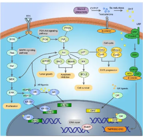
Figure 1.3 The cellular biology of prostate cancer. The complex underlying cellular
9.2 DNA repair Defects
Both germline and somatic genomic aberrations of DNA repair genes have been reported in prostate cancer. Causative germline mutations in DNA defect repair genes occur in 5% of hereditary prostate cancer carries[64], and strikingly, in 8-15% of mCRPC. Somatic mutations of DNA repair genes occur in almost 23% of mCRPC[111]. The most commonly aberrant genes are BRCA1/2 and ATM.
In 2005, studies showed that PARP inhibition could lead to death of BRCA1/2 deficient tumor cells [232]. Several studies over the past decade have demonstrated the utility of PAPR inhibitors in different types of tumors[233-235]. The PARP inhibitor olaparib has been approved by the FDA for the treatment of women with advanced ovarian cancer. While for mCRPC, recent clinical trials showed that PARP inhibitor (Lynparza/a/olaparib) significantly improved disease-free progression survival in patients with BRCA1/2, ATM mutations, compared with Enzaluramide and Arbitrone[190].
PARP (Poly ADP-ribose polymerase) detect and initiate an immediate cellular response to single-strand DNA breaks. Inhibition of PARP in the DNA repair deficient tumor results in synthetic lethality of the tumor cells [232, 236]. There are also studies suggesting that PARP inhibitors may have broader anti-tumor activity as PARP interacting with ETS and AR [237, 238].
In addition, DNA mismatch genes aberrations such as MSH6, MSH2, PSM2 occur in prostate cancer and potentially other defects in DNA repair [90, 239, 240]. These aberrations impair DNA mismatch repair, lead to an increase in mutation burden of tumors. There is also evidence showing microsatellite instability in tumors associated with DNA mismatch mutation, therefore, MMR defects can sensitize cancer to immunotherapy (Anti-PD-1, anti-CTLA4 therapeutics)[241, 242]. So far, patient selection approaches in CRPC for immune-checkpoint targeting have yet to be pursued.
9.3 PIK3-AKT pathway
PIK3-AKT signaling pathway is altered in 19% of local prostate cancer and 30% ofmCRPC. PTEN deletion is the most common aberration affecting 12% of local prostate cancers and 25% of mCRPC[112, 243]. Animal model experiments confirmed that the deletion of PTEN resulted in the formation of precursor prostate cancer lesions [244] and promoted disease progression when such features are combined with abnormalities in ERG, TPP53 [245, 246].
In tumors lacking PTEN, PI3KCA activity is suppressed while PI3KCB signaling is active. But in clinical studies with PI3KCB inhibitors, whether it was used alone or in
combination with docetaxel, the overall 1-year survival rate have not been shown to improve[247]. The reason for this may be that the inhibition of PI3KCB only results in the inhibition of AKT-mechanistic target of mTOR signaling which relives the feedback inhibition onupstream substrates and thus causes activation of PI3KCA and a rebound in downstream signaling[248]. However, Dactolisib, an inhibitor of multiple targets including PI3K and mTOR also have not shown any therapeutic advantage [249].
Inconsistent with PI3K and mTOR inhibitors, ATK inhibitor Ipatasertib combined with abiraterone showed better anti-tumor activity than abiraterone alone, especially in prostate cancer patients with PTEN deletion [227]. Studies have shown that the anti-tumor activity of ATK inhibitors benefit from PTEN-AKT-FOXO1 axis instead of AKT-mTOR signaling [225].
Recent studies also indicate that PTEN loss induces cellular senescence and myeloid-derived suppressor cell infiltration can block this senescence [245, 250, 251]. In the PTEN-null mouse model, infiltration of CD11b+, glucocorticoid receptor 1-positive myeloid cells protect a population of proliferating tumor cells from senescence. These myeloid-derived suppressor cells appear to infiltrate the prostate along a chemokine-chemokine receptor (CXCR2), and release IL-1 receptor antagonist, which inhibits senescence and drives proliferation. These findings
suggested that, targeting innate immunity may be a new therapeutic approach for PTEN-loss prostate cancer [252, 253].
9.4 ETS gene rearrangements
The transcription factors of ETS family such as ERG, ETV1, ETV4, FLI1 have important oncogenic roles in many prostate cancers. ETS rearrangements were found in about 40-60% prostate cancer patients of European ancestry. The most common rearrangement are ERG (46%), followed by ETV1 ( 8%), ETV4 (4% )and FL1( 1%)[254].
ERG usually fuses with TMPRSS2 which is regulated by an androgen -regulated promoter element[103]. TMPRSS2-ERG fusion leads to ERG overexpression, resulting in AR expression and tumor cell proliferation[255]. Overexpression of ETS induces the formation of prostatic intraepithelial neoplasia(PIN) in a genetically engineered mouse model[153]. When combined with increased AR signaling or PTEN loss,Overexpression of ETS leads the progression of tumors[153]. Therefore, inhibiting ETS oncogene signaling is a promising therapeutic strategy to treat prostate cancer.
Transcription factors are generally considered as undruggable targets. However, new strategies of modulating the activity of transcription factors have shown promise, including disrupting the interaction between transcription factors and other proteins andthe interaction between proteins and DNA, or restricting the binding of transcription factors by epigenetic modification of chromosome[256]. Currently, such as dithiophene diamidine compounds and DB1255 which inhibit ERG-DNA interactionsare under development[257]. In addition, clinical trials using optimized liposome-encapsulated siRNA to silence ERG expression in prostate cancer is ongoing[258].Another potential therapeutic strategy is to target the downstream effectors of TMPRSS2-ERG. Evidence shows that PLA2G7 is up-regulated in ERG-positive cancer and PLA2G7 silencing by siRNA sensitized ERG-rearrangement-positive VCaP cells to oxidative stress, reducing cell viability [259, 260].
In addition to ERG rearrangement, YK-4-279, a small molecule drug targeting the FLI1 rearrangement, has reached phase I clinical trials for Ewing sarcoma treatment [261]. YK-4-279 inhibits the binding of EWS-FL1 to RNA helicase and induces apoptosis of cancer cells.YK-4-279 also shows the ability to inhibit ERG and ETV1 rearrangements in vitro [262].
9.5 TP53
As the most common aberration, TP53 mutations are carried in 41% of pan-cancers, especially in HGSOC (high-grade serous ovarian cancer) (98%), esophageal adenocarcinoma (89%) and small cell lung cancer (85%). Almost 15% of mCRPCcarry TP53 mutation[263].
TP53 is a tumor suppression gene, encodes the p53 protein which maintain a low level by MDM2 regulated post-translational ubiquitin degradation in normal cells. When DNA is damaged, cellular stress induces phosphorylation of MDM2 and acetylation of p53, leading to accumulation and activation of p53. Activated p53 proteins stopproliferating cells in G1/S phase to repair the DNA damage[264]. Oncogenic stress triggers a DNA damage response involving p53, which constitutes a major barrier against tumor development. However, recent studies have shown that this effect of p53 is dispensable in tumors, and that p53 maintaining the homeostasis of cellular metabolism and redox balance in cell is even more important[265]. Moreover, many mutant p53 proteins have acquired gain-of-function (GOF) activities [266-268], which enable them to, for example, inactivate other p53 family members, in particular the tumor proteins p63 and p73 [269].(Figure 1.4)
Therefore, a drug development strategy for p53 is to use small molecules that promote proper folding and/or reactivation of common missense-mutant p53 proteins. Several of these compounds show significant anti-tumor activity in vitro and in vivo models. Clinical trials of two of the mutant-p53-targeting compounds are ongoing. APR-246 is being tested in phase II trial s[270], while the molecule COTI-2 is being studied in a phase I trial [271]. Although the current clinical trials do not include the treatment of prostate cancer, the efficacy of these drugs in the treatment of prostate cancer with TP53 mutation should also be investigated in the future.
Figure1.4Mutant p53 proteins have acquired gain-of-function (GOF) activities, inactivate other p53 family members, in particular the tumor proteins p63 and inhibit the transcription activities of p63.
9.6 WNT signaling
Aberrations that result in WNT pathway activation, such as loss of function of APC (adenomatous polyposis coli protein), mutations in genes encoding beta-catenin and
mutations in RNF43 have been reported in 15% of the mCRPC[254]. RNA-seq revealed that WNT beta-catenin signaling is a functionally important pathway for androgen-independent prostate cancer progression [272, 273]. Therefore, targeting WNT signaling in the subset of the mCRPC with activation of this pathway is promising. There are multiple compounds engaged in clinical trials for solid tumors [274]. As well the efficacy of these drugs to treat mCRPC with WNT pathwayaberrations should also be investigated in the future.
9.7 The RAS-RAF-MEK signaling
Arguably less common in prostate cancer, but nevertheless still clinically relevant and potentially targetable, is oncogenic activation of RAS-RAF-MEK signaling [275, 276]. Such activation includes uncommon (1-2%) recurrent BRAF and RAF1 rearrangements as well as rare mutations of these genes [277] and other aberrations of genes activating this pathway, including HRAS, SPRED, SPROUTY, FGF, and FGFR[112, 113].
As ETS proteins are downstream effectors of RAS-RAF-MEK-extracellular signal-regulated kinase(ERK) signaling, resistance to AR blockade in ETS-rearrangedprostate cancer has been postulated to involve RAS-RAF-MEK signaling[278]. Activation of the MAPK pathway could also activate ETS signaling in