R E S E A R C H A R T I C L E
Open Access
Experimentally induced cartilage degeneration
treated by pulsed electromagnetic field
stimulation; an in vitro study on bovine cartilage
Francesca Veronesi
1*, Milena Fini
1,2, Gianluca Giavaresi
1,2, Alessia Ongaro
3, Monica De Mattei
3, Agnese Pellati
3,
Stefania Setti
4and Matilde Tschon
1,2Abstract
Background: Osteoarthritis (OA) is the final result of progressive alterations to articular cartilage structure, composition and cellularity, followed by an increase in the concentration of pro-inflammatory cytokines in joint synovial fluid. Even though the effect of pulsed electromagnetic field (PEMF) stimulation in counteracting OA progression and inflammation is of increasing interest, because of its anabolic and anti-inflammatory properties, the present study aimed to improve the knowledge on cartilage extracellular matrix (ECM) and chondrocyte changes related to the exposure of PEMF, from a histological and histomorphometric point of view.
Methods: Anin vitro OA model was realized, culturing bovine cartilage explants with a high dose of interleukin 1β (IL1β, 50 ng/ml) at different experimental times (24 h, and 7 and 21 days). The effects of PEMFs (75 Hz, 1.5 mT) were evaluated in cartilage explants treated with IL1β or not (control), in terms of cartilage structure, cellularity and proteoglycans, glycosaminoglycans, collagen II and transforming growth factorβ1 synthesis by using histology, histomorphometry and immunohistochemistry.
Results: Making a comparison with control cartilage, IL1β-treated explants showed a decrease in cartilage matrix, structure and cellularity parameters. PEMFs were able to counteract the progression of OA acting on both cartilage cellularity and ECM in cartilage previously treated with IL1β. Normal distribution (Kolmogroc-Smirnov test) and homoscedasticity (Levene test) of data were verified, then, the non-parametric Kruskal Wallis test
followed by Mann-WhitenyU test for pairwise comparisons were performed. The p-value was adjusted according
to the Dunn-Sidak correction.
Conclusions: These results, obtained by culturing and treating cartilage explants from two different joints, confirmed that PEMF stimulation can be used as adjuvant therapy to preserve cartilage from detrimental effects of high inflammatory cytokine levels during OA.
Keywords: Osteoarthritis, Cartilage, Explants, Inflammation, Microenvironment, Pulsed electromagnetic field Background
The presence of pro-inflammatory cytokines in the joint microenvironment favors cartilage degeneration and osteoarthritis (OA) progression with alterations of proteoglycans (PGs) and collagen fibers, prevalently collagen type II (Coll II) [1]. Among pro-inflammatory
cytokines, interleukin1β (IL1β) plays a pivotal role in inducing degradation of extracellular matrix (ECM) components and synthesis of other pro-inflammatory cytokines, chemokines and proteolitic enzymes. Since cartilage has a poor intrinsic reparative capability if not treated, OA leads to progressive disability and require-ment of joint arthroplasty [2].
Current therapeutic strategies to prevent OA include the use of nonsteroidal anti-inflammatory drugs (NSAIDs) [3], that act at articular and not cellular levels, intra-articular injection of hyaluronic acid (HA) and physical
* Correspondence:[email protected]
1Department Rizzoli RIT, Rizzoli Orthopedic Institute, Laboratory of Biocompatibility, Innovative Technologies and Advanced Therapies, Bologna 40136, Italy
Full list of author information is available at the end of the article
© 2015 Veronesi et al. Open Access This article is distributed under the terms of the Creative Commons Attribution 4.0 International License (http://creativecommons.org/licenses/by/4.0/), which permits unrestricted use, distribution, and reproduction in any medium, provided you give appropriate credit to the original author(s) and the source, provide a link to the Creative Commons license, and indicate if changes were made. The Creative Commons Public Domain Dedication waiver (http://creativecommons.org/publicdomain/zero/1.0/) applies to the data made available in this article, unless otherwise stated.
exercises [4], that act only in the muscle of the joint. These treatments relieve pain and inflammation and improve functionality, but do not resolve the patho-logical process once triggered, thus the outcomes are inconclusive and remain as long as these therapies are administered. For this reason, orthopedic research in-vestigates chondroprotective treatments able to reduce the local inflammatory microenvironment and to favor damaged articular cartilage anabolic activity.
Pulsed electromagnetic field (PEMF) stimulation has been already studied and proposed for the regeneration of musculoskeletal tissues such as cartilage [5], bone [6, 7], tendon [8] and ligament [9]. As far as cartilage tissue is concerned, PEMFs positively affect chondrocyte prolifera-tion and ECM component synthesis [10]. However, the existing studies, performed with different PEMF physical parameters and exposure times, gave contradictory results [11–15], in terms of chondrocyte viability, DNA content, glycosaminoglycans (GAGs), Coll II, PGs and aggrecan gene expression and production. In a previous in vitro study, on bovine cartilage explants, we conducted a dose– response study to find the optimal dosage, in terms of PEMF frequency and exposure intensity, able to stimulate significantly PG synthesis [16]. Furthermore, the stimula-tory effect of PEMFs on PG synthesis in presence of IL1β and in combination with insulin-like growth factor 1 (IGF-I) was observed [17, 18]. PEMF and IGF-I showed an additive effect on PG synthesis in human OA cartilage explants cultured in the absence or presence of IL1β [19]. Inin vivo studies, PEMF stimulation was able to limit the progression of OA of increasing severity [20–22], to reduce IL1β and tumor necrosis factor-α (TNF-α) concen-trations and to increase transforming growth factor-β1 (TGFβ1) in the synovial fluid of sheep treated with autolo-gous osteochondral grafts [23]. In clinical studies, PEMF stimulation was successfully used to treat pain, knee swell-ing and functionality, after total knee arthroplasty [24, 25] and in early knee OA patients [26]. In other studies, bene-ficial symptomatic effects [27, 28] or additional effects to physical treatments [29] were not observed in OA patients.
Even if PEMFs act on the joint microenvironment and have anabolic and chondroprotective actions, the mecha-nisms by which PEMFs exert their effects on biological systems remain not completely understood. To our know-ledge, no histological and histomorphometric evaluations have assessed the effects of PEMF stimulation in an inflammatory OA microenvironment obtained in vitro with the addition of a high IL1β dose.
The present study aimed to analyze whether PEMF stimulation (75 Hz, 1.5 mT), applied to bovine cartilage explants, derived from two different joints of the same animals, was able to counteract the catabolic effect of a high dose of IL1β. In comparison to 2D monolayer
chondrocyte cultures, the cartilaginous 3D explants cultured with IL1β are a well-accepted method to measure and evaluate the effect and mechanism of action of a therapy, by simultaneously evaluating chon-drocytes and cartilage matrix and by closely mimicking the clinical situation of OA joint, in which a high dose of IL1β is found in the early stages of OA [30] and its concentration can be further increased by surgical intervention [2].
It was hypothesized that the PEMF stimulation alone, without the addition of other biological stimuli, might improve the production of the most important cartilage ECM components (PGs, GAGs, COLL II) and anabolic factor (TGF-β1) and preserve cartilage structure, thus controlling OA development. To demonstrate this hypothesis it was adopted an already set and validated in vitro model of OA [18, 31], which used a high dose of IL1β (50 ng/ml) to create an inflammatory OA micro-environment. After 24 h, 7 and 21 days of culture, PEMF effect, in combination or not with IL1β, was evalu-ated through histological, histomorphometric and immu-nohistochemical analyses. In this study it was observed that PEMFs were able to counteract the progression of OA acting on both cartilage cellularity and ECM in cartil-age previously treated with IL1β.
Methods
Cartilage explant cultures and treatment conditions
Full-thickness explants of bovine articular cartilage were aseptically dissected, by using a 4 mm dermal punch (Stiefel Laboratories, Milan, Italy), from the metacarpo-phalangeal(MC) and metatarsophalangeal (MT) joints of 14-18-month-old animals (Limousine breed), as previ-ously described [16–18]. The abattoir gave permission for the use of cartilage explants in this study and no eth-ical approval, from a recognized ethics committee, was required for this study. The cartilage was explanted shortly after the bovine slaughter and cartilage discs (three discs in each well) were cultured in 0.5 ml culture medium in multiwells (Nunc, Denmark, 1.6 cm the diameter of each well). Before PEMF exposure and IL1β administration, all explants were allowed to equilibrate in culture for 48 h in DMEM/F12 (Life Technologies, Monza, Italy) supplemented with 10 % FBS and antibi-otics (penicillin 100 units/ml, streptomycin 0.1 mg/ml) (complete medium) and for an additional 48 h in medium without serum, at 37 °C in an atmosphere of 5 % CO2. During the experiments, explants were cul-tured in complete medium in the absence and presence of IL1β (50 ng/ml) [18]. Half cultures were exposed to PEMF throughout the entire culture period (24 h, 7 and 21 days). Medium was changed at the beginning of the exposure (time 0) and every 3 days.
For each experimental time, 4 experimental groups were set up and each group was composed of 3 explant discs from MC and 3 more from MT joints:
– Control group (CTR): cartilage explants; – IL1β group: cartilage explants submitted to IL1β
administration;
– PEMF group: cartilage explants exposed to PEMFs; – IL1β + PEMF group: cartilage explants submitted to
IL1β and exposed to PEMFs.
Characteristics of PEMFs and exposure conditions
The PEMF generator system was the same as that used in previous studies [9, 16–18, 32, 33]. It consisted of a pair of circular Helmoltz coils of copper wire placed opposite to each other and in a signal generator (Igea S.p.A., Carpi, Italy). The multiwell plates were placed be-tween this pair of Helmoltz coils, so that the plane of the coils was perpendicular to the multiwell plates, and the direction of the induced electric field was perpen-dicular to the direction of the magnetic field. The power generator produced a pulsed signal with the following parameters: the pulse duration was 1.3 ms and the fre-quency was 75Hz, yielding a duty cycle of 1/10. The intensity peak of the magnetic field was 1.5mT and the induced electric field, as detected with a standard coil probe (50 turns, 0.5 cm internal diameter of the coil probe, 0.2 copper diameter), was 0.07 mV/cm.
The intensity of the magnetic field was detected between two coils from one side to the other, by the Hall probe of the Gaussmeter (LE, Gaussmeter DG500, USA), with a reading sensitivity of 0.2 %. Inside this area, where the multiwell plate was placed, the magnetic field was uniform. Cartilage explants were exposed to con-tinuous PEMF for 24 h, 7 or 21 days. PEMF unexposed cultures were placed inside the same incubator at a dis-tance where no difference from background magnetic field was observed when the PEMF generator was turned on. In each experiment, controls and treatments were performed in triplicate wells.
Histology
At the end of the experimental times, cartilage explants, cultured in the above mentioned conditions, were rou-tinely processed for paraffin embedding procedures. Briefly, samples were fixed for 24 h in 10 % neutral buff-ered formalin solution in PBS, extensively rinsed in dis-tilled water, dehydrated in graded alcohol solutions (70, 95 %, two times and 100 %, three times, one hour for each step), cleared in xylene e finally paraffin embedded. Sections (5 ± 1μm) were cut along the longitudinal axis of the samples by a semi-automated microtome (Microm H340E, Germany) and stained with Toluidine Blue, for the PG quantification and Safranin O-Fast green staining, for
the GAG quantification. Three non-consecutive sections for each sample were evaluated, by 2 independent his-tologists through a semi-quantitative score (modified O’Driscoll score) [34]. The modified O’Driscoll score (Table 1: minimum 0 = osteoarthritic cartilage - max-imum 11 = normal intact cartilage) analyzed 4 cartilage aspects: Safranin O staining, surface regularity, cellular-ity and chondrocyte clustering.
Immunohistochemical stainings (TGFβ1 and Coll II)
Cartilage sections were dewaxed in decreasing graded ethanol solutions until PBS rinsing for 20 min and then immunostained for TGFβ1 and Coll II. Briefly, after fix-ation, sections were extensively rinsed in PBS and permeabilized by incubation in 0.3 % hydrogen peroxide in PBS solution for 15 min. For Coll II immunostaining, sections were pre-treated for antigen unmasking with 0.2 % Pronase (P-8811, Sigma, Mo, USA) solution in PBS for 30 min at 37 °C. Then, 10 % normal serum was added for 1 h at room temperature to block nonspecific antibody binding and the primary antibodies (rabbit polyclonal antibody anti TGFβ1, sc-146 and mouse monoclonal antibody anti collagen II, sc-52658, 1:50 dilutions, Santa Cruz Biotechnology, CA, USA) were applied and incubated overnight at 4 °C. After rinsing in PBS, slides were incubated with appropriate biotinylated secondary antibody and with horseradish peroxidase-streptavidin complex for 1 h each (ABC Staining System, Biotechnology, CA, USA). Sample reaction was developed with 3,3-diaminobenzidine substrate and permanently mounted. Negative controls, by omitting the primary antibody, were included to check proper specificity and performance of the applied reagents.
Table 1 Modified O’Driscoll score
Parameter Feature Grade
Safranin O staining Normal or nearly normal 3
Moderate 2
Slight 1
None 0
Surface regularity Smooth and intact 3
Superficial horizontal lamination 2 Fissures 25–100 % of the thickness 1 Severe disruption, including fibrillation 0
Cellularity Normal cellularity 3
Slight hypocellularity 2
Moderate hypocellularity 1
Severe hypocellularity 0
Chondrocyte clustering No clusters 2
<25 % of the cells 1
Histomorphometric measurements
Slices stained with Toluidine Blue or Safranin O-Fast Green and immunostained for TGFβ1 or Coll II were observed by a light microscope (BX51, Olympus Italia Srl, Segrate-Milano, Italy) and three Regions of Interest (ROI) for each slide were grabbed at a 40× magnifica-tion. Image analysis by Leica Q-Win Software (Leica Microsystem, Wetzlar, Germany) was performed and the threshold applied was One-dimensional (LUTs: Lookup Table Transforms) based on selected ranges of RGB channels to measure the following parameters expressed as percentage:
– GAGs, as the ratio between the Safranin O stained areas and the total ROI areas;
– PGs, as the ratio between the Toluidine Blue stained areas and the total ROI areas;
– TGFβ1, as the ratio between the immunopositive stained areas and the total ROI areas;
– Coll II, as the ratio between the immunopositive stained areas and the total ROI areas.
Statistical analysis
Statistical analysis was performed using the IBM SPSS Statistics v.21.0 software (IBM Corp, USA). Firstly, nor-mal distribution (Kolmogroc-Smirnov test) and homo-scedasticity (Levene test) of data were verified, then, the non-parametric Kruskal Wallis test followed by Mann-Whiteny U test for pairwise comparisons were per-formed. The P-value was adjusted according to the Dunn-Sidak correction [adj p = 1 - (1 – p)k, where k is the number of pairwise comparisons]. Since the prelim-inary Kruskal-Wallis test by distinguish MC and MP joints did not show significant differences between the two harvesting sites within each type of treatment, data were analyzed when merged.
Results
Thein vitro cartilage OA model after IL1β administration
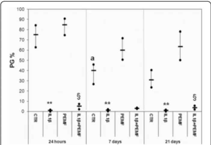
In the CTR Group the production of PGs, GAGs and COLL II significantly decreased at 7 days in comparison with cultures at 24 h by −47 % (p = 0.022), −58 % (p = 0.008) and−27 % (p = 0.038), respectively (Figs. 1, 2, 3).
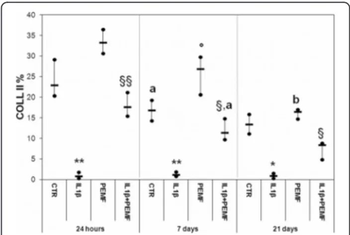
As expected, the treatment with IL1β significantly decreased the PGs, GAGs, Coll II and TGFβ1 produc-tion in cartilage explants as compared to CTR at each experimental time, except for TGFβ1 at 21 days (Figs. 1, 2, 3, 4). Specifically, PGs decreased by 96– 99 % (at 24 h p = 0.0005, at 7 days p = 0.0005 and at 21 days p = 0.0005), GAGs by 88 % at 24 h (p = 0.0005) and about 73 % at 7 (p = 0.008) and 21 (p = 0.03) days and Coll II by 92–96 % (at 24 h p = 0.0005, at 7 days p = 0.0005 and at 21 daysp = 0.04) (Figs. 1, 2, 3). The produc-tion of TGFβ1 significantly decreased at 24 h (p = 0.0005,
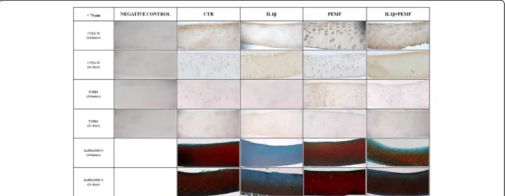
47 %) and 7 days (p = 0.017, 68 %) (Fig. 4). Figure 5 shows the immunohistochemical and histological images of explants stained for Coll II, TGFβ1 and Safranin O at 24 h and 21 days.
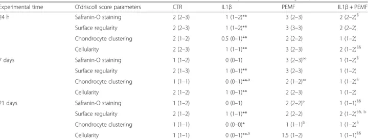
Because of the IL1β effect, the O’Driscoll score pro-gressively worsened by 50–60 %, during the experimen-tal times, in comparison to CTR (at 24 h p = 0.0005, at 7 days p = 0.0005 and at 21 days p = 0.04) (Fig. 6). By analyzing each score parameter (Table 2), it was noted
Fig. 1 Boxplots of histomorphometric measurements of PGs expressed as percentage in bovine cartilage explants. Black line = median; extreme values = minimum-maximum. Control (CTR), exposed to PEMF (PEMF), treated with IL1β (IL1β) or treated with IL1β and stimulated with PEMF (IL1β + PEMF) explants. 24 h, 7 and 21 days (n = 6). Mann-Whitney U test: - Within each experimental time: IL1β versus CTR (**, p < 0.005); IL1β + PEMFversus IL1β (§, p < 0.05); - Within each group: 7 days versus 24 h (a,p < 0.05)
Fig. 2 Boxplots of histomorphometric measurements of GAGs expressed as percentage in bovine cartilage explants. Black line = median; extreme values = minimum-maximum. Control (CTR), exposed to PEMF (PEMF), treated with IL1β (IL1β) or treated with IL1β and stimulated with PEMF (IL1β + PEMF) explants. 24 h, 7 and 21 days (n = 6). Mann-Whitney U test: - Within each experimental time: IL1β versus CTR (*, p < 0.05; **, p < 0.005); PEMF versus CTR (°,p < 0.05; °°, p < 0.005); IL1β + PEMF versus IL1β (§, p < 0.05; §§, p < 0.005); - Within each group: 7 daysversus 24 h (a,p < 0.05)
that, the cellularity (p = 0.025) and chondrocyte cluster-ing (p = 0.025) decreased significantly by 50 % in the CTR group from 7 to 21 days and from 24 h to 7 days, respectively. At 24 h, the reduction induced by IL1β on each O’Driscoll score parameter in comparison with CTR was 50 % for Safranin-O staining (p = 0.0005), sur-face regularity (p = 0.002) and cellularity (p = 0.0005) and 75 % for chondrocyte clustering (p = 0.0005), whereas at 7 and 21 days it was 50 % for surface regularity (at 7 days p = 0.0005 and at 21 days p = 0.0005), 50–100 % for
cellularity (at 7 days p = 0.001 and at 21 days p = 0.001) and 100 % for chondrocyte clustering (at 7 days p = 0.001 and at 21 daysp = 0.02).
Effects of PEMF stimulation
As also observed by histology in Fig. 5, in bovine cartilage explants stimulated by PEMF, no significant increases in PGs were found in comparison with CTR explants at each experimental time (Fig. 1). Conversely, PEMF stimulated GAG production at 7 (p = 0.0005, 170 %) and 21 (p = 0.005, 151 %) days, as well as Coll II (p = 0.005, 59 %) and TGFβ1 (p = 0.005, 58 %) at 7 days (Figs. 2, 3, 4). By analyz-ing data over time, Coll II in the PEMF group diminished from 7 to 21 days (p = 0.004, 38 %).
When bovine cartilage explants were cultured with IL1β and were stimulated with PEMFs, very high increases in PG production compared to the IL1β group were ob-served at 24 h (p = 0.01, 439 %) and 21 days (p = 0.04, 235 %) (Fig. 1). The IL1β + PEMF group also showed in-creases in GAG production in comparison to the IL1β group at 24 h (p = 0.0005, 684 %), 7 (p = 0.02, 250 %) and 21 days (p = 0.01, 307 %) (Fig. 2). Coll II increased mas-sively in the IL1β + PEMF group at 24 h (p = 0.002, 1890 %), 7 (p = 0.02, 835 %) and 21 days (p = 0.007, 734 %) in comparison to IL1β (Fig. 3). GAG and Coll II synthesis decreased by 57 % (p = 0.02) and 35 % (p = 0.02), respectively, from 24 h to 7 days in the IL1β + PEMF group. Regarding TGFβ1 production, it in-creased progressively at all experimental times (at 24 h p = 0.005, 71 %, at 7 days p = 0.02, 184 % and at 21 days p = 0.007, 204 %) in comparison to the IL1β group (Fig. 4).
The O’Driscoll score results of the PEMF-treated group did not differ from those of the CTR group at each experi-mental time (Fig. 6). Taking into account each O’Driscoll score parameter (Table 2), PEMF stimulation improved chondrocyte clustering at 7 days (p = 0.0005, 100 %) and Safranin-O staining at 7 (p = 0.0005, 100 %) and 21 days (p = 0.03, 200 %) compared to that of the CTR group.
The effect of PEMF stimulation, applied to IL1β-treated cartilage explants (IL1β + PEMF group), improved the O’Driscoll score in comparison to the IL1β group, at both 24 h (p = 0.02, 75 %) and 21 days (p = 0.03, 150 %) (Fig. 6). A decrease in O’Driscoll score was highlighted between 24 h and 7 days in the IL1β + PEMF group (p = 0.007, 43 %). The IL1β + PEMF group improved by about 100 % for Safranin-O staining (p = 0.02) and cellularity (p = 0.003) at 24 h, for chondrocyte clustering (p = 0.008) and Safranin-O staining (p = 0.008) at 7 days, and for surface regularity (p = 0.003), Safranin-O staining (p = 0.001), chondrocyte clustering (p = 0.005) and cellularity (p = 0.001) at 21 days compared to those of the IL1β group (Table 2). In addition, surface regularity of the IL1β + Fig. 3 Boxplots of histomorphometric measurements of Coll II
expressed as percentage in bovine cartilage explants. Black line = median; extreme values = minimum-maximum. Control (CTR), exposed to PEMF (PEMF), treated with IL1β (IL1β) or treated with IL1β and stimulated with PEMF (IL1β + PEMF) explants. 24 h, 7 and 21 days (n = 6). Mann-Whitney U test: - Within each experimental time: IL1β versus CTR (*, p < 0.05; **, p < 0.005); PEMF versus CTR (°, p < 0.05); IL1β + PEMFversus IL1β (§, p < 0.05; §§, p < 0.005); - Within each group: 7 daysversus 24 h (a,p < 0.05); 21 days versus 7 days (b,p < 0.005)
Fig. 4 Boxplots of histomorphometric measurements of TGF-β1 expressed as percentage in bovine cartilage explants. Black line = median; extreme values = minimum-maximum. Control (CTR), exposed to PEMF (PEMF), treated with IL1β (IL1β) or treated with IL1β and stimulated with PEMF (IL1β + PEMF) explants. 24 h, 7 and 21 days (n = 6). Mann-Whitney U test: - Within each experimental time: IL1β versus CTR (*, p < 0.05; **, p < 0.005); PEMF versus CTR (°,p < 0.05); IL1β + PEMF versus IL1β (§, p < 0.05)
PEMF group decreased by 50 % (p = 0.003) from 24 h to 7 days (Table 2).
Discussion
The present study aimed to evaluate the effects and mech-anisms of action of PEMF stimulation (75 Hz, 1.5 mT) in preserving cartilage from OA structural deterioration. The in vitro effects were investigated on bovine cartilage ex-plants harvested from two different joints. To simulate the OA microenvironment, a high IL1β concentration (50 ng/ ml) was administered in explant cultures, as set in an our previousin vitro study [30], for a longer period of 21 days
and the changes in the structure and cellularity of CTR and IL1β-treated cartilage explants, either exposed or not to PEMFs, were analyzed. The concentration of IL-1β found in the synovial fluid of patients affected by OA is between 0.068 and 0.33 pg/ml [35]. However, most of the in vitro literature studies, that aimed to re-create an in-flammatory microenvironment, added 10 ng/ml of IL-1β in the medium of cartilage explants [36–38], while there are other authors that employed 50 or 100 ng/ml [39–41]. The so high concentration of IL-1β used in the present study was chosen to employed an intermediate cytokine dosage and to evaluate if PEMF stimulation is able to restore cartilage ECM biological and morphological prop-erties also with a huge concentration of IL-1β than the most employed one (10 ng/ml). The current data con-firmed the hypotheses of the study. The addition of a high dose of IL1β to the culture medium induced a significant reduction, not only in structural parameters, but also ECM components and TGFβ1 synthesis, a decline observed at all experimental times until 21 days. These results are in agreement with those of previous in vitro studies, that observed GAGs, PGs and Coll II loss with the addition of IL1β in the culture medium of human, bo-vine and horse cartilage explants [30, 42, 43]. Although the joint inflammatory microenvironment of OA consists of different inflammatory mediators, such as matrix metal-loproteinases (MMPs), the disintegrin and metalloprotein-ases with thrombospondin motifs (ADAMTs) and other pro-inflammatory cytokines, TNF-α and Interleukin-6 (IL-6), data suggested that IL1β might have the strongest effect. Indeed, with the addition of IL1β in the culture medium, the most important components of cartilage ECM and TGFβ1 synthesis were reduced and other cartil-age structural parameters (matrix and cellularity) were Fig. 5 Immunohistochemical and histological images of bovine cartilage explants. Control (CTR), exposed to PEMF (PEMF), treated with IL1β (IL1β) or treated with IL1β and stimulated with PEMF (IL1β + PEMF) explants. 24 h and 21 days. Magnification at 20x. Bar = 70 μm. Negative control column contains 4 images of the negative control of the relative markers (COLL II and TGFβ1)
Fig. 6 Boxplots of total O’Driscoll modified score in bovine cartilage explants. Black line = median; extreme values = minimum-maximum. Control (CTR), exposed to PEMF (PEMF), treated with IL1β (IL1β) or treated with IL1β and stimulated with PEMF (IL1β + PEMF) explants. 24 h, 7 and 21 days (n = 6). Mann-Whitney U test: - Within each experimental time: IL1β versus CTR (*, p < 0.05; **, p < 0.005); IL1β + PEMFversus IL1β (§, p < 0.05); - Within each group: 7 days versus 24 h (a,p < 0.05)
compromised. Coll II, PGs and GAGs are the most abun-dant components of cartilage ECM, which are responsible for the ECM organization and give shear and tensile prop-erties and the ability to resist compressive loads to cartilage.
Secondly, the stimulation with PEMFs, applied to healthy explants, did not modify PG content or the structural parameters at any of the experimental times, and moreover, increases in GAGs and Coll II were ob-served after 24 h of culture. Instead, PEMFs improved all the analyzed ECM proteins and the structural param-eters in IL1β-treated explants, also at 21 days of culture. Similar results were published by Brighton et al., in 2006 and 2008 using capacitively coupled electric fields (CCEF). They observed an increase in Coll II and PG production in human and bovine cartilage explants, treated or not with IL1β, after stimulation [44, 45]. Finally, in this study it was also observed that IL1β and PEMFs have an opposite effect on TGFβ1, the anabolic growth factor that induces matrix production, mesen-chymal stem cell chondrogenic differentiation and chon-drocyte proliferation [46].
This study allowed a more complete view of the role of PEMFs that has been already investigated in previous our in vitro studies [18, 31]. The strengths of the study were: 1) the histological and histomorphometric evalua-tions on in vitro cartilage explants, stimulated with PEMF parameters already used in patients suffering from knee pain due to cartilage degeneration [47]; 2) the long-term culture of cartilaginous explants (21 days) and 3) the comparative analysis in two different joints. A limitation of the study was the absence, in the culture explants model, of the subchondral bone, that is recog-nized to play a role in OA development. It is noteworthy
that the score employed in this study is a modification of the score proposed by O’Driscoll SW, et al., in 1988 [34]: only the parameters that regard the matrix, the structure and the chondrocyte hypocellularity and clus-tering could be evaluated.
In comparison to 2D monolayer chondrocyte cultures andin vivo situation, the 3D cartilage explants, adopted in this study, are a more homogenous model to evaluate car-tilage metabolism. Chondrocytes are maintained in their physiological microenvironment, the de-differentiation phenomenon is prevented and the inter-individual vari-ables are reduced [48]. In addition, the use of cartilage explants is a more suitablein vitro method than chondro-cytes embedded in alginate cultures to study PEMF and IL1β effects, because the chondrocytes are embedded in their native ECM. In fact the changes in ECM compos-ition may play an important role in maintaining osteo-arthritis damage and the alterations in hyaluronate degradation can be an alternative mechanism for the regulation of proteoglycan release from cartilage fol-lowing IL1β stimulation [49]. 3D explants are reliable even at long experimental times because the effects of IL1β treatment and PEMF stimulation were evident also at 21 days of culture. Manyin vitro culture studies evaluated bovine cartilage because of the advantageous easy availability and similar thickness to human cartil-age [50]. Moreover, the metabolic activity and struc-tural composition of bovine cartilage is reported to be quite similar to those of humans [50].
Conclusions
This study shows that the addition of IL1β to the culture medium has a detrimental effect on cartilage explants by reducing PG, GAG, Coll II and TGFβ1 quantification and
Table 2 Median values (minimum and maximum values) of O’Driscoll modified score parameters in cartilage explants
Experimental time O’driscoll score parameters CTR IL1β PEMF IL1β + PEMF
24 h Safranin-O staining 2 (2–3) 1 (1–2)** 3 (2–3) 2 (2–2)§
Surface regularity 2 (2–3) 1 (1–2)** 3 (3–3) 2 (2–2)
Chondrocyte clustering 2 (1–2) 0.5 (0–1)** 2 (2–2) 1 (1–2)
Cellularity 2 (2–3) 1 (1–1)** 3 (2–3) 2 (1–2)§§
7 days Safranin-O staining 1 (1–2) 0 (0–1) 3 (2–3)°° 1 (1–2)§
Surface regularity 2 (1–3) 1 (0–1)** 3 (2–3) 1 (1–2)
Chondrocyte clustering 1 (1–1) 0 (0–1)**,a 2 (1–2)°° 1 (1–2)§
Cellularity 2 (1–2) 1 (0–1)** 2 (2–3) 1 (1–2)
21 days Safranin-O staining 1 (1–2) 0 (0–1) 2 (2–2)° 1 (1–1)§§
Surface regularity 2 (1–2) 1 (1–1)** 2 (2–2) 2 (1–2)§§, b
Chondrocyte clustering 1 (1–1) 0 (0–0)* 1 (1–1)b 1 (1–2)§
Cellularity 1 (1–1) 0 (0–1)**,a 1.5 (1–2) 1 (1–1)§§
Control (CTR), exposed to PEMF (PEMF), treated with IL1β (IL1β) or treated with IL1β and stimulated with PEMF (IL1β + PEMF) explants. 24 h, 7 and 21 days (n = 6) Mann-Whitney U test: - Within each experimental time: IL1β versus CTR (*, p < 0.05; **, p < 0.005); PEMF versus CTR (°, p < 0.05; °°, p < 0.005); IL1β + PEMF versus IL1β (§, p < 0.05; §§, p < 0.005); Within each group: 7 days versus 24 h (a
, p < 0.05); 21 days versus 7 days (a
, p < 0.05;b
O’Driscoll modified score parameters. PEMF stimulation is able to counteract these catabolic effects both on ECM and chondrocyte features. It was also found that PEMFs have a greater effect on cartilage explants compromised by IL1β in comparison to normal tissues. These results confirm the role of PEMFs in chondroprotection and, as shown by the long-term results, they suggest their ability to limit OA progression.
Abbreviations
OA:Osteoarthritis; PEMF: Pulsed electromagnetic field; ECM: Extracellular matrix; IL1β: Interleukin 1β; PGs: Proteoglycans; Coll II: Collagen type II; NSAIDs: Nonsteroidal anti-inflammatory drugs; HA: Hyaluronic acid; GAGs: Glycosaminoglycans; IGF-I: Insulin-like growth factor 1; TNF-α: Tumor necrosis factor-α; TGFβ1: Transforming growth factor-β1;
MC: Metacarpophalangeal; MT: Metatarsophalangeal; CTR: Control group; MMPs: Metalloproteinases; ADAMTs: The disintegrin and metalloproteinases with thrombospondin motifs; IL-6: Interleukin-6; CCEF: Capacitively coupled electric fields.
Competing interests
The authors declare that they have no competing interests. Stefania Setti is employed in IGEA, that developed the PEMF generator.
Authors’ contributions
FV has been involved in acquisition, analysis and interpretation of data. MF has made substantial contributions to conception and design of the study and she has been involved in revising the manuscript critically for important intellectual content. GG has made substantial contributions to analysis and interpretation of data and carried out statistical analysis. AO has made substantial contributions to conception and design of the study and was involved in the in vitro culture of cartilage explants. MDM has made substantial contributions to conception and design of the study and revised the manuscript. AP has made substantial contributions to acquisition and analysis of data. SS provided the PEMF generator and has made substantial contributions to analysis and interpretation of data. MT has made substantial contributions to analysis and interpretation of data and has been involved in revising the manuscript critically for important intellectual content. All authors have been involved in drafting the manuscript, have given final approval of the version to be published and agree to be accountable for all aspects of the work in ensuring that questions related to the accuracy or integrity of any part of the work are appropriately investigated and resolved. Acknowledgements
Funding for the study was provided by“Pathogenesis and molecular targets in degenerative musculoskeletal diseases” project FIRB No. RBAP10KCNS – 2010 and was supported partly by grants from Programma di Ricerca Regione Emilia Romagna-Università 2010–2012 to MT (Biological and Biophysical Stimulation on Implant Osteolysis and Aseptic Loosening Conditions: Effects of Pulsed Electromagnetic Fields and Platelet Derivatives Project).
Author details
1Department Rizzoli RIT, Rizzoli Orthopedic Institute, Laboratory of Biocompatibility, Innovative Technologies and Advanced Therapies, Bologna 40136, Italy.2Laboratory of Preclinical and Surgical Studies, Rizzoli Orthopedic Institute, Bologna 40136, Italy.3Department of Morphology, Surgery and Experimental Medicine, University of Ferrara, Ferrara 44121, Italy.4IGEA– Clinical Biophysic, Carpi (Modena) 41012, Italy.
Received: 25 July 2015 Accepted: 7 October 2015
References
1. Gao Y, Liu S, Huang J, Guo W, Chen J, Zhang L, et al. The ECM-cell interaction of cartilage extracellular matrix on chondrocytes. Biomed Res Int. 2014;2014:648459. doi:10.1155/2014/648459.
2. Fitzsimmons RJ, Gordon SL, Kronberg J, Ganey T, Pilla AA. A Pulsing Electric Field (PEF) Increases Human Chondrocyte Proliferation through a
Transduction Pathway Involving Nitric Oxide Signaling. J Orthop Res. 2008;26:854–9. doi:10.1002/jor.20590.
3. Braund R, Abbott JH. Recommending NSAIDs and paracetamol: a survey of New Zealand physiotherapists’ knowledge and behaviours. Physiother Res Int. 2011;16:43–9. doi:10.1002/pri.472.
4. Karatosun V, Unver B, Ozden A, Ozay Z, Gunal I. Intra-articular hyaluronic acid compared to exercise therapy in osteoarthritis of the ankle. A prospective randomized trial with long-term follow-up. Clin Exp Rheumatol. 2008;26:288–94.
5. van Bergen CJ, Blankevoort L, de Haan RJ, Sierevelt IN, Meuffels DE, d'Hooghe PR, et al. Pulsed electromagnetic fields after arthroscopic treatment for osteochondral defects of the talus: double-blind randomized controlled multicenter trial. BMC Musculoskelet Disord. 2009;10:83. doi:10.1186/1471-2474-10-83.
6. Hannemann PF, van Wezenbeek MR, Kolkman KA, Twiss EL, Berghmans CH, Dirven PA, et al. CT scan-evaluated outcome of pulsed electromagnetic fields in the treatment of acute scaphoid fractures: a randomised, multicentre, double-blind, placebo-controlled trial. Bone Joint J. 2014;96-B:1070–6. doi:10.1302/0301-620X.96B8.33767.
7. Ongaro A, Pellati A, Bagheri L, Fortini C, Setti S, De Mattei M. Pulsed electromagnetic fields stimulate osteogenic differentiation in human bone marrow and adipose tissue derived mesenchymal stem cells.
Bioelectromagnetics. 2014;35:426–36. doi:10.1002/bem.21862. Epub 2014 Aug 6. 8. de Girolamo L, Viganò M, Galliera E, Stanco D, Setti S, Marazzi MG, et al. In vitro functional response of human tendon cells to different dosages of low-frequency pulsed electromagnetic field. Knee Surg Sports Traumatol Arthrosc. 2014. [Epub ahead of print].
9. Benazzo F, Zanon G, Pederzini L, Modonesi F, Cardile C, Falez F, et al. Effects of biophysical stimulation in patients undergoing arthroscopic reconstruction of anterior cruciate ligament: prospective, randomized and double blind study. Knee Surg Sports Traumatol Arthrosc.
2008;16:595–601. doi:10.1007/s00167-008-0519-9.
10. Chang SH, Hsiao YW, Lin HY. Low-frequency electromagnetic field exposure accelerates chondrocytic phenotype expression on chitosan substrate. Orthopedics. 2011;34:20. doi:10.3928/01477447-20101123-10. 11. Štolfa S, Škorvánek M, Štolfa P, Rosocha J, Vaško G, Sabo J. Effects of Static
Magnetic Field and Pulsed Electromagnetic Field on Viability of Human Chondrocytes in vitro. Physiol Res. 2007;56 Suppl 1:S45–9.
12. Schmidt-Rohlfing B, Silny J, Woodruff S, Gavenis K. Effects of pulsed and sinusoid electromagnetic fields on human chondrocytes cultivated in a collagen matrix. Rheumatol Int. 2008;28:971–7. doi:10.1007/s00296-008-0565-0. 13. Chang CH, Loo ST, Liu HL, Fang HW, Lin HY. Can low frequency
electromagnetic field help cartilage tissue engineering? J Biomed Mater Res. 2010;92A:843–51. doi:10.1002/jbm.a.32405.
14. Sadoghi P, Leithner A, Dorotka R, Vavken P. Effect of Pulsed Electromagnetic Fields on the Bioactivity of Human Osteoarthritic Chondrocytes.
Orthopedics. 2013;36:e360–5. doi:10.3928/01477447-20130222-27. 15. Bobacz K, Graninger WB, Amoyo L, Smolen JS. Effect of pulsed
electromagnetic fields on proteoglycan biosynthesis of articular cartilage is age dependent. Ann Rheum Dis. 2006;65:949–51.
16. De Mattei M, Fini M, Setti S, Ongaro A, Gemmati D, Stabellini G, et al. Proteoglycan synthesis in bovine articular cartilage explants exposed to different low-frequency low-energy pulsed electromagnetic fields. Osteoarthritis Cartilage. 2007;15:163–8.
17. De Mattei M, Pellati A, Pasello M, Ongaro A, Setti S, Massari L, et al. Effects of physical stimulation with electromagnetic field and insulin growth factor-I treatment on proteoglycan synthesis of bovine articular cartilage. Osteoarthritis Cartilage. 2004;12:793–800.
18. De Mattei M, Pasello M, Pellati A, Stabellini G, Massari L, Gemmati D, et al. Effects of electromagnetic fields on proteoglycan metabolism of bovine articular cartilage explants. Connect Tissue Res. 2003;44:154–9. 19. Ongaro A, Pellati A, Masieri FF, Caruso A, Setti S, Cadossi R, et al.
Chondroprotective effects of pulsed electromagnetic fields on human cartilage explants. Bioelectromagnetics. 2011;32:543–51. doi:10.1002/bem.20663. 20. Veronesi F, Torricelli P, Giavaresi G, Sartori M, Cavani F, Setti S, et al. In vivo
effect of two different pulsed electromagnetic field frequencies on osteoarthritis. J Orthop Res. 2014;32:677–85. doi:10.1002/jor.22584. 21. Fini M, Giavaresi G, Torricelli P, Cavani F, Setti S, Canè V, et al. Pulsed
electromagnetic fields reduce knee osteoarthritic lesion progression in the aged Dunkin Hartley guinea pig. J Orthop Res. 2005;23:899–908. 22. Fini M, Torricelli P, Giavaresi G, Aldini NN, Cavani F, Setti S, et al. Effect of
and epyphiseal trabecular bone of aged Dunkin Hartley guinea pigs. Biomed Pharmacother. 2008;62:709–15.
23. Benazzo F, Cadossi M, Cavani F, Fini M, Giavaresi G, Setti S, et al. Cartilage repair with osteochondral autografts in sheep: effect of biophysical stimulation with pulsed electromagnetic fields. J Orthop Res. 2008;26:631–42. doi:10.1002/ jor.20530.
24. Adravanti P, Nicoletti S, Setti S, Ampollini A, de Girolamo L. Effect of pulsed electromagnetic field therapy in patients undergoing total knee arthroplasty: a randomised controlled trial. Int Orthop. 2014;38:397–403. doi:10.1007/s00264-013-2216-7.
25. Moretti B, Notarnicola A, Moretti L, Setti S, De Terlizzi F, Pesce V, et al. I-ONE therapy in patients undergoing total knee arthroplasty: a prospective, randomized and controlled study. BMC Musculoskelet Disord. 2012;13:88. doi:10.1186/1471-2474-13-88.
26. Nelson FR, Zvirbulis R, Pilla AA. Non-invasive electromagnetic field therapy produces rapid and substantial pain reduction in early knee osteoarthritis: a randomized double-blind pilot study. Rheumatol Int. 2013;33:2169–73. doi:10.1007/s00296-012-2366-8.
27. Thamsborg G, Florescu A, Oturai P, Fallentin E, Tritsaris K, Dissing S. Treatment of knee osteoarthritis with pulsed electromagnetic fields: a randomized, double-blind, placebo-controlled study. Osteoarthritis Cartilage. 2005;13:575–81.
28. Ay S, Evcik D. The effects of pulsed electromagnetic fields in the treatment of knee osteoarthritis: a randomized, placebo-controlled trial. Rheumatol Int. 2009;29:663–6. doi:10.1007/s00296-008-0754-x.
29. Ozgüçlü E, Cetin A, Cetin M, Calp E. Additional effect of pulsed electromagnetic field therapy on knee osteoarthritis treatment: a randomized, placebo-controlled study. Clin Rheumatol. 2010;29:927–31. doi:10.1007/s10067-010-1453-z.
30. Clutterbuck AL, Mobasheri A, Shakibaei M, Allaway D, Harris P. Interleukin-1β– Induced Extracellular Matrix Degradation and Glycosaminoglycan Release Is Inhibited by Curcumin in an Explant Model of Cartilage Inflammation. Ann N Y Acad Sci. 2009;1171:428–35. doi:10.1111/j.1749-6632.2009.04687.x.
31. Stabellini G, De Mattei M, Calastrini C, Gagliano N, Moscheni C, Pasello M, et al. Effects of interleukin-1beta on chondroblast viability and extracellular matrix changes in bovine articular cartilage explants. Biomed Pharmacother. 2003;57:314–9.
32. De Mattei M, Varani K, Masieri FF, Pellati A, Ongaro A, Fini M, et al. Adenosine analogs and electromagnetic fields inhibit prostaglandin E2 release in bovine synovial fibroblasts. Osteoarthritis Cartilage. 2009;17:252–62. doi:10.1016/ j.joca.2008.06.002.
33. Ongaro A, Pellati A, Setti S, Masieri FF, Aquila G, Fini M, et al. Electromagnetic fields counteract IL-1β activity during chondrogenesis of bovine mesenchymal stem cells. J Tissue Eng Regen Med. 2012. doi:10.1002/term.1671.
34. O’Driscoll SW, Keley FW, Salter RB. Durabilty of regenerated articular cartilage produced by free autogenous periosteal grafts in major full-thickness defects in joint surfaces under the influence of continuous passive motion. A follow-up report at one year. J Bone Joint Surg Am. 1988;70:595–606.
35. Vincent HK, Percival SS, Conrad BP, Seay AN, Montero C, Vincent KR. Hyaluronic Acid (HA) Viscosupplementation on Synovial Fluid Inflammation in Knee Osteoarthritis: A Pilot Study. Open Orthop J. 2013;7:378–84. doi:10.2174/1874325001307010378. eCollection 2013.
36. Blaney Davidson EN, van Caam AP, Vitters EL, Bennink MB, Thijssen E, van den Berg WB, et al. TGF-β is a potent inducer of Nerve Growth Factor in articular cartilage via the ALK5-Smad2/3 pathway. Potential role in OA related pain? Osteoarthritis Cartilage. 2015;23:478–86. doi:10.1016/j.joca.2014.12.005. 37. Li P, Raitcheva D, Hawes M, Moran N, Yu X, Wang F, et al. 20 maintains
cartilage integrity and decreases osteophyte formation in osteoarthritis through both anabolic and anti-catabolic mechanisms. Osteoarthritis Cartilage. 2012;20:1336–46. doi:10.1016/j.joca.2012.07.004.
38. Stradner MH, Angerer H, Ortner T, Fuerst FC, Setznagl D, Kremser ML, et al. The immunosuppressant FTY720 (fingolimod) enhances glycosaminoglycan depletion in articular cartilage. BMC Musculoskelet Disord. 2011;12:279. doi:10.1186/1471-2474-12-279.
39. Kim J, Xu M, Xo R, Mates A, Wilson GL, Pearsall 4th AW, et al. Mitochondrial DNA damage is involved in apoptosis caused by pro-inflammatory cytokines in human OAchondrocytes. Osteoarthritis Cartilage. 2010;18:424–32. doi:10.1016/j.joca.2009.09.008.
40. Rai MF, Graeve T, Twardziok S, Schmidt MF. Evidence for regulated interleukin-4 expression in chondrocyte-scaffolds under in vitro
inflammatory conditions. PLoS One. 2011;6:e25749. doi:10.1371/ journal.pone.0025749.
41. Clérigues V, Guillén MI, Gomar F, Alcaraz MJ. Haem oxygenase-1 counteracts the effects of interleukin-1β on inflammatory and senescence markers in cartilage-subchondral bone explants from osteoarthritic patients. Clin Sci (Lond). 2012;122:239–50. doi:10.1042/CS20100519.
42. Li P, Raitcheva D, Hawes M, Moran N, Yu X, Wang F, et al. Hylan G-F 20 maintains cartilage integrity and decreases osteophyte formation in osteoarthritis through both anabolic and anti-catabolic mechanisms. Osteoarthritis Cartilage. 2012;20:1336–46. doi:10.1016/j.joca.2012.07.004. 43. Julovi SM, Yasuda T, Shimizu M, Hiramitsu T, Nakamura T. Inhibition of
interleukin-1beta-stimulated production of matrix metalloproteinases by hyaluronan via CD44 in human articular cartilage. Arthritis Rheum. 2004;50:516–25.
44. Brighton CT, Wang W, Clark CC. Up-regulation of matrix in bovine articular cartilage explants by electric fields. Biochem Biophys Res Commun. 2006;342:556–61.
45. Brighton CT, Wang W, Clark CC. The effect of electrical fields on gene and protein expression in human osteoarthritic cartilage explants. J Bone Joint Surg Am. 2008;90:833–48. doi:10.2106/JBJS.F.01437.
46. Lim EH, Sardinha JP, Myers S, Stevens M. Latent transforming growth factor-beta1 functionalised electrospun scaffolds promote human cartilage differentiation: towards an engineered cartilage construct. Arch Plast Surg. 2013;40:676–86. doi:10.5999/aps.2013.40.6.676.
47. Negm A, Lorbergs A, Macintyre NJ. Efficacy of low frequency pulsed subsensory threshold electrical stimulation vs placebo on pain and physical function in people with knee osteoarthritis: systematic review with meta-analysis. Osteoarthritis Cartilage. 2013;21:1281–9. doi:10.1016/ j.joca.2013.06.015.
48. Clutterbuck AL, Smith JR, Allaway D, Harris P, Liddell S, Mobasheri A. High throughput proteomic analysis of the secretome in an explant model of articular cartilage inflammation. J Proteomics. 2011;74:704–15. doi:10.1016/ j.jprot.2011.02.017.
49. Sztrolovics R, Recklies AD, Roughley PJ, Mort JS. Hyaluronate degradation as an alternative mechanism for proteoglycan release from cartilage during interleukin-1beta-stimulated catabolism. Biochem J. 2002;362(Pt 2):473–9. 50. Hargrave-Thomas EJ, Thambyah A, McGlashan SR, Broom ND. The bovine
patella as a model of early osteoarthritis. J Anat. 2013;223:651–64. doi:10.1111/joa.12115.
Submit your next manuscript to BioMed Central and take full advantage of:
• Convenient online submission • Thorough peer review
• No space constraints or color figure charges • Immediate publication on acceptance
• Inclusion in PubMed, CAS, Scopus and Google Scholar • Research which is freely available for redistribution
Submit your manuscript at www.biomedcentral.com/submit