Overexpression of HIPK2 circumvents the blockade of apoptosis in
chemoresistant ovarian cancer cells
Rosa Puca
a, Lavinia Nardinocchi
a, Giuseppa Pistritto
b, Gabriella D'Orazi
a,c,⁎
a
Department of Experimental Oncology, Molecular Oncogenesis Laboratory, Regina Elena Cancer Institute, 00158 Rome, Italy
b
Department of Neuroscience, University“Tor Vergata”, 00133 Rome, Italy
c
Department of Oncology and Neurosciences, University“G. d'Annunzio”, 66013 Chieti, Italy Received 4 December 2007
Available online 18 April 2008
Abstract
Objective. Chemoresistance, due to inhibition of apoptotic response, is the major reason for the failure of anticancer therapies. HIPK2 regulates
p53-apoptotic function via serine-46 (Ser46) phosphorylation and activation of p53 is a key determinant in ovarian cancer cell death. In this study
we determined whether HIPK2 overexpression restored apoptotic response in chemoresistant cancer cells.
Methods. Using cisplatin chemosensitive (2008) and chemoresistant (2008C13) ovarian cancer cell lines we compared drug-induced activation
of the HIPK2/p53Ser46 apoptotic pathway. The levels of HIPK2, Ser46 phosphorylation, and PARP cleavage were detected by Western blotting.
The p53Ser46 apoptotic commitment was evaluated by luciferase assay using the Ser46 specific AIP1 target gene promoter. The apoptotic
pathway was detected by caspase-3, -8, and -9 activities.
Results. HIPK2 was expressed differently in sensitive versus chemoresistant cells in response to different chemotherapeutic drugs (i.e., cisplatin
and adriamycin), though the p53Ser46 apoptotic pathway was not defective in chemoresistant 2008C13 cells. Thus, 2008C13 cells were resistant to
cisplatin but sensitive to adriamycin-induced apoptosis through activation of the HIPK2/p53Ser46 pathway. HIPK2 knock-down inhibited the
adriamycin-induced apoptosis in 2008C13 cells. Exogenous HIPK2 triggered apoptosis in chemoresistant cells, associated with induction of
p53Ser46-target gene AIP1.
Conclusions. HIPK2 is an important regulator of p53 activity in response to a chemotherapeutic drug. These results suggest that different
drug-activated pathways may regulate HIPK2 and that HIPK2/p53Ser46 deregulation is involved in chemoresistance. Exogenous HIPK2 might
represent a novel therapeutic approach to circumvent inhibition of apoptosis in treatment of chemoresistant ovarian cancers with wtp53.
© 2008 Elsevier Inc. All rights reserved.
Keywords: HIPK2; p53; Ovarian cancer; Chemotherapeutic drugs; Apoptosis; Caspase activity
Introduction
Ovarian cancer is the primary cause of death from
gyne-cological malignancies. Although cisplatin (CDDP) has been
used successfully to treat several types of cancers, including
ovarian cancer, both intrinsic and acquired resistance to
cisplatin occurs frequently to severely limit treatment success
[1]
. However, molecular mechanisms that underlie
chemoresis-tance are largely unknown. As for many anticancer drugs,
CDDP exerts its antitumoral effects by inducing apoptosis;
therefore functional cell death machinery is necessary to allow
successful treatment.
The mechanisms of chemoresistance appear to be
multi-factorial and include among others, inhibition of apoptosis and
dysfunction of p53 oncosuppressor
[2,3]
. P53 is a key regulator
of apoptosis through induction of both the intrinsic and extrinsic
apoptotic pathways
[4]
. P53 is activated by cellular and
geno-toxic stresses through a large number of posttranscriptional
modifications such as phosphorylation, acetylation,
methyla-tion, and ubiquitylation [reviewed in ref.
5,6
], meaning that a
very large number of proteins can regulate upstream p53 for an
efficient oncosuppressor function. Importantly, p53 regulatory
proteins are themselves in turn regulated by other interacting or
Gynecologic Oncology 109 (2008) 403–410
www.elsevier.com/locate/ygyno
⁎ Corresponding author. Fax: +39 06 52662505. E-mail address:[email protected](G. D'Orazi).
0090-8258/$ - see front matter © 2008 Elsevier Inc. All rights reserved. doi:10.1016/j.ygyno.2008.02.018
modification proteins induced differently from various processes
(such as DNA damage)
[7]
. Among the multiple proteins that
regulate p53 activity in response to genotoxic stress is the
homeodomain interacting protein kinase 2 (HIPK2) that induces a
p53 apoptotic pathway. HIPK2 is a serine
–threonine kinase that
belongs to a family of transcriptional co-repressors
[8]
. HIPK2 is
activated in response to various types of DNA-damaging agents,
including ultraviolet (UV) and ionizing irradiation (IR), cisplatin,
adriamycin (ADR) and roscovitine chemotherapeutic drugs
[9–
14]
. HIPK2 phosphorylates p53 at Ser46 for specific activation of
proapoptotic target genes, including p53AIP1, PIG3, Bax, Noxa,
and KILLER/DR5
[9–11,15]
and contributes to the regulation of
p53-induced apoptosis. Recent work elucidated some
mechan-isms regulating HIPK2 function during the initiation and
execution phases of apoptosis
[16]
. After activating p53 HIPK2
is subsequently cleaved by p53-dependent caspase activation and
thus the HIPK2 cleaved form shows a higher capacity to
phosphorylate p53Ser46 before disappearing at the end of the
apoptotic phase
[16]
. This regulatory pathway allows a tight and
highly integrated control of p53-apoptotic activity
[17]
. On the
other hand, the signalling pathways and the molecules involved in
HIPK2 activation in response to different stimuli are very poorly
understood. This consideration led us to examine the role of
HIPK2 in response to chemotherapeutic drugs in a
cisplatin-sensitive 2008 ovarian cancer cell line and in its resistant
coun-terpart 2008C13 and whether HIPK2 overexpression could
cir-cumvent the inhibition of apoptosis induced by chemoresistance.
In this study, we have used chemosensitive-2008 and
chemoresistant-2008C13 ovarian cancer cell lines as an in vitro
model. We found that the HIPK2 apoptotic function appeared to
be differently regulated in response to different chemotherapeutic
drugs (i.e., cisplatin and adriamycin) in sensitive versus
chemoresistant cells, although the p53Ser46 apoptotic pathway
was not defective in chemoresistant-2008C13 cells. These results
suggest that different drug-activated pathways might regulate
HIPK2 and that HIPK2/p53Ser46 deregulation was involved in
chemoresistance. Finally, HIPK2 overexpression circumvented
inhibition of apoptosis in chemoresistant cells. Thus, exogenous
HIPK2 might represent a novel therapeutic approach to induce
apoptosis in chemoresistant ovarian cancers with wtp53.
Materials and methods
Cell lines and treatments
Cisplatin sensitive human ovarian carcinoma cell line 2008 and its resistant variant 2008C13, both carrying endogenous wtp53 (kindly provided by Dr. S.B. Howell, University of San Diego, La Jolla, CA, USA) were maintained in RPMI-1640 (GIBCO-BRL, Life Technology, Grand Island, NY) medium supplemented with 10% heat-inactivated fetal bovine serum (FBS, GIBCO-BRL) plus glutamine and antibiotics, in a humidified atmosphere with 5% CO2at 37 °C. The 2008 cell line, established from a
patient with serous cystadenocarcinoma of the ovary, and its resistant subclone 2008C13, derived from 2008 cells by in vitro exposure to CDDP, have been previously characterized by Howell et al.[18]and Chaney et al.
[19]. Unless otherwise specified, 5μg/ml CDDP and 2 μg/ml ADR were added to the cells in culture medium at 24 h before harvest. The 5μg/ml dose of CDDP and the 2μg/ml dose of ADR were chosen because of their ability to induce HIPK2 activation, p53Ser46 phosphorylation, and apoptosis, as previously shown[11,20].
Western immunoblotting
Cell were washed in ice-cold phosphate-buffered saline (PBS), collected by trypsinization, rinsed with PBS, and lysed for 20 min on ice in lysis buffer. For total cell extracts, cells were lysed in lysis buffer (50 mM Tris–HCl pH 7.5; 1 mM ethylene diamine tetraacetic acid— EDTA; 150 mM NaCl; 0.5% sodium deoxycholate; 0.1% sodium dodecyl sulfate— SDS; 1% Nonidet P-40) or in high-salt lysis buffer (for HIPK2 detection) (50 mM Tris–HCl pH 7.5; 300 mM NaCl; 5 mM EDTA pH 8; 1% NP-40) plus a mix of protease inhibitors (Sigma Chemical Company, St. Louise, MO), sonicated and spun at top speed for 15 min. For subcellular fractionations, cells were lysed in hypotonic buffer (10 mM HEPES, pH 7.9; 10 mM KCl, 0.1 mM EDTA, 0.1 mM ethylene glycol tetraacetic acid— EGTA) plus NP-40 to a final concentration of 0.5%. After 15 min in ice cells were spun at top speed to collect the cytoplasmic fraction (supernatant). The remaining pellet was washed with hypotonic buffer, resuspended in lysis buffer, sonicated and spun at top speed for 20 min to remove debris and collect the nuclear fraction (supernatant). Equal amounts of total cell lysates were mixed with Laemmli sample buffer, resolved on 9–12% SDS-polyacrylamide gel (SDS-PAGE) and transferred to polyvinylidene fluoride membrane (PVDF, Millipore, Bedford, MA, USA). After transfer, membranes were stained with ponceau stain (Sigma) to verify uniform loading and transfer. Western immunoblot was performed by incubating with primary antibodies followed by anti-immunoglobulin–G-horseradish peroxidase anti-body (BioRad Laboratories, Inc., Hercules, CA, USA).
The antibodies used were: anti-p53 (DO1) mouse monoclonal (Santa Cruz Biotechnology), anti-p53Ser46 rabbit polyclonal (Cell Signaling Technology, Danvers, MA, USA), anti-PARP mouse monoclonal (BD Pharmingen, San Diego, CA, USA), anti-HIPK2 rabbit polyclonal (kindly provided by M.L. Schmitz, Justus-Liebig-University, Giessen, Germany), and anti-tubulin mouse monoclonal (Immunological Sciences). Immunoreactivity was detected by the enhanced chemiluminescence (ECL) reaction kit (Amersham Corp., Arlington Heights, IL) in accordance with the manufacturer's instructions.
Transfection and plasmids
Transient transfection was carried out using the N,N-bis-(2-hydroxyethyl)-2-amino-ethanesulphonic acid-buffered saline (BBS) version of the calcium phosphate procedure[21]. The amount of plasmid DNA in each sample was equalized by supplementing with empty plasmid. The comparable transfection efficiency between 2008 and 2008C13 cells was assayed by the use of a fluorescent microscope after co-transfecting with a green fluorescent protein (GFP) tagged vector. The expression vectors used in this study were: wild-type HIPK2-Flag and its kinase defective K221R-Flag mutant[9]; pCAG3.1wtp53, p53S46A (nonphosphorylatable Ser46) (kindly provided by Dr. E. Appella, NIH, Bethesda, MD, USA)[9], and p53S46D mutants (phosphorylation mimic) (kindly provided by Dr. L. Mayo, Case Western Reserve University, Cleveland, Ohio, USA); dominant-negative mutants specific for caspase-8 (DN-caspase-8) and caspase-9 (DN-caspase-9) (kindly provided by Dr. Valerie Castle, University of Michigan, MI, USA); and pSUPER vectors carrying HIPK2 or aspecific RNA-interfering sequences[11].
Viability assay
Exponentially proliferating cells were transfected with HIPK2, kinase defective K221R mutant and Flag-empty expression vectors. Soon after transfection, cells were trypsinized and re-plated in duplicate for assessment of cell death at daily intervals by direct counting with a hemocytometer. Both floating and adherent cells were collected and cell viability was determined by trypan blue exclusion. The percentage of viable cells, i.e. blue/total cells, was determined by scoring 100 cells per field for three times. Unless specified numbers are means of three different experiments.
Transactivation assay
Cells were plated in 60 mm dishes and transiently transfected with the luciferase reporter driven by the p53-dependent promoter AIP1 (kindly provided by H. Arakawa, National Cancer Center, Tokyo, Japan), using the BBS-version
of the calcium phosphate procedure and either treated with CDDP and ADR for 24 h or co-transfected with HIPK2 and K221R expression vectors. The amount of plasmid DNA was equalized in each sample by supplementing with empty vector. Thirty-six hours after transfection luciferase activity was assayed. Transfection efficiency was normalized with the use of a co-transfected CMVβ-galactosidase (β-gal) plasmid. Luciferase activity was assayed on whole-cell extract and the luciferase values were normalized toβ-gal activity and protein content. At least three independent experiments were performed in duplicate.
Caspase activity
For caspase activity both adherent and floating cells were collected by centrifugation at 1100 rpm for 10 min. After washing with PBS, the cell pellets were lysed in ice-cold lysis buffer provided by the caspase assay kit (Biovision, Mountain View, CA, USA), according to the manufacturer's instructions. Total cell lysates were centrifuged at top speed for 20 min at 4 °C. The resulting supernatants were analysed for protein concentration using the Lowry method for normalization of assay results on a protein basis. The caspase fluorometric enzymatic activity assay was carried out following the manufacturer's instruc-tions. The fluorogenic substrates were DEVD-amino-4-trifluromethyl coumarin (AFC) (caspase-3), IETD-AFC (caspase-8), and LEHD-AFC (caspase-9) (Biovision, Mountain View, CA, USA). At least three independent experiments were performed.
RNA extraction and reverse transcriptase-PCR (RT-PCR)
Equal number of cells was harvested in TRIzol Reagent (Invitrogen) and total RNA was isolated following the manufacturer's instructions. The first strand cDNA was synthesized using the Moloney murine leukemia virus reverse transcriptase enzyme according to the manufacturer's instructions (Applied). Semi-quantitative Reverse-Transcriptase PCR was carried out with HOT-MASTER Taq enzyme (Eppendorf) using 2μl cDNA reaction and HIPK2 specific oligonucleotides[11]under conditions of linear amplification. PCR was performed in duplicate in two different sets of cDNA. PCR products were run on a 2% agarose gel and visualized with ethidium bromide. The housekeeping GAPDH mRNA, used as internal standard, was amplified from the same cDNA reaction mixture.
Results
Deregulation of HIPK2/p53Ser46 expression in
cisplatin-resistant cells
HIPK2 takes a significant part in mediating apoptotic
response, including that induced by p53Ser46 activation
follow-ing cisplatin treatment
[11]
. To investigate the role of HIPK2 in
chemoresistance we first analysed gene expression in
cisplatin-sensitive 2008 and its resistant counterpart 2008C13 cells and
found similar HIPK2 mRNA expression levels (
Fig. 1
A). Next,
HIPK2 protein levels were analysed in 2008 and 2008C13 cells in
the presence or absence of CDDP treatment as it is known that the
levels of HIPK2 protein increase after DNA damage such as UV
irradiation, cisplatin, and ADR treatments
[9–11,14]
. As shown in
Fig. 1
B, the administration of different doses of CDDP to 2008
and 2008C13 cells resulted in comparable induction of the
endogenous HIPK2 protein levels; however, the 5
μg/ml dose of
CDDP induced upregulation of HIPK2 only in resistant 2008C13
cells while the sensitive 2008 cells showed downregulation of
HIPK2 that correlated with cleavage of the apoptotic marker
PARP. (
Fig. 1
B). CDDP induced apoptosis in a
concentration-dependent manner in chemosensitive 2008 cells but had no effect
in its resistant counterpart 2008C13, as previously reported
[22]
.
In agreement with the induction of apoptosis, the 5
μg/ml dose of
CDDP induced phosphorylation of p53 at Ser46 only in 2008
cells compared to the resistant 2008C13 cells (
Fig. 1
C). These
findings are in agreement with the proposed regulatory loop
between p53 and HIPK2 in response to an apoptotic dose of a
drug that is: activation of HIPK2 activates p53Ser46 which in turn
induces caspase-mediated degradation of HIPK2 allowing for the
rapid amplification of the apoptotic response
[16]
. Therefore,
Fig. 1. HIPK2/p53Ser46 expression in cisplatin-sensitive 2008 and cisplatin-resistant 2008C13 cells. (A) The results show that HIPK2 mRNA was equally expressed in 2008 and 2008C13 cells. GAPDH was amplified as internal control. (B) The expression of HIPK2 and PARP cleavage as a result of CDDP (0.8, 1.6, 2.5, and 5μg/ ml) treatment. Cells were harvested 24 h after treatment and the expression of HIPK2 and PARP cleavage was determined by Western blotting with whole-cell lysates. Anti-tubulin was used as protein loading control. (C) 2008 and 2008C13 cells treated with 5μg/ml of CDDP for 24 h were harvested and the expression of p53 and p53Ser46 phosphorylation was assessed by Western blotting with whole-cell lysates. Anti-tubulin was used as protein loading control (D) HIPK2 subcellular localization. 2008 and 2008C13 cells treated with 5μg/ml of CDDP for 24 h were subjected to nuclear and cytoplasmic extraction and expression of HIPK2 was assessed by Western blotting. Anti-tubulin was used as a cytoplasmic marker. The results show representative bands from at least two independent experiments.
among of the mechanisms that contribute to the inhibition of p53
apoptotic function in 2008C13 chemoresistant cells might be the
lack of proper HIPK2 activation with impairment of
p53Ser46-induced apoptosis.
To test whether the lack of HIPK2 function in 2008C13 cells
following CDDP treatment might depend on its cytoplasmic
relocalization that impairs p53 phosphorylation, as previously
shown
[23]
, analysis of nuclear/cytoplasmic cell extracts was
performed. As shown in
Fig. 1
D, HIPK2 protein was mainly
localized in the nucleus of both 2008 and 2008C13 cells;
moreover, it was downregulated only in sensitive 2008 cells
following apoptotic dose of CDDP treatment, in agreement with
the results described above. Altogether, these data suggest that
the impairment of HIPK2/p53Ser46 function in resistant
2008C13 cells, following an apoptotic dose of CDDP treatment,
does not depend on HIPK2 cytoplasmic relocalization.
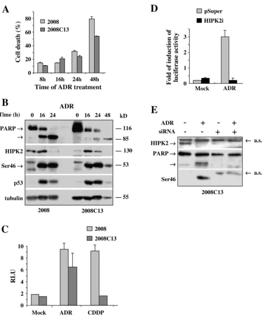
Fig. 2. P53Ser46 activity is not impaired in 2008C13 chemoresistant cells. (A) Time-course analysis of ADR treatment (2μg/ml) on cell viability in 2008 and 2008C13 cells. Trypan blue exclusion assay was performed to quantify cell viability at 8, 16, 24 and 48 h after treatment. The results are the mean of two independent experiments performed in triplicate. Standard deviation is indicated. (B) 2008 and 2008C13 cells were treated with 2μg/ml ADR for 16, 24, and 48 h, harvested and the expression of HIPK2, p53, p53Ser46, and PARP cleavage was determined by Western blotting with whole-cell lysates. Anti-tubulin was used as protein loading control. The protein molecular weights are indicated. (C) 2008 and 2008C13 cells were transfected with AIP1-luc reporter and treated with CDDP (5μg/ml) and ADR (2μg/ml) for 24 h. Relative Luciferase Activity (RLU) normalized to β-gal is shown. The shown data represent the mean±SD from three independent experiments performed in duplicate. (D) 2008C13 cells were transfected with pSUPER or pSUPER-HIPK2-interfering vectors and 48 h later transfected with AIP1-luc reporter and treated with ADR (2μg/ml) for 24 h. Luciferase activity normalized to β-gal is shown. The result is shown as fold of induction of relative luciferase activity. Data are representative of three independent experiments performed in duplicate. Standard deviation from the mean is indicated. (E) 2008C13 cells were transiently transfected with siRNA to knock-down HIPK2 and treated with ADR (2μg/ml for 24 h). The expression of HIPK2, p53Ser46 phosphorylation, and PARP cleavage was assessed by Western blotting with whole-cell lysates. Arrows indicate aspecific signals (a.s.).
The p53Ser46 pathway is not compromised in 2008C13
cisplatin-resistant cells
To evaluate whether 2008C13 cells were resistant to other
chemotherapeutic drugs, cells were treated with ADR that has
been shown to induce Ser46 phosphorylation and apoptosis
[20]
and to accumulate HIPK2 at protein level
[14]
. Moreover, we
have recently shown that ADR can induce apoptosis in 2008
cells and that HIPK2 knock-down inhibits this effect
[15]
. As
shown in
Fig. 2
A, ADR treatment induced cell death in 2008
cells and in a similar extent in 2008C13 cells. Immunoblot
analysis of 2008 and 2008C13 cell extracts treated with ADR
for 16, 24, and 48 h showed cleavage of the apoptotic marker
PARP from its 116 kDa to 85 kDa fragment in both cell lines,
although the 2008C13 cells showed a slightly slower kinetics;
it also showed p53Ser46 phosphorylation in both cell lines, and
HIPK2 downregulation concomitant to PARP cleavage
(
Fig. 2
B). These findings suggest that the 2008C13 cells were
sensitive to ADR with induction of HIPK2/p53Ser46 apoptotic
pathway.
The involvement of p53 function in drug-induced apoptosis
was then evaluated by luciferase assay using the AIP1 promoter
that is a specific target of p53Ser46 phosphorylation
[20]
. To this
end 2008 and 2008C13 cells were transfected with the AIP1-luc
vector and treated with CDDP and ADR. As shown in
Fig. 2
C,
the reporter activity of the AIP1 promoter was induced in 2008
cells after treatment with both drugs while it was induced in
2008C13 cells only following ADR treatment. To investigate
the role of HIPK2 in p53-induced AIP1 activation, a similar
luciferase assay was performed in 2008C13 cells depleted of
endogenous HIPK2 by transient transfection with pSUPER
vectors carrying HIPK2, or aspecific RNA-interfering
se-quences. A shown in
Fig. 2
D, the ADR-induced reporter activity
of the AIP1 promoter was inhibited by HIPK2 knock-down; in
agreement, PARP cleavage and Ser46 phosphorylation were not
detected in 2008C13 cells depleted of HIPK2 and treated with
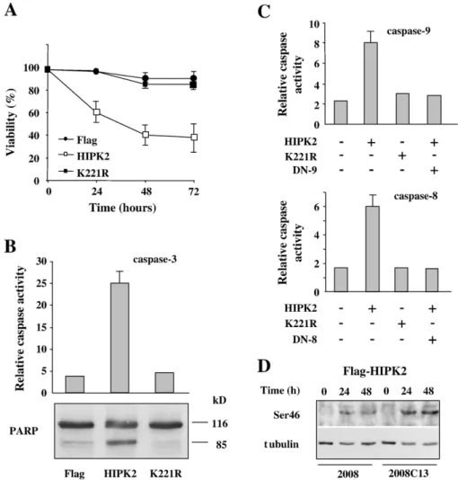
Fig. 3. HIPK2 overexpression induces apoptosis in 2008C13 chemoresistant cells. (A) 2008C13 cells were transfected with HIPK2, K221R, and Flag-empty expression vectors and cell viability was measured by trypan blue exclusion 24, 48, and 72 h post-transfection. The results shown are representative of two independent experiments performed in triplicate. Standard deviation is indicated. (B) The expression of caspases-3 and PARP cleavage as result of HIPK2 overexpression. 2008C13 cells were transfected as in (A) and 24 h later the caspase-3 activity was detected by fluorogenic assay (upper panel) and the expression of PARP cleavage was assessed by Western blotting of whole-cell lysates (lower panel). The uncleaved (116 kDa) and cleaved (85 kDa) forms of PARP are indicated. (C) 2008C13 cells were co-transfected with HIPK2 and dominant-negative expression vectors for caspases-8 (DN-8) and -9 (DN-8). Caspase-8 and -9 activities were detected by fluorogenic assay 24 h post-transfection. Data are representative of three independent experiments. Standard deviation is indicated. (D) The p53Ser46 phosphorylation after HIPK2 overexpression. 2008 and 2008C13 cells were transfected with HIPK2 and the expression of Ser46 phosphorylation was assessed 24 and 48 h after transfection by Western blotting with whole-cell lysates. Anti-tubulin was used for loading control. The results show representative bands from two independent experiments.
ADR (
Fig. 2
E). Similar results were obtained in 2008 cells (not
shown), as previously reported
[15]
. These experimental results
reveal that the p53Ser46 apoptotic pathway is not defective
in 2008C13 cells and that HIPK2 takes part in this
regula-tion following ADR treatment; they also suggest that HIPK2/
p53Ser46 deregulation is involved in chemoresistance.
HIPK2 overexpression induces p53Ser46 apoptotic pathway in
chemoresistant 2008C13 cells
To investigate the possibility to evade inhibition of apoptosis
and circumvent chemoresistance, exogenous HIPK2 was
over-expressed in 2008C13 cells. HIPK2 overexpression reduced cell
viability, compared to Flag-empty or K221R mutant expression
vectors (
Fig. 3
A). Similar results were obtained previously in
2008 cells
[15]
. Induction of apoptosis was evident after
overexpression of HIPK2 but not of K221R mutant, as assessed
by caspases-3 activity, a downstream effector of caspases-8 and -9
(
Fig. 3
B, upper panel), and PARP cleavage (
Fig. 3
B, lower panel).
The caspases' involvement was then evaluated by fluorogenic
assays of 2008C13 cells co-transfected with HIPK2 and K221R,
along with specific caspase-8 and caspase-9 dominant-negative
(DN) expression vectors. As shown in
Fig. 3
C, specific induction
of both caspase-8 and -9 activities was reached only after HIPK2
overexpression, and inhibited by specific dominant-negative
mutants co-expressed with HIPK2.
The involvement of p53 in HIPK2-mediated apoptosis was
evaluated by Western blot analysis and by luciferase assay. As
shown in
Fig. 3
D, p53Ser46 phosphorylation was comparably
induced in both 2008 and 2008C13 cells after HIPK2
over-expression. Luciferase assay showed that AIP1 promoter was
induced in 2008C13 cells only after HIPK2 overexpression,
compared to K221R mutant (
Fig. 4
A). Finally, a luciferase assay
was performed in 2008C13 cells transiently transfected with
AIP1-luc reporter and wtp53, or Ser46A (nonphosphorylatable
Ser46) and Ser46D (phosphorylation mimic) mutants. As shown
in
Fig. 4
B, the AIP1 promoter reporter activity was induced in a
bigger extent by Ser46D compared to wtp53 while the Ser46A
mutant did not induce AIP1 reporter activity. Altogether, these
results show that exogenous HIPK2 exerts p53Ser46-induced
apoptosis with involvement of both caspase-8 and -9 pathways,
no matter whether the cells are drug resistant or not.
Discussion
Apoptosis is the unique outcome that may lead to a successful
cancer therapy. Indeed, the failure to die in response to several
genotoxic and/or chemotherapeutic agents due to defects in one
or more components of the apoptotic pathway, is a determinant
of tumor cell resistance to antineoplastic treatments. Therefore,
genetic restoration of the apoptotic pathway or introduction of
proapoptotic molecules is an attractive approach for treating
cancers.
Resistance to p53-induced apoptosis is an important
phenotype of tumor cells expressing wtp53. We have previously
shown that HIPK2 regulates p53 oncosuppressor activity
through Ser46 phosphorylation and induction of apoptotic
target genes in response to genotoxic stress including that
induced by chemotherapeutic drugs
[9,11,15]
. In the present
study, we have found that HIPK2 is differently expressed at
protein level, in response to CDDP, in sensitive 2008 versus
chemoresistant 2008C13 cells with impairment of downstream
p53Ser46 phosphorylation. It has been reported that 2008C13
cells express a wild-type p53 that is necessary for the induction
of apoptosis
[22]
, however the presence of wild-type p53 does
not necessarily ensure a chemosensitive phenotype because
when p53 is not mutated, deregulated upstream molecules can
inhibit its function
[24–27]
. In this regard, it has been recently
found that HIPK2 cytoplasmic localization can inhibit p53
activation
[23]
. However we have found that HIPK2 was
mainly nuclear in both 2008 and 2008C13 cells, suggesting that
the impairment of HIPK2/p53Ser46 function in resistant
2008C13 cells following apoptotic dose of CDDP treatment,
does not depend on HIPK2 cytoplasmic relocalization. HIPK2
inactivation might instead depend on deregulation of upstream
molecules induced by different stimuli, as we have found that
HIPK2-mediated p53Ser46 activation was induced in sensitive
2008 cells by both CDDP and ADR while it was induced in
Fig. 4. HIPK2 overexpression induces p53Ser46-dependent AIP1 promoter. (A) 2008C13 cells were co-transfected with AIP1-luc reporter and HIPK2 or K221R kinase defective mutant. Relative Luciferase Activity (RLU) normalized to β-gal is shown. The shown data represent the mean ± SD from three independent experiments performed in duplicate. (B) 2008C13 cells were co-transfected with AIP1-luc reporter and wtp53, S46A, or S46D mutants. Relative Luciferase Activity (RLU) normalized toβ-gal activity is shown. Data are representative of three independent experiments performed in duplicate. Standard deviation from the mean is indicated.
2008C13 cells only by ADR. These findings suggest that the
HIPK2/p53Ser46 complex was not defective in 2008C13 cells
but rather it was deregulated by chemoresistance induced by
CDDP. How HIPK2 regulation is involved in chemoresistance
awaits further investigations. Thus, the existence of different
signalling pathways and regulatory proteins that finally lead to
HIPK2 activation in response to different signals is very poorly
understood. In this regard, recent findings showed that
IR-induced HIPK2 accumulation might be regulated by the ATM
pathway, and the authors suggest the existence of an essential
HIPK2 cofactor which is required to form an active p53Ser46
kinase complex
[13]
. Therefore, it is important to understand the
upstream role of individual as well as combinatorial
post-translational modifications or protein/protein interaction events
in regulating HIPK2 response that may be altered in acquired
chemoresistance to better understand the impact of HIPK2 on
p53 activity.
As mentioned, HIPK2-induced apoptosis is mainly due to
positive regulation of p53 function
[9
–13,15]
. Here we have
shown that overexpression of HIPK2 induced apoptosis in
chemoresistant ovarian cancer cells with activation of p53Ser46
target gene AIP1, supporting the HIPK2 potential application in
chemoresistant ovarian cancer therapy. It has been shown that
p53 phosphorylation at Ser46 is a late event after DNA damage
and is a necessary step for inducing irreversible apoptosis with
activation of a specific target gene such as AIP1
[20,28]
. It has
been shown that a defect in Ser46 phosphorylation contributes
to the acquisition of the p53 resistance in an oral squamous cell
carcinoma cell line
[29]
. Therefore, the lack of p53Ser46
phos-phorylation by deregulation of activating kinases may be
responsible of inhibition of p53-induced apoptosis and
devel-opment of chemoresistance. In this regard, we have previously
found that silencing of endogenous HIPK2 reduces p53Ser46
phosphorylation and p53 apoptotic function in response to
CDDP
[11]
and found here that HIPK2 knock-down reduced
p53Ser46-dependent apoptosis in response to ADR, indicating
that impaired HIPK2 function could contribute to the
develop-ment of chemoresistance. Further work is required to determine
the feasibility of using HIPK2 overexpression for treatment of
human ovarian cancer with wtp53. However, recent studies
have explored the possibility to treat tumors with p53Ser46
downstream mediators. Thus, if the p53 protein is insufficiently
modified in cancer cells due to alterations of phosphorylation
kinases a Ser46 mutant (Ser46-phenilalanine) or the
down-stream mediator AIP1 target gene offer the advantage of
en-hancing transcription of p53 target genes and induce apoptosis
[30,31]
.
In conclusion, the data presented here indicate that HIPK2/
p53Ser46 can be deregulated in chemoresistance, suggesting that
various stress stimuli activate distinct signalling pathways leading
to HIPK2 regulation that eventually activates p53 apoptotic
response. Our results also suggest that the use of exogenous
HIPK2 might be able to circumvent inhibition of apoptosis in
chemoresistant tumors that harbour wtp53. Collectively, our
findings might have important contributions for cancer treatment.
HIPK2 inhibition reduces the efficacy of chemotherapeutic agents
and inhibits p53Ser46 apoptotic pathway therefore HIPK2 might
be a potential target for gene therapy of chemoresistant ovarian
cancer with wtp53.
Conflict of interest statement
The authors have no conflicts of interest to declare.
Acknowledgments
This work was supported by Grants from Associazione Italiana
per la Ricerca sul Cancro (AIRC) and Ministero dell'Universita' e
Ricerca (Cofin-Miur). We thank Dr. A. Sacchi for her scientific
support.
References
[1] Borst P, Jonkers J, Rottenberg S. What makes tumors multidrug resistant? Cell Cycle 2007;6:2782–7.
[2] Fraser M, Leung BM, Yan X, Dan HC, Cheng JQ, Tsang BK. p53 is a determinant of X-linked inhibitor of apoptosis protein/Akt-mediated chemoresistance in human ovarian cancer cells. Cancer Res 2003;63:7081–8. [3] Siddik ZH. Cisplatin: mode of cytotoxic action and molecular basis of
resistance. Oncogene 2003;22:7265–79.
[4] Haupt S, Berger M, Goldberg Z, Haupt Y. Apoptosis—the p53 network. J Cell Sci 2003;116:4077–85.
[5] Xu Y. Regulation of p53 responses by post-translational modifications. Cell Death Differ 2003;10:400–3.
[6] Brooks CL, Gu W. Ubiquitination, phosphorylation and acetylation: the molecular basis for p53 regulation. Curr Opin Cell Biol 2003;2:164–71. [7] Giaccia AJ, Kastan MB. The complexity of p53 modulation: emerging
patterns from divergent signals. Genes Dev 1998;12:2973–83.
[8] Kim YH, Choi CY, Lee S, Conti MA, Kim Y. Homeodomain-interacting protein kinases, a novel family of co-repressors for homeodomain transcription factors. J Biol Chem 1998;273:875–9.
[9] D'Orazi G, Cecchinelli B, Bruno T, Manni I, Higashimoto Y, Saito S, et al. Homeodomain-interacting protein kinase-2 phosphorylates p53 at Ser46 and mediates apoptosis. Nat Cell Biol 2002;4:11–9.
[10] Hofmann TG, Moller A, Sirma H, Zentgraf H, Taya Y, Droge W, et al. Regulation of p53 activity by its interaction with homeodomain-interacting protein kinase-2. Nat Cell Biol 2002;4:1–10.
[11] Di Stefano V, Rinaldo C, Sacchi A, Soddu S, D'Orazi G. Homeodomain-interacting protein kinase-2 activity and p53 phosphorylation are critical events for cisplatin-mediated apoptosis. Exp Cell Res 2004;293:311–20. [12] Wesierska-Gadek J, Schmitz ML, Ranftler C. Roscovitine-activated HIP2
kinase induces phosphorylation of wtp53 at Ser-46 in human MCF-7 breast cancer cells. J Cell Biochem 2007;100:865–74.
[13] Dauth I, Kruger J, Hofmann TG. Homeodomain-interacting protein kinase 2 is the ionizing radiation-activated p53 serine 46 kinase and is regulated by ATM. Cancer Res 2007;67:2274–9.
[14] Choi DW, Seo Y-M, Kim E-A, Sung KS, Ahn JW, Park S-J, et al. Ubiquitination and degradation of homeodomain-interacting protein kinase 2 (HIPK2) by WD40-repeat/SOCS box protein WSB-1. J Biol Chem 2007,doi:10.1074/jbc.M708873200.
[15] Pistritto G, Puca R, Nardinocchi L, Sacchi A, D'Orazi G. HIPK2-induced p53Ser46 phosphorylation activates the KILLER/DR5-mediated caspase-8-extrinsic apoptotic pathway. Cell Death Differ 2007, doi:10.1038/sj. cdd.4402186.
[16] Gresko E, Roscic A, Vichalkovski S, del Sal G, Schmitz ML. Autoregulatory control of the p53 response by caspases-mediated processing of HIPK2. EMBO J 2006;25:1883–94.
[17] Harris SL, Levine AJ. The p53 pathway: positive and negative feedback loops. Oncogene 2005;24:2899–908.
[18] Andrews PA, Velury S, Mann SC, Howell SB. cis-Diamminedichloropla-tinum(II) accumulation in sensitive and resistant human ovarian carcinoma cells. Cancer Res 1998;48:68–73.
[19] Delmastro DA, Li J, Vaisman A, Solle M, Chaney SG. DNA damage inducible-gene expression following platinum treatment in human ovarian carcinoma cell lines. Cancer Chemother Pharmacol 1997;39:245–53. [20] Oda K, Arakawa H, Tanaka T, Matsuda K, Tanikawa C, Mori T, et al.
P53AIP1, a potential mediator of p53-dependent apoptosis, and its regulation by Ser-46-phosphorylated p53. Cell 2000;102:849–62. [21] Chen C, Okayama H. High-efficiency transformation of mammalian cells
by plasmid DNA. Mol Cell Biol 1987;7:2754–6.
[22] Yan X, Fraser M, Qiu Q, Tsang BK. Over-expression of PTEN sensitizes human ovarian cancer cells to cisplatin-induced apoptosis in a p53-dependent manner. Gynecol Oncol 2006;102:348–55.
[23] Pierantoni GM, Rinaldo C, Mottolese M, Di Benedetto A, Esposito F, Soddu S, Fusco A. High-mobility group A1 inhibits p53 by cytoplasmic relocalization of its proapoptotic activator HIPK2. J Clin Invest 2007;117:693–702. [24] Hollstein M, Sidransky D, Vogelstein B, Harris CC. P53 mutations in
human cancers. Science 1991;253:49–53.
[25] Greenblatt MS, Bennet WP, Hollstein M, Harris CC. Mutations in the p53 tumor suppressor gene: clues to cancer etiology and molecular pathogen-esis. Cancer Res 1994;54:4855–78.
[26] Hollstein M, Hergenhahn M, Yang Q, Bartsch H, Wang ZQ, Hainaut P. New approaches to understanding p53 gene tumor mutation spectra. Mutat Res 1999;432:199–209.
[27] Vousden KH, Prives C. P53 and prognosis: new insights and further complexity. Cell 2005;120:7–10.
[28] Mayo LD, Rok Seo Y, Jackson MW, Smith ML, Rivera Guzman JR, Koegaonkar CK, et al. Phosphorylation of human p53 at serine 46 determines promoter selection and whether apoptosis is attenuated or amplified. J Biol Chem 2005;280:25953–9.
[29] Ichwan SJA, Yamada S, Sumreijkanchanakij P, Ibrahim-Auerkari E, Eto K, Ikeda MA. Defect in serine 46 phosphorylation of p53 contributes to acquisition of p53 resistance in oral squamous cell carcinoma cells. Oncogene 2006;25:1216–24.
[30] Yoshida K, Monden M, Nakamura Y, Arakawa H. Adenovirus-mediated p53AIP1 gene transfer as a new strategy for treatment of p53-resistant tumors. Cancer Sci 2004;95:91–7.
[31] Nakamura Y, Futamura M, Kamino H, Yoshida K, Nakamura Y, Arakawa H. Identification of p53-46F as a super p53 with an enhanced ability to induce p53-dependent apoptosis. Cancer Sci 2006;97:633–41.