Abstract.
Aim: To evaluate, in human immunodeficiency
virus-hepatitis C virus co-infected patients, the impact of C-C
chemokine receptor type 5 (CCR5) antagonist
maraviroc-based antiretroviral therapy on the carotid intima media
thickness and on atheromasic plaques. Patients and Methods:
In this pilot prospective study, 12 HIV-HCV co-infected
patients underwent color-Doppler ultrasonography before and
48 weeks after switching to a dual therapy based on
maraviroc plus protease inhibitors boosted with ritonavir.
Changes of intima media thickness, inflammatory and
endothelial adhesion biomarkers levels, Veterans Aging
Cohort Study index and Framingham risk score were
evaluated. Results: At baseline 11 (91.6%) patients showed
pathological ultrasonographic findings. After 48 weeks, two
patients showed an amelioration of intima media thickness. Of
the remaining patients with plaques, four showed a reduction
of the previously diagnosed plaque; no patients worsened.
Conclusion: Our data suggest that CCR5 inhibition could
reduce the development of atherosclerosis especially in the
non-calcific stage and could play an important role in the
blockade of atheromasic plaque progression.
HIV CCR5 co-receptor is involved in HIV viral entry, but
has also a prominent role in many inflammatory pathways.
CCR5 regulates the migration of monocytes, natural killer
cells and T helper 1 (Th1) cells into sites of inflammation.
CCR5 expression on monocytes/macrophage and T-cells and
both endothelial and vascular smooth muscle cells suggests
a key role in the atherosclerotic process. Several studies
demonstrated that inhibition of this co-receptor produces an
anti-inflammatory and anti-atherogenic effect (1-5). A body
of evidence has documented that in patients with HIV,
atherosclerosis is accelerated and chronic inflammatory
processes are activated (6-9); furthermore, certain evidence
indicates that hepatitis C (HCV) co-infection can augment
the risk of cardiovascular events (10, 11). Moreover, hepatic
stellate cells, that play an important role in the pathogenesis
of liver fibrosis, express both HIV CCR5 and C-X-C
chemokine receptor type 4 (CXCR4) co-receptors. HIV-1
glycoprotein 120 (Gp120) binding to CXCR4 and CCR5
activates hepatic stellate cells, with a fibrogenic and
pro-inflammatory effect (12, 13).
The aim of our pilot prospective study was to evaluate, in
a group of HIV-HCV co-infected patients, the impact of a
CCR5 antagonist maraviroc (MVC)-based dual antiretroviral
therapy (ART) on cardiovascular risk assessed with echo
color-Doppler evaluation of the epi-aortic vessels,
inflammatory and adhesion cytokines dosage and risk scores
Veterans Aging Cohort Study (VACS) index and
Framingham risk score (FRS) in a 48-week follow-up. The
effects of a CCR5 antagonist-based ART on the progression
of liver fibrosis were also evaluated.
Patients and Methods
Twelve consecutive HIV-HCV co-infected outpatients attending the Institute of Infectious Diseases of the University of Bari, Italy, were enrolled in our study between January 2013 and April 2014, the follow-up was concluded in April 2015. Inclusion criteria were: stable ART and undetectable plasma HIV-1 RNA for more than 12 months and with the most recent undetectable HIV-RNA <1 month from baseline visit, R5 tropism ascertained on peripheral blood mononuclear cell DNA by V3 sequencing assessed until the previous 6 months and detectable serum HCV-RNA. Exclusion criteria were: negative HCV-RNA, positive hepatitis B surface antigen (HBsAg), detectable HIV-RNA, or alcohol abuse. All the patients were experiencing the following conditions requiring a shift from their nucleoside/nucleotide reverse transcriptase inhibitor (NRTIs) backbone therapy, as indicated by the current guidelines: nine had high cardiovascular risk (FRS more than 10% or carotid changes on echoic images), one had renal impairment (low estimated glomerular filtration, with proteinuria and hypophosphatemia) and two had bone loss (osteopenia or This article is freely accessible online.
Correspondence to: Paolo Maggi, Institute of Infectious Diseases,
University of Bari, piazza Giulio Cesare 11, 70125 Bari, Italy. Tel: +39 0805592134, Fax: +390805478333, e-mail: [email protected]
Key Words: CCR5, maraviroc, HIV, HCV, atheromasic plaques.
Effects of Therapy with Maraviroc on the Carotid Intima Media
Thickness in HIV-1/HCV Co-infected Patients
PAOLO MAGGI, GIUSEPPE BRUNO, FRANCESCO PERILLI, ANNALISA SARACINO,
ANNA VOLPE, CARMEN SANTORO, NICOLETTA LADISA and GIOACCHINO ANGARANO
osteoporosis). All the patients shifted to MVC plus protease inhibitors boosted with ritonavir (PI/r) from their current antiretroviral therapy (ART) based on three NRTIs, or two NRTIs plus a PI/r, or two NRTIs plus a non-NRTI. Immunovirological data were monitored after 1 month and then every 3 months. During the visits, the adherence to the therapy was verified. If patients were previously treated with a PI, the same PI was maintained after switching to MVC. All drugs were prescribed at standard doses.
Patients underwent echo color-Doppler ultrasonography before and 48 weeks after antiretroviral therapy shifting. Inflammatory and endothelial adhesion biomarkers levels interleukin-6 (IL6), high-sensitivity C-reactive protein (hs-CRP), D-dimers, intercellular adhesion molecule 1 (ICAM1) and vascular cell adhesion protein 1 (VCAM1) were evaluated at the same timepoints. VACS index and FRS were also calculated.
Inflammation and enhotelial biomarkers. Plasma D-dimers were
quantified by means of an enzyme-linked fluorescent assay (Vidas D-Dimer Exclusion II, Biomerieux Marcy-L’Etoile France; normal values: 0-500 ng/m) at the Central Laboratory of the University Policlinico Hospital of Bari. Hs-CRP (Biokit Quantex CRP, Netherlands; normal values: 0-300 ng/), IL-6 (Boster Biological Immunoleader, China; detection limit: 0.3 pg/ml, assay range: 4.69-300 pg/ml), ICAM1 and VCAM1 (Boster Biological Immunoleader; detection limit: 10 pg/ml, assay range: 156-10,000 pg/ml) were measured at the Infectious Diseases Laboratory of the University Policlinico Hospital of Bari by commercially available assays.
GP120 sequencing and co-receptor tropism CRT assignment.
HIV-DNA was extracted and amplified from peripheral blood mononuclear cell and GP120 sequencing on proviral DNA was performed as previously described (14). CRT was inferred with the geno2pheno [co-receptor] algorithm (http://co-receptor.bioinf.mpi-inf.mpg.de/), setting the false-positive rate at 10% to classify isolates as R5.
IMT and plaque assessment. The primary outcome measure was the
evaluation and description of any eventual changes of carotid intima
media thickness (IMT). Ultrasonography of the epi-aortic vessels
was performed using a power color-Doppler instrument with 7.5 mgHz probes (Esaote Biomedica, Genoa, Italy). IMT changes were studied considering the following measurement sites: common carotid: 1 cm before the carotid bifurcation, at carotid bifurcation; internal carotid: 1 cm after the carotid bifurcation. Characteristics of the intima, pulsation index, resistance index, minimal speed, peak speed, and mean speed, were evaluated. An IMT of more than 1 mm were considered to be pathological. Atherosclerotic plaques, if present, were described. A carotid was classified as being affected by plaque if there was a localized thickening >1.2 mm that did not uniformly involve the whole left or right common carotid bifurcation with or without flow disturbance (15, 16).
In order to better evaluate the characteristics of the plaques that showed improvements with respect to those that remained unmodified, we applied a method already described in other issues to distinguish inflammatory from atheromasic patterns (17, 18). In brief, the following ultrasound color-Doppler characteristics were examined: echogenicity of the lesion with respect to the vessel wall; presence of acoustic shadows inside the lesion; homogeneity of the lesion wall; profile of the endoluminal surface; and the presence or absence of a cleavage plain. Characterization of lesions was consistent with methods describing echogenicity and plaque surface
appearance previously established by an International Consensus Conference concerning plaque morphology and risk (19).
An iso-hypoechogenic structure, a homogeneous wall, a smooth endoluminal surface, and the absence of a cleavage plain were considered as characteristics of an inflammatory pattern. In contrast, a hyperechogenic structure, the presence of shadow cones, an irregular endoluminal surface and a cleavage plain were considered characteristics of atheromasic plaques.
The ultrasonographic examination were performed by a single physician (PM). All the images were revised by FP, who was blinded to the patient’s treatment history and status. Both the physicians had more than 10 year’s experience in the ultrasound color-Doppler technique and had carried at least 1,000 documented epi-aortic examinations.
Liver fibrosis assessment. Liver stiffness was evaluated by a single
certified operator using transient elastography (FibroScan®;
Echo-Sens, Paris, France). Liver fibrosis was also determined using FIB-4 as surrogate marker. FIB-FIB-4 was calculated using Sterling’s formula, as follows: age (years) × AST (IU/l)/platelet count (expressed as platelets ×109/l]× [ALT1/2(IU/l).
Statistical methods. Descriptive statistics were produced for all
variables. Chi-square or Fisher’s exact test and t-test for unpaired samples were used for statistical analysis. A p-value of less than 0.05 was considered significant.
Ethic considerations. Written informed consent was obtained from
all patients. The local Ethics Committee (Comitato Etico Indipendente Locale – Azienda Ospedaliera Ospedale Policlinico Consorziale Bari – Italy) approved the study, within the PREVALEAT cohort study. PREVALEAT (PREmature VAscular LEsions and Antiretroviral Therapy) is a multicenter, longitudinal cohort study aimed at evaluation of cardiovascular risk in HIV-infected patients since 2004. All procedures were in accordance with the ethical standards of the institutional and National Research Committee and with the 1964 Helsinki declaration and its later amendments or comparable ethical standards.
All images of the echo-color Doppler were photographed and properly archived, together with all the other data in the archives of the Institute.
Results
The clinical characteristics at baseline are summarized in Table
I. The median age was 50 year. Among the 12 patients
enrolled, 11 (92%) were males, nine (75%) smoked and five
(41.7%) had high blood pressure. All patients had a long
history of HIV and HCV, respectively a median of 25 and 20
years from the first HIV and HCV testing. Median ART
exposure was 15.5 years. Median time with HIV suppression
was 4.5 years. Only one patients was being treated with
rosuvastatin and acetylsalicylic acid. No patient had diabetes.
At baseline, 11 (91.6%) patients showed pathological
ultrasonographic findings; of these, two had abnormal IMT and
nine had plaques. Findings for one patient were normal. Before
shifting to MVC plus PI/r, 10 patients (83%) were receiving
two NRTIs plus PI/r, one patient two NRTIs plus non-NRTI
and another one three NRTIs. Antiretroviral treatment and
switching to MVC plus PI/r are described in Table II.
After 48 weeks of MVC-based therapy, the patient with
normal features remained normal; the two patients with
abnormal IMT (maximum observed values 1.1 and 1 mm,
respectively) showed an amelioration (to 0.9 and 0.76 mm,
respectively). Of the remaining nine patients with plaques,
four (44%) showed a reduction of the previously diagnosed
atheromasic plaque; five (56%) did not show any
modification of their baseline ultrasonographic findings; no
patients worsened.
Among the nine smokers, eight had plaques at baseline.
At follow-up, four (50%) showed an improvement at
ultrasonographic examination. Four out of the five patients
with high blood pressure had plaques at baseline but only
one patient had an improvement at follow-up. The plaque
pattern patient being treated with rosuvastatin and
acetylsalicylic acid did not change.
The relationship between outcome of the plaques and their
characteristics is described in Table III. No significant changes
in inflammatory and endothelial adhesion biomarkers, VACS
index and FRS were detected, as shown in Table IV. Before
shifting to MVC plus PI/r among the four patients with plaques
who showed an improvement at 48 weeks, two patients were
receiving abacavir/lamivudine with PI/r (lopinavir/r and
atazanavir/r), while the other two patients were receiving
tenofovir/emtricitabine with darunavir/r once daily.
Liver stiffness. At baseline, one patient had no/mild fibrosis
(F0-F1), an F2-F3 stage of fibrosis was observed in eight and
a F4 stage in three. After 48 weeks of treatment, three
patients (25%) showed an improvement in liver stiffness,
whereas in nine (75%) the fibrosis grade using Fibroscan did
not change. Median liver stiffness decreased from 9.2 kPa
(interquartile range, IQR=8.1-13.7) at baseline to 7.6 kPa
(IQR=6.8-11.4) after 48 weeks without achieving statistical
significance. No change was observed between median FIB4
value at baseline [1.69 (IQR=1.1-2.8)] and after a follow-up
of 48 weeks [1.6 (IQR=1.0-2.8)].
Immunovirological outcome. One patient experienced an
HIV virological failure (350 copies of HIV-RNA) after 6
months of treatment with MVC plus atazanavir/r and it was
necessary to strengthen the therapy adding tenofovir/
emtricitabine. In the following period, HIV remained
suppressed and no ultrasonographic changes were observed.
All the other patients remained aviremic during the whole
period of follow-up.
Discussion
In HIV patients, atherosclerosis is accelerated and chronic
inflammatory processes are activated. Cardiovascular disease
is one of the most common non-acquired immune deficiency
syndrome events, with overall increased morbidity and
mortality (20). Likewise a large number of studies suggest
an increased risk of cardiovascular disease events in
HCV-monoinfected and HIV-HCV co-infected patients compared
with uninfected controls (21-24). The risk of cardiovascular
disase in HIV-HCV-positive patients is due to several
mechanisms: aging, insulin resistance, hepatic steatosis,
chronic inflammation, microbial translocation and immune
activation (25).
HIV CCR5 co-receptor, which plays a crucial role in HIV
viral entry, is also involved in many inflammatory pathways
(1-5). MVC is the only available CCR5 antagonist currently used
in clinical practice for treating HIV infection. The blockade of
CCR5 can have an important effect on plaque progression and
systemic inflammation as previously described (1).
In a recent study, Cipriani et al. demonstrated that in a
murine model of genetic dyslipidemia, MVC reduced
Table I. Baseline characteristics of the study population (n=12).Characteristic Value Age, years 50 (48-52) Male gender 11 (92%) CDC clinical stage A 4 (33.4%) B 7 (58.4%) C 1 (8.2%) Source of HIV infection
Intravenous drug user 11 (92%) Other 1 (8%) Source of HCV infection
Intravenous drug user 11 (92%) Other 1 (8%) Nadir CD4, cells/ml 198 (98.2-271) CD4 cell count, cells/ml 660 (579.7-931.7) Time since HIV diagnosis (years) 25 (21-27) Time since HCV diagnosis (years) 20 (15.7-21.7) Time on ART (years) 15.5 (8.5-18.7) Time with HIV viral suppression (years) 4.5 (3-8.7) HCV RNA, IU×106/ml 2.1245 (1.426-5.25075)
HCV genotype 1 7 (58.4%) 3 2 (16.6%) 4 3 (25%) Previous history of failure
with anti-HCV therapy 6 (50%) Smoker 9 (75%) High blood pressure 5 (41.7%) Use of statins 1 (9%) Use of acetylsalicylic acid 1 (9%) CDC: Centre for disease control and prevention; HIV: human immunodeficiency virus; HCV: hepatitis C virus; ART: antiretroviral therapy; CD4: CD4 cells. All values are expressed as median and interquartile range or number (percentage), unless otherwise specified.
Table III. Baseline characteristics of carotid intima media thickness (IMT) and plaques, and outcome at follow-up in nine patients with pathological
ultrasonography.
Patient no. Iso-hypoechogenicity Homogeneity Smooth surface Cleavage plain Shadow cones After 48-week follow-up 1 − − − + + No improvement 2 − − − + − No improvement 3 − − − + − No improvement 5 + + + − − Improvement 6 + + + − − Improvement 7 − − − + − No improvement 10 + + + − − Improvement 11 + + + − − Improvement 12 − − − + + No improvement +: Present; −: absent.
Table II. Antiretroviral therapy (ART) of each patient at the time of switching and ultrasonographic outcome.
Ultrasonography
Patient no. Pre-switch ART Study ART FPR at baseline (%) At baseline Improvement 1 3TC/ABC+ATV/r MVC+ATV/r 45.1 Pathological No 2 TDF/FTC+EFV MCV+ATV/r 94.5 Pathological No 3 3TC+ABC+AZT MVC+DRV/r 48.7 Pathological No 4 TDF/FTC+LPV/r MVC+LPV/r 29.8 Normal No 5 3TC/ABC+ATV/r MVC+ATV/r 30.1 Pathological Yes 6 3TC/ABC+LPV/r MVC+LPV/r 39.6 Pathological Yes 7 3TC/ABC+ATV/r MVC+ATV/r 95.5 Pathological No 8 TDF/FTC+DRV/r MCV+DRV/r 33.2 Abnormal IMT No 9 3TC/ABC+LPV/r MVC+LPV/r 46.5 Abnormal IMT No 10 TDF/FTC+ DRV/r MVC+DRV/r 13.5 Pathological Yes 11 TDF/FTC+DRV/r MVC+DRV/r 59.2 Pathological Yes 12 TDF/FTC +LPV/r MVC+LPV/r 35.3 Pathological No 3TC: Lamivudine; ABC: abacavir; ATV: atazanavir; DRV: darunavir; FPR: false-positive rate; FTC: emtricitabine; IMT: intima media thickness; LPV: lopinavir; MVC: maraviroc; r: ritonavir; TDF: tenofovir.
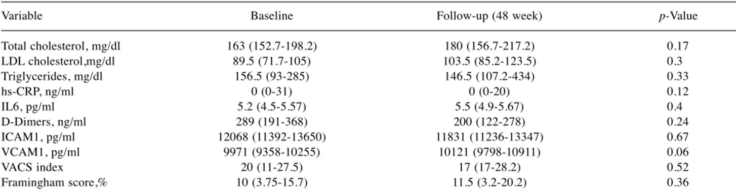
Table IV. Modification of serum levels of metabolic, immunological and inflammatory biomarkers at baseline and after a follow-up of 48 weeks. Variable Baseline Follow-up (48 week) p-Value Total cholesterol, mg/dl 163 (152.7-198.2) 180 (156.7-217.2) 0.17 LDL cholesterol,mg/dl 89.5 (71.7-105) 103.5 (85.2-123.5) 0.3 Triglycerides, mg/dl 156.5 (93-285) 146.5 (107.2-434) 0.33 hs-CRP, ng/ml 0 (0-31) 0 (0-20) 0.12 IL6, pg/ml 5.2 (4.5-5.57) 5.5 (4.9-5.67) 0.4 D-Dimers, ng/ml 289 (191-368) 200 (122-278) 0.24 ICAM1, pg/ml 12068 (11392-13650) 11831 (11236-13347) 0.67 VCAM1, pg/ml 9971 (9358-10255) 10121 (9798-10911) 0.06 VACS index 20 (11-27.5) 17 (17-28.2) 0.52 Framingham score,% 10 (3.75-15.7) 11.5 (3.2-20.2) 0.36 LDL: Low-density lipoprotein; hs-CRP: high-sensitivity C-reactive protein; IL6: interleukin-6; ICAM1: intercellular adhesion molecule 1; VCAM1: vascular cell adhesion protein 1; VACS index: Veterans Aging Cohort Study index. Data are median (interquartile range) values.
atherosclerosis progression by lowering macrophage
infiltration and expression of adhesion molecules and
RANTES (regulated on activaction, normal T-cell expressed,
and secreted) inside the plaques and reversed the
proinflammatory profile in a mouse model characterized by a
ritonavir–induced inflammation (1).
In the present study, the potential benefit of MVC on
atherosclerosis progression and on the inflammatory profile
was evaluated in a group of HIV-HCV co-infected patients
who were shifted to a MVC plus PI/r dual therapy from their
triple ART. Our data revealed that a number of patients
shifted to a MVC plus PI/r regimen showed an improvement
of their baseline carotid lesions: both the patients with
pathological IMT findings showed an amelioration, as did
4/9 (44%) of the patients with plaques.
No difference in impact of the previous ART was observed
between patients who achieved an improvement. Of the four
patients with improved plaques, two patients were receiving
abacavir/lamivudine with PI/r (lopinavir/r and atazanavir/r)
at baseline, while the other two patients were receiving
tenofovir/emtricitabine with darunavir/r once a day.
However, because of the small number of patients, a possible
role of the prior ART in those achieving an improvement of
the atherosclerotic plaque cannot be definitely excluded. In
fact, after 48 weeks, an increase of low-density lipoprotein
cholesterol was observed however, without, a statistical
significance. This might be explained by the loss of
lipid-lowering effect observed with tenofovir but also by a slight
improvement in liver function. Only one patient with plaque
was treated with rosuvastatin and acetylsalicylic acid, but no
improvement was evident.
Aging in HIV-positive patients is associated with an
increased frequency of non-HIV-related comorbidities. This
appears to be related to chronic inflammation, immune
activation and immunosenescence (26). Furthermore levels
of inflammatory and immune activation such as IL6, hs-CPR
and D-dimer are associated with increased risk for
non-HIV-related events such as cardiovascuar disease and
atherosclerosis progression, and all-cause mortality (20). All
patients in our study had a long history of HIV and HCV
infection, as shown in Table I, but they also had a long
period of HIV viral suppression. For this reason, it is likely
that the long period of HIV undetectability and the
persistence of viral suppression during the period of
observation did not show an impact on changes of
inflammation, endothelial dysfunction and immune
activation biomarkers.
Nine patients were smokers and eight had plaques at
baseline. At follow-up, four (50%) showed an improvement
at ultrasonographic examination. Undiagnosed high blood
pressure was found in five patients, four of them had plaques
at baseline but only one patient had an improvement,
highlighting the importance of a periodic proper control of
blood pressure. Only one patient with arterial hypertension
started anti-hypertensive therapy (ramipril), however no
modification in ultrasonographic finding was observed at
follow-up.
Among the four patients with improved plaques, two
showed an improvement in liver stiffness and in two there
was no modification in their fibrosis grade using Fibroscan.
The possible association between progression of
atherosclerosis and liver stiffness modification is worthy of
future investigation.
Even if no worsening was registered in our study, only
plaques with specific characteristics such as
iso-hypoechogenic structure, smooth endoluminal surface and
absence of a cleavage plain had an improvement, as showed
in Table III. These characteristics of the ultrasound images
are due to the inflammatory aspect of the lesions:
inflammatory plaques are mainly composed of watery and
cellular content. On the other hand, the atheromasic plaque is
a phenomenon involving only the endothelium and the
luminal portion of the intima, thus accounting for the typical
presence of a cleavage plain between the lesion and the
underlying tissues. In contrast, a higher degree of
echogenicity is proportional to the fiber content and shadow
cones are due to the presence of calcifications. Moreover, the
endoluminal surface of a typical atheromasic plaque is
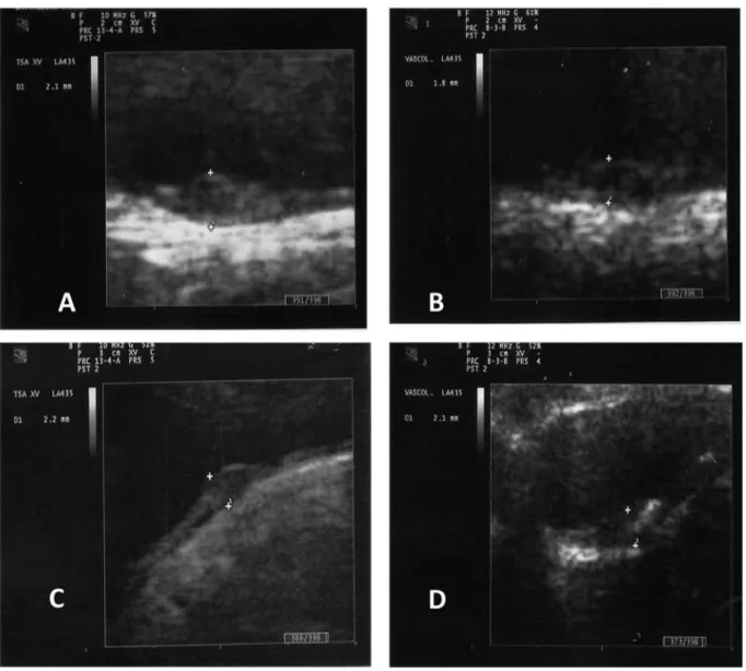
generally irregular (27). Two examples of plaques are shown:
at baseline, the first patient had a plaque with an
inflammatory aspect (Figure 1A) which had improved at
follow-up (Figure 1B); conversely, the second patient had a
plaque with atheromasic characteristics at baseline (Figure
1C) that did not show improvement at follow-up (Figure 1D).
This evidence suggests that CCR5 inhibition might reduce
the development of atherosclerosis, especially in the
inflammatory and non-calcific stage, and could play a role
in the blockade of atheromasic plaque progression. Of note,
the amelioration of plaques is a phenomenon that does not
involve the classic risk factors, as documented by VACS
index and FRS, which remained unchanged. For this reason,
we could hypothesize a direct effect of the pharmacological
CCR5 abrogation not mediated by other inflammatory or
endothelial adhesion biomarkers, which did not show any
modification during the study period. However, the role of
an NRTI-sparing dual regimen in ameliorating vascular
lesions cannot be excluded and further investigations to
clarify this point are warranted.
The main limitation of our study is the small number of
patients enrolled. This was also a single-arm study and there
was no control group. It would be interesting in further
investigations to compare our end-points in patients who
switch to an MVC-based regimen to those who do not switch
in a larger population for a longer time. To the best of our
knowledge, even if the potential activity of MVC in reducing
the progression of plaque has been already described (1) and a
recent study of Piconi et al. showed that MVC reduces arterial
stiffness in a patients with similar characteristics (28), this is
the first study evaluating the impact of MVC on the IMT and
on the progression of plaque using echo color-Doppler
ultrasonography in a clinical practice setting. An evaluation on
a larger population is warranted to confirm our observations.
References
1 Cipriani S, Francisci D, Mencarelli A, Renga B, Schiaroli E, D’Amore C, Baldelli F and Fiorucci S: Efficacy of the CCR5 antagonist maraviroc in reducing early, ritonavir-induced atherogenesis and advanced plaque progression in mice. Circulation 127: 2114-2124, 2013.
2 Maguire JJ, Jones KL, Kuc RE, Clarke MC, Bennett MR and Davenport AP: The CCR5 chemokine receptor mediates vasoconstriction and stimulates intimal hyperplasia in human vessels in vitro. Cardiovasc Res 101: 513-521, 2014.
3 Combadière C, Potteaux S, Rodero M, Simon T, Pezard A, Esposito B, Merval R, Proudfoot A, Tedgui A and Mallat Z: Combined inhibition of CCL2, CX3CR1, and CCR5 abrogates Ly6C(hi) and Ly6C(lo) monocytosis and almost abolishes atherosclerosis in hypercholesterolemic mice. Circulation 117: 1649-1657, 2008.
4 Afzal AR, Kiechl S, Daryani YP, Weerasinghe A, Zhang Y, Reindl M, Mayr A, Weger S, Xu Q and Willeit J: Common CCR5-del32 frameshift mutation associated with serum levels of inflammatory markers and cardiovascular disease risk in the Bruneck population. Stroke 39: 1972-1978, 2008.
Figure 1. Two examples of plaque evolution from baseline to follow-up. Patient at baseline (A) and improvement after 48 weeks (B). Patient at
5 Muntinghe FL, Verduijn M, Zuurman MW, Grootendorst DC, Carrero JJ, Qureshi AR, Luttropp K, Nordfors L, Lindholm B, Brandenburg V, Schalling M, Stenvinkel P, Boeschoten EW, Krediet RT, Navis G and Dekker FW: CCR5 deletion protects against inflammation-associated mortality in dialysis patients. J Am Soc Nephrol 20: 1641-1649, 2009.
6 Freiberg MS, Chang CC, Kuller LH, Skanderson M, Lowy E, Kraemer KL, Butt AA, Bidwell Goetz M, Leaf D, Oursler KA, Rimland D, Rodriguez Barradas M, Brown S, Gibert C, McGinnis K, Crothers K, Sico J, Crane H, Warner A, Gottlieb S, Gottdiener J, Tracy RP, Budoff M, Watson C, Armah KA, Doebler D, Bryant K and Justice AC: HIV infection and the risk of acute myocardial infarction. JAMA Intern Med 173: 614-622, 2013.
7 Durand M, Sheehy O, Baril JG, Lelorier J and Tremblay CL: Association between HIV infection, antiretroviral therapy, and risk of acute myocardial infarction: a cohort and nested case-control study using Québec’s public health insurance database. J Acquir Immune Defic Syndr 57: 245-253, 2011.
8 Friis-Møller N, Thiébaut R, Reiss P, Weber R, Monforte AD, De Wit S, El-Sadr W, Fontas E, Worm S, Kirk O, Phillips A, Sabin CA, Lundgren JD, Law MG; DAD Study Group: Predicting the risk of cardiovascular disease in HIV-infected patients: the data collection on adverse effects of anti-HIV drugs study. Eur J Cardiovasc Prev Rehabil 17: 491-501, 2010.
9 Hsue PY, Deeks SG and Hunt PW: Immunologic basis of cardiovascular disease in HIV-infected adults. J Infect Dis
205(Suppl 3): S375-382, 2012.
10 Fernández-Montero JV, Barreiro P, de Mendoza C, Labarga P and Soriano V: Hepatitis C virus coinfection independently increases the risk of cardiovascular disease in HIV-positive patients. J Viral Hepat 23: 47-52, 2015.
11 McKibben RA, Haberlen SA, Post WS, Brown TT, Budoff M, Witt MD, Kingsley LA, Palella FJ Jr., Thio CL and Seaberg EC: A Cross-sectional Study of the Association Between Chronic Hepatitis C Virus Infection and Subclinical Coronary Atherosclerosis Among Participants in the Multicenter AIDS Cohort Study. J Infect Dis 213: 257-265, 2015.
12 Hong F, Tuyama A, Lee TF, Loke J, Agarwal R, Cheng X, Garg A, Fiel MI, Schwartz M, Walewski J, Branch A, Schecter AD and Bansal MB: Hepatic stellate cells express functional CXCR4: role in stromal cell-derived factor-1alpha-mediated stellate cell activation. Hepatology 49: 2055-2067, 2009. 13 Bruno R, Galastri S, Sacchi P, Cima S, Caligiuri A, DeFranco
R, Milani S, Gessani S, Fantuzzi L, Liotta F, Frosali F, Antonucci G, Pinzani M and Marra F: gp120 modulates the biology of human hepatic stellate cells: a link between HIV infection and liver fibrogenesis. Gut 59: 51320, 2010.
14 Monno L, Saracino A, Scudeller L, Punzi G, Brindicci G, Altamura M, Lagioia A, Ladisa N and Angarano G: Impact of mutations outside the V3 region on co-receptor tropism phenotypically assessed in patients infected with HIV-1 subtype B. Antimicrob Agents Chemother 55: 5078-5084, 2011. 15 Belcaro G, Nicolaides AN, Laurora G, Cesarone MR, De Sanctis
M, Incandela L and Barsotti A: Ultrasound morphology classification of the arterial wall and cardiovascular events in a 6-year follow up study. Arterioscler Thromb Vasc Biol 16: 851-856, 1996.
16 Nicolaides AN, Shifrin EG, Bradbury A, Dhanjil S, Griffin M, Belcaro G and Williams M: Angiographic and duplex grading of internal carotid stenosis: can we overcome the confusion? J Endovasc Surg 3: 158-165, 1996.
17 Maggi P, Perilli F, Lillo A, Carito V, Epifani G, Bellacosa C, Pastore G and Regina G: An ultrasound-based comparative study on carotid plaques in HIV-positive patients vs atherosclerotic and arteritis patients: atherosclerotic or inflammatory lesions? Coronary Artery Disease 18: 23-29, 2007.
18 Maggi P, Perilli F, Lillo A, Volpe A, Pastore G and Regina G: B-Mode ultrasound study of carotid plaques in HIV+ patients for detection of inflammatory endothelial lesions. Current HIV Research 7: 541-546, 2009.
19 De Bray JM, Baud JM and Duzat M: Consensus concerning the morphology and the risk of carotid plaques. Cerebrovasc Dis 7: 289-296, 1997.
20 Duprez DA, Neuhaus J, Kuller LH, Tracy R, Belloso W, De Wit S, Drummond F, Lane HC, Ledergerber B, Lundgren J, Nixon D, Paton NI, Prineas RJ, Neaton JD; INSIGHT SMART Study Group: Inflammation, Coagulation and Cardiovascular Disease in HIV-Infected Individuals. PLoS ONE 7(9): e44454, 2012. 21 Butt AA, Xiaoqiang W, Budoff M, Leaf D, Kuller LH and
Justice AC: Hepatitis C virus infection and the risk of coronary disease. Clin Infect Dis 49: 225-232, 2009.
22 Freiberg MS, Chang CC, Skanderson M, McGinnis K, Kuller LH, Kraemer K, Rimland D, Goetz MB, Butt AA, Rodriguez Barradas MC, Gibert C, Leaf D, Brown ST, Samet J, Kazis L, Bryant K, Justice AC; Veterans Aging Cohort Study: The risk of incident coronary heart disease among veterans with and without HIV and hepatitis C. Circ Cardiovasc Qual Outcomes 4: 425-432, 2011.
23 Tsui JI, Whooley MA, Monto A, Seal K, Tien PC and Shlipak M: Association of hepatitis C virus seropositivity with inflammatory markers and heart failure in persons with coronary heart disease: data from the Heart and Soul study. J Card Fail
15: 451-456, 2009.
24 Liao CC, Su TC, Sung FC, Chou WH and Chen TL: Does hepatitis C virus infection increase risk for stroke? A population-based cohort study. PLoS ONE 7(2): e31527, 2012.
25 Ampuero J and Romero-Gómez M: Assessing cardiovascular risk in hepatitis C: An unmet need. World J Hepatol 7: 2214-2219, 2015.
26 Deeks SG: HIV infection, inflammation, immunosenescence, and aging. Annu Rev Med 62: 141-155, 2011.
27 De Brai JM and Glatt B: Quantification of atheromatous stenosis in the extracranial internal carotid artery. Cerebrovasc Dis 5: 414-426, 1995.
28 Piconi S, Pocaterra D, Rainone V, Cossu M, Masetti M, Rizzardini G, Clerici M and Trabattoni D: Maraviroc reduces arterial stiffness in pi-treated hiv-infected patients. Sci Rep 6: 28853, 2016.