Fantoni’s translaryngeal tracheotomy complications.
Personal experience
Complicanze della tracheotomia translaringea secondo Fantoni.
Esperienza personale
G. NERI, D. ANGELUCCI1, O. LEONE, R. ORTORE, A. CROCE
Department of Experimental and Clinical Surgical Sciences, Otorhinolaryngology Section, “G. D’Annunzio” University, Chieti; 1Pathology Institute, “SS. Annunziata” Hospital, Chieti, Italy
Key word Tracheotomy • Percutaneous tracheotomy • Complications • Surgical treatment
Parole chiave
Tracheotomia • Tracheotomia percutanea • Complicanze • Trattamento chirurgico
Summary
Tracheotomy is a surgical procedure which, in conditions of acute respiratory emergency, guarantees an adequate airway through the trachea whereas, in cases of chronic respiratory failure, it is used to improve ventilation through the reduction of the dead respiratory space. Over the last few years, surgical techniques used in tracheotomy have been considerably modi-fied, not only to respond to the needs of clinical indications but also on account of problems related to management of the pa-tient and tracheostomy tube, particularly in the home setting. Besides traditional surgical techniques, in fact, in the Intensive Care Unit, percutaneous dilatative procedures are being used with increasing frequency, in particular, translaryngeal tra-cheotomy according to Fantoni. The latter, however, according to reports in the literature, has been shown to be followed by a higher peri-operative complication rate (40%) which involves maintenance of good function of the tracheostomy, a condition which is particularly dangerous in the management of patients in the home setting. Personal experience is described in the management of 6 patients submitted to tracheotomy according to Fantoni and in combined home treatment, who, some time af-ter the operation, presented ‘embedding’ of the tracheostomy tube in the tracheostomy opening. The six patients were treated at home with ventilatory support using automatic ventilation system and were submitted, in our Clinic, to a surgical review with preparation of a tracheotomy according to the convention-al method. Our experience showed a particular feature of the difficulty in the management of patients presenting respiratory diseases, submitted to translaryngeal tracheotomy and, there-after, maintained in combined home treatment: in these sub-jects, in fact, the presence of the tube, the difficulty in cleaning the peristomial skin, the reduced autonomy from the automatic ventilation system and the frequent coexistence of mucopuru-lent tracheo-bronchial inflammatory diseases, trigger micro-le-sions of the stoma and, therefore, scar keloid, narrowing of the lumen and embedding of the tube itself. In conclusion, in our personal experience, we are of the opinion that translaryngeal tracheotomy, since it is easily carried out and is a slightly inva-sive procedure, plays a very important role in the management of the Intensive Care Unit patient but should be reserved for the few cases requiring tracheostomy for limited periods of time, in low risk patients and within the first 18 days after the acute damaging event.
Riassunto
La tracheotomia è una tecnica chirurgica che in condizioni di emergenza respiratoria acuta garantisce la pervietà tracheale mentre nei casi d’insufficienza respiratoria cronica è utiliz-zata per migliorare la ventilazione attraverso la riduzione dello spazio morto respiratorio. Nel corso degli ultimi anni le tecniche chirurgiche di tracheotomia hanno subito notevoli modifiche, sia per far fronte all’estensione delle indicazioni cliniche, sia a causa dei problemi legati alla gestione del paziente e della cannula, soprattutto in sede domiciliare. Accanto alle tecniche chirurgiche tradizionali (TT) infatti, nei reparti di Terapia Intensiva sono utilizzate sempre più di frequente le tecniche dilatative percutanee (TP) ed in parti-colare la tracheotomia translaringea (TLT) secondo Fantoni. Quest’ultima però, da quanto risulta dalla letteratura, è gravata da una più alta percentuale di complicanze periop-eratorie (40%) compromettenti il mantenimento di una buona pervietà della tracheostomia, condizione particolarmente peri-colosa nella gestione domiciliare del paziente. Gli Autori ripor-tano l’esperienza relativa a 6 pazienti, tracheotomizzati con la metodica di Fantoni e trattati in assistenza domiciliare inte-grata, che hanno presentato, a distanza di poco tempo dall’in-tervento, un “incarceramento” della cannula nel tramite tracheostomico. I 6 pazienti erano trattati a domicilio con ventilazione assistita mediante respiratore automatico e sono stati sottoposti a revisione chirurgica con il confezionamento di una tracheotomia eseguita con metodica tradizionale. La nostra esperienza documenta un particolare aspetto della diffi-coltà di gestione del paziente affetto da patologia respiratoria, trattato con TLT e successivamente mantenuto in assistenza domiciliare integrata: in questi soggetti infatti la presenza stessa della cannula, la difficoltà di pulizia della cute peris-tomiale, la ridotta autonomia dal respiratore automatico e la frequente coesistenza di affezioni flogistiche tracheo-bronchiali mucopurulente, favoriscono microlesioni della stomia e, di conseguenza, retrazioni cicatriziali, restringimento del lume ed incarceramento della cannula stessa. In conclusione, ci sembra di poter affermare che la TLT, per la semplicità di esecuzione e la scarsa invasività riveste un ruolo molto impor-tante nel management del paziente in terapia intensiva, ma andrebbe riservata ai soli casi che necessitano di tracheostomia per periodi di tempo limitati, in pazienti a basso rischio ed entro i primi 18 giorni dall’evento lesivo acuto.
Introduction
Tracheotomy, as is well known, is a surgical proce-dure in which an opening is made in the anterior wall of the trachea, which is sufficiently large to permit the passage of air1.
This may be necessary in cases of real emergency as, for instance, in acute respiratory insufficiency1-3, or
may be used as an elective procedure in cases of chronic respiratory insufficiency inasmuch as, by re-ducing the respiratory dead space, alveolar ventilation is improved.
The most common causes leading to emergency pro-cedures are inhalation of foreign bodies, acute suffo-cating laryngitis associated or not with subglottic oedema, acute trauma with crushing of the thyroid or post-surgical laryngeal spasm. Election treatment is, instead, reserved for subjects with chronic respiratory difficulty due to lung diseases, such as lung fibrosis, or paralytic neurological syndromes, in patients sub-mitted to prolonged treatments which require long-term intubation, as in ICUs, in patients submitted to surgical procedures in the upper aereo-digestive path-ways aimed at avoiding respiratory insufficiency2-4.
Various changes have been made in tracheotomy, over the last few years, not only concerning indications which have been extended, but also the problems re-lated to management of the patient and the tra-cheostomy tube. Alongside the traditional surgical procedures, percutaneous dilation methods have re-cently been introduced in which, following an “inci-sion” of the tracheal wall, dilators are introduced, ei-ther externally or internally, until an inter-anular dila-tion is produced this is sufficiently wide to allow in-troduction of the tube with an adequate diameter3 5 6.
Conventional surgical techniques differ, not only re-garding the type of anaesthesia but also the mode of performance related to the cutaneous incision (horizon-tal, or more rarely vertical), tracheal incision (supra-sub-trans-isthmic, a Π or I shape etc. in exceptional cases cricothyroidal or cricotracheal), the cutaneous-tracheal opening (partial, total, complete stomas)1 3.
Surgical technique
CONVENTIONALTECHNIQUES(CT)
Inter-crico-thyroidal tracheotomy or laryngotomy These techniques are rarely performed, in conditions of extreme urgency by means of Hukermann tracheal trocar, consisting in the opening of the crico-thyroidal membrane with a “minimal” skin incision7.
Simple tracheotomy
Following cutaneous incision, in the adult, the
open-ing of the trachea is usually performed at interanular level, whereas, in paediatric patients, resection is usu-ally vertical and median involving two or more tra-cheal rings. In elective conditions, in the normal adult, the best approach in the opening of the trachea is through the 2nd and 4th tracheal ring, which can be
reached with resection of the thyroid isthmus (trans-isthmic tracheotomy). In emergency conditions, tra-cheotomy may be performed above the 2nd tracheal ring (supra-isthmic tracheotomy); however, an exces-sively cranial positioning of the cannula may, in this case, cause cricoidal lesions with subsequent laryn-geal stenosis. A tracheotomy below the thyroidal isth-mus (subisthmic tracheotomy) is performed in anatomic situations that limit the surgical ap-proach3 5 8 9as, for example, in the case of thyroid
hy-pertrophy, or prior to partial laryngectomy.
The cutaneous-tracheal opening is generally main-tained by means of a suture between the lower borders of the surgical opening and that of the tracheal open-ing. Management of the patient, and in particular, of the tracheal tube, is thus much easier, inasmuch a preferential route is created to introduce the tube, an entry channel which is always open and mandatory (partial stomia)10 11.
PERCUTANEOUSTRACHEOTOMIES(PT)
The early techniques of PT, described by Shelden in 1957 and by Toye and Weinstein in 1969 using Seldinger’s method, were abandoned on account of severe complications which followed (vascular le-sions, posterior tracheal and oesophageal perfora-tions)5 6.
In 1985, Ciaglia et al.12 described a personal
tech-nique of percutaneous dilatational tracheotomy with a tracheal approach, between the first and third ring, employing dilators of increasing calibre which pro-gressively enlarge the diameter of the tracheotomy opening in order to be able to introduce a tube of a suitable size. In a variation of this technique a dilating biopsy (Griggs, Schackner) is used which is intro-duced into the trachea in such a way as to later allow positioning of the cannula mounted on an introducer-dilator similar to those of Ciaglia.
An alternative to the technique of Ciaglia is TLT, which has been used to a great extent, over the last few years, particularly in ICUs.
In this technique, a metallic guide inserted, via the percutaneous approach, into the trachea, is cranially guided and emerges through the mouth of the patient or within the orotracheal tube (rigid according to the traditional technique or conventional according to the more recent variations of the technique) or alongside the latter. All the manoeuvres should be carried out under endoscopic control. An appropriate tracheosto-my tube, with a conic steel tipped dilator (conotube) is anchored to the metallic guide, which is guided
through the vocal cords and the larynx until it reaches the anterior tracheal wall. With an appropriate traction manoeuvre on the guide and external digital counter-pressure on the trachea, the tube is brought to the sur-face, with the aid of the conic dilator which is cut, and then correctly positioned with a rotation of 180°, fi-nally the connector for the automatic ventilator is in-troduced13 14; briefly, dilation occurs “from the inside
to the outside” of the trachea. Variants of the TLT have been proposed, which differ from the basic method as far as secondary features are concerned, such as the different type of endoscopic control and ventilation support, but which enable us to adapt the technique to the different types of patients15.
TRACHEOTOMY COMPLICATIONS
Complications following tracheotomy (CT and PT) are not only numerous but also differ in severity. These may be classified as:
– intraoperative (haemorrhage, lesions of the adja-cent anatomic structures, disorders in heart rhythm and stroke);
– early post-operative period (haemorrhage, pneu-mothorax, subcutaneous emphysema, dislocation or obstruction of the tube, infections, etc. …); – late post-operative period (tracheal stenosis,
tra-cheo-oesophageal or tracheo-cutaneous fistula, obstruction and dislocation of the tube, infections, granulomas, etc …).
The increase in complications recorded over the last 10 years could be influenced by the fact that most of tracheotomies, performed in elective conditions, are of the percutaneous types, for patients with respirato-ry insufficiency in Intensive Care, in which the con-trol of the airways is often secondary to changes in conscious status and laryngeal reflex16 17.
The total percentage of complications reported in the literature is 51% in conventional tracheotomy (of which 3% severe or of intermediate severity) and 49% in percutaneous tracheotomy (of which 20% severe or of intermediate severity), with a greater percentage of peri-operative complications with the percutaneous technique (40%) and post-operative with the conven-tional surgical technique18 19. In a recent study20, a
comparison of the type and percentages of complica-tions secondary to the different techniques, performed with the Chi-Square Pearson test, revealed a signifi-cant difference in morbidity, the percutaneous tech-niques being advantageous, particularly with respect to the sequelae and early post-operative complica-tions, whereas the difference related to intra- and/or peri-operative as well as late post-operative complica-tions (0.58 and 0.29%, respectively) was not signifi-cant. From this study, it can be seen that besides de-layed timing, i.e., late performing (>18 days) of the CT, it is correlated with a greater percentage of se-quelae than following TLT.
In this report we present a complication of TLT re-peatedly observed and treated with conventional tra-cheotomy.
Patients and methods
Over the last 2 years, we have observed 6 patients (4 female, 2 male, mean age 61 years (range, 54-67)), treated with TLT in the ICUs of various hospitals and later discharged with a BPAP type of automatic venti-lation system at intermittent pressure.
Of these patients, 4 were autonomous, from automat-ic ventilation system, for more than 1 hour, and in par-ticular, 3 of these (2 female, 1 male) presented lung fi-brosis with PCO2values >50 mmHg while one patient presented ependimoma; the other two patients had amyotrophic the lateral sclerosis (ALS) and they were autonomous from automatic ventilation system for less than 5 minutes.
Moreover, 4 subjects (3 female, 1 male) had a cuffed tube Shiley n. 8 type introduced upon discharge from the ICU; as far as the two patients with LAS are con-cerned, the first still had a tube of Fantoni, while in the other, the tracheal tube had been changed in the hospital and, upon reintroduction, it was necessary, due to the narrowing of the lumen, to reduce the di-ameter of the cannula which had been introduced and, thus, at the time of the visit, the patient was using a cuffed Shiley size 6 tube.
All patients were subject to the treatment a few months previously (mean three months); 4 of these had substituted the tube only once, upon discharge from the ICU; of the 2 patients with LAS, the first had never substituted the conotube while the second had already substituted the tube a second time. All patients were followed in the home setting since they were in-cluded in an integrated home assistance (IHA) pro-gramme and all had requested otorhinolaryngologic consultancy for substitution of their tube.
CT was carried out in all patients following hospitali-sation, 4 of these in loco-regional anaesthesia while in the two patients with LAS, due to the reduced auton-omy from automatic ventilatory system, general anaesthesia was preferred.
Biopsies were collected in all patients from the cuta-neous-tracheal orifice and bacteriological examina-tions were carried out on the stoma and on the lavage liquid of the tube.
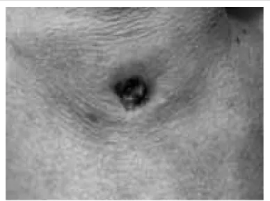
The tracheostomy tube, upon physical examination, was found to be still “embedded” due to a narrow stenosis of the surgical orifice which prevented its substitution (Fig. 1).
A total of 5 patients had a Shiley type cuffed tube, one patient still had a conotube which had been introduced during hospitalisation in the ICU.
ap-peared to be stenotic and rigid, with peristomal fibro-sis (Fig. 2); in the patient with a conotube, the tra-cheostomy, besides the fibrosis and stenosis, appeared to present acute inflammation and, moreover, crusted mucopurulent secretions were present both on the bor-ders of the tracheostomy and within the tube. The conventional surgical technique was used in all cases, with a suture being placed between the lower border of the tracheal orifice and the skin in order to create a preferential route for later substitutions of the cannula (partial lower stoma).
In 4 cases, the thyroidal isthmus was not sectioned, whereas, in the other two, the surgical procedure was performed via the trans-isthmic route due to the size of the thyroid gland. In all cases, diastasis was ob-served between the cutaneous and the tracheal plans
(Fig. 3). Stitches were removed on the 10thoperative
day, at the patient’s home.
Follow-up, one month after surgery, showed good opening of the tracheostoma without signs of inflam-mation or accumulation of secretions and thus substi-tution of the tube was carried out without any diffi-culty.
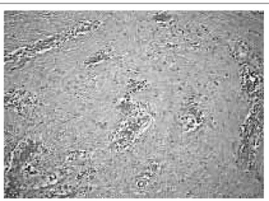
Histological examination of the peristomal tissue re-vealed, in all cases, diffuse microerosive features of the epidermis with local lympho-granulocyte infiltra-tions of the elastosic derma associated with giant-cell reaction, epidermic parakeratosis with erosive fea-tures and marked scar fibrosis (Fig. 4).
Bacteriologic examination demonstrated the presence of colonies of coagulase-negative staphilococcus both on the biopsy specimen and culture broth prepared on
Fig. 1. Embedded tracheostomy cannula (n. 6): pre-op-erative view.
Fig. 2. Following removal of cannula, tracheostoma ap-pears hardened, with granulomas and completely stenotic.
Fig. 3. Diastasis between cutaneous and tracheal planes is visible. Intra-operative view.
Fig. 4. Epidermic layer is interrupted by a large granulo-matous ulceration.
the lavage liquid of the tracheal tube, while the as-sessment for mycetes was negative.
Discussion and conclusions
PT is a surgical technique which, even if proposed several years ago, is increasingly used in clinical prac-tice, in particular in ICUs; it is currently estimated that 80% of the tracheotomies carried out in the above-mentioned divisions, are, indeed, PT1 2.
Of the various methods, TLT is considered the easiest to perform, the least invasive and, according to the “author”, the tracheal mucosa should be brought close to the skin with minimal trauma and virtually without bleeding, inasmuch as the passage of the reinforced
tube, is from the inside to the outside of the trachea, and would, thus, contribute to sealing the tracheal mu-cosa to the skin13 14.
Albeit, in the last 2 years, in our Clinic, we observed 6 cases coming from various ICUs in which use of the method led to embedding of the tube in the tra-cheostomy orifice, a severe complication especially for patients being cared for in the home setting inas-much as substitution is impossible and, therefore, emergency treatment is required.
Thus, the danger related to substitution of the tube at home, or its removal in emergency conditions, in these cases, is due to the certainty that, once having removed the tube from its location, forcing the stenot-ic stoma, it is impossible to then introduce the new tube, resulting in lack of ventilation and hypoxia; in these patients, in whom respiratory autonomy is <5 minutes, this would mean exposing them to a life-threatening situation.
Furthermore, among the cases observed, there was one subject still equipped with a conotube, who also presented severe purulent peristomal inflammation. If this problem is present to a lesser extent in hospi-talised patients, it is still more serious in patients treat-ed at home inasmuch as daily care, as previously ob-served, is performed by family members who often do not have the know-how or adequate technical ability to meet the needs of these patients.
In conclusion, from personal experience and a review of the literature, it can be seen that percutaneous tra-cheotomy techniques may be advantageous as far as concerns rapid performance, lower incidence of tra-cheal stenosis and the good aesthetic results. On the other hand, this may induce complications, being se-vere in many cases, such as dislocation or obstruction of the tube20, which require a prompt solution. In these
patients, this possibility could have even been fatal
Fig. 5. Scarring collagen deposits, trapping vessels and inflammatory infiltrate are visible.
Fig. 6. Marked degeneration of the elastic trama is visi-ble within the ulcerative process.
Fig. 7. Phagocytic processes with giant cells containing phagocyted material in cytoplasm.
References
1 Bernad AC, Kenady DE. Conventional surgical
tracheosto-my as the preferred method of airway management. J Oral
Maxillofac Surg 1999;57:310-5.
2 Bonner S, Taylor M. Airway obstruction in head and neck
surgery. Anesthesia 2000;55:290-1.
3 Dubin J. Tracheotomia. In: Encycl. Méd. Chir. Tecniche
Chirurgiche – testa e collo. Vol 2. Paris (France): Elsevier;
1993. p. 46-430, 1-9.
4 Head JM. Tracheostomy in the management of respiratory
problems. N Engl J Med 1960;264:587-91.
5 Dulguerov P, Gysin C, Perneger TV, Chevrolet JC.
Percuta-neous or surgical tracheostomy: A meta-analysis. Crit Care
Med 1999;27:1617-25.
6 Friedman Y, Mizok BA. Percutaneous versus surgical
tra-cheostomy: procedure of choice or choice of procedure. Crit
Care Med 1999;27:1684-5.
7 Matthews HR, Hopkins RB. Treatment of sputum retention
by minitracheostomy. Br J Surg 1984;71:147-50.
8 Reibel JF, Heffner JE, Durbin Watson CB, Bishop MJ,
Stauf-fer JL, et al. Tracheotomy/Tracheostomy. Respiratory Care 1999;44:820-7.
9 Gysin C, Dulguerov P, Guyot JP, Perneger TV, Abajo B,
Chevrolet JC. Percutaneous versus surgical tracheostomy: a
double-blind randomized trial. Ann Surg 1999;230:708-14.
10 Griggs WM, Myburg JA, Worthley LI. A prospective
com-parison of a percutaneous tracheostomy technique with stan-dard surgical tracheostomy. Int Care Med 1999;17:261-3.
11 Heikkinen M, Aarnio P, Hannukainen J. Percutaneous
di-latational tracheostomy or conventional surgical tra-cheostomy? Crit Care Med 2000;28:1399-402.
12 Ciaglia P, Firsching R, Syniec C. Elective percutaneous
di-latational tracheostomy. A new simple bedside procedure; preliminary report. Chest 1985;87:715-9.
13 Fantoni A, Ripamonti D, Lesmo A, Zanoni CI.
Translaryn-geal tracheostomy. A new era? Minerva Anestesiol
1996;62:313-25.
14 Fantoni A, Ripamonti D. A non-derivative, non surgical
tra-cheostomy: the translaryngeal method. Int Care Med
1997;23:386-92.
15 Byhahn C, WiIke HJ, Lischke V, Westphal K.
Translaryn-geal tracheostomy: two modified techniques versus the basic technique – early experience in 75 critically ill adults. Int
Care Med 2000;26:457-61.
16 Muttini S, Melloni G, Gemma M, Casati A, Carretta A,
Giu-dici A, et al. Percutaneous or surgical tracheostomy.
Prospec-tive, randomized comparison of the incidence of early and late complications. Minerva Anestesiol 1999;65:521-7.
17 Chew JY, Cantrell RW. Tracheostomy. Complications and
management. Arch Otolaryngol 1972;96:538-5.
18 Glass WW, King OJ jr, Lui A. Complications of
tracheosto-my. Arch Surg 1962;85:72-9.
19 Guarino A. Complicanze immediate della tracheotomia
chirurgica. Anest Rianim Intens 1994;15:304-7.
20 Succo G, Crosetti E, Pecorari GC, Nadalin J, Ragona R,
Donadio PP, et al. Complications of tracheostomy in
criti-cally patients: comparison of dilation and surgical tech-niques. Acta Otorhinolaryngol Ital 2002;22:1-11.
21 Gelosa G, Rosa G, Colombo S. Complicanze delle
tra-cheotomie. In: Colombo E, editor. Le tratra-cheotomie. Lecce:
Torgraf; 2001. p. 117-35. inasmuch as embedding of the tube in the
tracheostom-al site would have made substitution of the tube in emergency conditions impossible with subsequent acute respiratory insufficiency and risk of death. In these dramatic cases, it is mandatory to have trained personnel, confirmed opening of the tra-cheostoma, since in the event autonomous respiration is reduced, prolonged removal of the tube would not be possible since during the manoeuvre to reintroduce the tube, there is a risk of creating a false route or of abruptly interrupting the tracheal rings, thus creating mucoperichondrial flaps likely to cause stenosis20.
Thus, considering that PT is followed by a higher probability of moderate-severe complications than the conventional surgical technique20 21, the former is, in
our opinion, a useful low cost alternative, in patients at low risk, in whom the procedure is performed with delay (timing >18 days) and who require a tra-cheostomy for a limited time period, whilst the latter should be reserved for patients with an unfavourable anatomy, with coagulation disorders or other risk fac-tors, in whom the surgical procedure is performed ear-lier (timing <18 days) and in whom tracheostomy is expected to be maintained for a long time. Thus com-plications, due to stenosis of the orifice and opening of the tracheostomy could be avoided, which, can-celling the benefits of TLT, lead the patient to the like-lihood of another surgical procedure.
■ Received December 10, 2002. Accepted March 10, 2003.
■ Address for correspondence: Dr. G. Neri, c/o Clinica ORL, Policlinico Universitario “SS. Annunziata”, via dei Vestini, 66100 Chieti, Italy. Fax: +39 0871 552033. E-mail: [email protected]