Terminal Transferase
Positive Rat Thymocytes are Resistant
to Steroid-induced
Apoptosis
Oriana Trubiani*,
F.J.
Bollum#,
and Roberto
Di Primio§
Istituto
di Morfologia Umana Normale, Universita
di Chieti,
Via dei Vestini
12, 66100 Chieti,
Italy*;
Supertechs,
96100 Medical Center Drive, Rockville,
MD. 20850, USA#; Istituto
di Morfologia Umana Normale,
Universita
di Ancona, Monte D'Ago, 60131 Ancona, Italy§
Keywords: apoptosis/steroids/terminal
deoxynucleotidyl transferase/Bcl-2/thymocytes
ABSTRACT.
Apoptosis is a prominent mechanism of programmed cell death in the immunesystem. In the
thymus apoptosis is responsible
for the deletion
of autoreactive
T-cells during thymic differentiation.
The
typi-cal features of apoptosis are characterized by nuclear and cytoplasmic morphologic changes, along with
cleav-age of chromatin at regularly
spaced sites.
Terminal deoxynucleotidyl
transferase
(TdT) is a DNApolymerizing
enzyme found at an early stage of T and B lymphocyte differentiation,
which generates diversity
in the
DNAse-quence of immunoglobulin (Ig) or T cell receptor (TCR). The combined evaluations
of thymocyte
morphologi-cal features,
immunephenotype and thymic topography associated to TdTexpression allow the recognition of
three different
thymocyte subpopulations,
characterized
by small-size,
intermediate-size
and large-size.
The
re-sults of this study show that dexamethasone (Dx)-treatment
induces cell death via apoptosis involving distinct
transformations related to differentiation
stages of thymic subpopulations. Intermediate and small-size
thymo-cytes that are TdT-negative or weakly positive at nuclear level are Dx sensitive.
In contrast the large-size
thymo-cytes, highly TdT positive,
corresponding
to the undifferentiated
cells,
do not show significant
morphological
modifications
and TdT positivity
to Dx-treatment.
Immunocytochemical analysis
shows that Dx-treatment does
not affect TdT synthesis
but morphological changes, occurring during apoptotic process, are responsive to
intra-cellular
movementand intranuclear arrangement of the TdT.
The process of negative and positive clonal selection
occurring in the thymus leads to death in a plurality of
thymocyte subpopulations
(22, 28, 29, 35). Apoptosis
or programmedcell death occurs at an intermediate
stage of differentiation
of thymocytes showing a CD4+,
CD8+ phenotype
and avery low density
TCR (6, 15, 17,
27, 34). These cells, localized
in the deep and in the
inter-mediate cortical areas of the thymus, represent most of
the thymic population. Previous works suggested that
apoptosis can be artificially
induced by irradiation
or by
glucocorticoids
(2, 4, 14, 30, 39, 40), while
physiologi-cally
this
mechanism can be induced
prevalently
by
TCR or CD3-associated
complex activation
(5, 32, 33).
The high rate of death within the thymus appears to
reflect
the molding of the initial
TCRrepertoire.
Ma-ture T cells respond to the antigen in self major
histo-compatibility
(MHC)molecules. During differentiation
in the thymus, the TCRrepertoire
is shaped by both
pos-itive and negative events involving MHCmolecules (6).
Recently the role of steroids in programmed cell death
has been shown and since thymic epithelial
cells contain
To whomcorrespondence to be addressed.
the enzymes required
for steroidogenesis,
the thymus
itself can produce pregnenolone and
deoxycorticoster-one. These thymic steroids could play a key role in
thym-ocyte survival since it has been shownthat the
simultane-ous presence of TCRand the intranuclear receptors for
glucocorticoids
prevent, by mutual antagonism,
thymo-cytes death (41). Otherwise, TCRor glucocorticoids
in
a singular wayactivated, indece the apoptotic process
(32, 33, 40). The treatment
of thymocytes with
glucocor-ticoids provides a convenient way to study cell death in
vitro.
The bcl-2 gene was identified at the chromosomal
breakpoint
of t(14, 18) bearing
B cell lymphomas (34).
bcl-2 is novel amongproto-oncogenes that displays an
important functional
role of blocking programmed cell
death in selected hematopoietic cell lines (38). Bcl-2 is
detected to the inner mitocondrial membraneand this
position is novel for proteins with an oncogenic role.
The localization
of Bcl-2 suggests that the fundamental
metabolic functions of the inner mitochondrial
mem-brane,
which include
oxidative
phosphorylation
and
electron and metabolite transport, are implicated in the
survival mechanism (23). The Bcl-2 protein appearsthen geographically
restricted
in tissues
characterized
by apoptotic
cell death (24). In the thymus regional
dis-tribution
suggests that Bcl-2 is differentially
regulated
during T cell maturation and involved in the
preserva-tion of T cells.
Terminal deoxynucleotidyl
transferase
(TdT) is a
"cre-ative" DNApolymerase able to generate a somatic
diver-sification
between T and B cell clones (9, ll, 12). In the
T-cell compartment the presence of TdT was found
lated to gene rearrangement coding the T-cell antigen
re-ceptor (7, 8, 12). It has been previously
explained
(8, 20)
that TdT positive cells in the rat thymus belong to
dis-tinct subsets. Ultrastructural
analysis of TdTsuggests
that the localization of the enzyme is related to
thymo-cyte morphological features and TdTexpression
corre-sponds to the maturational stages of T cells. In this
work we have analysed, according to cell-size
and TdT
expression, the apoptotic process induced by
Dx-treat-ment.
MATERIALS AND METHODS
Male Sprague-Dawley rats were from Charles River
(Mila-no, Italy). Rabbit polyclonal antibody against 32-KDa calf
thymus TdT protein was produced as described (10). Bcl-2
an-ti-rat
antibody was from Santa Cruz Biotechnology,
Inc.
(San-ta Cruz, Ca., USA). Dexamethasone, culture media and all
reagent grade materials
were from Sigma (St. Louis, Mo.,
USA). Reagents for electron microscopy were from
Polysci-ence (Warrington, PA., USA).
Thymocytes preparation and dexamethasonetreatment.
Male Sprague-Dawley rats, 3-5 wks old, were used. The
thy-mus was removed, washed in PBS, and placed in cold RPMI
1640 supplemented
with 2 mML-Glutamine,
100
mMNa-Pyruvate. The thymus was minced and cells were put in
sus-pension. Cell suspensions
were filtered
and centrifuged
for 4
min at 400 g, at room temperature. Pellets were resuspended
in pre-warmed RPMI 1640 supplemented
with \Q% FBS. Cell
concentration was adjusted at 1 x 10⁶ cells/ml.
Dexametha-sone (Dx) was added as previously
reported (31) to the cell
sus-pension at 1 fiM concentration.
Cells were cultured at 37°C,
in a range between 4 to 16 hrs in an atmosphere containing
5% CO2. Control cultures were done in absence of
dexametha-sone.
Morphological study. Cell suspensions were fixed with
1.25% glutaraldehyde
in 0.1 M cacodylate
buffer,
pH 7.6 for
1 hour at 4°C. After rinsing in the same buffer the fixed cells
were post fixed with 1% OsO4 in cacodylate
buffer for 1 hour
in a dark cold room. The cells were then washed with buffer
and double distilled
water and stained overnight using a
satu-rate uranyle acetate aqueous solution. The cell suspensions
were then washed, coated with agar, dehydrated with alcohol
and toluene, and then embeddedin Spurr medium. Thin
sec-tions were obtained and observed as described above.
Immunocytochemistry.
Immunocytochemistry
for TdT
lo-calization
was carried out as previously described (19-21).
Briefly, for electron microscopy, cell suspensions were fixed
with 2% paraformaldehyde
in 0.1 M Na-cacodylate
buffer,
pH 7.4, for 2 hrs and subsequently permeabilized
with 0.4%
saponin in Na-cacodylate buffer for 15 min. After several
changes of Na-cacodylate buffer containing 0. 1 Mglycine and
2%sucrose, fixed and permeabilized cells were incubated
over-night with a rabbit anti-calf
TdT polyclonal
antibody (5 fjtg
/ml). Cells were then reacted with peroxides-conjugated
goat
anti-rabbit
IgG for 2 hours and incubated,
after washing, for
10 min at room temperature with a substrate for peroxidase
detection
(0.05 mg/ml DABin 0.05 M Tris-HCl buffer pH 7.6
and 0.015% H2O2). Cell suspensions
were then treated
with
1% OSO4 for 1 hr, dehydrated
and embedded in Spurr
medi-um. Thin sections were stained with lead citrate and observed
with a Zeiss 109 electron microscope. In negative controls, the
primary or secondary antibodies were omitted.
DNAextraction. At the end of the incubation
period,
thymocyte cultures
were centrifuged for 4 min at 400 g. The
pellet was resuspended in lysis buffer.
DNAwas extracted
with ethanol chloroform/isoamyl
alcohol (24: 1) and
precipi-tated with 2.5 vol. of absolute ethanol. DNAconcentrations
were detected by spectrophotometric absorption at 260 nm.
Ten fig of DNA/samplewas loaded into a 1.8% agarose gel
and electrophoresed.
DNAwas detected using
UVillumina-tion after ethidium bromide staining.
Fluorescence-activated
cell sorter (FA CS) analysis.
Thym-ocytes were analyzed accordingly to the physical parameters
or stained for 30 min at 4°C with anti-rat Bcl-2 mouse
mono-clonal antibody at 1 : 100 dilution and revealed using goat
anti-mouse FITC conjugated. Quantitative fluorescence analysis
of stained cells was performed on a Coulter Epics Elite flow
cy-tometer (Coulter Inc., USA).
RESULTS
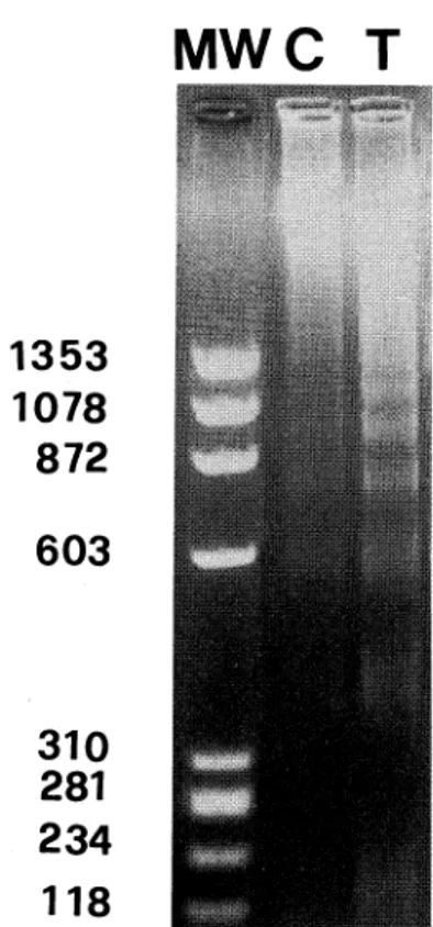
Agarose gel electrophoresis
of DNA(Fig. 1),
recov-ered from Dx-treated thymocytes, shows the
DNApat-tern characteristic
of apoptosis, a chromatin ladder of
DNAfragments in multiple,
of 180-200 bp. Untreated
thymocytes also show manyapoptotic bands due to
spontaneous apoptotis.
Both reacted and untreated
thymocytes exhibited similar patterns of
DNAfragmen-tation.
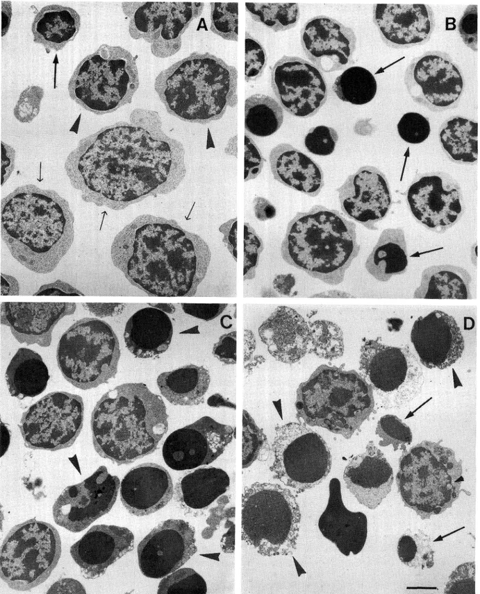
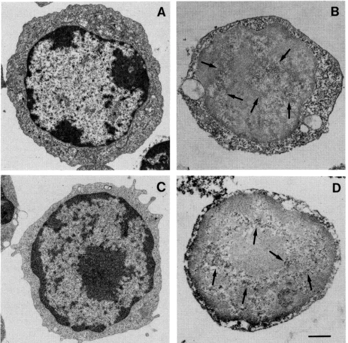
Ultrastructural
analyses of thymocytes allowed
identification
of three distinct
subpopulations:
small,
intermediate
and large thymocytes, as reported in Fig.
2A. These subpopulations
show different
sensitivity
to Dx-treatment.
Four hours later (Fig. 2B), small-size
thymocytes appear mostly apoptotic, otherwise
interme-diated and large-seze cells are normally arranged. At
eight hours (Fig. 2C) intermediate
thymocytes show
fea-tures of apoptotic process and at 16 hours (Fig. 2D)
ex-tensive degenerative processes are recognizable in small
and intermediate
apoptotic
thymocytes.
During
Dx-treatment large thymocytes revealed only few
modifica-MWC T
Fig. 1. Agarose gel analysis of DNAfrom Dx treated (T) and un-treated thymocytes (C). The DNAsamples were fractionated by elec-trophoresis in a 1.8% agarose gel and stained with ethidium bromide. The characteristic ladder-like pattern of DNAbanding indicative of apoptotic endogenous nuclease activity is observed prevalently in Dx-treated thymocytes. MW:DNAmolecular weight.
tions in the cytoplasmic compartmentand the nucleus
appeared normally arranged (Fig. 2B, C, D).
Dx-treat-ment producing morphological changes did not affect
the TdTsynthesis but induces an intracellular
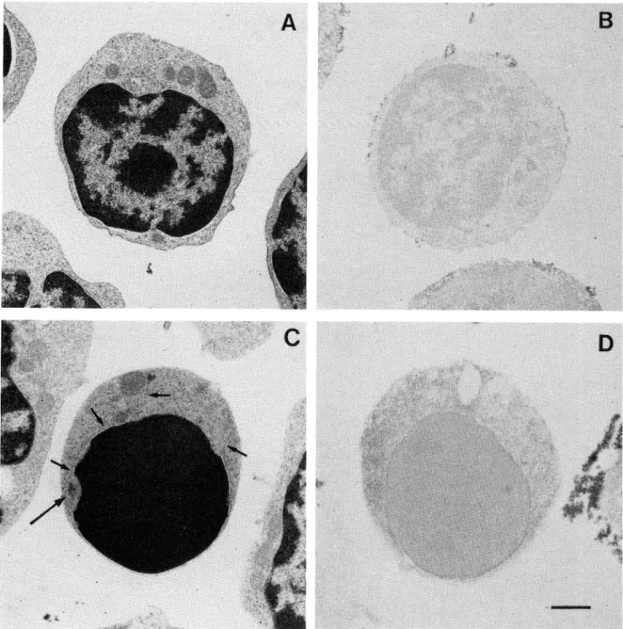
redistribu-tion of TdT. In fact, small cells (Fig. 3A) exhibited
a
smooth surface and their cytoplasm contained only few
mitochondria.
The nucleus
contained
predominantly
marginated heterochromatin, which often formed a
cen-tral mass. Small thymocytes were TdT negative both at
the nuclear and at the cytoplasmic level (Fig. 3B). After
Dx-treatment (Fig. 3C) nuclear changes typical of
apop-tosis were detected in small thymocytes, lacking major
cytoplasmic damage. Nucleolar material was frequently
detected
at the nuclear
periphery
and nuclear
mem-brane was sometimes interrupted
(Fig. 3C). Dx-treated
small thymocytes were TdTnegative as untreated cells
(Fig.
3D).
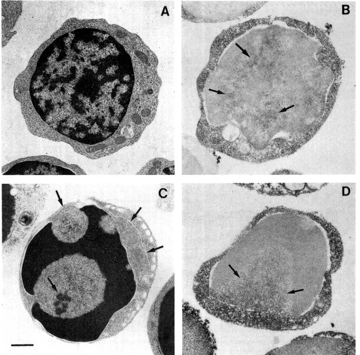
Intermediate
thymocytes
(Fig.
4A) were a
consistently
heterogeneous
thymic
subpopulation
of
cells
showing a smooth surface.
The cytoplasm
con-tained some mitochondria, a Golgi apparatus and
iso-lated profiles
of rough endoplasmic reticulum (RER).
The nucleus contained a large amountof
heterochroma-tin.
Untreated
intermediate
thymocytes
were weakly
TdT positive,
predominantly at the cytoplasmic
com-partment. Sometimeit was possible to observe a
resid-ual amount of the enzyme present at the nuclear level
confined to the euchromatin domains (Fig. 4B).
Dx-treated intermediate thymocytes (Fig. 4C) showed
apop-totic changes both at the cytoplasmic and at the nuclear
levels. The cytoplasm was condensed and contained
oc-casional vacuoles. Apoptotic nuclei displayed
peripher-al chromatin condensation forming either toroid caps
or crescents and the nucleoli showa characteristic
pat-tern of disaggregation.
Dx-treated
thymocytes (Fig. 4D)
remained TdT-positive and the nuclear level reaction
was predominantly localized in the residual nuclear
ma-trix. Large thymocytes (Fig. 5A) showed an irregularsurface and the abundant cytoplasm contained several
mitochondria, ribosomes and RERprofiles.
The
nucle-us consisted largely of euchromatin with scanty
hetero-chromatin opposed to the nuclear envelope. One or
more nucleoli were also present. These cells appeared to
contain a large amount of TdT, diffusely
distributed
in
the cytoplasm and bound to the nuclear
interchroma-tinic region (Fig. 5B). Dx-treated cells (Fig. 5C)
dis-played a moderate heterochromatin margination.
More-over, typical apoptotic patterns were not detected.
Im-munocytochemical
analyses (Fig. 5D) showed that TdT
waslocalized in the samenuclear euchromatin areas as
in the untreated cells. The cytoplasm also displayed a
diffuse positivity
for TdT. Accordingly to
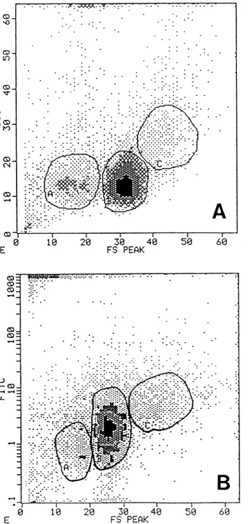
ultrastructur-al results in flow cytometry, by the anultrastructur-alysis of forward
light
scatter,
three
different
subpopulations
were
re-cognizable.
Fig.
6A shows that
small thymocytes
are
15± 10% of total population,
while intermediate
and
large cells are respectively
65± 10% and 8±3% of total
thymocytes. These results are the meansof five different
experiments and correspond to the date obtained at
elec-tron microscope level. Flow cytometry
immunefluores-cence analysis showed (Fig. 6B) the different
expression
of Bcl-2 protein connected to size of thymocyte
subpop-ulations.
Large-size thymocytes
were bright
positives,
shile
intermediate
cells
constituted
a heterogeneous
population
showing different
score of positivity
from
bright to negative and the small-size
cells were
prevalent-ly Bcl-2 negative.
DISCUSSION
Cell
death
takes
two distinct
forms,
necrosis
and
apoptosis. Necrosis is considered as a degenerative
phe-nomenonwhile apoptosis is an active endogenous
proc-esses implicated in the regulation of normal, and
neo-plastic cells (3, 13). In the ontogeny of the
immunesys-tem cell death it occurs by deletion of autoreactive
T
cell clones during thymic maturation and deletion of B
cells occurs in the germinal center without
antigen-cod-ing positive
selection
of centrocytes
(37, 16, 17, 26). The
Fig. 2. Morphological analysis of Dx-treated thymocytes. A: Untreated thymocyte sub-populations. Arrows: small thymocytes. Head arrows: intermediate thymocytes. Light arrows: large thymocytes. B: 4 hours of Dx-treatment; it is possible to observe apoptotic features only in small-size thymocytes (arrows). C: 8 hours of treatment; intermediate cells show typical modification of nuclear compartment (arrowheads). D: 16 hours of treatment; small (arrows) and intermediate thymocytes (arrowheads) show extensive necrotic process. Large cell shows a reduction of cell-size with a light modification of ultrastructure. Bar 5 //m.
B
D
#*
Fig. 3. Transmission electron microscope analysis of glutaraldehyde-osmium tetroxide fixed untreated (A) and treated (C) small-size thymo-cytes. Immunocytochemical investigation of TdT, performed as described show that untreated (B) and Dx-treated (D) small thymocytes are com-pletely unstained. Nucleolus: arrow. Nuclear membrane:small arrow. Bar 2 ^m.
proto-oncogene
bcl-2 has recently
been implicated
as
a component of the molecular processes that decide
whether some cell lives or dies (35). In the thymus bcl-2
expression protects immature thymocytes from
gluco-corticoid,
radiation
and anti-CD3-induced
apoptosis
and its presence is related to the differentiation stages of
thymocytes (31, 35, 38). Since terminal transferase
has
been found in both pre-T and pre-B lymphocytes and
its role consists in producing, at an early stage of
differ-entiation,
a diversification
of the immune-cells by
addi-tion of N nucleotides to the termini segment during
V-D-J rearrangement
(1, 7, 8, 18). By in situ
hybridiza-tion, it has been shown that TdT mRNAis confined to
the thymic
cortex (8),
and quantitative
analysis
using
the polymerase
chain reaction
(PCR)
has established
that TdT disappears along with up-regulation
of the
TCRand down-regulation
of CD4 or CD8 after
pos-itive
selection
(8).
Commitment to the CD4 or CD8
X.
à": jS å
.**à"<; v.'iV> v<«^, i
sr
Fig. 4. Analysis of mediumsize thymocytes. A: infrastructure of treated cells. B: immunocytochemistry reaction of TdT show a specific
stain-ing at the cytoplasmic and nuclear levels (arrows). C: Dx-treated thymocytes display condensed chromatin, nuclear membrane(small arrows) and
nucleolus with a characteristic
pattern of disintegration
(arrows). D: immunocytochemicalstudies of Dx-treated cells showing dark
immunopre-cipitate localized in non condensed nuclear chromatin (arrows) and at cytoplasmic level. Bar 2.5 /mi.
lineage was known to occur in the thymus: imma-ture CD4 CD8 TCR~ thymocytes differentiate into CD4+CD8+TCR10 cells, a small percentage of which mature into either CD4+CD8 TCRhi or CD4 CD8+TCRhi thymocytes. Entry into these single
positive populations required expression of the
appro-priate positively
selecting
MHCclass I or II molecules
on epithelial cells of the thymus (5, 25). Therefore, a
given cell can then engage MHCmolecules on thymic
stromal cells with both its TCRand the appropriate
coreceptor,
along
with
differentiation;
otherwise,
it
dies.
Since
apoptosis
has been described
as a clonal
mechanism of deletion of thymocyte subsets expressing
inappropriate TCR, in this study we propose that
large-size undifferentiated
Bcl-2 positive thymocytes
express-ing TdT at the nuclear level and showexpress-ing as described
,/.A'.^--*à"*':'*.-,;'* "."--M 'MM& tf?: "**K r. -*3*'*'/à" *.å à"''. B å ''>-:.i -*-i»&M-?:~J? ' >^-r aM /. '^-'^ <;~
Ki
;k^fh >%*.'à"à"à" :ai». r* '7;t? JW»3fià"à"#:& :
.*t'. i;.. ^^^^^^^^^^3mam
Hi å Py:'*J&8&
.-km
à"4 ^^:<v^ *i j #, !< ^ m w g - j !サ> 蝣 II K m 1 II ,^ *"i i 蝣 a m . 蝣 jm &f mi II m m 7*-:* ~"- -"\.ife>y ,'*V*V-à"i§:/
Fig. 5. Analysis of large-size thymocytes. A: morphological features of untreated cells. B: TEMimmunocytochemical analysis of TdT shows a large amount of enzyme localized at the nuclear level linked to interchromatinic regions (arrows). C: Dx-treated thymocytes exhibit only a light re-duction of cell-size. D: immunocytochemical study of TdT in Dx-treated cells. Immunoprecipitate is localized at the nuclear (arrows) and cyto-plasmic levels, as observed in untreated cells. Bar 3 ftm.
(8, 20) the phenotype CD4 CD8 TCR~ are not
sensi-tive to corticosteroid
treatments or in detail are not
com-mitted to positive or negative selection process.
Inter-mediate and small-size
thymocytes, Bcl-2 low positive
or negative, which have rearranged the TCRsegments
contain, at this stage of differentiation,
an intrinsic
apoptotic programthat can be regulated by several
spe-cific biochemical events. Dx-treatment, moreover, does
not affect the molecular machinery of TdT metabolism,
regulated
at the early step of thymocyte differentiation,
but intracellular
movementof the enzymeappears
sub-ordinate to the modification of nuclear structure.
The
experiments reported here suggest that the different
re-sponse to corticosteroids
are related
to the stage of
thymocyte differentiation
and the identification
of
apop-totic subpopulation will greatly simplify gene
expres-30 40
FS PEAK
39 40
FS PEAK
50 69
Fig. 6. Flow cytometry analysis of rat thymocytes. Section A: Scat-ter analysis shows three distinguishable populations: small
thymo-cytes (A), intermediate thymothymo-cytes (B) and large-size
thymocytes (C).
Section B: Flow cytometry alalysis of Bcl-2 protein performed as de-scribed in materials and methods section. A: small thymocytes. B: in-termediate thymocytes. C: large thymocytes. It is possible to observe the different score of positivity related to the size of the cells. The re-sults showed in section A and B are the same obtained in five differentexperiments.
sion and other mechanistic studies of apoptosis.
Acknowledgments. This study was supported by Italian
MURST
and CNRgrants.
REFEREN CES
Alt, F. and Baltimore, D. 1982. Joining of immunoglobulin chain gene segments: Implications from a chromosomewith
evi-dence of three D-JH fusions. Proc. Natl. Acad. Sci. USA, 79:
4118-4122.
Arends, M.J. and Wyllie, A.H. 1991. Apoptosis: mecha-nism and roles in pathology. Int. Rev. Exp. Pathol, 32:
223-254.
Ashwell, J.D., Berger, N.A., Cidlowski, J.A., Lane, D.P., and Korsmeyer, S.J. 1994. Coming to terms with death: apop-tosis in cancer and immune development. Immunol. Today, 15:
147-151.
Bansal, N., Houle, A.G., and Melnylovych, G. 1990. Dexa-methasone- induced killing of neoplastic cells of lymphoid deri-vation: lack of early calcium involvement. /. Cell PhysioL , 143:
105-109.
Berg, L.J., Pullen, A.M., Fazecas de St. Groth, B., Mathis D., Benoist, C, and Davis, M.M. 1989. Antigen/MHC-spe-cific T cells are preferentially exported from the thymus in the
presence of their MHCligand. Cell, 58: 1035-1046.
Blackman, M., Kappler, J., and Marrack, P. 1990. The role of T cell receptor in positive and negative selection of
devel-oping T cells. Science, 248: 1335-1341.
Bogue, M., Mossmann, H., Stauffer, U., Benoist, C, and
Mathis, D. 1993. The level of N-region diversity in T cell re-ceptors is not pre-ordained in the stem cell. Eur. J. Immunol,
23: 1185-1188.
Bogue, M., Gilfillan, S., Benoist, C, and Mathis, D.
1992. Regulation of N-region diversity in antigen receptors through thymocyte differentiation and thymus ontogeny. Proc.
Natl. Acad. ScL Usa, 89: 11011-11015.
Bollum, F.J. 1974. Terminal deoxynucleotidyl Transferase. In Boyer, R.D. ed., The Enzymes. New York: Academic Press,
10: 145-151.
Bollum, F.J. 1975. Antibody to Terminal deoxynucleotidyl
Transferase. Proc. Nat. Acad. Sci. USA, 72: 4119-4122. Bollum, F.J. 1963. Progress in nucleic acid research. Vol.1, Academic Press, New York, pp.1-66.
Cayre, Y., de Sostoa, A., and Silverstone, A.E. 1981. Isola-tion of subset of thymocytes inducible for Terminal Transferase
biosynthesis. /. Immunol. , 126: 553-556.
Cohen, J.J. and Duke, R.C. 1992. Apoptosis and
program-med cell death in immunity. Annu. Rev. Immunol, 10:
267-293.
Cohen, J.J. and Duke, R.C. 1984. Glucocorticoid activation of a calcium-dependent endonuclease in thymocyte nuclei leads
to cell death. /. Immunol, 132: 38-42.
Cohen, G.M., Sun, X.-M., Snowden, R.T., Ormerod, M.G., and Dinsdale, D. 1993. Identification of a transitional
pre-apoptotic population of thymocytes. /. Immunol, 151:
566-574.
Crompton, T., Ohashi, P., Schneider, S.D., Pircher, H., and MacDonald, H. 1991. A cortisone sensitive CD3lowsub-set of CD4+CD8"thymocytes represents an intermediate stage
in intrathymic repertoire selection. Int. Immunol, 4: 153-161.
S.M. 1993. Evidence for programmed cell death of self-reac-tive T cell receptor-posiself-reac-tive2482-2487. thymocytes. Eur. J. Immunol, 23:
Desiderio,
S.V.,
Yacopoulos,
G.D., Paskind,
ML, Thomas,
E., Boss, M.A., Landau, N., Alt, F.W., and Baltimore, D. 1984. Insertion of N region into heavy-chain genes is corre-lated with expression of Terminal deoxynucleotydyl Transferase
in B cells. Nature, 311: 752-755.
Di Primio, R. and Bollum, F.J. 1987. Immunoenzymatic
method for detection of Terminal deoxynucleotidyl Transferase by light and electron microscopy. Hemat. Pathol., 3: 173-181.
Di Primio, R., Trubiani, O., and Bollum, F.J. 1992. Ultra-structural localization of Terminal deoxynucleotydyl
Transfer-ase (TdT) in rat thymocytes. Thymus, 19: 183-190.
Di Primio, R., Trubiani, O., and Bollum, F.J. 1992.
Intracel-lular localization of Terminal Transferase during the cell cycle.
Exp. Cell Res., 202: 405-411.
Ezine, S. and Ceredig, R. 1994. Haemopoiesis and early
T-cell differentiation. Immunol. Today, 15: 151-154.
HOCKENBERY, D., HUNEZ, G., MlLLIMAN, C, SCHREIBER, R.D.,
and Korsmeyer, S.J. 1990. Bcl-2 is an inner mitochondrial membrane protein that blocks programmed cell death. Nature,
348: 334-336.
Hockenbery, D., Zutter, D.M., Hickey, W., Nahm, M., and Korsmeyer, S.J. 1991. Bcl-2 protein is topographically
re-stricted in tissue characterized by apoptotic cell death. Proc. Natl. Acad. Sci. USA, 88: 6961-6965.
Kisielow, P., Teh, H.S., Bluthmann, H., and von Boehmer, H. 1988. Positive selection of antigen-specific T cells in
thy-mus by restricting MHCmolecules. Nature, 335: 730-733. Liu, Y.J., Joshua, D.F.E., Williams, G.T., Smith, C.A.,
Gordon, J., and MacLennan, I.CM. 1989. Mechanism of antigen-driven selection in germinal centres. Nature, 342:
929-931.
Murphy, K.M., Heimberger, A.B., and Loh, D.Y. 1990.
Induction by antigen of intrathymic apoptosis of
CD4+CD8+TCRlow thymocytes in vivo. Science, 250: 1720-1723.
Reinherz, E.L. and Schlossman, S.F. 1980. The differentia-tion and fucdifferentia-tion of human T lymphocytes. Cell, 19: 821-827. Rothenberg, E.M. 1992. The development of fuctionally re-sponsive T cells. Adv. Immunol., 51: 185-214.
Sellins, K.S. and Cohen, J.J. 1987. Gene induction by ^-ir-radiation leads to DNAfragmentation lymphocytes. /.
Immu-nol., 139: 3199-3206.
31. Sentman, C.L., Shutter,
J.R., Hockenbery, D., Kanagawa,
O., and Korsmeyer, S.J. 1991. Bcl-2 inhibits multiple forms of apoptosis but not negative selection in thymocytes. Cell, 67:
879-888.
32. Shi, Y.F., Bissonnette, R.P., Parfrey, N., Szalay, M.,
Kubo, R.T., and Green, D.R. 1991. In vivo administration of monoclonal antibodies to the CD3T cell receptor complex
in-duces cell death (apoptosis) in immature thymocytes. /.
Immu-nol, 146: 3340-3346.
33. Smith, C.A., Williams, G.T., Kingston, R., Jenkinson, E.J., and Owen, J.J.T. 1989. Antibodies to CD3/T-cell receptor complex induce death by apoptosis in immature T cells in
thym-ic cultures. Nature, 337: 181-184.
34. Snodgrass, H.R., Kisielov, P., Kiefer, M., Steinmetz, M.,
and von Boehmer, H. 1985. Ontogeny of the T-cell antigen re-ceptors within the thymus. Nature, 313: 592-595.
35. Strasser, A., Harris, A.W., and Cory, S. 1991. Bcl-2 trans-gene inhibits T cell death and perturbs thymic self-censorship.
Cell, 67: 889-899.
36. Tsujimoto, J., Gorham, J., Comman, J., Jaffe, E. and Croce, CM. 1985. The t(14; 18) chromosome translocations involved in B cell neoplasm results from mistakes in VDJjoining. Sci-ence, 229: 1390-1393.
37. Turka, L.A., Linsley P.S., Paine III, R., Schieven, G.R., Thompson, C.B., and Ledbetter, J.A. 1991. Signal transduc-tion via CD4, CD8, and CD28in mature and immature
thymo-cytes: implications for thymic selection. /. Immunol., 146:
1428-1436.
38. Vaux,D.L., Cory, S.,andAdams, J.M. 1988. Bcl-2genepro-motes haemopoietic cell survival and cooperates with c-myc to immortalize pre-B cells. Nature, 335: 440-442.
39. Wyllie, A.H. 1980. Glucocorticoid-induced thymocyte apop-tosis is associated with endogenous endonuclease activation.
Na-ture, 284: 555-556.
40. Yamada, T. and Ohyama, H. 1988. Radiation-induced
inter-phase death of rat thymocytes in internally
programmed(apop-tosis). Int. J. Radiah Biol. Relat. Stu. Phys. Chem. Med., 53:
65-75.
41. Zacharchuk, CM., Mercep, M., Chakraborti, P.K.,
Si-mons, S.S. Jr., andAshwell, J.D.. 1990.
ProgrammedTlym-phocyte death. Cell activation and steroid-induced pathways are mutually antagonistic. /. Immunol , 145: 4037-4045.