1
Università degli studi del Piemonte Orientale
“Amedeo Avogadro”
Dipartimento di Scienze del Farmaco
Dottorato di ricerca in Biotecnologie Farmaceutiche ed Alimentari
XXVI ciclo a.a. 2010-2013
New pathways of protumor “emergency“
granulo/monocytopoiesis
2
Contents
Chapter1 1
Introduction1.1 Inflammation and cancer 1.1.2 Myeloid cells in cancer 1.1.3 MDSCs
1.1.4 MDSCs expansion 1.1.5 TAM
1.2 IL-17 cytokine family 1.2.1 IL-17A and IL-17F
1.2.2 IL-17A and IL-17F producer cells 1.3 Nuclear receptor (NR) superfamily
1.4 Retinoid-related orphan receptors (RORs) 1.4.1 ROR structure and activity
1.4.2 ROR gene structure 1.4.3 ROR protein structure
1.4.4 RORs: ligand-dependent transcription factors
1.4.5 Role of RORγ in the development of secondary lymphoid tissues 1.4.6 Critical functions of RORγ in thymopoiesis
1.4.7 RORs and cellular metabolism
1.4.8 RORs and T cell lineage specification 1.4.9 Role of RORs in Th17 cell differentiation
1.4.10 Transcriptional regulation of IL-17 positive cell 1.4.11 Suppression of ROR transcriptional activity by FOXP3 1.4.12 Interaction with coregulatory proteins
1.5 RORs and cancer
1.5.1 RORs and increased cancer susceptibility 1.5.2 Tumour-associated Th17 cells
3 1.5.3 Evidence for antitumour activity
1.5.4 Pro-tumour role of Th17 cell-associated cytokines 1.6 Hematopoiesis
1.6.1 Bone marrow stem cell niche
1.7 T cell in regulation of hematopoiesis 1.7.1 Effects of IL-17 on hematopoietic cells 1.7.2 Effects of IL-17 on mesenchimal stem cell 1.7.3 The role of IL-17 in hematopoiesis
1.8 Steady state and emergency hematopoiesis
1.8.1 Demand-adapted regulation of early hematopoiesis in infection and inflammation
Chapter 2 55
Outline of the thesis
Chapter 3 61
New pathways of protumor “emergency“ granulo/monocytopoiesis
Chapter 4 109
Discussion
Chapter 5 117
“Effects of the chemotherapeutic drug Topotecan on anti-tumor immunity”
Chapter 6 143
5
7
1.1 Inflammation and cancer
The idea of a relationship between inflammation and cancer dates back 1863, when Rudolf Virchow observed leukocyte infiltration in neoplastic tissues and hypothesized that the origin of cancer was at sites of chronic inflammation.
Yet, it was only during the last decade that several evidences undoubtedly demonstrated the critical role of inflammation in tumorigenesis, and that some of the underlying molecular mechanisms have been elucidated (1).
In response to tissue injury, a network of chemical signals initiates and maintains a host response designed to “heal” the afflicted tissue. This involves activation and direct migration of leukocytes (neutrophils, monocytes and eosinophils) from the venous system to sites of damage. A family of chemotactic cytokines, named chemokines, which possess a relatively high degree of specificity for chemoattraction of specific leukocyte populations (2, 3), recruits downstream effector cells and dictates the natural evolution of the inflammatory response. Inflammation is usually self-limiting; however, disregulation of any of the converging factors can lead to abnormalities and, ultimately, pathologies including cancer. Indeed, in chronically inflamed tissues, a subversion of cell death and/or repair programmes might occur, resulting in uncontrolled proliferation of cells carring DNA mutations. It is estimated that 20% of all cancers is associated with chronic infection and inflammation (4). The connection between inflammation and cancer can be viewed as consisting of two pathways: an extrinsic pathway, driven by inflammatory conditions (such as inflammatory bowel disease) that increase cancer risk; and an intrinsic pathway, driven by genetic alterations (such as oncogenes activation) that cause both inflammation and neoplasia.
The two pathways converge, resulting in the activation of transcription factors, mainly nuclear factor-κB (NF-κB), signal transducer and activator of transcription 3 (STAT3) and hypoxia-inducible factor 1α (HIF1α), in tumour cells.
8
These transcription factors coordinate the production of inflammatory mediators, including cytokines and chemokines, as well as the production of cyclooxygenase 2 (COX2) (which, in turn, results in the production of prostaglandins).
These factors recruit and activate various leukocytes, most notably cells of the myelomonocytic lineage. The cytokines activate the same key transcription factors in inflammatory cells, stromal cells and tumour cells, resulting in more inflammatory mediators being produced and a cancer-related inflammatory microenvironment being generated. “Smouldering” cancer-related inflammation has many tumour-promoting effects including induction of genomic instability, alteration in epigenetic events and subsequent inappropriate gene expression, enhanced proliferation and resistance to apoptosis of initiated cells, induction of tumour angiogenesis and tissue remodelling with consequent promotion of tumour cells invasion and metastasis (5). Despite these evidences, genetic studies of mouse models have demonstrated that the inflammatory response supported by innate immune cells is crucial for the activation of an adaptive immune response capable to eliminate nascent tumors (6). It is generally accepted that immune cells continuously recognize and destroy nascent tumor cells but, due to the genetic instability that characterized neoplastic cells, the arising of new variants able to evade the immune surveillance results in tumor establishment and progression (immunoediting process) (7). In this regard, several studies have emphasized that the “smouldering” inflammation associated with tumors is mainly oriented to tune the adaptive immune response. Indeed, tumor-associated dendritic cells mainly show an immature phenotype (8) and myelomonocytic cells recruited in tumors express an alternative M2 functional phenotype mainly oriented towards the suppression of the adaptive immune response (9, 10). In agreement, clinical studies suggest that an established type-2 “suppressive” immunological profile correlates with poor prognosis, as shown in colorectal, hepatocellular and pancreatic carcinomas and in Hodgkin’s lymphoma (11).
9
1.1.2 Myeloid cells in cancer
Neoplastic cells condition distant sites, such as the bone marrow and spleen, by releasing soluble factors that drive the accumulation of myeloid cells; these myeloid cells subsequently promote neovascularization and metastasis.
This creates a tumour-driven ‘macroenvironment’. As discussed above, this macroenvironment conditions DCs, macrophages and granulocytes to become immunosuppressive. However, the most prominent effect is the accumulation of highly immunosuppressive, immature myeloid cells (iMCs).
These cells were named myeloid derived suppressor cells (MDSCs) to highlight their common myeloid origin and immunoregulatory properties (12).
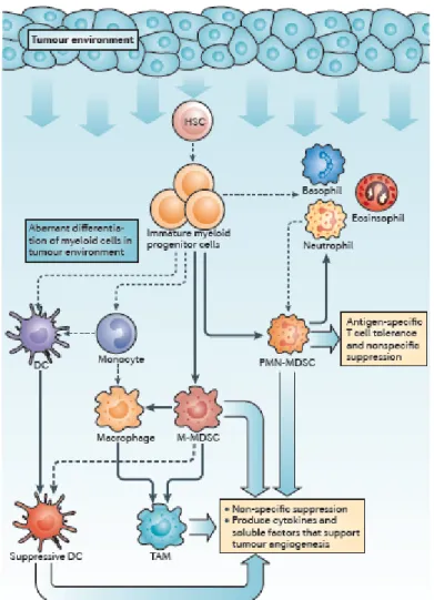
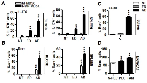
The increase in circulating myeloid cells in tumour-bearing hosts, originally termed “reactive neutrophilia” or “emergency granulopoiesis”, was associated with an increased frequency of iMCs. Immature myeloid cells with the same phenotype as MDSCs are continually generated in the bone marrow of healthy individuals and differentiate into mature myeloid cells without causing detectable immunosuppression. However, in cancer, myeloid cell differentiation is diverted from its normal pathway which leads to the terminal differentiation of mature macrophages, DCs and granulocytes towards a pathway that favours the differentiation of pathological MDSCs (Figure 1).
10
Figure 1- Changes that occur in myeloid cells in cancer.
Factors produced in the tumour microenvironment by tumour cells and stromal cells promote the aberrant differentiation of myeloid lineage cells. The dotted lines show the normal pathways of myeloid cell differentiation from immature myeloid precursor cells to dendritic cells (DCs), macrophages and granulocytes. The solid bold lines indicate the aberrant pathways of myeloid cell differentiation that occur in cancer, in which the tumour environment can promote the development of various immunosuppressive populations, including monocytic myeloid-derived suppressor cells (M-MDSCs), polymorphonuclear myeloid-derived suppressor cells (PMN-MDSCs), suppressive DCs and tumour-associated macrophages (TAMs) (13).
11
1.1.3 MDSCs
Myeloid-derived suppressor cells (MDSCs) are a heterogeneous population of cells of myeloid origin that comprises myeloid progenitor cells and immature macrophages, immature granulocytes and immature dendritic cells.
The two major MDSC subsets include the monocyte-like
CD11b+Gr1+Ly6ChiLy6Glow (M-MDSCs) and the granulocyte (neutrophil)-like CD11b+Gr1+Ly6GhiLy6Clow (PMN-MDSCs). They are present in an activated state that is characterized by the increased production of reactive oxygen and nitrogen species, and of arginase 1 (14,15). They are potent suppressors of various T-cell functions. In mice, the phenotype of MDSCs is CD11b+GR1+, although functionally distinct subsets within this population have been identified (16).
In humans, the phenotype of MDSCs is LIN–HLA-DR–CD33+ or
CD11b+CD14–CD33+; human cells do not express a marker that is homologous to mouse GR1 (17,18). MDSCs have also been identified within a CD15+ population in human peripheral blood. MDSCs were first characterized in tumour-bearing mice and in patients with cancer. These cells have been shown to markedly expand systemically when mice are inoculated with transplantable tumour cells and when tumours spontaneously develop in transgenic mice with tissue-restricted oncogene expression. In addition, up to a tenfold increase in MDSC numbers was detected in the blood of patients with different types of cancer (17,18).
In several mouse tumour models, as many as 20–40% of nucleated splenocytes are MDSCs (in contrast to the 2–4% seen in normal mice). In addition, MDSCs are found in tumour tissues and in the lymph nodes of tumour-bearing mice.
Although initial observations and most of the current information on the role of MDSCs in immune responses has come from studies in the field of cancer research, accumulating evidence has shown that MDSCs also regulate immune responses during bacterial and parasitic infections, acute and chronic inflammation, traumatic
12
stress, sepsis and transplantation. Recent studies (19) indicate that MDSCs have characteristics of both M1 and M2 macrophages (see paragraph “TAM”).
Indeed, it was described in tumor-bearing mice a population of circulating CD11b+Gr-1+ inflammatory monocytes expressing IL-4Rand able to release both IL-13 and IFN-γ(19), characteristics that are compatible with a function intermediate between those of M1 and M2 macrophages.
Cooperation between IL-13 and IFN-γled to sustained activation of both ARG1 and NOS2 in MDSC populations, causing dysfunctional T cell responses.
These results suggest that MDSCs and TAMs respond with an M2 macrophage– oriented program to classic signals driving macrophage activation in tumor-bearing hosts.
1.1.4 MDSCs expansion
Factors that induce MDSC expansion can include cyclooxygenase 2 (also known as PTGS2), prostaglandins (20-22), stem-cell factor (SCF) (20), macrophage colony-stimulating factor (M-CSF), IL-6 (23), granulocyte/macrophage CSF (GM-CSF) (22) and vascular endothelial growth factor (VEGF) (23).
Most of these factors trigger signaling pathways in MDSCs that converge on Janus kinase (JAK) protein family members and signal transducer and activator of transcription 3 (STAT3), which are signalling molecules that are involved in cell survival, proliferation, differentiation and apoptosis (24). STAT3 is arguably the main transcription factor that regulates the expansion of MDSCs.
MDSCs from tumour-bearing mice have markedly increased levels of phosphorylated STAT3 compared with IMCs from naïve mice (25).
Exposure of haematopoietic progenitor cells to the supernatant from tumour-cell cultures resulted in the activation of JAK2 and STAT3, and was associated with an expansion of MDSCs in vitro. However, this expansion was abrogated when STAT3 expression in haematopoietic progenitor cells was inhibited (26).
13
Moreover, ablation of STAT3 expression through the use of conditional knockout mice or selective STAT3 inhibitors markedly reduced the expansion of MDSCs and increased T-cell responses in tumour-bearing mice (25,27).
STAT3 activation is associated with increased survival and proliferation of myeloid progenitor cells, probably through the upregulation of the expression of B-cell lymphoma XL, cyclin D1, MYC and survivin. One such pathway involves the calcium-binding pro-inflammatory proteins S100A8 and S100A9 (29). STAT3-mediated upregulation of these proteins in myeloid progenitors inhibits DC differentiation and promotes MDSC accumulation (30).
In addition, STAT3 regulates the transcription factor CCAAT/enhancer-binding protein-β (C/EBPβ). C/EBPβ regulates myelopoiesis in healthy individuals and has a crucial role in controlling the differentiation of myeloid progenitors to functional MDSCs (28). STAT3 is responsible, at least in part, for inducing the expansion of MDSCs populations via upregulation of C/EBPβ and also has an indirect role in myeloid cell mobilization, accumulation and survival (31).
So, abnormal and persistent activation of STAT3 in myeloid progenitor cells prevents their differentiation into mature myeloid cells and thereby promotes MDSC expansion.
1.1.5 TAMs
Tumor Associated Macrophages (TAMs) represent the major population of leucocytes infiltrating tumors and several studies indicate that TAMs express crucial tumor-promoting functions (e.g. induction of tumor cell proliferation and angiogenesis, incessant matrix turnover, repression of adaptive immunity), which ultimately have an important impact on disease progression (32).
According clinical studies have demonstrated a correlation between high frequency of TAMs and the poor prognosis for many different human tumors including lymphoma, cervix, bladder, breast and lung cancers (33).
14
Macrophages are highly plastic cells able to finely modulate their programs in response to different microenvironmental conditions (34).
In response to diverse signals, macrophages undergo polarized activation (35). Classically activated (M1) macrophages are pro-inflammatory and elicit tissue destructive reactions. Alternatively activated (M2) macrophages are oriented to tissue repair and remodelling, immunoregulation, and tumor promotion.
Despite their potential anti-tumor activities several evidence indicate that tumors co-opt macrophages to promote their own development and invasion through the surrounding stroma (32, 36). Indeed, in many cancers TAMs express an M2 like phenotype which supports immune escape, tumor growth and malignancy (34, 37). TAMs express high levels of M2 macrophage markers (IL-10, TGF-β, ARG1, and the mannose receptor) and low levels of mediators of M1 macrophage–mediated inflammation (IL-12, TNF-α, and IL-6) (32).
1.2 IL-17 cytokine family
Interleukin 17 (IL-17) family is a recently identified group of cytokines sharing homology in amino acid sequences with highly conserved cysteine residues critical to their 3-dimensional shape [38]. So far, six members, IL-17A (commonly referred as to IL-17), IL-17B, IL-17C, IL-17D, IL-17E (also called IL-25) and IL-17F, have been identified [39-42]. Murine IL-17A is a 21 kDa glycoprotein containing 147 amino acid residues that shares 63% amino acid identity with human IL-17A (155 amino acids), and both mouse and human IL-17A are secreted as disulfide-linked homodimers. Each member of the IL-17 family shares
16%–50% amino acid identity with IL-17A (43,44). Among the family members, IL-17A and IL-17F share the highest amino acid sequence identity (50%), whereas IL-17E is the most divergent, with 16% identity to IL-17A. The IL-17 receptor (IL-17R) family includes five members (IL-17RA to IL-17RE).
15
Functional receptors for IL-17 family cytokines are thought to consist of homodimers or heterodimers. For example, the heterodimer of IL-17RA and IL-17RC is a receptor for homodimers and heterodimers of IL-17A and IL-17F, whereas the heterodimer consisting of IL-17RA and IL-17RB serves as a receptor for IL-17E. Of note, IL-17A, IL-17B, IL-17C, and IL-17F, but not IL-17E, can induce the expression of proinflammatory cytokines such as tumor necrosis factor (TNF) and IL-1β from fibroblasts and peritoneal exudate cells and promote neutrophil migration, suggesting that these family members play similar roles in the development of certain diseases. Alternatively, IL-17E appears to be involved in promoting Th2 cell-type immune responses. However, the functional roles of other members of the IL-17 family have not been as well characterized as for IL-17A.
1.2.1 IL-17A and IL-17F
IL-17A and IL-17F are highly homologous and bind to the same receptor. Furthermore, IL-17A and IL-17F can both be secreted as disulfide-linked homodimers or heterodimers. Thus, these two molecules are likely to have similar biological activities (45,46). Indeed, both IL-17A and IL-17F are involved in the development of inflammation and host defense against infection by inducing the expression of genes encoding proinflammatory cytokines (TNF, IL-1, IL-6, G-CSF, and GM-CSF), chemokines (CXCL1, CXCL5, CXCL8, CCL2 and CCL7), antimicrobial peptides (defensins and S100 proteins), and matrix metalloproteinases (MMP1, MMP3, and MMP13) from fibroblasts, endothelial cells, and epithelial cells. IL-17A also promotes stem cell factor (SCF-) and G-CSF-mediated granulopoiesis and recruits neutrophils to the inflammatory sites. IL-17A also induces the expression of intercellular cell adhesion molecule 1 (ICAM-1) in keratinocytes as well as iNOS and cyclooxygenase-2 in chondrocytes.
16
However, IL-17F is a weaker inducer of proinflammatory cytokine expression and is produced by a wider range of cell types, including innate immune cells and epithelial cells. Moreover, the tissue distribution of IL-17RA and IL-17RC is different. These differences may result in some functional specialization of these cytokines.
1.2.2 IL-17A- and IL-17F-producer cells
There are several type of hematopoietic cells known to produce IL-17.
IL-17A and IL-17F were initially reported to be predominantly expressed in Th 17 cells. In addition to IL-17A and IL-17F double-positive cells, populations that are only IL-17A or IL-17F positive have been identified. The mechanisms that regulate IL-17A and IL-17F production also differ; IL-17F is expressed earlier than IL-17A during Th17 cell development (47). In addition to Th17 cells, a wide variety of T cells also produce IL-17A and IL-17F. These cytokines are produced by cytotoxic CD8+ T cells (Tc17) under conditions that are similar to those required by Th17 cells, but different from those required by IFN-γ producing CD8+ T cells (Tc1). Similarly, distinct populations of γfT (γf-17) cells and NKT (NKT-17) cells produce IL-17A and IL-17F (48). However, IL-23 and IL-1 can directly induce γδ-17 cell development in the absence of IL-6 and TCR ligation because, unlike naïve CD4+ and CD8+ T cells, these cells constitutively express IL-23R, IL-1R, and RORγt. Likewise, NKT cells produce IL-17A in the presence of IL-1 and IL-23 in combination with TCR stimulation. These two T cell populations (γδ-17 and NKT-17) can rapidly produce IL-17A and IL-17F in response to proinflammatory cytokine stimulation and may therefore provide an essential initial source of these two cytokines. More recently, innate lymphoid populations of neutrophils, monocytes, natural killer cells, and lymphoid tissue inducer (LTi)-like cells have been shown capable of rapidly producing IL-17A and IL-17F (48).
17
IL-17 producing cells with macrophage morphology have been described in patient with breast cancer (8). In addition, IL-17A is produced by intestinal Paneth cells (49), whereas IL-17F mRNA, but not IL-17A mRNA, is expressed in colonic epithelial cells (50), suggesting that IL-17A and IL-17F from nonlymphoid cells may also regulate immune responses. Substantial efforts are underway to clarify the mechanisms that control IL-17A and IL-17F production in these cell types, and the relative contributions of the resulting cytokines in immune response.
1.3 Nuclear receptor (NR) superfamily
The nuclear receptor (NR) superfamily is a highly conserved family of transcription factors. Humans, mice, and rats have respectively 48, 49, and 47 nuclear receptors each. NRs function as ligand-dependent transcription factors and share considerable amino-acid sequence homology (51).
General structural characteristics of NRs are a variable amino-terminal A/B region, a central, highly conserved DNA binding domain (DBD), also termed C region, a hinge region (D), and a carboxy-terminal ligand binding domain (LBD, or E region). The LBD is responsible for the recognition and binding of the receptor’s ligand as well as ligand-dependent transcriptional activity.
Some receptors contain an additional C-terminal region (F region) whose function is poorly understood. Approximately half of the NR superfamily have well characterized natural ligands whereas the remaining receptors are considered “orphan” receptors and remain the focus of intense research (52).
The majority of NRs with identified natural ligands are also validated targets for clinical purposes and are a rich source of therapeutics aimed at the treatment of a great number of diseases, including inflammation, cancer, and metabolic disorders. Orphan NRs are an active area of research due to the potential for identification of ligands that may be used to modulate these receptors with the goal of developing targeted therapeutics for various diseases (53).
18
Over the past few years, there have been significant breakthroughs in the identification of novel ligands, both natural and synthetic, for several orphan NRs.
1.4 Retinoid-related orphan receptors (RORs)
The cloning of several steroid hormone receptors in the 1980s led to an intense search by many laboratories for additional, novel members of the steroid hormone superfamily (54,55). This resulted in the identification of a number of orphan receptors, including members of the retinoid-related orphan receptor (ROR) subfamily, which consists of RORα (NR1F1, RORA or RZRα) (56), RORβ (NR1F2, RORB or RZRβ) (57) and RORγ (NR1F3, RORC or TOR) (58). ROR genes have been cloned from several mammalian species and zebrafish.
1.4.1 ROR structure and activity
1.4.2 ROR gene structure
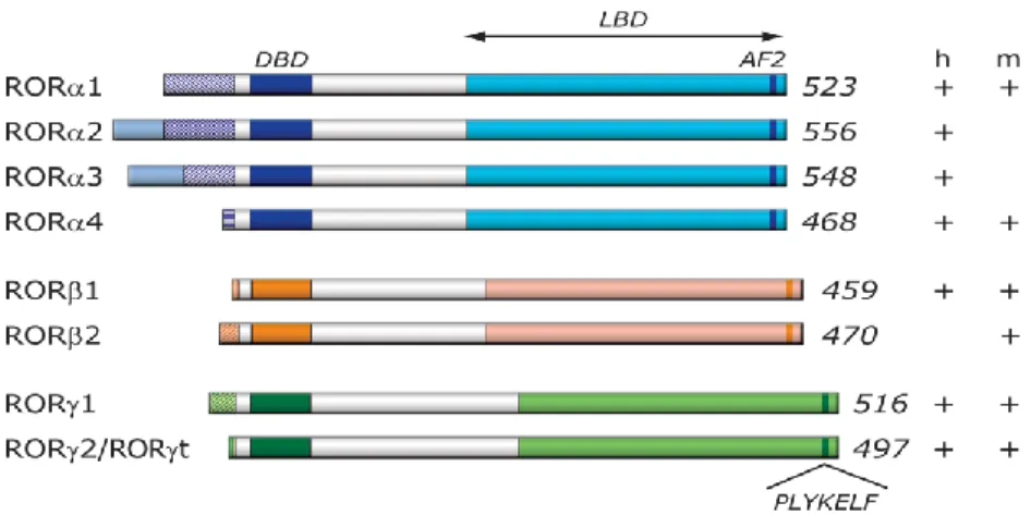
The RORα human gene spans a relatively large 730 kb genomic region. The RORβ and RORγ genes cover approximately 188 and 24 kb, respectively. As a result of alternative promoter usage and exon splicing, each ROR gene generates several isoforms that differ only in their amino-terminus (57,59)
19
Figure 2- Schematic representation of ROR family members. Schematic structure of the various ROR isoforms.
The different ROR isoforms identified in human and mouse are shown on the right (+/-) (109)
Four human RORα isoforms, referred to as RORα1-4, havebeen identified, while only two isoforms, α1 and α4, have been reported for mouse.
The mouse RORβ gene generates two isoforms, β1 and β2, while humans appearto express only the RORβ1 isoform (57). Both the mouse and human RORγ gene generate two isoforms, γ1 and γ2 (60). Most isoforms exhibit a distinct pattern of tissue-specific expression and are involved in the regulation of different physiological processes and target genes. For example, human RORα3 is only found in human testis (61). RORα1 and RORα4 are both prominently expressed in mouse cerebellum, while other mouse tissues expresspredominantly RORα4 (62). In the mouse, expression ofRORβ2 is restricted to the pineal gland and the retina, while RORβ1 is the predominant isoform in cerebral cortex, thalamus, and hypothalamus (57). RORγ2, most commonly referred to as RORγt, is exclusively detected in a few distinct cell types of the immune system, while RORγ1 expression is restricted to several other tissues (63).
Expression of the nuclear hormone receptor RORC has been reported in Th17 cells, Th22 cells, γδ T cells, NKT cells and CD4+CD8+ thymocytes.
20
In addition, RORC is expressed by cells that do not belong to the T or B
cell lineage, are thymus and recombinase activating geneindependent, yet have a lymphoid-like morphology (64,65). These cells are currently referred to as RORC expressing innate lymphoid cells (RORC+ ILC) (66).
Although most ROR isoforms are under the control of different promoters, little is known about the transcriptional regulation of their tissue-specific expression.
1.4.3 ROR protein structure
The ROR genes encode proteins of 459 to 556 amino acids (Figure 1).
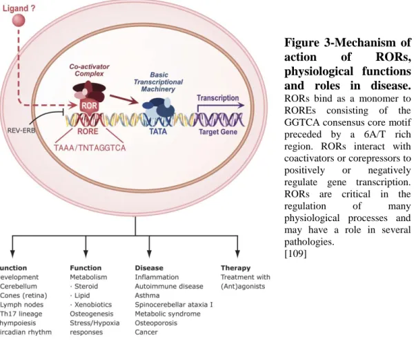
RORs exhibit a typical nuclear receptor domain structure consisting of four major functional domains: an N-terminal (A/B) domain followed by a highly conserved DNA-binding domain (DBD), a hinge domain, and a C-terminal ligand-binding domain (LBD). RORs regulate gene transcription by binding to specific DNA response elements (ROREs), consisting of the consensus RGGTCA core motif preceded by a 6-bp A/T-rich sequence, in the regulatory region of target genes (67). RORs bind ROREs as a monomer and do not form heterodimers with retinoid-X receptors (RXRs) (57) (Figure 3).
21
Figure 3-Mechanism of
action of RORs,
physiological functions and roles in disease. RORs bind as a monomer to ROREs consisting of the GGTCA consensus core motif preceded by a 6A/T rich region. RORs interact with coactivators or corepressors to positively or negatively regulate gene transcription. RORs are critical in the
regulation of many
physiological processes and may have a role in several pathologies.
[109]
Although RORα-γ and their different isoforms recognize closely-related ROREs, they exhibit distinct affinities for different ROREs. The amino-terminus (A/B domain) has been shown to play a critical role in conferring DNA binding specificity to the various ROR isoforms. In addition to the RORE sequence and the amino terminus, the promoter context may play an important factor in determining which ROR is recruited to a particular RORE. The LBDs of nuclear receptors are multifunctional and play a role in ligand binding, nuclear localization, receptor dimerization, and provide an interface for the interaction with coactivators and corepressors. X-ray structural analysis demonstrated that RORs have a secondary domain structure that is characteristic of that of nuclear receptors (68).
22
The LBDs of RORs contain, in addition to the typical 12 canonical α-helices (H1-12), two additional helices, H2’ and H11’. The activation function 2 (AF2) in H12 consists of PLYKELF, which is 100% conserved among RORs (Figure 1). Deletion of the H12 or point mutations within H12 causes loss of the ROR transactivation activity and results in a dominant-negative ROR (69).
It is believed that H10 plays a critical role in the homo- and heterodimerization of nuclear receptors. Structure analyses revealed the presence of a link in H10 of the LBD of RORα and RORβ that would greatly affect the dimerization capability of RORs (68). This is consistent with the conclusion that RORs do not form homodimers or heterodimers with other RORs or RXRs.
1.4.4 RORs: ligand-dependent transcription factors
The ligand binding domains of NRs are multifunctional.
Typically, ligand binding induces a conformational change in the receptor resulting in dissociation of corepressors and recruitment of co-activators (62).
However, RORs are constitutively active meaning that they are in an active conformation in the absence of ligand and that ligand binding might actually repress receptor activity (inverse agonist). While identification of the endogenous ligands for RORs has been controversial, recent evidence suggests that, similar to the liver X receptor’s (LXRs), oxygenated sterols may function as high affinity ligands. Indeed, 7- oxygenated sterols (7α-OHC, 7β-OHC, and 7-ketocholesterol) function as inverse agonists to both RORs. The 7-oxygenated sterols bind to both RORα and RORγ isoforms with an affinity significantly greater than the affinity for cholesterol and cholesterol sulfate, and suppress their transactivation properties. It was also shown that both RORα and RORγ are constitutively active in the absence of ligand, able to bind co-activator peptides, and activate transcription. Several other endogenous RORα and RORγ ligands have been described recently. 24Shydroxycholesterol (24S-OHC) is a high affinity ligand for RORα and RORγ,
23
and similar to the 7-oxygentated sterols, 24S-OHC acts as an inverse agonist and dose dependently reduces the RORα and RORγ constitutive activity (70).
X-ray structure analysis of the RORβ(LBD) identified stearic acid as a fortuitously-captured ligand that appeared to act as a stabilizer by filling the ligand-binding pocket, rather than as a functional ligand (71). Subsequently, several retinoids, including all-trans retinoic acid (ATRA) and the synthetic retinoid ALRT 1550 (ALRT), were identified as functional ligands for RORβ (72).
ATRA and ALRT 1550 were able to bind RORβ(LBD) reversibly and with high affinity and reduced RORβ-mediated transcriptional activation, suggesting that they act as partial antagonists. These retinoids were also able to bind RORγ and inhibit RORγ-mediated transactivation, but did not bind RORα or affect RORα-induced transactivation (72). Although future research needs to determine whether in vivo ROR activity is regulated by endogenous ligands, these crystallographic and structural studies do support the concept that ROR activity can be modulated by specific endogenous and/or synthetic (ant)agonists.
This conclusion is highly relevant to the emerging roles of RORs in several pathologies, including inflammation, various autoimmune diseases, obesity, and asthma, and the promise that these receptors might serve as potential targets for pharmacological intervention in these diseases (Figure 3).
24
1.4.5 Role of RORγ in the development of secondary
lymphoid tissues
RORγ1 and RORγt (RORγ2) exhibit distinct patterns of tissue-specific expression. RORγ1 is expressed in many tissues, including liver, adipose, skeletal muscle, and kidney, while the expression of RORγt is exclusively expressed in a few distinct cell types of the immune system [73,74]. Mice deficient in RORγ expression lack lymphnodes and Peyer's patches (PPs), suggesting that RORγ is indispensable for lymph node organogenesis and development of PPs [73,74].
Lymphoid tissue inducer (LTi) cells play a critical role in the development of lymph nodes and PPs. LTi cells originate from hematopoietic precursor cells in the fetal liver and have been recently characterized as CD45intCD4+CD3-CD127 (IL-7Rα)+Lin- cells in mice and in humans as lineage-negative CD45intCD4-CD3 -CD127hiLin- cells [73]. LTi cells are absent from spleen, mesentery, and intestine of RORγ-/- E18.5 embryos, indicating that RORγt plays a critical role in the generation and/or survival of LTi cells [73]. As a consequence, the lack of lymph nodes and PPs in RORγt- or RORγ-deficient mice is due to the absence of LTi cells.Besides PPs and mesenteric lymph nodes, the intestinal immune system contains several other lymphoid cell compartments, including cryptopatches and isolated lymphoid follicles (ILFs) [75]. ILFs develop from cryptopatches in response to inflammatory innate immune signals generated by the colonization of the intestine by bacteria. Lin-cKit+CD127+CD44+ cells in cryptopatches and in ILFs express high levels of RORγt and appear to constitute the adult counterpart of LTi cells [74,75]. The deficiency in cryptopatches and ILFs observed in RORγ -/-mice indicate that RORγt is essential for the development of these cell compartments and is due to the absence of these LTi-like cells in these mice.
25
1.4.6 Critical functions of RORγ in thymopoiesis
Several studies have demonstrated that RORγt plays a critical role in the regulation of thymopoiesis [76]. During thymopoiesis, T cell precursor CD25-CD44+CD4 -CD8- cells (DN1) differentiate successively via two intermediate stages, CD25+CD44+ (DN2) and CD25+CD44- (DN3), into CD44-CD25- (DN4) thymocytes. These cells then differentiate via immature single positive (ISP) cells (CD3-CD4-CD8low) into double positive CD4+CD8+ (DP) thymocytes.
After successful T cell receptor α (TCRα) gene rearrangement, DP cells expressing TCRαβ receptor undergo a careful selection process to eliminate thymocytes expressing nonfunctional or autoreactive TCR. The positive selected DP thymocytes mature into single positive (SP) CD4+CD8- helper and CD4-CD8+ cytotoxic T cells that then colonize the secondary lymphoid organs, including the spleen, lymph nodes, and PPs. RORγt is transiently expressed during thymopoiesis [76]. It is undetectable in DN thymocytes and highly induced when ISP cells differentiate into DP thymocytes and again down-regulated when DP thymocytes differentiate into mature T lymphocytes. At birth and during early stages of life, RORγ null mice have a significantly smaller thymus compared to wild type mice as a result of a drastic reduction in the number of double positive DP and SP thymocytes, while the percentage of ISP thymocytes is greatly increased [73]. The accumulation of ISP cells in RORγ-/- mice appears to be due to a delay in the differentiation of ISP into DP cells, suggesting a role for RORγt in the regulation of the ISP-DP transition [76]. In addition, DP thymocytes undergo massive apoptosis in vivo and in vitro [78]. The accelerated apoptosis of RORγ-/- DP thymocytes is related to reduced expression of the anti-apoptotic gene Bcl-XL.
This repression is an early and key event in accelerated apoptosis in RORγ -/-thymocytes [78]. Thus, RORγ functions as a positive regulator of Bcl-XL expression and, as a result, promotes the survival of DP thymocytes, thereby enabling gene rearrangement
26
1.4.7 RORs and cellular metabolism
Accumulating evidence indicates that RORs play an important role in the regulation of several metabolic pathways, particularly lipid and steroid metabolism [79]. Initial characterization of RORγ−/− mice revealed that they display normal cholesterol and triglyceride levels, with slightly lower blood glucose levels than their wt counterparts [80]. However, recent evidence suggests that RORγ may indeed have a role in metabolism through regulation of adipogenesis and insulin sensitivity. Meissburger et al. demonstrate that RORγ is a negative regulator of adipocyte differentiation in vitro. When overexpressed during adipocyte differentiation, RORγ decreases the amount of differentiated adipocytes.
However, in vivo differentiation of adipocyte precursors in RORγ−/− mice was enhanced but showed decreased size. The smaller adipocytes were insulin sensitive and protected the mice from obesity induced hyperglycemia and insulin resistance [81]. Moreover, analysis of adipose stromal-vascular fractions from obese human subjects demonstrated a positive correlation between RORγ expression and adipocyte size that was negatively correlated with adipogenesis and insulin sensitivity. These findings suggest that RORγ may be a novel target for the treatment of obesity-associated insulin resistance [80]. The deletion of both RORα and RORγ exhibit similar changes in cholesterol, triglyceride, and blood glucose levels as the single knockout mice. Gene expression analysis from livers of double knock out (DKO) mice suggests a degree of functional redundancy between RORα and RORγ which is most likely due to the similarities in RORE binding affinities. However, the recent evidence regarding obesity and insulin resistance in the RORγ−/− mice highlights the differences between the two NRs in metabolic processes.
27
1.4.7 RORs and T cell lineage specification
1.4.8 Role of RORs in Th17 cell differentiation
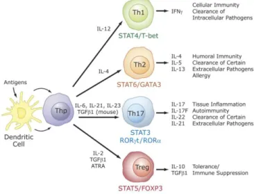
Recent studies identified a critical role for RORs in the regulation of lineage specification in helper T cells (82). Differentiation into different effector CD4+ T cell lineages, T helper (Th) 1, Th2, Th17, and T regulatory (Treg or Th3) cells is initiated through an interaction of dendritic cells with uncommitted (naïve) CD4+ T helper cells (Thp). Th1, Th2, Treg, and Th17 are characterized by their synthesis of specific cytokines and their immuno-regulatory functions (Figure 4).
Figure 4- Specific role for RORs in T cell lineage specification. Differentiation into different effector CD4+ T cell lineages, T helper (Th) 1, Th2, Th17, and T regulatory (Treg) cells. [109]
Interferon γ is the signature cytokine produced by Th1 cells, while IL-4, IL-5 and IL-13 are major cytokines produced by Th2 cells. The recently discovered Th17 cells produce IL-17A (IL-17), IL-17F, IL-21, and IL-22 as major cytokines, while Treg cells synthesize IL-10 and TGFβ1 (83,84). T helper and Treg cells play a critical role in several inflammatory responses, including adaptive immune responses to various pathogens.
28
Host defense is coordinated by the proinflammatory Th1, Th2, and Th17 cells, while Treg cells are involved in the down regulation and contraction of an immune inflammatory response. Th17 cells are believed to be the major proinflammatory cells involved in autoimmunity, while Treg cells protect against autoimmunity (84). Differentiation into Treg cells and Th17 cells is often reciprocal, involving several positive and negative regulatory networks that favor one or the other lineage (74). This includes a yin-yang relationship between RORγt and FOXP3 expression and their regulation by several cytokines, transforming growth factor β (TGFβ), and ATRA. Littman, Cua, and their colleagues were the first to report that RORγt is required for the differentiation of naïve CD4+ T cells into Th17 cells (85). This was supported by findings from several other laboratories (84).
RORγt is induced during differentiation of antigen-stimulated Thp cells along the Th17 lineage in response to IL-6 or IL-21 and TGFβ. Cells deficient in IL-6 do neither express RORγt nor IL-17F and IL23R (86). IL-6 mediates its action through activation of STAT3. Deficiency in STAT3 greatly impaired the activation of RORγt expression and Th17 differentiation, suggesting that this induction is STAT3-dependent (87). Inversely, deficiency in suppressor of cytokine signaling 3 (SOCS3), a negative regulator of STAT3 activity, enhances Th17 differentiation (88). Whether STAT3 regulates RORγt expression directly by binding to the RORγt promoter needs to be established. Additional evidence for a role of RORγt in Th17differentiation came from studies showing that Thp cells isolated from RORγ null mice exhibited a marked reduction in their ability to undergo differentiation along the Th17 lineage (89). Conversely, exogenous expression of RORγt in Thp cells greatly increased the expression of IL-17 cytokines and IL23R in the absence of cytokines. Subsequent studies showed that, like RORγt, RORα is highly induced during Th17 cell differentiation in a STAT3-dependent manner (90).
29
Deficiency in RORα reduced IL-17 and IL23R, but not IL-17F or IL-22 expression, while exogenous expression of RORα in Thp cells or Jurkat cells enhanced IL-17, IL-17F, IL-22, and IL23R expression (90). These observations indicate that RORα also functions as a positive regulator of Th17 differentiation, suggesting a degree of functional redundancy between RORγt and RORα. This was consistent with findings showing that Th17 differentiation was completely impaired in Thp cells deficient in both RORα and RORγ (90). Thus, both RORα and RORγt are required for optimal differentiation of Thp cells into Th17 cells; however, RORγt appears to be the major player in this process because RORγt deficiency has a more pronounced effect on the expression of Th17 cytokines than loss of RORα. Whether RORα and RORγ have any unique roles in the regulation of gene expression during or after Th17 differentiation needs further study.
In addition to the induction of RORγt, IL-17, and IL-17F, differentiation along the Th17 lineage is associated with increased expression of IL-21 and IL23R (86). IL-21 expression is not affected in RORγ-deficient mice, suggesting that this regulation occurs upstream of RORγ. In contrast to IL-21, the induction of IL23R is greatly reduced in RORγ-deficient mice, suggesting that its expression is regulated by RORγt.
30
1.4.10 Transcriptional regulation of IL-17 positive cell
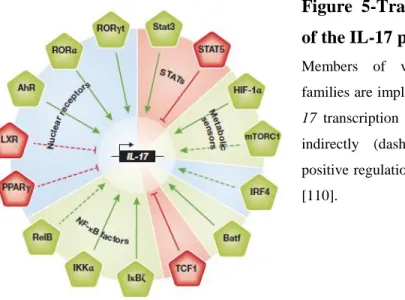
A substantial amount of work concentrated on the signaling transcriptional events that occurring during Th 17 development (Figure 5).
Figure 5-Transcriptional regulation of the IL-17 program.
Members of various transcription factor families are implicated in the regulation of
IL-17 transcription either directly (solid line) or
indirectly (dashed line). Green indicates positive regulation and red negative regulation [110].
The transcription factors Rorγt, AhR and IκBζ are specifically induced during Th17-cell development and have key roles in their differentiation and function (91,92). STAT3, IRF4, Runx1 and Batf, on the other hand, influence Th17 development, but their expression is not restricted to Th17 cells (93,94).
As anticipated, the transcriptional modulators that target these key transcription factors can either enhance or impair the differentiation of Th17 cells.
The positive and negative regulators of the key transcription factor IRF4—ROCK2 and IRF4 binding protein, respectively were clearly shown to alter the activity of IRF4, thereby resulting in the spontaneous development of autoimmune disorders in mice with aberrantly activated ROCK2 and deficient in IRF4 binding protein (95).
31
The nuclear receptor LXR negatively regulates the IL-17 immune response by upregulating the transcription factor Srebp-1c, which interacts physically with AhR to interfere with its function in promoting Th17 development (96).
In addition, the nuclear receptor PPARγ was shown to negatively regulate Th17 differentiation by repressing the induction of Rorγt. Similarly to the differential role of STAT4 and STAT6 for Th1 and Th2 differentiation, respectively, STAT3 downstream from IL-6 signalling has a central role in the initiation of Th17 development. The components of the NF-κB signalling pathway regulate numerous immune responses, and their dysregulation is linked to inflammatory and autoimmune diseases, as well as cancer (97). Interestingly, several of these molecules regulate Rorγt and IL-17A expression. Thymic γδT cells express the surface lymphotoxin-β-receptor and, after its ligation, RelB mediates the induction of Rorγt in γδ T cells, but not αβ T cells, which is required for the differentiation of IL-17-producing γδ T cells and an innate IL-17 response to Escherichia coli infection (98). Besides NF-κB, kinase inhibitors for NF-κB signalling also play a role in Th17 differentiation. Although IKKα was originally identified as an inhibitor of NF-κB, IKKα can promote IL-17 transcription in Th17 cells by binding to the IL-17 locus in a NF-κB-independent way (99).
Furthermore, IκBζ is specifically induced during Th17 differentiation and, in cooperation with Rorγt, is essential for robust IL-17 transcription (91).
Several groups have demonstrated recently that the kinase mammalian target of rapamycin (mTOR) and the transcription factor HIF1-α, which are cellular metabolic sensors, control Th17 fate determination. The mTOR complex 1 (mTORC1), but not mTORC2, regulates the small GTPase Rheb signalling pathway, which promotes Th1 and Th17, but not Th2, differentiation (100).
HIF-1α is upregulated during Th17 differentiation, possibly in a STAT3-and mTOR-dependent manner, which in turn enhances Rorγt expression.
After this event, a molecular complex with STAT3, HIF-1α, Rorγt and P300 induces the robust expression of TH17-associated genes (101).
32
1.4.11 Suppression of ROR transcriptional activity by
FOXP3
Treg and Th17 cells are reciprocally regulated, involving several positive and negative regulatory networks [83,84]. The balance between the expression of RORγt and FOXP3 plays a critical role in determining whether uncommitted CD4+ T helper cells differentiate into Treg or Th17 cells (Figure 6).
Regulation of FOXP3 expression by TGFβ and IL-6 plays a critical role in Treg and Th17 lineage determination [83,84]. In the presence of TCR engagement, TGFβ induces FOXP3 expression and Treg differentiation in murine Thp cells. Although TGFβ can moderately enhance the expression of RORα and RORγt, FOXP3 is induced at much higher levels, thereby shifting the ROR/FOXP3 balance towards FOXP3 and Treg differentiation [86,102]. Addition of IL-6 or IL-21 inhibits the induction of FOXP3 by TGFβ and activates RORα and RORγt expression, thereby shifting the ROR/FOXP3 balance in favor of Th17 differentiation. These observations indicate that FOXP3 represses Th17 differentiation by antagonizing RORγt function rather than inhibiting its expression [83]. This antagonism is mediated through a direct interaction of FOXP3 with RORα and RORγt.
33
1.4.12 Interaction with coregulatory proteins
For many receptors, binding of a ligand functions as a switch that induces a conformational change in the receptor that involves a repositioning of H12 (AF2) [83]. When RORs are in a transcriptionally-active conformation, H12 with H3 and H4 form a hydrophobic cleft and a charge clamp that involves the participation of a conserved Lys in H3 and a conserved Glu in H12 within RORs [68].
The clamp serves as an interaction interface for LXXLL motifs present in coactivators and related motifs in corepressors [95].
Receptor:coactivator complexes, through their histone acetylase activity, induce histone acetylation that results in decompaction of chromatin and increased transcription of target genes [94], while receptor association with corepressor complexes leads to histone deacetylation and subsequently compaction of chromatin and repression of gene expression [95]. RORs interact with corepressors, as well as coactivators, suggesting that they can function both as repressors and activators of gene transcription. Recently, repression of ROR-mediated transcriptional activation by the forkhead box transcription factor p3 (FOXP3) was shown to depend on a direct interaction between RORs and FOXP3 [103].
34
1.5 RORs and cancer
1.5.1 RORs and increased cancer susceptibility
A number of studies have provided evidence a role for RORs in cancer.
Mice deficient in the expression of RORγ exhibit a high incidence of thymic lymphomas that metastasize frequently to liver and spleen [104]. As a consequence, the lifespan of RORγ null mice is greatly reduced. The enhanced lymphoma formation may be related to changes in thymic homeostasis observed in RORγ KO mice [105]. Although the molecular mechanism underlying enhanced lymphoma formation has not yet been established, it may be related to the dysregulation of differentiation and proliferation in RORγ-/- thymocytes.
Interestingly, type B leukemogenic virus (TBLV), which causes T-cell lymphomas in mice, was found to be frequently integrated at the RORγ locus [106].
However, in contrast to RORγ null mice, RORγ expression correlated positively with lymphomas. Whether RORγ is implicated in human lymphomas requires further study. Another potential link between RORγ and cancer is emerging from studies showing increased expression of Th17-associated genes, including RORγ, IL-17, and IL-23, in gastric tumors, an increase in the population of circulating Th17 cells in gastric cancer patients [107], and a high incidence of Th17 cells at sites of ovarian cancer [108]. These elevated levels of proinflammatory cytokines may contribute to cancer pathogenesis. Alterations in RORγt expression or activity may cause changes in the Th17 cell population and production of proinflammatory cytokines, thereby affecting cancer progression positively or negatively.
35
1.5.2 Tumour-associated Th17 cells
Recently, TH17 cells have been investigated in patients with diverse cancer types, including ovarian cancer and prostate cancer.
Although not the predominant T cell subset within the tumour, Th17 cells are present in the tumour microenvironment. Th17 cells do not home to lymphoid tis-sues, but some of the above homing molecules might be involved in Th17 cell migrate to inflammatory tissues and tumours [111]. Th17 cells express other cytokines in addition to IL-17, and this might be functionally relevant in several physiological and pathological settings. Human tumour-infiltrating Th17 cells express negligible levels of the anti-inflammatory cytokine IL-10, but around 50–90% of Th17 cells produce high levels of effector cytokines such as IL-2, (GM-CSF), interferon-γ (IFNγ) and tumour necrosis factor (TNF) [111].
Therefore, tumour-associated TH17 cells exhibit an effector T cell cytokine profile similar to that of effector T cells that have been described in infectious diseases [112,113]. A similar cytokine profile has been observed in TH17 cells associated with distinct human tumour types, including carcinomas of the skin, intestine, pancreas, liver and ovaries [111]. These data indicate that TH17 cells might have a protective role in tumour immunopathology by promoting antitumour immunity.
1.5.3 Evidence for antitumour activity
The relationship between Th17 cells and tumour immunopathology is controversial [114]. However, there are several lines of evidence suggesting that Th17 cells can promote protective antitumour immune responses. Firstly, tumour-infiltrating Th17 cells express several effector cytokines, similar to that observed in patients with infectious diseases [112,113]. This suggests that tumour-associated Th17 cells might be functional effector T cells. Consistent with this possibility, Th17 cells are negatively correlated with the presence of T-Reg cells[111] and are positively correlated with effector immune cells, including IFNγ+ effector T cells, cytotoxic
36
CD8+ T cells and natural killer (NK) cells, in the same tumour microenvironment [111]. These observations are supported by data from both human and mouse tumours [111]. Transgenic T cells polarized to a Th17 cell phenotype following treatment with TGFβ and IL-6 were shown to induce tumour eradication in mice [115]. In addition, IL-17-deficient mice show accelerated tumour growth and lung metastasis in many tumour models, and forced expression of IL-17 in tumour cells was shown to suppress tumour progression [116]. In patients with prostate cancer, a significant inverse correlation is found between Th17 cell differentiation and tumour progression [117]. Treatment with specific antibody against cytotoxic T lymphocyte antigen 4 (CTLA4) [118] induces Th17 cells in patients with melanoma and the levels of IL-17 detected in tumour-associated ascites positively predicts patient survival. Taken together, these data provide strong evidence that Th17 cells can have protective roles in tumour immunity.
1.5.4 Pro-tumour role of Th17 cell-associated cytokines
Th17 cells infiltrate into tumor sites and draining lymph nodes in cancer patients [119]. The number of IL-17A-producing cells correlates with poor survival in patients with hepatocellular carcinoma, whereas the number was decreased in patients with advanced ovarian cancer, lung cancer, or non-Hodgkin’s lymphoma. Thus, Th17 cells and IL-17A may play different roles depending on the tumor type and stage. Transplantation of IL-17A-overexpressing tumor cells into immunodeficient mice induced angiogenesis through induction of vascular endothelial growth factor (VEGF) expression, resulting in enhanced tumor growth [120]. T cellderived IL-17A also dramatically increases the release of angiogenic and chemoattractive IL-8 from tumor cells [121]. Delayed growth of subcutaneously transplanted B16 melanoma cells and MB49 bladder carcinoma cells was observed in IL17a_/_ mice, whereas IFNγ_/_ mice showed accelerated growth and augmented IL-17A production in the tumor [122].
37
In this case, IL-6 was expressed in response to IL-17A-activated Stat3 in the tumor cells, upregulating the expression of prosurvival and proangiogenic genes.
IL-17A is also involved in the development of colonic cancer in multiple intestinal neoplasia (Min) mice, a model of familial adenomatous polyposis [123]. Interestingly, enterotoxigenic Bacterioides fragilis (ETBF), an intestinal commensal bacteria, can accelerate colonic inflammation and tumor formation in Min mice [124]. In this model, ETBF selectively induced Th17 cell differentiation via Stat3 activation, and blocking IL-17A as well as IL-23R dramatically reduced tumor development indicating that a Stat3- and Th17 cell-dependent pathway contributed to the disease. It was shown that IL-17 is required for the development of MDSCs in tumor-bearing mice. A defect in IL-17R reduces the number of MDSCs in the blood, spleen, and tumors. This is further supported by data showing that the administration of exogenous IL-17 increases the number of MDSCs in wild-type bearing mice, whereas neutralization of IL-17 in wild-type tumor-bearing mice reduced the number of MDSCs [125].
TAM and MDSCs both generally recognized as dominant promoting forces, produce large amounts of the Th17 cell driving cytokines TGFbeta and IL-6 [126]. IL-23 is an IL-12 cytokine family member, which is produced by APCs and
promotes the expansion and survival of Th17 cells. It has been reported that IL-23-deficient mice are resistant to chemically induced tumours [127].
This resistance is associated with decreased expression of matrix metalloproteinase 9 (MMP9) in the skin, a decrease in the expression of angiogenic markers and high levels of CD8+ T cell infiltration. Given the close relationship between IL-23 and Th 17 cells, it has been proposed that Th17 cells or IL-17 derived from Th17 cells can promote tumorigenesis in this model. However, this hypothesis remains to be tested and antitumour effects of IL-23 have been observed in several mouse tumour models [128].
38
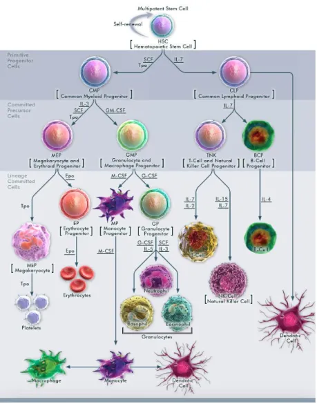
1.6 Hematopoiesis
Hematopoiesis is a process of blood cell production and maturation in the bone marrow hematopoietic stem cells. The process begins with the hematopoieti stem cell. (HSCs). Because mature blood cells are predominantly short lived, stem cells are required throughout life to replenish multilineage progenitors and the precursors committed to individual hematopoietic lineages. HSCs reside as rare cells in the bone marrow in adult mammals and sit atop a hierarchy of progenitors (common myeloid progenitor (CMP), common lymphoid progenitor (CLP)) that become progressively restricted to several or single lineages. These progenitors yield blood precursors devoted to unilineage differentiation and production of mature blood cells, including red blood cells, megakaryocytes, myeloid cells (monocyte/macrophage and neutrophil), and lymphocytes. As with all other stem cells, HSCs are capable of self-renewal the production of additional HSCs and differentiation, specifically to all blood cell lineages. HSCs are defined operationally by their capacity to reconstitute the entire blood system of a recipient. Developmentally, there are two waves of hematopoiesis, so probably with two corresponding origins for HSCs [130]. Primitive hematopoiesis, distinguished from definitive hematopoiesis by large and nucleated erythrocytes and specifically expressed fetal hemoglobin isoforms, occurs as a transient wave preceding the advent of definitive hematopoiesis. Primitive hematopoiesis in mammals begins within the blood islands in yolk sac. Independently, the stem cells for definitive hematopoiesis originate within embryonic SP/AGM (splanchnopleur, aorta, gonads, and mesone-phros). Only these definitive hematopoietic stem/progenitor cells originating from AGM, but not primitive HSCs are able to repopulate the entire hematopoietic system in the lethally irradiated adult recipient mice [131].
39
Definitive HSCs expand in number in the AGM, and then migrate to and colonize fetal liver and spleen where they continue to differentiate into recognizable hematopoietic precursors. After birth, definitive hematopoiesis is primarily confined to bone marrow, and in some pathological conditions also to extramedullary sites such as spleen, liver, and occasionally lung and brain.
Hematolymphoid differentiation and maturation from HSCs to terminally differentiated cells involves loss of self-renewal capacity, transient acquisition of high proliferative potential, and subsequent restriction to a mature blood cell lineage, that, with the exception of memory T and B cells as well as some tissue macrophages and Langerhans cells, is postmitotic. The sequence of differentiation events (Figure 6) goes from HSCs to nonself-renewing multipotent progenitors (MPPs). MPPs, via intermediate populations debated in their specific details elsewhere, give rise to CMPs, and CLPs. The CLP and CMP are considered as primitive progenitor cells which then give rise to more-differentiated progenitors (also termed as committed precursor cells). Ultimately, these cells give rise to unilineage committed progenitors. Each step of the Hematopoietic progression that is survival, proliferation or differentiation is well supported by cytokines and growth factors.
40
Figure 6- Hematopoiesis from multipotent stem cell
Hematopoiesis is a hierarchical progression of Multipotential HSCs that gradually lose one or more developmental options, becoming progenitor cells committed to a single lineage; these progenitors then mature into the corresponding types of peripheral-blood cells. Each step of the Hematopoietic progression that is survival, proliferation or differentiation is well supported by cytokines and growth factors (ex. GM-CSF (Granulocyte-Macrophage Colony-Stimulating Factor), G-CSF (Granulocyte Colony Stimulating Factor), M-CSF(Macrophage Colony-Stimulating Factor)). [129]
41
1.6.1 Bone marrow stem cell niche
Key properties of stem cells such as their self-renewal and developmental capacity can be controlled in a nonautonomous manner by their cellular microenvironment. Bone marrow microenvironment is usually referred to as a stem cell niche. A niche is a group of stromal cells that allows a stem cell to maintain its identity [132]. The cells of a niche will prevent a previously specified cell from losing its stemness through loss of quiescence and potency or precocious differentiation. Bone marrow stromal cells are known to provide microenvironmental support for hematopoietic stem cells through the secretion of growth factors and cytokines (Figure 6), but also through direct contact in cell–cell interactions and through production of extracellular matrix [133]. All the stromal cells in bone marrow, except macrophages, originate from pluripotent bone marrow mesenchymal stem cell (BM-MSCs) that exhibit ability to self-renew as well as to differentiate into cells of mesodermal lineage, including osteocytes, adipocytes, chondrocytes, myocytes, tenocytes, myocardiocytes, and hematopoietic supportive stroma [133]. Although the concept of niches is well accepted and experimentally proven in various invertebrate systems, mammalian stem cell niches remain poorly understood, as the precise location of the stem cells themselves often remains elusive. Several studies have attempted to localize HSCs in bone sections or by using confocal/two-photon intravital imaging based on three-color fluorescence microscopy [134]. Putative HSCs have been found near the endosteum lined by OBs (endosteal niche) or in association with sinusoidal endothelium (perivascular niche). Moreover, the location of some important cellular components of the niches may not be restricted to the endosteum or the perivascular niche area, but may be part of both environments, raising the possibility that both niches and the location of HSCs may not be as distinct as is currently assumed.
42
1.7 T cell in regulation of hematopoiesis
The involvement of the immune system in regulation of hematopoiesis was suggested during 1970s of the last century.
Different studies pointed to the cooperation between hematopoietic cells and T cells for the maintenance of optimal hematopoiesis in mice [135,136].
Further research has demonstrated that thymus-derived T lymphocytes can produce cytokines that have powerful effects on hematopoiesis.
Understanding how T cells regulate both steady-state hematopoiesis and induced hematopoiesis during an immune response is critically important for increasing our knowledge of both hematopoietic control and immune regulation.
The potential of the specific T helper cell subsets, Th1 and Th2 cells, to modulate the hematopoietic response in different ways, although not well characterized, has been demonstrated and reported [137]. However, defining the role of newly recognized subset of IL-17- secreting T cells, Th17 cells, as well as the regulatory T cells (T-regs), in regulation of hematopoiesis is still in its infancy.
Only recent data implied to the presence of T-regs in hematolymphoid compartments and demonstrated the ability of T-regs to modulate hematopoietic progenitor cells activity, suggesting that T-regs are not restricted in their regulatory actions within the adaptive and innate immune systems [138].
As for the IL-17, even the early studies pointed to this cytokine as an example of bridging between T-cell function and haematopoiesis, demonstrating that IL-17 induces production of various pro-inflammatory cytokines, but also those with haematopoietic effects, such as GM-CSF, G-CSF (granulocyte-colony-stimulating factor), and IL-6 [139]. As the understanding of its function improved, it was shown that IL-17 is at the heart of a regulatory circuit controlling neutrophil homeostasis, while other studies demonstrated that IL-17 also affects other cells of hematopoietic system, such as erythroid progenitors, as well as mesenchymal stem cells.
43
These findings focused the attention on molecular mechanisms underlying
the effects of IL-17 on hematopoietic and mesenchymal stem cells in order to better understand the regulatory role of IL-17 in hematopoiesis, both during the normal, steadystate hematopoiesis and disturbed, altered hematopoietic responses.
1.7.1 Effects of IL-17 on hematopoietic cells
Initial evidence of IL-17’s stimulatory effect on myeloid cells was obtained after in vitro experiments which demonstrated that IL-17 can induce the proliferation of CD34 + human stem cells, as well as their maturation into neutrophils, only when cocultured with fibroblasts [140]. This way it was suggested that IL-17 affects hematopoietic stem cells indirectly, through the induction of fibroblasts to release secondary cytokines with hematopoietic effects, including G-CSF and IL-6.
Further on, the in vivo expression of IL-17 in an experimental model of adenovirus-mediated gene transfer of the murine IL-17 cDNA induced a profound stimulation of both bone marrow and splenic granulopoiesis and led to expansion of myeloid hematopoietic stem and progenitor cells (high proliferative potential colonies—HPPC, colony-forming-unit–granulocyte-erythrocyte-megakaryocyte-monocyte—CFU-GEMM, and colony-forming-unit-granulocyte–macrophage— CFUGM) and neutrophilia, in part through release of secondary cytokines, such as G-CSF [141]. Data obtained in the same experimental model also showed that for optimal granulopoiesis, beside G-CSF release, IL-17-mediated effects require the presence or induction of the transmembrane form of stem cell factor [142] and confirmed that IL-17 stimulates the production and differentiation of granulocyte lineage cells by inducing secondary hematopoietic cytokines [143].
Data, obtained in a different experimental approach, demonstrated that even a single injection of IL-17 recombinant protein elicits a cascade of biological
44
changes in vivo, affecting the cells of granulocytic lineage, as well as the levels of cytokines released, primarily IL-6, in both murine bone marrow and spleen [144]. The most obvious in vivo response of granulocytic cells to IL-17 administration was observed at the level of bone marrow morphologically recognizable proliferative granulocytes and spleen metamyelocytes, indicating that more mature progenitors respond first to its action. The role of IL-17 in erythropoiesis has not been as extensively studied as its role in granulopoiesis.
Different studies demonstrated that hematopoietic effects of IL-17 were highly dependent on the microenvironment, since the effects on the erythroid compartments in mouse spleen were opposite to those observed in the bone marrow. The differences were attributed to different cytokine profiles induced by IL-17 related to the tissue microenvironment in which hematopoiesis occurs [144]. Beside the influence of IL-17 during steady-state hematopoiesis, its effects on hematopoietic cells during the state of disturbed or altered hematopoiesis were also analyzed. Tan W. et al. 2006 showed that following sublethal radiation-induced myelosuppression, in vivo overexpression of murine IL-17 substantially enhances granulopoietic restoration, characterized by increase in neutrophils, as well as both splenic and bone marrow progenitor cells [145]. Data obtained during the naturally acquired Syphacia obvelata infection in mice confirmed that the activity of IL-17 differs depending on physiological/pathological status of the organism.
It was demonstrated that this pinworm parasite induces significant hematopoietic changes during infection, characterized by increased myelopoiesis and erythropoiesis in infected animals. This stimulation of hematopoiesis was accompanied by altered sensitivity of the bone marrow myeloid and erythroid progenitors from infected mice to IL 17, as compared to non-infected controls, and the changes in reactivity were manifested both at the cellular and molecular level [146,147].