Michele Abate Matteo Guelfi Andrea Pantalone Daniele Vanni Cosima Schiavone Isabel Andia Vincenzo Salini
Department of Medicine and Science of Aging, University “G. d’Annunzio” Chieti - Pescara, Chieti, Italy
Corresponding author: Michele Abate
Department of Medicine and Science of Aging, University “G. d’Annunzio” Chieti - Pescara Via dei Vestini 31
66013 Chieti, Italy E-mail: [email protected]
Summary
Background: Hormones can modify tendon home-ostasis, some of them leading to tendon damage, while others are essentials for healing. This nar-rative review summarizes the current knowledge on the topic, focusing on the hormones normally secreted by endocrine glands.
Methods: A search in PubMed, Web of Knowledge and EMBASE, using the terms tendinopathy or tendon, combined with estrogens, testosterone, thyroid and parathyroid hormones, glucocorti-coids and growth hormone, independently, was performed. Relevant articles focusing on the cor-relation between hormones and tendons, and their therapeutic use in tendinopathies, were se-lected.
Results: Tendon abnormalities observed in sub-jects with hyperparathyroidism, hypercortisolism and acromegaly are described. At present, experi-mental studies and preliminary observations in humans suggest that parathyroid and growth hor-mones, locally administered, are promising thera-peutic tools in specific tendon disorders. Local injections of glucocorticoids are useful in several tendinopathies, exploiting their anti-inflammatory and anti-proliferative properties, but carry the risk of further tendon degeneration and ruptures, due
to the detrimental direct effect of glucocorticoids on the tendon structure.
Conclusion: Because tendons injuries are fre-quent, often with long lasting sequels, it is impor-tant to improve our understanding concerning the therapeutic potential of hormones on healing. Level of evidence: IV.
KEY WORDS: estrogens, growth hormone, parathor-mon, steroids, thyroid, tendinopathy.
Introduction
Tendons are dynamic structures, characterized by complex metabolic activities. Mechanical loading dur-ing exercise is essential to maintain tendon home-ostasis. However, when the individual physiologic threshold of loading incidence and magnitude is ex-ceeded, the tendon reaction reverses from favorable towards degenerative. Inflammatory molecules are released and fuel the disease progression. After the failure of the healing process, a smoldering fibrogen-esis occurs with matrix turnover and cell activation without normal maturation.
The biological milieu surrounding the tendon compo-nents strongly influences the reaction to loading. It has been shown that adverse metabolic situations (diabetes1,2, obesity3and hypercholesterolemia4) may
alter the normal tendon response, and favor early de-generation5 . The frequent association between
hu-man tendinopathies and endocrine disorders, as well as experimental data, suggest that also hormones are involved in modifying tendon homeostasis6,7. The
interest in understanding their mechanism of action strives in developing therapeutic strategies for patho-logic tendon conditions.
A full comprehensive review on this topic is not con-ceivable, thus in this paper we aim to summarize the current knowledge, focusing on the hormones normally secreted by endocrine glands. We performed a search in PubMed, Web of Knowledge and EMBASE using the terms tendinopathy or tendon, combined with estro-gens, testosterone, thyroid, parathyroid, glucocorticoids and growth hormone, independently. Studies were deemed relevant if they were published in English and contained original research providing relevant knowl-edge related to the correlation between hormones and tendons, were selected8. Therefore we gathered in vivo
and in vitro informations that may provide clues on how to use hormones for tissue healing purposes.
Therapeutic use of hormones on tendinopathies:
a narrative review
Clinical evidence
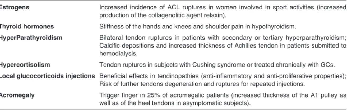
Several clinical observations suggest that hormonal disorders can be associated with tendinopathies (Tab. I). In some cases the evidence is strong, in oth-ers it is less consistent9.
There is no evidence that high or low levels of sexual hormones (estrogens, progesterone, testosterone) can be direct cause of tendinopathies. However, an increased incidence of anterior cruciate ligaments (ACL) ruptures has been reported in women involved in sport activities compared to males10. Indeed, the
sex-linked biomechanical and neuromuscular differ-ences, which contribute to explain this higher inci-dence5, are negligible before puberty, but become
ev-ident in the post-pubertal period11. However, studies
on the adaptations of ligaments and tendons across the menstrual cycle have provided inconclusive data5,
and recent research suggests that the higher inci-dence of ACL injuries in females should be attributed to relaxin, which works as a collagenolitic agent. Fe-males, but not Fe-males, show specific receptors for this substance, which has been found increased in ath-letes who underwent tendon ruptures12.
The clinical evidence of a relationship between thy-roid disorders and tendinopathies is also limited. Stiff-ness of the hands and knees is frequently observed in patients suffering from hypothyroidism13 and an
as-sociation with shoulder pain has been hypothesized on the basis of epidemiological data9. Recently, a
case of spontaneous rupture of the long head of the biceps tendon in a woman with hypothyroidism has been reported14.
Bilateral tendon ruptures (patellar and Achilles) may occur in patients with secondary or tertiary hyper-parathyroidism, the latter condition being character-ized by an autonomous parathyroid function, inde-pendent of calcium serum concentration. In this case the parathormon (PTH) levels are very high and the clinical pattern is characterized by general osteoporo-sis, with sub-periosteal bone resorption and resultant weakness of bone-tendon junction15. Ultrasound
ex-amination in patients submitted to regular hemodialy-sis allows to identify precociously those at risk,
show-ing tenderness durshow-ing probshow-ing, calcific depositions, and increased thickness of Achilles tendon. Tendon ruptures have been reported in subjects with Cushing syndrome or in patients treated chronically with GCs, mainly when associated to fluoroquinolone antibi-otics16. An epidemiologic survey has shown an
in-creased odds ratio around 3.0 for continous oral GCs, which declined shortly after therapy cessation. Simi-larly a single short-term high-dose GCs treatment course was sufficient transiently to increase the risk of tendon ruptures17.
On the other hand, local GCs injections are frequently performed in subjects with tendinopathies, and show beneficial effects in several cases, because their anti-inflammatory and anti-proliferative properties, but car-ry the risk of further tendon deterioration and ruptures after repeated courses of treatment18.
Similarly, in acromegaly, due to long-term exposure to elevated levels of growth hormone (GH) and In-sulin-like Growth Factor-1 (IGF-1), besides muscolo-skeletal abnormalities (coarsened facial features, growth of hands and feet and soft tissue hypertrophy, articular involvement of the axial and peripheral joints), tendon disorders have been put in evidence19.
Symptomatic flexor tenosynovitis (trigger finger) is an early manifestation found in 25% of patients. This condition, which affects the flexor pollicis longus or flexor digitorum tendons, is caused by the entrapment of the affected tendon at first annular pulley, due to proliferation and fibrosis of the sheath and/or to some localized tendon thickening20. Also in asymptomatic
subjects the thickness of the A1 pulley is higher than in the normal population, as well as the thickness of the heel tendons20. The increased tendon thickness
is reversible by normalizing GH/IGF-1 excess20.
Experimental evidence
EstrogensThe clinical observations previously reported have prompted a huge amount of experimental research aimed to explore the intimate mechanisms of action of hormones on tendons (Tab. II).
Table I. Clinical effects of hormonal disorders on tendons.
Estrogens Increased incidence of ACL ruptures in women involved in sport activities (increased production of the collagenolitic agent relaxin).
Thyroid hormones Stiffness of the hands and knees and shoulder pain in hypothyroidism.
HyperParathyroidism Bilateral tendon ruptures in patients with secondary or tertiary hyperparathyroidism; Calcific depositions and increased thickness of Achilles tendon in patients submitted to hemodialysis.
Hypercortisolism Tendon ruptures in subjects with Cushing syndrome or treated chronically with GCs. Local glucocorticoids injections Beneficial effects in tendinopathies (anti-inflammatory and anti-proliferative properties);
Risk of further tendons degeneration and ruptures for repeated injections.
Acromegaly Trigger finger in 25% of acromegalic patients (increased thickness of the A1 pulley as well as of the heel tendons in asymptomatic subjects).
The effects of estrogens are not clear. In vitro studies revealed that estradiol has an inhibiting effect upon collagen formation, increasing the expression of ma-trix metalloproteinase (MMP)-13, which is responsible for tendon collagen degradation10. An alternative
hy-pothesis suggests a negative effect of estrogens on the activity of the enzyme lysyl-oxidase, which regu-lates the addition of lysine and hydroxylysine-based cross-links into collagen fibrils, with ensuing in-creased ligaments laxity. Further research has shown that estradiol and raloxifene, a specific estrogen re-ceptor modulator, increase the expression of type III collagen and elastin, responsible for tendon elastic properties. These changes can lead to a reduction in tendons’ tensile strength because type III collagen fibers are thinner and more flexible than type I fibers21.
Paradoxically, similar conclusions have been reached in studies performed in ovariectomized rats, i.e. in conditions of hormonal deprivation22. The activity of
the extracellular MMP-2 is increased, as well as cell apoptosis both in muscles and tendons23. The
syn-thesis of type I collagen, fibronectin and elastin is de-creased, and a significantly lower healing rate in a micro-wound healing model has been found22,23. In
conclusion, both increased or reduced receptor stim-ulation could influence negatively the biomechanical tendon properties.
Testosterone
Specific receptors for testosterone have been demon-strated in tendons24. In vitro studies have shown that
progressive concentrations of dihydrotestosterone enhance the proliferation of tenocytes harvested from
healthy supraspinatus tendons25. The cultured cells
increase in number after 48 and 72 hours after treat-ment, and acquire a more de-differentiated aspect, becoming more flattened and polygonal with round nuclei, in comparison with control cultures, that show an elongated shape25. Therefore, it may be
speculat-ed that testosterone regulates lineage determination and differentiation in mesenchymal pluripotent cells resident in tendons25.
These results are indirectly supported by the finding that men with low testosterone levels show a signifi-cant reduction in both circulating and endothelial pro-genitor cells, while testosterone replacement therapy induces a significant increase in these progenitors with respect to baseline. This effect can be important during tendon-healing and repair, when active prolif-eration is required25.
Thyroid hormones
Thyroxine has an important role both in collagen syn-thesis and matrix metabolism. Hyperthyroidism is ac-companied by increased catabolism of both soluble and insoluble collagen, whereas hypothyroidism has opposite effects. Indeed, in subjects with hypothy-roidism, glycosaminoglycans deposition in the extra-cellular matrix explains the pathogenesis of stiff joints and carpal tunnel syndrome and may predispose to tendon calcification13.
Thyroid hormone receptors seem to be ubiquitous. Basic observations in vitro have shown that thyroid hormones promote the proliferation, in a dose depen-dent manner, of tenocytes obtained from tendon biop-sies of patients who underwent rotator cuff tears surgery9. Moreover, these hormones have a
protec-Table II. Effects of hormones on tendons in experimental conditions.
Estrogens Inhibiting effect upon collagen formation (increased expression of MMP-13);
Decreases synthesis of type I collagen, fibronectin and elastin in conditions of estrogen depriva-tion with a significant lower healing rate in a micro-wound healing model;
Negative influence on the biomechanical tendon properties (both with increased or reduced es-trogen receptor stimulation).
Testosterone Enhanced proliferation of tenocytes. Thyroid hormones Enhanced proliferation of tenocytes; Protective action against apoptosis.
Parathyroid hormone Homeostatic control of fibrochondrocytes at tendon insertion sites;
Production of type I and II (at low mechanical strain) and type X (high mechanical strain) collagens by fibrochondrocytes.
Glucocorticoids Reduced proliferation and viability of fibroblasts;
Decreased stem cell pool (formation of non-tendinous tissues);
Irreversible senescence in human tenocytes (inhibition of Sirtuin 1 and activation of the p53/p21 pathway);
Reduced tendon mechanical properties.
Growth hormone Higher expression for collagen mRNA and collagen fractional synthesis rate in acromegalics (compared to GH deficient patients);
Higher mRNA expression of collagen types I and III in giant transgenic mice with high circulating levels of GH and IGF-I (compared to dwarf mice with a disrupted GH receptor gene and to a wild-type control group);
Increased number of stem cells in vivo and preserved multipotency in vitro; Inhibition of cell death induced by anoxia.
tive action against apoptosis induced by serum depri-vation, which should be considered an important fac-tor for the failed healing response observed in human tendinopathies9. These observations reinforce the
idea of a physiological action of thyroid hormones in tendons homeostasis.
Parathormon
PTH has a relevant role in calcium homeostasis, in-ducing general osteoporosis, with sub-periosteal bone resorption and resultant weakness of bone-ten-don junction15.
Parathyroid Hormone-related Protein (PTHrP) be-longs to the same PTH gene family, but functions as predominately an autocrine/paracrine regulatory molecule. PTHrP has been identified in the perios-teum and tendon insertion sites (entheses), where it controls the biology of fibrochondrocytes26,
influenc-ing the recruitmnent and/or the activities of underlyinfluenc-ing bone cell populations27. Studies in vitro have shown
that cyclic tensile strain increases PTHrP expression. At low mechanical strain, PTHrP induces the produc-tion of type I and type II collagens by fibrochondro-cytes, while at high mechanical strain it stimulates type X collagen production, hence tendon mineraliza-tion28.
Glucocorticoids
Given the evidence of the healing properties of locally injected GCs in several tendinopathies, however as-sociated to the risk of further tendon deterioration and ruptures in some cases, a plethora of studies has been performed, aiming to elucidate the effects of GCs in isolated tendons, in tendon cell cultures, and in intact animals. Histologically, a loss of collagen or-ganization and an increase of collagen necrosis was observed, followed by a reparative response charac-terized by an inflammatory cells infiltrate29. The
prolif-eration and viability of fibroblasts was also reduced30.
GCs treatment depletes the stem cell pool and leads to the formation of non-tendinous tissues (e.g. fatty and cartilage-like tissues)31. There is in vitro and in
vivo evidence that GCs induce irreversible senes-cence in human tenocytes by inhibition of Sirtuin 1 and activation of the p53/p21 pathway. Tenocytes apoptosis resulted unaffected in some studies, in-creased in others18. Biochemical investigations have
shown that collagen synthesis was decreased with an increased ratio of type III to type I collagen32. Small
numbers of studies demonstrated changes in matrix enzymes (MMPs/TIMPs), proteoglycans, cytokines and other substances including FOX-01 and Sirtuin-132. The exposure of human tendon cells to
dexam-ethasone results in a time-dependent reduction of mRNA for Substance P, and the induction of Sub-stance P by Interleukin-1 beta and by cyclic mechani-cal loading is prevented. This finding could explain why GCs may alleviate pain in tendinopathies. Stud-ies investigating the mechanical propertStud-ies of tendon after GC injection reported conflicting results. Some Authors observed a decrease in mechanical proper-ties, others an increase, or no significant change33.
However, pooling the results, a metaanalysis de -monstrated a clear trend towards reduced mechani-cal properties in tendon after GC injections18. GH/IGF-1 system
A plethora of experimental data emphasizes the con-cept that the GH/IGF-1 system is crucial in maintain-ing muscle-tendon homeostasis. In a study the colla-gen mRNA expression and collacolla-gen protein fractional synthesis rate (FSR) was evaluated locally, in mus-cles and tendons, by means of microdialysis tech-nique, in acromegalics, relative to GH deficient pa-tients34. A higher expression for collagen and IGF1
mRNA was found in acromegalics. Moreover, there was a tendency towards a higher collagen protein FSR and a smaller collagen fibril diameter in acrome-galics in comparison to GH deficient patients. The conclusions coming by human studies are sup-ported by animal experiments. Giant transgenic (GT) mice, with high circulating levels of GH and IGF-I, and dwarf mice with a disrupted GH receptor gene (GHR−/−), leading to GH resistance and low circulat-ing IGF-I, were compared to a wild-type control group (CTRL). GHR−/− mice had significantly lower colla-gen fibril volume fraction in Achilles tendon, as well as decreased mRNA expression of IGF-I isoforms and collagen types I and III in muscle compared to CTRL. In contrast, the mRNA expression of IGF-I iso-forms and collagens in GT mice was generally high in both tendon and muscle compared to controls. Mean collagen fibril diameter was significantly decreased with both high and low GH/IGF-I signaling, but the GHR−/− mouse tendons were most severely affected with a total loss of the normal bimodal diameter distri-bution35.
GH plus resistance exercise attenuate structural changes in rat myotendinous junctions resulting from chronic unloading36. In addition, GH/IGF-I axis
in-creases the number of stem cells in vivo37, and
pre-serves their multipotency in vitro. In addition, the cell death induced by anoxia is inhibited by the addition of IGF-1 through activation of PI3K signaling, suggest-ing that IGF-1 can be considered as a survival fac-tor38. The controversy about whether anabolic tendon
adaptations are due to the systemic increase or to lo-cal IGF production, or to both, is topic of current re-search and discussion. Indeed the GH/IGF-1 effects are mediated through endocrine as well as paracrine/ autocrine mechanisms. This system includes the binding proteins, namely GH binding proteins and IGF-1 binding proteins (IGFBP)39. In fact, increased
levels of circulating IGF-1 do not always entail biolog-ical actions at the local tendon level since only free IGF and binary complexes can leave the blood stream. In fact, most IGF (70-80%) circulates as ternary complexes composed by IGF-I, IGFBP3 and Acid Labile Subunit. These complexes are too big to leave the blood stream. Only the free form (less than 2% of circulating IGF-1) or binary compounds (20-25% of 1 associated to binding proteins, IGF-BP1-6) can reach the tendon39. Several experiments
lev-els of GH or IGF-1 are responsible of the stimulation and regulation of collagen synthesis in human tendon tissue. Indeed, studies measuring local IGF-1 re-sponse by means of interstitial fluid sampling method-ologies have shown that, after exercise, peritendi-nous values are consistently higher than those of the circulating hormone40,41. To add complexity, it is also
feasible that the ability for IGF-1 to bind to its recep-tor in skeletal muscle and tendon is mediated indi-rectly by the binding proteins. If this is the case, acute exercise can increase the IGF-I receptor bind-ing capacity and affinity to IGF-1, and there may be certain maximal thresholds at which additional circu-lating IGF-1 is no longer effective.
Therapeutic perspectives
Because injuries to tendons are frequent and costly, and occur at all ages, mainly in subjects practicing sport activities at professional or amateur level, it is important to improve our understanding concerning the therapeutic potential of hormones in tendon heal-ing. In this section the future perspectives of hormon-al therapies are briefly summarized (Tab. III).
The effects of the pill on the incidence of ACL rup-tures in females athletes have been evaluated with conflicting results. Indeed, besides studies which re-port a lower rate of lesions in women who use the pill42, in other studies this evidence is lacking43. To
add complexity, the dosage of estrogens and proges-terone vary widely among the pills, and as a conse-quence the endogenous hormonal levels vary accord-ingly. Therefore no conclusion can be drawn about the protective effects of contraceptives against ACL injuries.
As far as PTH is concerned, several studies have shown that recombinant PTH (rhPTH) accelerates bone healing and increases chondrocyte recruitment and differentiation44. In rats submitted to detachment
and immediate repair of the right supraspinatus ten-don, using bone tunnel suture fixation, receiving daily subcutaneous injections of 10 mg/kg of rhPTH, histo-logical analysis revealed, in comparison to controls, at 28 and 56 days, more fibrocartilage, osteoblasts, and blood vessels, and a significantly better collagen
fiber orientation45. These findings have been
con-firmed in a rat model of ACL reconstruction with auto-graph, where intermittently administered PTH has shown to enhance the thickness and micro-architec-ture of trabecular bone46. Unfortunately these
promis-ing results did not translate into improved biomechan-ical properties. More recently, experiments have been performed to evaluate whether rhPTH has healing potential at tendon level. In mice, the deep digital flexor tendon was transected and immediately re-paired. PTH (40 mg/kg) was therefore administered, by either subcutaneous or intraperitoneal injections. In comparison to controls, PTH promoted an early formation of reparative tissue associated with an creased expression of extracellular matrix genes, in-cluding fibrillar collagens, type I and type III, and fi-bronectin47. Similarly to previous experiments, no
sig-nificant differences in tensile strength were seen. An incomplete integration/remodeling of the newly formed tissue at the microstructural level, as well the lack of an appropriate mechanical loading and/or the short observation period, are possible reasons that may explain these unsatisfactory results. Therefore, further research is needed to confirm the potential therapeutic effect of PTH in tendon-to-bone or ten-don-to-tendon healing.
The use of GCs in the treatment of tendinopathies is an intriguing issue. Intratendinous and peritendinous corticosteroids injections are highly beneficial in trig-ger fintrig-ger and De Quervain syndrome, whereas in ro-tator cuff, patellar and Achilles tendon diseases the results are deceiving and short lasting. Moreover, tendon ruptures have been reported after GC admin-istration. It is evident that the positive effects of GC are probably related to the well-known anti-inflamma-tory and anti-proliferative properties of these hor-mones on pathologic tendons, whereas adverse ef-fects must be reported to the detrimental effect of GCs on the tendon structure, when degenerative fea-tures are present. Therefore, the boundary between the good and the evil remains uncertain, and caution is required in patients with relapses of chronic overuse tendinopathies. Indeed, in these situations, GC administration, although beneficial in the short term, can worsen tendon degeneration.
Research evaluating the healing properties of GH/ Table III. Therapeutic perspectives.
Estrogens+Progesterone No firm evidence that the pill can reduce the incidence of ACL ruptures in female athletes. Parathormone Increased tendon healing in experimental conditions (but not improved biomechanical
properties).
Glucocorticoids Beneficial effects in tendinopathies (but deterioration and possible ruptures after repeated courses of treatment).
GH/IGF-1 system Increased tenocytes proliferation and collagen production in animals and humans; Increased stem cells number and prevention of their death induced by anoxia;
Positive influence on tendon healing in experimental conditions (collagenase-induced tissue atrophy or tendon disruption) and in spontaneous tendinopathies in horses;
Increased cross sectional area and biomechanical properties after local injection of recombinant GH in elderly subjects during rehabilitation.
IGF-1 is in a more advanced stage. As shown in the previous paragraph, basic studies both in humans and animals have largely demonstrated the positive effects of GH/IGF-1 on tendons (increased tenocytes proliferation and collagen production, increased stem cells number and prevention of their death induced by anoxia). Moreover, studies have shown that this ac-tivity is mainly due to local production of IGF rather than to circulating levels of GH/IGF-1 complex. This finding is relevant because suggests the possibility of IGF-1 delivering in areas of tendon pathology, avoid-ing the side effects of a systemic administration. These studies have prompted specific experiments to ascertain whether GH-IGF1 could be an useful tool for tendon healing. The results in animal models are promising. IGF-1 injection promotes tendon and liga-ment healing after collagenase-induced tissue atro-phy or ligament disruption48. These findings are
sub-stantiated by a retrospective study performed in 40 cases of horse superficial digital flexor tendonitis. The intra-lesional administration of IGF-1 (25 or 50 μg ev-ery other day for 4 or 5 treatments) enhanced heal-ing, as shown by the decrease of ultrasonographic le-sions severity, although not to the point to return the horses to sport activities48.
Several human studies confirm animal experiments. Human tendon cells cultured in 3D with IGF-I supple-mentation showed an increased gene expression for collagen, tenomodulin and scleraxis49. Healthy
hu-mans, who receive either weeks of GH administration or acute injection of IGF-1, demonstrate increased expression and synthesis of collagen in muscle and tendon32. The local injections of recombinant IGF-1
into the patella tendon increased the collagen FSR and procollagen type I N-terminal propeptide (a mark-er for type I collagen synthesis) in the pmark-eritendinous fluid of healthy individuals50. Not only IGF-1 but also
GH provides positive results. In a randomized study, two injections of GH in the patellar tendon of ageing subjects increased collagen synthesis rate 3-4 hours after the second injection, and showed a tendency to higher collagen FSR. Systemic IGF-1 remained un-changed, but interstitial IGF-I increased in GH treated tendons compared with saline treated tendons. This confirms that GH stimulatory effects on tendinous col-lagen synthesis involves local IGF-1 production50.
Be-sides these observations, patients with Ehlers-Danlos syndrome (an inherited connective tissue disorder), treated with 1 mg of IGF-1 injected in the patellar ten-don, showed an increase in the protein synthesis rate in comparison with the controlateral tendon used as control35.
On the other hand, exercise leads to activation of the systemic GH/IGF-1 axis39. Engineered ligaments
treated with serum obtained from young healthy men after exercise resulted in more collagen and improved tensile strength in comparison to those treated with serum from resting men. In synthesis, results ob-tained in different experimental conditions lead to the concordant conclusion that GH/IGF-1 supplementa-tion has positive effects which can favour tendon healing in humans.
Especially elderly people, characterized by an age-dependent decline in the GH/IGF-1 activity, may ben-efit from local GH-IGF-1 administration. It is well known that elderly individuals show lower content of structural proteins in tendons, reduced magnetic res-onance imaging tendon signal intensities, and an in-crease in tendon cross-links due to advanced glyca-tion end products deposiglyca-tion. The GH/IGF-1 axis seems to be important for the maintenance of the structural proteins within these tissues. To assess whether GH administration could hasten rehabilita-tion, aged individuals had one leg immobilized two weeks, followed by 6 weeks of retraining. Therefore, the legs immobilized and retrained were divided in two groups, the first receiving daily injections of re-combinant GH, and the second saline solution as placebo. Several properties of the patellar tendon, in-cluding the cross sectional area, and biomechanical properties were measured, and the ratios type I/III and IGF-1 levels were evaluated in tendon biopsies. The group receiving GH injections showed an in-crease of patellar tendon cross sectional area and stiffness, associated to an increase of IGF-1. From these data it may be inferred that local GH adminis-tration may be useful to improve healing in patients during rehabilitation of tendon and ligament injuries and postsurgery51.
In conclusion, the relationships between hormones and tendons are complex and not fully understood. Howev-er, several experimental data suggest that some hor-mones (mainly GH/IGF-1) may offer significant ad-vances in addition to present conservative treatments for human tendon diseases. A crucial point is to ascer-tain whether the local administration can be therapeuti-cal, to avoid systemic interactions and undesirable ef-fects. Because tendons injuries are frequent, often with long lasting sequels, this promising therapeutic ap-proach deserves further clinical research.
Conflict of interest
All the Authors have no disclosure.
References
1. Boivin GP, Elenes EY, Schultze AK, Chodavarapu H, Hunter SA, Elased KM. Biomechanical properties and histology of db/db diabetic mouse Achilles tendon. Muscles Ligaments Tendons J. 2014;4(3):280-284.
2. Snedeker JG, Gautieri A. The role of collagen crosslinks in ageing and diabetes - the good, the bad, and the ugly. Muscles Ligaments Tendons J. 2014; 4(3):303-308.
3. Abate M. How obesity modifies tendons (implications for ath-letic activities). Muscles Ligaments Tendons J. 2014;4(3):298-302.
4. Hast MW, Abboud JA, Soslowsky LJ. Exploring the role of hy-percholesterolemia in tendon health and repair. Muscles Liga-ments Tendons J. 2014;4(3):275-279.
5. Abate M, Schiavone C, Salini V, Andia I. Occurrence of tendon pathologies in metabolic disorders. Rheumatology (Oxford). 2013;52:599-608.
6. Frizziero A, Vittadini F, Gasparre G, Masiero S. Impact of oe-strogen deficiency and aging on tendon: concise review. Mus-cles Ligaments Tendons J. 2014;4(3):324-328.
7. Galdiero M, Auriemma RS, Pivonello R, Colao A. Cushing, acromegaly, GH deficiency and tendons. Muscles Ligaments Tendons J. 2014;4(3):329-332.
8. Padulo J, Oliva F, Frizziero A, Maffulli N. Muscles, Ligaments and Tendons Journal - Basic principles and recommendations in clinical and field Science Research: 2016 Update. MLTJ. 2016;6:(1)1-5.
9. Oliva F, Piccirilli E, Berardi AC, Frizziero A, Tarantino U, Maf-fulli N. Hormones and tendinopathies: the current evidence. Br Med Bull. 2016;117(1):39-58.
10. Slauterbeck JR, Fuzie SF, Smith MP, et al. The Menstrual Cy-cle, Sex Hormones, and Anterior Cruciate Ligament Injury. J Athl Train. 2002;37(3):275-278.
11. Andrish JT. Anterior cruciate ligament injuries in the skeletally immature patient. Am J Orthop (Belle Mead NJ). 2001;30:103-110.
12. Dehghan F, Haerian BS, Muniandy S, Yusof A, Dragoo JL, Salleh N. The effect of relaxin on the musculoskeletal system. Scand J Med Sci Sports. 2014;24(4):e220-229.
13. Harvie P, Pollard TCB, Carr AJ. Calcific tendinitis: Natural his-tory and association with endocrine disorders. J Shoulder El-bow Surg. 2007;16:169-173.
14. Pantazis K, Roupas ND, Panagopoulos A, Theodoraki S, Tsintoni A, Kyriazopoulou V. Spontaneous rupture of the long head of the biceps tendon in a woman with hypothyroidism: a case report. J Med Case Rep. 2016;10(1):2.
15. Shiota E, Tsuchiya K, Yamaoka K, Kawano O. Spontaneous major tendon ruptures in patients receiving longterm hemo -dialysis. Clin Orthop Relat Res. 2002;(394):236-242. 16. Claessen FM, de Vos RJ, Reijman M, Meuffels DE. Predictors
of primary Achilles tendon ruptures. Sports Med. 2014;44(9): 1241-1259.
17. Spoendlin J, Meier C, Jick SS, Meier CR. Oral and inhaled glu-cocorticoid use and risk of Achilles or biceps tendon rupture: a population-based case-control study. Ann Med. 2015;47 (6):492-498.
18. Dean BJ, Lostis E, Oakley T, Rombach I, Morrey ME, Carr AJ. The risks and benefits of glucocorticoid treatment for tendinopathy: a systematic review of the effects of local gluco-corticoid on tendon. Semin Arthritis Rheum. 2014;43(4):570-576.
19. Colao A, Marzullo P, Vallone G, et al. Ultrasonographic evi-dence of joint thickening reversibility in acromegalic patients treated with lanreotide for 12 months. Clin Endocrinol (Oxf). 1999;51(5):611-618.
20. Tagliafico A, Resmini E, van Holsbeeck MT, et al. Sonograph-ic depSonograph-iction of trigger fingers in acromegaly. J Ultrasound Med. 2009;28(11):1441-1446.
21. Irie T, Takahata M, Majima T, et al. Effect of selective estrogen receptor modulator/raloxifene analogue on proliferation and collagen metabolism of tendon fibroblast. Connect Tissue Res. 2010;51:179-187.
22. Circi E, Akpinar S, Balcik C, et al. Biomechanical and histolog-ical comparison of the influence of oestrogen deficient state on tendon healing potential in rats. Int Orthop. 2009;33(5):1461-1466.
23. Aydin A, Kenar H, Atmaca H, et al. The short- and long- term effects of estrogen deficiency on apoptosis in musculoskeletal tissues: an experimental animal model study. Arch Iran Med. 2013;16:271-276.
24. Khalkhali-Ellis Z, Handa RJ, Price RH Jr, Adams BD, Callaghan JJ, Hendrix MJ. Androgen receptors in human syn-oviocytes and androgen regulation of interleukin 1beta (IL-1beta) induced IL-6 production: a link between hypoandro-genicity and rheumatoid arthritis? J Rheumatol. 2002;29
(9):1843-1846.
25. Denaro V, Ruzzini L, Longo UG, et al. Effect of dihydrotestos-terone on cultured human tenocytes from intact supraspinatus tendon. Knee Surg Sports Traumatol Arthrosc. 2010;18:971-976.
26. Chen X, Macica C, Nasiri A, Judex S, Broadus AE. Mechani-cal regulation of PTHrP expression in entheses. Bone. 2007;41(5):752-759.
27. Kim YJ, Kim HJ, Im GI. PTHrP promotes chondrogenesis and suppresses hypertrophy from both bone marrow-derived and adipose tissue-derived MSCs. Biochem Biophys Res Com-mun. 2008;373:104-108.
28. Han X, Guo L, Wang F, Zhu Q, Yang L. Contribution of PTHrP to mechanical strain-induced fibrochondrogenic differentiation in entheses of Achilles tendon of miniature pigs. J Biomech. 2014;47(10):2406-2414.
29. Akpinar S, Hersekli MA, Demirors H, Tandogan RN, Kayasel-cuk F. Effects of methylprednisolone and betamethasone in-jections on the rotator cuff: an experimental study in rats. Adv Ther. 2002;19(4):194-201.
30. Tempfer H, Gehwolf R, Lehner C, et al. Effects of crystalline glucocorticoid triamcinolone acetonide on cultered human supraspinatus tendon cells. Acta Orthop. 2009;80(3):357-362. 31. Zhang J, Keenan C, Wang JH. The effects of dexamethasone on human patellar tendon stem cells: implications for dexam-ethasone treatment of tendon injury. J Orthop Res. 2013;31 (1):105-110.
32. Muto T, Kokubu T, Mifune Y, et al. Temporary inductions of matrix metalloprotease-3 (MMP-3) expression and cell apop-tosis are associated with tendon degeneration or rupture after corticosteroid injection. J Orthop Res. 2014;32(10):1297-1304.
33. Shapiro PS, Rohde RS, Froimson MI, et al. The effect of local corticosteroid or ketorolac exposure on histologic and biome-chanical properties of rabbit tendon and cartilage. Hand (N Y). 2007;2(4):165-172.
34. Doessing S, Holm L, Heinemeier KM, et al. GH and IGF1 lev-els are positively associated with musculotendinous collagen expression: experiments in acromegalic and GH deficiency patients. Eur J Endocrinol. 2010;163(6):853-862.
35. Nielsen RH, Holm L, Jensen JK, et al. Tendon protein synthe-sis rate in classic Ehlers-Danlos patients can be stimulated with insulin-like growth factor-I. J Appl Physiol. 2014;117 (7):694-698.
36. Curzi D, Lattanzi D, Ciuffoli S, et al. Growth hormone plus re-sistance exercise attenuate structural changes in rat my-otendinous junctions resulting from chronic unloading. Eur J Histochem. 2013;57(4):e37.
37. Heinemeier KM, Mackey AL, Doessing S, et al. GH/IGF-I axis and matrix adaptation of the musculotendinous tissue to exer-cise in humans. Scand J Med Sci Sports. 2012;22(4):e1-7. 38. Scott A, Khan KM, Duronio V. IGF-I activates PKB and
pre-vents anoxic apoptosis in Achilles tendon cells. J Orthop Res. 2005; 23:1219-1225.
39. Gatti R, De Palo EF, Antonelli G, Spinella P. IGF-I/IGFBP sys-tem: metabolism outline and physical exercise. J Endocrinol Invest. 2012;35(7):699-707.
40. Olesen JL, Heinemeier KM, Gemmer C, et al. Exercise-de-pendent IGF-I, IGFBPs, and type I collagen changes in human peritendinous connective tissue determined by microdialysis. J Appl Physiol (1985). 2007;102(1):214-220.
41. Nindl BC, Scofield DE, Strohbach CA, et al. IGF-I, IGFBPs, and inflammatory cytokine responses during gender-integrat-ed Israeli Army basic combat training. J Strength Cond Res. 2012;26 Suppl 2:S73-81.
42. Martineau PA, Al-Jassir F, Lenczner E, Burman ML. Effect of the oral contraceptive pill on ligamentous laxity. Clin J Sport Med. 2004;14(5):281-286.
43. Agel J, Bershadsky B, Arendt EA. Hormonal therapy: ACL and ankle injury. Med Sci Sports Exerc. 2006;38:7-12.
44. Weiss S, Hennig T, Bock R, Steck E, Richter W. Impact of growth factors and PTHrP on early and late chondrogenic dif-ferentiation of human mesenchymal stem cells. J Cell Physiol. 2010;223(1):84-93.
45. Hettrich CM, Beamer BS, Bedi A, et al. The effect of rhPTH on the healing of tendon to bone in a rat model. J Orthop Res. 2012;30(5):769-774.
46. Bi F, Shi Z, Jiang S, Guo P, Yan S. Intermittently administered parathyroid hormone promotes tendon-bone healing in a rat model. Int J Mol Sci. 2014;15(10):17366-17379.
47. Lee DJ, Southgate RD, Farhat YM, et al. Parathyroid hormone 1-34 enhances extracellular matrix deposition and organiza-tion during flexor tendon repair. J Orthop Res. 2015;33(1):17-24.
48. Witte TH, Yeager AE, Nixon AJ. Intralesional injection of in-sulin-like growth factor-I for treatment of superficial digital flex-or tendonitis in Thflex-oroughbred racehflex-orses: 40 cases (2000-2004). J Am Vet Med Assoc. 2011;239:992-997.
49. Herchenhan A, Bayer ML, Eliasson P, et al. Insulin-like growth factor I enhances collagen synthesis in engineered human tendon tissue. Growth Horm IGF Res. 2015;25(1):13-19. 50. Vestergaard P, Jørgensen JO, Olesen JL, et al. Local
admin-istration of growth hormone stimulates tendon collagen syn-thesis in elderly men. J Appl Physiol (1985). 2012;113(9):1432-1438.
51. Boesen AP, Dideriksen K, Couppé C, et al. Effect of growth hormone on aging connective tissue in muscle and tendon: gene expression, morphology, and function following immobi-lization and rehabilitation. J Appl Physiol (1985). 2014;116 (2):192-203.