Alzheimer´s disease-associated Aβ42 peptide: expression and purification
for NMR structural studies
Montserrat Serra-Batiste,1 Raquel Garcia-Castellanos,1 Martí Ninot-Pedrosa,1 Bernat Serra-Vidal,1 Nicholas Simon Berrow,1 Natàlia Carulla1
1Institute for Research in Biomedicine (IRB Barcelona), The Barcelona Institute of
Science and Technology, Baldiri Reixac 10, 08028 Barcelona, Spain
Both authors contributed equally to this work
*Corresponding author:
Natàlia Carulla, Baldiri Reixac 10, Barcelona 08028, Spain, telephone: +34 93 4037123, fax: +34 93 4037126, e-mail: [email protected]
ABSTRACT
The aggregation of the amyloid-beta peptide (Aβ) in the brain is strongly associated with Alzheimer´s disease (AD). However, the heterogeneous and transient nature of this process has prevented identification of the exact molecular form of Aβ responsible for the neurotoxicity observed in this disease. Therefore, characterizing Aβ aggregation is of utmost importance in the field of AD. Nuclear magnetic resonance spectroscopy (NMR) is a technique that holds great potential to achieve this goal. However, it requires the use of specific labels introduced through recombinant expression of Aβ. In this paper, we report on a straightforwardexpression and purification protocol to obtain [U-15N] and [U-2H,13C,15N] Aβ42. Aβ42 is expressed fused to Small Ubiquitin-like Modifier (SUMO), which prevents Aβ42 aggregation. The solubilizing capacity of SUMO has allowed us to design a purification protocol involving immobilized metal affinity chromatography (IMAC), a desalting step, and two size exclusion chromatography (SEC) purifications. This approach, which does not require the use of costly and time-consuming reversed phase high performance liquid chromatography (RP-HPLC), offers a much straightforward strategy to those previously described to obtain [U-15N] Aβ42 and it is the first protocol through which to achieve[U-2H,13C,15N] Aβ42. The peptides obtained are of high purity and have the required isotope enrichment to support NMR-based structural studies.
KEYWORDS
Alzheimer´s disease, Aβ42 peptide, isotope labeling, NMR, soluble expression, SUMO.
INTRODUCTION
Alzheimer’s disease (AD) is a progressive neurodegenerative disorder that robs us of our most human qualities. Beyond its devastating effects on patients, AD is also a major burden for patients’ families and implies enormous healthcare costs [1]. To date, no disease-modifying treatment is available, and therefore research on the molecular basis of AD must continue. The aggregation of the amyloid-beta peptide (Aβ) in the brain is strongly associated with AD [2]. Aβ is obtained from the transmembrane amyloid precursor protein (APP) through the sequential cleavage of β- and secretase. γ-secretase cleavage is not specific, leading to Aβ peptides of different lengths, Aβ38 to Aβ43, ranging from 38 to 43 residues. Among them, Aβ42 is considered to be the peptide most strongly linked to AD, as levels of this variant are an indication of AD progression [3], and several familial AD (fAD) mutations that give rise to early AD alter the Aβ40/Aβ42 ratio in favor of Aβ42 production [4]. Upon release from the membrane into the extracellular media, the Aβ peptide has a strong tendency to aggregate. This aggregation is a multistep process involving various aggregates that ultimately leads to the formation of amyloid fibrils. Due to the heterogeneity and transient nature of this process, the exact molecular form of Aβ responsible for the neurotoxicity observed in AD is not known. Therefore, characterizing Aβ aggregation is of utmost importance in the AD field. One of the techniques with the potential to characterize this process is nuclear magnetic resonance spectroscopy (NMR) [5-12], which requires the use of specific labels introduced through recombinant expression of Aβ.
A large number of strategies have been developed to produce recombinant Aβ42. However, due to the aggregation-prone nature of Aβ42, initial approaches resulted in the production of modified Aβ42 sequences rather than of the wild type. Modifications
included oxidation on the methionine 35 side chain of Aβ42 to methionine sulfoxide (Met-35(ox)Aβ42) [6,13], Aβ42 fused to a tag [14-16], and Aβ42 containing unnatural mutations in its sequence [6,17] or additional N-terminal residues [18-21]. More recently, several strategies have led to the production of wild-type Aβ42 with satisfactory yields [22-28], as well as isotopically labeled 15N and 15N, 13C Aβ42 [29,30]. One particularly attractive strategy initially proposed by Satakarni and Curtis relies on the expression of Aβ42 fused to Small Ubiquitin-like Modifier (SUMO) [27]. They showed that SUMO solubilizes Aβ42, since the SUMO-Aβ42 fusion protein was obtained from both soluble and insoluble cell lysates. However, in spite of the solubilizing power shown by SUMO, Weber et al. have recently reported on a purification protocol to obtain 15N and 15N, 13C Aβ42 starting from SUMO-Aβ42 accumulated in inclusion bodies, and thus used denaturing buffers for the lysis and initial purification steps [29].
Working under denaturing conditions makes proteins more vulnerable to chemical modifications, leading, for example, to the oxidation of the methionine side chain to methionine sulfoxide. Aβ42 has a methionine in position 35 (Met-35). Indeed, there is much controversy in the literature regarding the role of Met-35(ox)Aβ42 in AD [31,32]. Therefore, in any Aβ42 purification strategy, it is critical to separate Met-35(ox)Aβ42 from Aβ42. Since these molecules differ in hydrophobicity, the most efficient method to separate them relies on the use of costly and time-consuming preparative reversed phase high performance liquid chromatography (RP-HPLC). Indeed, most Aβ42 purification protocols use this technique during the purification steps [22-28] [29,30].
In this paper, and inspired by the work of Satakarni and Curtis [27], we exploit the capacity of SUMO to solubilize Aβ42 when these two molecules are fused and thus establish a new and efficient purification protocol. Rather than using highly denaturing
conditions, our approach requires mild conditions for the lysis and initial immobilized metal affinity chromatography (IMAC) purification steps, which are followed by a desalting step, cleavage of Aβ42 from SUMO using the efficient SUMO protease (Ulp1), and two size exclusion chromatography (SEC) steps. Using this approach, no oxidation of Met-35 was observed and therefore RP-HPLC was not required. This strategy afforded 6 mg of [U-15N] Aβ42 and 2 mg [U-2H,13C,15N] Aβ42 per L of culture. The chemical purity of the final product was assessed by analytical RP-HPLC, and the isotope incorporation of the labeled samples was determined by high-resolution mass spectrometry (HRMS). Both techniques indicated that the strategy provides peptides of sufficiently high purity and isotope enrichment to support NMR-based structural studies.
MATERIALS AND METHODS Reagents
The following labeled compounds were used: 15NH4Cl (99%, Cambridge Isotope
Laboratories), D-glucose-13C6, 1,2,3,4,5,6,6-d7 (97-98% D, 99% 13C, Euriso-top) and
D2O (99.9%, Euriso-top). All reagents were purchased from Sigma-Aldrich unless
otherwise specified. Cloning
The DNA encoding Aβ42 was synthesized by PCR with KOD polymerase (Novagen) methods and following the modular approach previously described [20], but with the following primers to add the 15 bp on each side for the In-Fusion method:
Fw 5’-GCGAACAGATCGGTGGTGATGCGGAGTTCCGTCATGATTCAG-3’
Rev 5’-ATGGTCTAGAAAGCTTTATTACGCTATGACAACACCACCCACCATGAGTCCA ATGATGGCACC-3’
The amplified fragment was further purified and cloned into a pOPINS vector [33] previously cut with KpnI and HindIII (New England Biolabs) restriction enzymes using the In-Fusion cloning method (Clontech). This yielded a plasmid for expression of Aβ42 in the Escherichia coli (E. coli) cytoplasm as a fusion protein with an N-terminal hexahistidine SUMO affinity tag (MGSSHHHHHHGSDSE
VNQEAKPEVKPEVKPETHINLKVSDGSSEIFFKIKKTTPLRRLMEAFAKRQGK EMDSLRFLYDGIRIQADQTPEDLDMEDNDIIEAHREQIGG↓-Aβ42), where ↓ represents the SUMO protease cleavage site).
Stock solutions
[U-15N] Aβ42 was produced using auto-induction. The following stock solutions were required. The 20x 15N-NPS and 50x 5052 solutions were prepared as previously described [34]. Briefly, the 20x 15N-NPS solution contained 142 g Na2HPO4, 136 g
KH2PO4, 50 g 15NH4Cl and 14.2 g Na2SO4 per L and the 50x 5052 solution contained
250 g glycerol, 25 g D-glucose and 100 g α-lactose per L. The 500x trace metal solution was also prepared based on previous descriptions with small adjustments [35]. Briefly, 1 L of 500x trace metal solution contained 8 mL 5 M HCl, 5 g FeCl2·4H2O, 184 mg
CaCl2·2H2O, 64 mg H3BO3, 18 mg CoCl2·6H2O, 4 mg CuCl2·2H2O, 340 mg ZnCl2, 605
mg Na2MoO4·2H2O, and 40 mg MnCl2·4H2O. These three solutions were
heat-sterilized and stored at room temperature until use. The 100x vitamin solution was prepared by dissolving 50 mg thiamine hydrochloride, 10 mg D-biotin, 10 mg choline chloride, 10 mg folic acid, 10 mg niacin, 10 mg pantothenic acid, 10 mg pyridoxal, and 1 mg riboflavin in 100 mL MilliQ water. This solution was sterilized using a 0.2-µm
filter, wrapped in aluminum foil, and stored at -20ºC until use.
[U-2H,13C,15N] Aβ42 was produced using M9 minimal media. Various stock solutions were required: the 30x salt solution was prepared as a 50-mL aliquot of D2O
containing 10.2 g anhydrous Na2HPO4, 4.5 g anhydrous KH2PO4, 0.75 g NaCl and 0.37
g MgSO4; the 100x vitamin solution and the 500x trace metal solution were prepared as
detailed above using D2O instead of H2O.
Media for growth and expression
For the production of [U-15N] Aβ42, 15N-labeled P-5052 medium for auto-induction was prepared from 2 mL of 1 M MgSO4 solution, 50 mL of the 20x 15N-NPS, 20 mL of
the 50x 5052, 2 mL of the 500x trace metal solution and 916 mL heat-sterilized MilliQ water. The resulting medium was heat-sterilized and subsequently 10 mL of the previously filtered 100x vitamin solution was added to it.
For the production of [U-2H,13C,15N] Aβ42, M9 minimal media was prepared from
33.3 mL of the 30x salt solution, 10 mL of the 100x vitamin solution, 1 g NH4Cl, and 4
g D-glucose and brought to 1 L with 50% H2O/50% D2O or 100% D2O for the
pre-cultures. For the final culture, 1 g 15NH4Cl and 2 g D-glucose-13C6, 1,2,3,4,5,6,6-d7
were used in 100% D2O. The resulting solutions were sterilized by filtering them
through a 0.2-µm filter. Afterwards, 2 mL of heat-sterilized 500x trace metal solution, freeze-dried and resuspended in D2O, were added to each of them.
Protein expression
Rosetta (DE3) pLysS E. coli cells (Novagen) were transformed with the expression vector and grown overnight at 37°C on Luria Bertani (LB)-agar plates containing 1% glucose. All cell cultures were also supplemented with 35 µg/mL chloramphenicol and 50 µg/mL kanamycin.
adapted from a previously described protocol [34]. Single colonies were picked and grown overnight in 2 x 12.5 mL LB, 1% glucose. The pre-cultures were centrifuged at 3,000 g for 10 min at 25ºC. Each pellet was transferred to 0.5 L 15N-labeled P-5052 auto-inducing media with the appropriate antibiotics using a 3-L Erlenmeyer flask. The resulting cultures were grown for 6 h at 37ºC and 180 rpm. The temperature was then lowered to 25ºC, and the culture was incubated 22 h more at 180 rpm. The cells were then harvested by centrifugation at 9,000 g for 15 min at 4ºC and then frozen at -80°C.
For [U-2H,13C,15N] Aβ42 expression, single colonies were picked and grown overnight in 4 x 3 mL LB, 1% glucose. The LB pre-cultures were centrifuged at 3,000 g for 10 min at 25ºC. The pellets were then transferred to 120 mL M9 medium, containing 50% H2O/50 % D2O and the corresponding antibiotics. The 50% D2O pre-culture was
grown for 7 h at 37ºC and centrifuged at 2,000 g for 20 min. The pellet was re-suspended and inoculated in 240 mL M9 medium in 100% D2O. The 100% D2O
pre-culture was grown overnight. The next morning, the pre-pre-culture was centrifuged at 2,000 g for 20 min, and the pellet was re-suspended and inoculated in 1 L M9100% D2O medium, containing 1 g/L 15NH4Cl and 2 g/L D-glucose-13C6, 1,2,3,4,5,6,6-d7
glucose. The culture was grown at 37°C and 180 rpm and induced at OD600 ~0.8 by the
addition of IPTG to a final concentration of 0.5 mM. After further growth for 3 h, the cells were harvested by centrifugation at 9,000 g for 15 min at 4ºC and then frozen at -80°C.
Protein isolation from the soluble and insoluble fractions
20 mg of cell pellet was resuspended in 1 mL buffer A (300 mM NaCl, 50 mM sodium phosphate, 20 mM imidazole, 1% Tween-20 and 1 mM tris(2-carboxyethyl)phosphine (TCEP) pH 8.0), supplemented with half a pill of ethylenediaminetetraacetic acid (EDTA)-free Complete protease inhibitor (Roche) and
1 mg DNAse (Roche). The resuspended cells were lysed using a 3-mm tapered microtip sonicator (VCX 750 Ultrasonic Processor, Sonics) for 10 min in an ice water bath. The cell extract was diluted with buffer A and then centrifuged at 15,000 g for 10 min at 4ºC, and the supernatant corresponding to the soluble fraction was kept. The pellet was resolubilized in buffer A containing 8 M urea using the same volume of the previously collected supernatant. The resulting solution was then sonicated in a bath (Bransonic® Ultrasonic cleaner B1510E) for 5 min and then centrifuged at 15,000 g for 10 min at 4ºC. The supernatant obtained, corresponding to the insoluble fraction, as well as that corresponding to the soluble fraction, were analyzed by Western Blot (WB).
Protein purification
The cell pellet was resuspended in 6 mL buffer A per g of cells, supplemented with 1 EDTA-free Complete protease inhibitor pill (Roche) and 1 spatula of DNAse (Roche) per 50 mL of buffer. The resuspended cells were lysed using a cell disruptor (Constant Systems Ltd. U.K.) operating at 20,000 psi. The cell extract was then centrifuged at 30,000 g for 30 min at 4ºC, the supernatant filtered using a 0.45 µm and subsequently purified by IMAC. The supernatant was loaded at 1 mL/min onto a HisTrap HP 5-mL Ni column (GE Healthcare), which was previously equilibrated with 5 column volumes of buffer A. After the loading step, the resin was washed with buffer B (300 mM NaCl, 50 mM sodium phosphate, 40 mM imidazole, 0.05% Tween-20 and 1 mM TCEP pH 8.0) for 10-15 column volumes, until UV absorbance was stable. The fusion protein was eluted at 2-5 mL/min using the following 3-step elution method: (a) a 15 mL linear gradient from 0 to 15% of buffer C (300 mM NaCl, 50 mM sodium phosphate, 500 mM imidazole, 0.05% Tween-20 and 1 mM TCEP pH 8.0), followed by (b) a 20 mL isocratic step at 15% buffer C and (c) a second isocratic step at 100% buffer C until UV absorbance was stable. IMAC fractions were analyzed by sodium dodecyl sulfate
polyacrylamide gel electrophoresis (SDS-PAGE), and those containing the fusion protein were pooled in batches of 10 mL. Subsequently, buffer was exchanged using a HiPrep 26/10 desalting column (GE Healthcare) equilibrated with 50 mM ammonium carbonate and 1 mM TCEP. Afterwards, the concentration and purity of protein was determined by nanodrop and RP-HPLC, respectively. Afterwards, to cleave Aβ42 from the SUMO fusion tag, samples were incubated overnight at 4ºC with SUMO protease (Ulp1) [33] in a 1:50 protease:protein ratio. The concentration of Aβ42 peptide after the cleavage was determined by RP-HPLC analysis. Subsequently, aliquots containing 3.75 mg Aβ42 were prepared and freeze-dried. Each of these aliquots was solubilized with 6.8 M guanidinium thiocyanate (GdnSCN) to 2.5 mg Aβ42/mL and sonicated for 5 min in an ice bath. Afterwards, the sample was further diluted with MilliQ water to 1.5 mg Aβ42/mL and 4 M GdnSCN, and centrifuged at 10,000 g for 6 min at 4ºC. Finally, 2.5 mL of the 1.5 mg Aβ42/mL was injected into a HiLoad Superdex 30 prep grade column (GE Healthcare), previously equilibrated with 50 mM ammonium carbonate, and eluted at 4ºC at a flow rate of 1 mL/min. The peaks corresponding to SUMO and monomeric Aβ42 were collected separately and their purity and concentration were determined by RP-HPLC. Both pools were freeze-dried, and the Aβ42 pool was subjected to the same GdnSCN solubilization protocol and SEC fractionation, as described. Pure Aβ42 was obtained after the two SEC steps. It was then aliquoted in the desired amounts, freeze-dried, and kept at -20ºC until use.
SDS-PAGE and WB
Samples for Fig. 1 were analyzed as follows: 10 µL of 3X sample buffer (SB) was added to 20 µL of supernatant and reconstituted pellet, boiled for 5 min at 95ºC. 20 µL of each of the resulting samples was electrophoresed using Mini-protean tetracell® system (Bio-Rad) in 0.75 mm-thick SDS-PAGE containing 15% acrylamide. Gels were
run at 120 V and stained with Coomassie Blue. For WB protein samples, SDS-PAGE gels were transferred to a 0.22 µm nitrocellulose (GE Healthcare) at 100 V for 2 h at 4ºC. Next, the membranes were washed in Tris-buffered saline and 0.1% Tween 20 (TBST), blocked in 5% (w/v) non-fat dried milk overnight at 4ºC, and incubated with primary antibody 6E10 (Covance) in 5% (w/v) non-fat dried milk overnight at 4ºC. Blots were treated with secondary horseradish peroxidase-conjugated mouse secondary antibody (GE Healthcare) using the chemiluminescence Immobilon ECL detection system (Millipore) and exposed to X-ray films (Super RX Medical X-Ray, Fujifilm), which were developed using the Hyper processor automatic film developer (Amersham Pharmacia Biotech).
Samples for Fig. 3A were analyzed as follows: 5µL of NuPAGE® LDS Sample Buffer (4X) were added to 15µL of both uncleaved and cleaved samples, and boiled for 2 min at 95ºC. 15µL of each of the resulting samples were electrophoresed using NuPAGE® Novex™ 10% Bis-Tris Midi Protein SDS-PAGE gels using NuPAGE® 2-(N-morpholino)ethanesulfonic acid (MES) SDS running buffer supplemented with NuPAGE® antioxidant (all NuPAGE® products are from Thermo Fisher Scientific). Gels were run at 200 V and stained with Coomassie Blue.
RP-HPLC
10-50 µL of sample obtained after desalting, Aβ42 cleaved from SUMO-Aβ42, and different fractions obtained after either of the two SEC steps were injected into a RP-HPLC (Waters Alliance 2695 equipped with 2998 photodiode array detector). Samples were analyzed using a Symmetry 300 C4 column (4.6 × 150 mm, 5 µm, 300 Å; Waters)
at a flow rate of 1 mL/min and a linear gradient from 0 to 60% B in 15 min (A = 0.045% trifluoroacetic acid (TFA) in water, and B = 0.036% TFA in acetonitrile) at 60ºC. The concentration of monomeric Aβ42 was determined by RP-HPLC using the
above-described conditions. A calibration curve was generated on the basis of an Aβ42 solution that had previously been quantified by amino acid analysis.
Mass spectrometry
The retention time of Met-35(ox)Aβ42 relative to that of Aβ42 in RP-HPLC was determined by analyzing a mixture of them by liquid chromatography coupled to MS (LC-MS). The sample was examined using a BioSuite pPhenyl 1000 RPC analytical column (10 µm, 2 × 75 mm; Waters) at a flow rate of 100 µl/min comprising a linear gradient running from 5 to 80% B in 60 min (A= 0.1% formic acid (FA) in water, B= 0.1% FA in acetonitrile). The column outlet was directly connected to an Advion TriVersa NanoMate, which was used as a splitter and as the nanospray source (250 nl/min) of an LTQ-FT Ultra mass spectrometer (Thermo Scientific). Positive polarity was used with a spray voltage in the NanoMate source set to 1.7 kV. The capillary voltage, capillary temperature, and tube lens on the LTQ-FT were tuned to 44 V, 200°C, and 100 V, respectively.
The molecular weight of the labeled Aβ42 samples was determined by LC-MS. Briefly, aliquots of pure [U-15N] Aβ42 and [U-2H,13C,15N] Aβ42 obtained after SEC fractionation were diluted 1/10 with H2O and 1% FA. 10 µL of the resulting samples
(20 pmols in column) were used for LC-MS analysis. Samples were injected to a BioSuite pPhenyl 1000 column (10 µm RPC 2.0 mm x 75 mm; Waters) at a flow rate of 100 µl/min using an Acquity UPLC system (Waters) provided with a Binary Solvent Manager and an autosampler. Peptides were eluted using a linear gradient from 5% to 80% B in 60 min (A = 0.1% FA in water, B = 0.1% FA in acetonitrile). The column outlet was directly introduced into the electrospray ionization (ESI) source of an LCT-Premier XE mass spectrometer (Waters). Capillary voltage, cone voltage, cone gas flow and desolvation gas flow were set to 3000 V, 100 V, 50 L/h and 600 L/h, respectively.
Desolvation and source temperatures were set to 350ºC and 120ºC, respectively. The mass spectrometer acquired full MS scans (400-4000 m/z) working in positive polarity and TOF-V mode. Data was acquired with MassLynx software, V4.1. (Waters). MS spectra corresponding to the chromatographic peak were summed. Charged protein species in the resulting spectrum were deconvoluted to their zero charged average masses using the integrated MaxEnt1 (maximum entropy) algorithm.
NMR spectroscopy
2D 1H-15N Heteronuclear Single Quantum Coherence (HSQC) spectra were recorded on 400 µM [U-15N] SUMO dissolved in 50 mM sodium phosphate, pH 7.4, 90% H2O/10% D2O at 25ºC and on 150 µM [U-15N] Aβ42 dissolved in 95%
dimethylsulfoxide-d6 (DMSO-d6) (Euriso-top), 5% D2O at pH* 4.6 (adjusted with
dichloroacetic acid-d2) and 25ºC on a Bruker 800 and 600 MHz spectrometer,
respectively, equipped with a cryogenic probe head. All data were processed and analyzed using TopSpin software from Bruker.
RESULTS AND DISCUSSION
SUMO solubilizes Aβ42 when the two molecules are fused
The expression of [U-15N] SUMO-Aβ42 fusion protein was accomplished through two steps, including an LB pre-culture and a final culture in 15N-labeled P-5052 auto-inducing media. Approximately 7-10 g (wet weight) of cells was obtained per L of culture. The use of auto-induction to produce [U-2H,13C,15N] Aβ42 required lactose-d14
and glycerol-d5 as metabolic precursors. The cost of these reagents is prohibitively
expensive so we opted for IPTG induction, which required the less expensive D-glucose-13C6, 1,2,3,4,5,6,6-d7. The expression of [U-2H,13C,15N] SUMO-Aβ42 required
four steps to adapt the cells to D2O media. These included a first pre-culture in LB, a
pre-culture in 100% D2O, and a final culture in 100% D2O containing 1 g/L 15NH4Cl and 2
g/L D-glucose-13C6, 1,2,3,4,5,6,6-d7 glucose. We obtained approximately 3-5 g (wet
weight) of cells per L of culture.
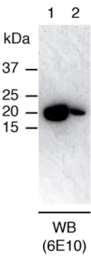
Satakarni and Curtis previously reported on the capacity of SUMO to solubilize Aβ42 when fused to it [27]. To examine this property under our expression conditions, we analyzed the supernatant corresponding to the soluble fraction, as well as the insoluble pellet resuspended in the same volume as that of the previously collected supernatant, by WB using 6E10, an antibody that recognizes residues 3 to 8 of the Aβ sequence (Fig. 1). Our results indicated that SUMO-Aβ42 was predominantly expressed in the soluble fraction. This finding contrasts with that described by Satakarni and Curtis, who reported obtaining the same volumetric productivity of SUMO-Aβ42 after IMAC purification of the soluble supernatant and the insoluble pellet [27]. These differences can be explained by the fact that we produced SUMO-Aβ42 using minimal media while they used richer LB media. Given that protein expression yields are usually lower in the former media and that aggregation is highly dependent on protein concentration, it is likely that the concentrations of SUMO-Aβ42 produced in minimal media are not high enough to promote extensive aggregation and accumulation of the fusion protein in inclusion bodies. This explanation is supported by a report describing the expression and purification of the 50-amino acid protein medin [36]. Similar to Aβ42, medin forms amyloid fibrils, which are the major protein component of Aortic Medial Amyloid. In the aforementioned report, the authors describe the successful expression and purification of [U-13C,15N] medin fused to His6-SUMO. As in the case
of SUMO-Aβ42, the SUMO-medin construct expressed in minimal media gives rise to a high level expression in the soluble fraction (90%). Notably, the final volumetric yield
of [U-13C,15N] medin obtained from the soluble fraction is the same as the one we report here for [U-15N] Aβ42.
Oxidation of methionine 35 is not observed during purification
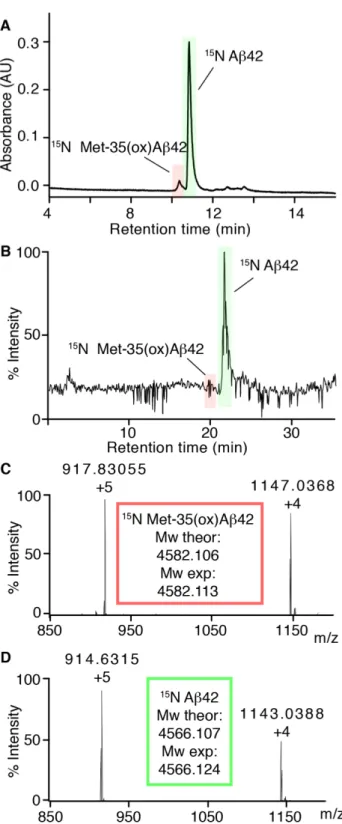
Next, to design the purification strategy, we took into account various considerations. First, under the minimal media, SUMO-Aβ42 is predominantly expressed in the soluble fraction. Second, the use of highly denaturing conditions makes proteins vulnerable to chemical modifications, including methionine oxidation. Third, the most widely reported undesired side product during Aβ purification is oxidized 35. Fourth, Met-35(ox)Aβ42 and Aβ42 differ mainly in terms of hydrophobicity, so the most efficient method to separate them is via costly and time-consuming preparative RP-HPLC. Given these considerations, we chose to focus on the purification of the soluble SUMO-Aβ42 fraction of the cell lysates, which allowed us to design a purification protocol that did not require the use of highly denaturing conditions in the stages of the purification when Aβ42 was still fused to SUMO. In doing so, our aim was to avoid Met-35 oxidation and thus the use of RP-HPLC. To ensure that Met-35 was not oxidized during the different stages of the Aβ42 purification, we used analytical RP-HPLC. Analysis of a mixture of 35(ox)Aβ42 and Aβ42 by analytical RP-HPLC and LC-MS revealed that Met-35(ox)Aβ42 elutes earlier than the reduced form (Fig. 2). This result supports the capacity of this method to detect the presence of Met-35(ox)Aβ42 in Aβ42 samples.
The first step of the purification relied on the (His)6 tag present at the N-terminal of
SUMO, which allowed a simple purification of SUMO-Aβ42 by IMAC using non-denaturing, degassed buffers in the presence of TCEP. Next, to cleave the SUMO-Aβ42 construct, the IMAC buffer was replaced by 50 mM ammonium carbonate and 1 mM TCEP at pH 9.0. This buffer allowed us to resolve the following two issues, namely to cleave Aβ42 from SUMO under basic pH—conditions reported to slow down Aβ42
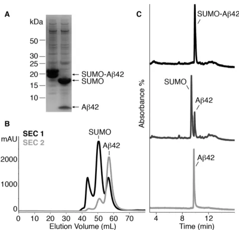
aggregation [37], and to subsequently lyophilize the sample, yielding a lyophilized powder free of insoluble salts. When present, the latter can promote Aβ42 aggregation upon subsequent resuspension. Under the 50 mM ammonium carbonate and 1 mM TCEP buffer, the cleavage of SUMO-Aβ42 using SUMO protease (Ulp1) [33] was highly effective, leading to SUMO and Aβ42, as analyzed by SDS-PAGE (Fig. 3A) and RP-HPLC (Fig. 3C). The two bands detected in the SDS-PAGE analysis of SUMO-Aβ42 (Fig. 3A, lane 1) were assigned to two populations of SUMO-SUMO-Aβ42 caused by either an incomplete denaturation of SUMO-Aβ42 prior to sample separation or subsequent partial renaturation during the separation. Products of incomplete translation during synthesis, and/or partial degradation during lysis, and/or sample processing of both SUMO and Aβ42 moieties were excluded as these would be evident in both the SDS-PAGE gels and the RP-HPLC profiles of the downstream SUMO protease-cleaved products (Fig. 3A and 3C).
Since the molecular weight of SUMO (12.4 kDa) is almost three-fold larger than that of Aβ42 (4.5 kDa), we proceeded with their separation by means of SEC (Fig. 3B). To obtain the best yield for monomeric Aβ42, it was critical to ensure the absence of Aβ42 aggregates at the time of injection. To this end, we resuspended the lyophilized powder obtained after SUMO protease cleavage at 2.5 mg/mL Aβ42 in 6.8 M GdnSCN. This strong chaotropic reagent is able to solubilize plaque cores from the brains of AD patients [2]. Therefore, performing this step ensured complete solubilization of the sample containing Aβ42 and SUMO. To minimize Aβ42 aggregation during elution from the column, SEC was performed at 4ºC. At this temperature, 6.8 M GdnSCN precipitates. To avoid this, the sample containing Aβ42 and SUMO was diluted to 4 M GdnSCN and 1.5 mg/mL Aβ42 before subjecting it into the SEC apparatus. To prevent the oxidation of Met-35, SEC was carried out using carefully degassed 50 mM
ammonium carbonate at pH 9.0. Again, we chose this buffer because its basic pH has been reported to minimize Aβ42 aggregation [37] and because its volatility allowed subsequent lyophilization of the fractions containing Aβ42 without leaving any insoluble salts in the lyophilized powder. To completely separate Aβ42 from SUMO, a second SEC purification was required (Fig. 3B). After this second SEC, the purity of the peptide and the absence of Met-35(ox)Aβ42 (compare to Fig. 2A) was confirmed by RP-HPLC (Fig. 3C). This expression and purification strategy allowed us to obtain 6 mg [U-15N] Aβ42 and 2 mg [U-2H,13C,15N] Aβ42 per L of culture. The purity of labeled peptides was determined by RP-HPLC and found to be > 98% (Fig. 4A). Moreover, their identity and label incorporation was determined by HRMS analysis (Fig. 4B) and reported in Table 1.
The labeled Aβ42 is amenable to NMR-based structural studies
Notably, apart from obtaining pure [U-15N] Aβ42 and [U-2H,13C,15N] Aβ42, the expression and purification strategy simultaneously allowed us to obtain pure [U-15N] SUMO and [U-2H,13C,15N] SUMO after the first SEC purification. Soluble and well-folded proteins are useful as standards to set up NMR experiments and are usually purchased from commercial sources. For example, 5 mg [U-15N] ubiquitin costs more than 1,000 € and 550 µL 0.5 mM [U-2H, 13C, 15N] maltose binding protein more than 6,000 €. Since SUMO is a soluble, well-folded protein, the [U-15N] SUMO and
[U-2H,13C,15N] SUMO obtained from this strategy could be useful for setting up NMR
experiments (Fig. 5A).
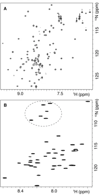
[U-15N] Aβ42 and [U-2H,13C,15N] Aβ42 peptides will be useful to characterize Aβ42 aggregation by means of NMR spectroscopy. As an example of a possible application, we measured 1H-15N HSQC NMR spectrum of monomeric Aβ42, obtained after dissolving [U-15N] Aβ42 in 95% DMSO-d6, 5% D2O at pH* 4.6 (Fig. 5B). This buffer
has been reported to disaggregate Aβ into its constituent monomers while preserving hydrogen deuterium exchange (HDX) information [6,38-42]. HDX experiments are among the techniques most used in the literature to obtain structural information about amyloid fibrils [6,38,39,41] and also about aggregates formed during fibril formation [42]. Under these conditions, we observed at least 37-38 N-H cross-peaks corresponding to the amides of the Aβ42 backbone. Among them, six peaks appeared at the characteristic chemical shifts of glycines, consistent with the six glycines present in the Aβ42 sequence. Moreover, we expect that the [U-2H,13C,15N] Aβ42 sample will pave the way for NMR studies of Aβ42 in the form of high molecular weight complexes, including those formed in a membrane environment.
Conclusions
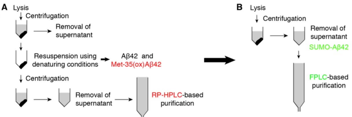
We have produced [U-15N] Aβ42 and [U-2H,13C,15N] Aβ42 using a novel and efficient expression and purification protocol (Figure 6). Our purification protocol involves an IMAC, a desalting step, and two SEC purifications. The ease of the purification strategy is based on the expression of Aβ42 fused to SUMO, which prevents Aβ42 aggregation. This strategy circumvents the requirement of denaturing conditions in the initial stages of purification, thus preventing the formation of Met-35(ox)Aβ42 and the need for costly and time-consuming preparative RP-HPLC purification. Consequently, recombinant expression of labeled Aβ42 is now accessible to many protein laboratories, including those that do not have access to preparative-RP-HPLC. Indeed, all the purification steps reported can be performed with the generally accessible fast protein liquid chromatography system (FPLC). Indeed, the cost of the reagents to produce 6 mg [U-15N] Aβ42 using this protocol is negligible; in this regard, we calculated it to be less than €15-20 per mg, thus being much cheaper than commercially available recombinant [U-15N] Aβ42, which is sold at $695 per mg
(https://www.rpeptide.com/products/labeled-peptides-and-proteins/beta-amyloid-labeled-peptides-recombinant/). The SUMO protease used in this study was prepared in-house, the methodology is very simple and can be performed by any lab wishing to produce large quantities of the enzyme to reduce costs [43]. SUMO protease is also available commercially as 100,000 U for €600 (less than one cent per unit) and, as we estimate that 5,000-10,000 U should be sufficient to cut 25mg of the SUMO-Aß42 fusion overnight, this digestion step should not therefore be seen as prohibitively expensive. Additionally, even in the case that a laboratory would need to invest in the required FPLC columns—we assume possession of an FPLC system—the cost of the columns would be recovered in the first Aβ42 purification, and the columns themselves, especially Superdex 30, can be used for many other purifications. Therefore, the expression and purification protocol described herein offers an alternative inexpensive approach to previously described methods to obtain [U-15N] Aβ42 [29,30] and the first method to achieve [U-2H,13C,15N] Aβ42.
Acknowledgements
M.N.P. and B.S. acknowledge the Spanish Government FPI and FPU programs, respectively, for predoctoral fellowships. We thank the Mass Spectrometry Core Facility at IRB Barcelona for technical support, Dr. Marga Gairí from the Scientific and Technological Centers, University of Barcelona (CCiTUB), for acquiring NMR spectra of [U-15N] SUMO, and J. Garcia for helpful advice. This work was supported by a Program Grant from the MINECO-FEDER (SAF2015-68789-R) to N.C.
Abbreviations
Alzheimer’s disease (AD); amyloid-beta peptide (Aβ); amyloid-beta peptide comprising 42 residues (Aβ42); amyloid precursor protein (APP); dimethylsulfoxide-d6
(fAD); formic acid (FA); fast protein liquid chromatography system (FPLC); guanidinium thiocyanate (GdnSCN), heteronuclear single quantum coherence (HSQC); high-resolution mass spectrometry (HRMS); hydrogen deuterium exchange (HDX); immobilized metal affinity chromatography (IMAC); liquid chromatography coupled to MS (LC-MS); Luria Bertani (LB); nano electrospray (nanoESI); nuclear magnetic resonance spectroscopy (NMR); oxidation of the Met-35 side chain of Aβ42 to methionine sulfoxide (Met-35(ox)Aβ42); reversed phase high performance liquid chromatography (RP-HPLC); sample buffer (SB); size exclusion chromatography (SEC); small Ubiquitin-like Modifier (SUMO); sodium dodecyl sulfate polyacrylamide gel electrophoresis (SDS-PAGE); trifluoroacetic acid (TFA); tris-buffered saline, 0.1% tween 20 (TBST); tris(2-carboxyethyl)phosphine (TCEP); Western blot (WB).
Conflict of Interest
References
[1] International, A.D. World Alzheimer Report 2015, The Global Impact of Dementia: An analysis of prevalence, incidence, cost and trends. 2015, 1–87.
[2] Masters, C.L.; Selkoe, D.J. Biochemistry of amyloid β-protein and amyloid deposits in Alzheimer disease. Cold Spring Harb. Perspect. Med, 2012, 2, a006262– a006262.
[3] Blennow, K.; Zetterberg, H.; Fagan, A.M. Fluid biomarkers in Alzheimer disease. Cold Spring Harb. Perspect. Med, 2012, 2, a006221.
[4] Citron, M.; Westaway, D.; Xia, W.; Carlson, G.; Diehl, T.; Levesque, G.; Johnson-wood, K.; Lee, M.; Seubert, P.; Davis, A.; Kholodenko, D.; Motter, R.; Sherrington, R.; Perry, B.; Yao, H.; Strome, R.; Lieberburg, I.; Rommens, J.; Kim, S.; Schenk, D.; Fraser, P.; Hyslop, P.S.G.; Selkoe, D.J. Mutant presenilins of Alzheimer's disease increase production of 42-residue amyloid β-protein in both transfected cells and transgenic mice. Nat. Med, 1997, 3, 67–72.
[5] Riek, R.; Güntert, P.; Dobeli, H.; Wipf, B.; Wuthrich, K. NMR studies in aqueous solution fail to identify significant conformational differences between the monomeric forms of two Alzheimer peptides with widely different plaque-competence, A beta(1-40)(ox) and A beta(1-42)(ox). Eur. J. Biochem, 2001, 268, 5930–5936.
[6] Lührs, T.; Ritter, C.; Adrian, M.; Riek-Loher, D.; Bohrmann, B.; Döbeli, H.; Schubert, D.; Riek, R. 3D structure of Alzheimer's amyloid-beta(1-42) fibrils. Proc. Natl. Acad. Sci. USA, 2005, 102, 17342–17347.
[7] Petkova, A.T.; Leapman, R.D.; Guo, Z.; Yau, W.-M.; Mattson, M.P.; Tycko, R. Self-propagating, molecular-level polymorphism in Alzheimer's beta-amyloid fibrils. Science, 2005, 307, 262–265.
Evidence of fibril-like β-sheet structures in a neurotoxic amyloid intermediate of Alzheimer's β-amyloid. Nat. Struct. Mol. Biol, 2007, 14, 1157–1164.
[9] Yu, L.; Edalji, R.; Harlan, J.E.; Holzman, T.F.; Lopez, A.P.; Labkovsky, B.; Hillen, H.; Barghorn, S.; Ebert, U.; Richardson, P.L.; Miesbauer, L.; Solomon, L.; Bartley, D.; Walter, K.; Johnson, R.W.; Hajduk, P.J.; Olejniczak, E.T. Structural characterization of a soluble amyloid β-peptide oligomer. Biochemistry, 2009, 48, 1870–1877.
[10] Fawzi, N.L.; Ying, J.; Ghirlando, R.; Torchia, D.A.; Clore, G.M. Atomic-resolution dynamics on the surface of amyloid-β protofibrils probed by solution NMR. Nature, 2011, 480, 1–24.
[11] Lopez del Amo, J.M.; Schmidt, M.; Fink, U.; Dasari, M.; Fändrich, M.; Reif, B. An asymmetric dimer as the basic subunit in Alzheimer’s disease amyloid β fibrils. Angew. Chem. Int. Ed, 2012, 51, 6136–6139.
[12] Lu, J.-X.; Qiang, W.; Yau, W.-M.; Schwieters, C.D.; Meredith, S.C.; Tycko, R. Molecular structure of β-amyloid fibrils in Alzheimer's disease brain tissue. Cell, 2013, 154, 1257–1268.
[13] Döbeli, H.; Draeger, N.; Huber, G.; Jakob, P.; Schmidt, D.; Seilheimer, B.; Stüber, D.; Wipf, B.; Zulauf, M. A biotechnological method provides access to aggregation competent monomeric Alzheimer's 1–42 residue amyloid peptide. Bio/Technology, 1995, 13, 988–993.
[14] Carrotta, R.; Di Carlo, M.; Manno, M.; Montana, G.; Picone, P.; Romancino, D.; San Biagio, P.L. Toxicity of recombinant beta-amyloid prefibrillar oligomers on the morphogenesis of the sea urchin Paracentrotus lividus. FASEB J, 2006, 20, 1916–1917. [15] Das, U.; Hariprasad, G.; Pasha, S.; Mann, A.; Ganguli, M.; Sharma, S.; Kaur, P.; Singh, T.P.; Srinivasan, A. Interface peptide of Alzheimer's amyloid beta:
application in purification. Biochem. Bioph. Res. Co, 2007, 362, 538–542.
[16] Caine, J.; Volitakis, I.; Cherny, R.; Varghese, J.; Macreadie, I. Aβ produced as a fusion to maltose binding protein can be readily purified and stably associates with copper and zinc. Protein Peptide Lett, 2007, 14, 83–86.
[17] Sharpe, S.; Yau, W.-M.; Tycko, R. Expression and purification of a recombinant peptide from the Alzheimer's beta-amyloid protein for solid-state NMR. Protein Expres. Purif, 2005, 42, 200–210.
[18] Wiesehan, K.; Funke, S.A.; Fries, M.; Willbold, D. Purification of recombinantly expressed and cytotoxic human amyloid-beta peptide 1-42. J. Chromatogr. B Analyt. Technol. Biomed. Life Sci, 2007, 856, 229–233.
[19] Macao, B.; Hoyer, W.; Sandberg, A.; Brorsson, A.-C.; Dobson, C.M.; Härd, T. Recombinant amyloid beta-peptide production by coexpression with an affibody ligand. BMC Biotechnol, 2008, 8, 82–11.
[20] Walsh, D.M.; Thulin, E.; Minogue, A.M.; Gustavsson, N.; Pang, E.; Teplow, D.B.; Linse, S. A facile method for expression and purification of the Alzheimer's disease-associated amyloid beta-peptide. FEBS J, 2009, 276, 1266–1281.
[21] Zhang, B.; Cheng, X.R.; da Silva, I.S.; Hung, V.W.S.; Veloso, A.J.; Angnes, L.; Kerman, K. Electroanalysis of the interaction between (-)-epigallocatechin-3-gallate (EGCG) and amyloid-β in the presence of copper. Metallomics, 2013, 5, 259–264. [22] Lee, E.K.; Hwang, J.H.; Shin, D.Y.; Kim, D.I.; Yoo, Y.J. Production of recombinant amyloid-beta peptide 42 as an ubiquitin extension. Protein Expres. Purif, 2005, 40, 183–189.
[23] Shahnawaz, M.; Thapa, A.; Park, I.-S. Stable activity of a deubiquitylating enzyme (Usp2-cc) in the presence of high concentrations of urea and its application to purify aggregation-prone peptides. Biochem. Bioph. Res. Co, 2007, 359, 801–805.
[24] Thapa, A.; Shahnawaz, M.; Karki, P.; Raj Dahal, G.; Golam Sharoar, M.; Yub Shin, S.; Sup Lee, J.; Cho, B.; Park, I.-S. Purification of inclusion body—forming peptides and proteins in soluble form by fusion to Escherichia coli thermostable proteins. Biotech, 2008, 44, 787–796.
[25] Garai, K.; Crick, S.L.; Mustafi, S.M.; Frieden, C. Expression and purification of amyloid-β peptides from Escherichia coli. Protein Expres. Purif, 2009, 66, 107–112. [26] Finder, V.H.; Vodopivec, I.; Nitsch, R.M.; Glockshuber, R. The recombinant amyloid-β peptide Aβ1–42 aggregates faster and is more neurotoxic than synthetic Aβ1–42. J. Mol. Biol, 2010, 396, 9–18.
[27] Satakarni, M.; Curtis, R. Production of recombinant peptides as fusions with SUMO. Protein Expres. Purif, 2011, 78, 113–119.
[28] Kim, E.-K.; Moon, J.C.; Lee, J.M.; Jeong, M.S.; Oh, C.; Ahn, S.-M.; Yoo, Y.J.; Jang, H.H. Large-scale production of soluble recombinant amyloid-β peptide Aβ42 using cold-inducible expression system. Protein Expres. Purif, 2012, 86, 53–57.
[29] Weber, D.K.; Sani, M.-A.; Gehman, J.D. A routine method for cloning, expressing and purifying Aβ(1–42) for structural NMR studies. Amino Acids, 2014, 46, 2415–2426.
[30] Sharma, S.C.; Armand, T.; Ball, K.A.; Chen, A.; Pelton, J.G.; Wemmer, D.E.; Head-Gordon, T. A facile method for expression and purification of 15N isotope-labeled human Alzheimer´s β-amyloid peptides from E. coli for NMR-based structural analysis. Protein Expres. Purif, 2015, 116, 1–8.
[31] Butterfield, D.A.; Bush, A.I. Alzheimer's amyloid beta-peptide (1-42): involvement of methionine residue 35 in the oxidative stress and neurotoxicity properties of this peptide. Neurobiol. Aging, 2004, 25, 563–568.
methionine 35 is not necessary for amyloid β-protein toxicity. J. Neurochem, 2010, 113, 1–21.
[33] Assenberg, R.; Delmas, O.; Graham, S.C.; Verma, A.; Berrow, N.; Stuart, D.I.; Owens, R.J.; Bourhy, H.; Grimes, J.M. Expression, purification and crystallization of a lyssavirus matrix (M) protein. Acta Crystallogr. Sect. F Struct. Biol. Cryst. Commun, 2008, 64, 258–262.
[34] Tyler, R.C.; Sreenath, H.K.; Singh, S.; Aceti, D.J.; Bingman, C.A.; Markley, J.L.; Fox, B.G. Auto-induction medium for the production of [15N]- and [13C, U-15N]-labeled proteins for NMR screening and structure determination. Protein Expres. Purif, 2005, 40, 268–278.
[35] Studier, F.W. Protein production by auto-induction in high-density shaking cultures. Protein Expres. Purif, 2005, 41, 207–234.
[36] Davies, H.A.; Wilkinson, M.C.; Gibson, R.P.; Middleton, D.A. Expression and purification of the aortic amyloid polypeptide medin. Protein Expres. Purif, 2014, 98, 32–37.
[37] Teplow, D.B. In Amyloid, Prions, and Other Protein Aggregates, Part C; Methods in Enzymology; Elsevier, 2006; Vol. 413.
[38] Olofsson, A.; Sauer-Eriksson, A.E.; Ohman, A. The solvent protection of Alzheimer amyloid-β(1– 42) fibrils as determined by solution NMR spectroscopy. J. Biol. Chem, 2005, 281, 1–7.
[39] Kheterpal, I.; Wetzel, R. Hydrogen/deuterium exchange mass spectrometry. A window into amyloid structure. Acc. Chem. Res, 2006, 39, 584–593.
[40] Carulla, N.; Zhou, M.; Arimon, M.; Gairi, M.; Giralt, E.; Robinson, C.V.; Dobson, C.M. Experimental characterization of disordered and ordered aggregates populated during the process of amyloid fibril formation. Proc. Natl. Acad. Sci. USA,
2009, 106, 7828–7833.
[41] Sánchez, L.; Madurga, S.; Pukala, T.; Vilaseca, M.; López-Iglesias, C.; Robinson, C.V.; Giralt, E.; Carulla, N. Aβ40 and Aβ42 amyloid fibrils exhibit distinct molecular recycling properties. J. Am. Chem. Soc, 2011, 133, 6505–6508.
[42] Serra-Vidal, B.; Pujadas, L.; Rossi, D.; Soriano, E.; Madurga, S.; Carulla, N. Hydrogen/deuterium exchange-protected oligomers populated during Aβ fibril formation correlate with neuronal cell death. ACS Chem. Biol, 2014, 9, 2678–2685. [43] Lee, C.-D.; Sun, H.-C.; Hu, S.-M.; Chiu, C.-F.; Homhuan, A.; Liang, S.-M.; Leng, C.-H.; Wang, T.-F. An improved SUMO fusion protein system for effective production of native proteins. Protein Sci, 2008, 17, 1241–1248.
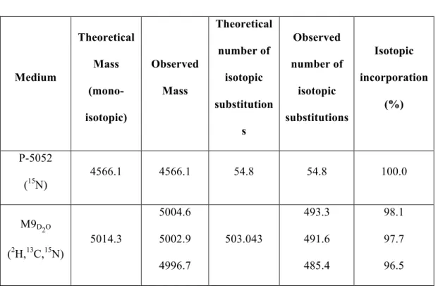
Tables: Medium Theoretical Mass (mono-isotopic) Observed Mass Theoretical number of isotopic substitution s Observed number of isotopic substitutions Isotopic incorporation (%) P-5052 (15N) 4566.1 4566.1 54.8 54.8 100.0 M9D2O (2H,13C,15N) 5014.3 5004.6 5002.9 4996.7 503.043 493.3 491.6 485.4 98.1 97.7 96.5
Table 1. Isotopic incorporation percentages. The theoretical monoisotopic masses were calculated using the Molecular Mass Calculator tool from the Biological Magnetic Resonance Data Bank website
(http://www.bmrb.wisc.edu/metabolomics/mol_mass.php?formula=C210H321N56O61 S2&subaction=Natural+Composition&updateIsoComp=1). In the case of [U-15N] Aβ42, they were obtained assuming 100% substitution of 14N isotopes for 15N and in the case of [U-2H,13C,15N] Aβ42 assuming 100% substitution of 12C, 14N and non-labile 1H isotopes for 13C, 15N and 2H. The theoretical and observed number of isotopic substitutions account for the difference between the theoretical mono-isotopic mass and the theoretical or observed mass of the different isotopically labeled Aβ42 peptides. Therefore, the theoretical and observed number of isotopic substitutions indicate the increase in mass due to the theoretical and experimental incorporation of 15N and 2H,
Figure Legends:
Fig. 1. SUMO-Aβ42 is predominantly expressed in the soluble fraction. WB analysis of the supernatants corresponding to the soluble (lane 1) and insoluble (lane 2) fraction of a crude lysate. Samples were blotted with 6E10 monoclonal antibody, which recognizes residues 3-8 of Aβ.
Fig. 2. Characterization of the retention time of Met-35(ox)Aβ42 and Aβ42 on RP-HPLC. (A) RP-HPLC chromatogram obtained for a sample containing Met-35(ox)Aβ42 (highlighted in red) and Aβ42 (highlighted in green). (B) LC-MS chromatogram obtained for the same sample containing Met-35(ox)Aβ42 (highlighted in red) and Aβ42 (highlighted in green). ESI-mass spectrum corresponding to the (C) first and the
(D) second peaks detected by LC-MS, assigned to Met-35(ox)Aβ42 and Aβ42, respectively. Mw theor refers to the theoretical monoisotopic mass and Mw exp to the experimental monoisotopic mass.
Fig. 3. Aβ42 purification. (A) SDS-PAGE analysis of SUMO-Aβ42 fusion protein before (lane 1) and after (lane 2) cleavage with SUMO protease (Ulp1). (B) SEC chromatograms corresponding to the first (black) and second (light gray) purification step of the SUMO and Aβ42 sample obtained after cleavage. SUMO and Aβ42 eluted at 50.5 mL and 57.1 mL, respectively. (C) RP-HPLC chromatograms at different stages of the Aβ42 purification: SUMO-Aβ42 before (black) and SUMO Aβ42 after cleavage (dark gray), and pure Aβ42 (light gray) obtained after the second SEC. SUMO-Aβ42, SUMO and Aβ42 eluted at 9.9 min, 9.3 min and 9.8 min, respectively.
Fig. 4. Purity and identity of [U-15N] Aβ42 and [U-2H,13C,15N] Aβ42. RP-HPLC
analysis of (A) [U-15N] Aβ42 and (B) [U-2H,13C,15N] Aβ42. HRMS analysis of (C)
[U-15N] Aβ42 and (D) [U-2H,13C,15N] Aβ42. ESI-mass spectrum (right) and deconvoluted
Fig. 5. Labeled SUMO and Aβ42 are suitable for NMR studies. (A) 1H-15N HSQC
NMR spectra of 400 µM [U-15N] SUMO in 50 mM sodium phosphate, pH 7.4, 90% H2O/10% D2O and (B) 150 µM [U-15N] Aβ42 in 100% DMSO-d6, 5% H2O at pH* 4.6.
The dotted circle indicates the position of the peaks appearing in the region characteristic of the glycine residues.
Fig. 6. Schematic diagram for purification of recombinant Aβ42. (A) Previous strategies report accumulation of the fusion-Aβ42 protein in inclusion bodies. This requires the use of denaturing conditions in the initial stages of purification, which leads to the formation of Met-35(ox)Aβ42. To separate Met-35(ox)Aβ42 from Aβ42, costly and time-consuming preparative RP-HPLC purification is required. (B) Proposed strategy based on the expression of Aβ42 fused to SUMO, which prevents Aβ42 aggregation. The solubility of SUMO-Aβ42 circumvents the requirement of denaturing conditions in the initial stages of purification, thereby preventing the formation of Met-35(ox)Aβ42. This protocol allows all the purification steps to be performed with the generally accessible fast protein liquid chromatography system (FPLC).
![Fig. 4. Purity and identity of [U- 15 N] Aβ42 and [U- 2 H, 13 C, 15 N] Aβ42](https://thumb-eu.123doks.com/thumbv2/123dokorg/4437307.29896/32.892.128.764.107.512/fig-purity-identity-u-n-aβ-u-aβ.webp)