UNIVERSITÀ DEGLI STUDI DI ROMA
"TOR VERGATA"
FACOLTA' DI SCIENZE M.F.N. DOTTORATO DI RICERCA IN SCIENZE CHIMICHE XXI CICLO“Synthesis and applications of porphyrinoids:
a journey into the multifaceted chemistry
of pyrrolic macrocycles”
Dott. Giuseppe Pomarico
A.A. 2008/2009 Docente Guida: Prof. Roberto Paolesse Coordinatore: Prof. Bruno Crociani
Index
Glossary
VIIIChapter 1
Porphyrins-based organic materials:
synthesis, characterization and exploitation
as chemical sensors
Introduction
1.1 A brief history of porphyrins chemistry 2
1.2 General features of porphyrins 4
1.3 Porphyrin derivatives: organic substrates for sensors
and materials chemistry 9
Results and discussion
1.4 Self assembled monolayer: the most straightforward way
to monomolecular film 16
1.4.1 Synthesis and characterization of porphyrin-based SAM 22
1.5 Porphyrinoids supramolecular aggregates 34
1.5.1 Synthesis and exploitation of self-assembled
Experimental section
1.6 Synthesis of thiol-derivatized porphyrin 52
1.6.1 Synthesis of 5-(4-carboxymethylphenyl)-10,15,20 -triphenylporphyrin (7a) 52 1.6.2 Synthesis of 5-(4-carboxyphenyl)-10,15,20 -triphenylporphyrin (8a) 52 1.6.3 Synthesis of 5-(4-carboxy-succinimidephenyl) -10,15,20-triphenylporphyrin (9a) 53 1.6.4 Synthesis of 5-(4-(N-(2-tert-butyloxycarbamoyl) -ethyl)-carboxyamidephenyl)-10,15,20-triphenylporphyrin 53 1.6.5 Synthesis of 5-(4-(N-(2-aminoethyl)-carboxyamidephenyl) -10,15,20-triphenylporphyrin (10a) 54
1.6.6 Synthesis of 12-Mercaptoacetyl-dodecanoic acid (12a) 54 1.6.7 Synthesis of N-(12-Mercaptoacetyl-dodecyloxyl) -succinimide (13a) 55 1.6.8 Synthesis of 5-(4-(N-(2-(12-mercaptoacetyl-dodecyl)-carbamoyl) -ethyl)-carboxyamidephenyl)-10,15,20-triphenylporphyrin 56 1.6.9 Synthesis of 5-(4-(N-(2-(12-mercapto-dodecyl)-carbamoyl) -ethyl)-carboxyamidephenyl)-10,15,20-triphenylporphyrin (14a) 56 1.6.10 Procedure for syntheses of Zn (II) and Co(II) complexes 57
1.7 Synthesis of SAM 57
1.7.1 Procedure for gold surfaces preparation 57
1.7.2 Procedure for porphyrin deposition onto gold 58
1.8 Synthesis of porphyrins nanotube 58
1.8.1 Synthesis of 5,10,15,20-[Tetra-(4-pyridyl)porphyrinato]SnCl2 (16a) 58
1.8.2 General procedure for porphyrins nanotube preparation 59
1.8.3 Polymer membrane preparation 59
1.8.5 AFM setup 60
1.8.6 Gas measurement setup 60
References
62Chapter 2
Corrole: chemistry and applications of an
unusual tetrapyrrolic macrocycle
Introduction
2.1 General properties of corroles 67
2.1.1 Spectroscopic properties of triarylcorroles 67
2.1.2 Corrole: acid-base equilibrium 70
2.2 Synthetic procedures for meso-triarylcorroles 72
2.3 Metallocorroles 75
2.3.1 Synthesis and properties of metallocorroles 75
2.3.2 Bonding geometries of metallocorroles 76
Results and discussion
2.4 Reactivity of triarylcorroles: from functionalization to new isomers 79 2.4.1 Isocorrole: unexpected product from oxidative synthetic step 82
2.5.1 Demetalation of corroles complexes: a starting point
towards new synthetic routes 93
Experimental section
2.6 Synthesis of meso-Arylcorroles 103
2.6.1 General procedure for syntheses of Triarylcorroles
(10b, 11b, 12b) 103 2.6.2 Purification of 5,10,15-Triphenylcorrole (10b) 103 2.6.3 Purification of 5,10,15-Tris-(4-methylphenyl)corrole (11b) 103 2.6.4 Purification of 5,10,15-Tris-(4-methoxylphenyl)corrole (12b) 104 2.6.5 Synthesis of 5,10,15-Tris-(4-nitrophenyl)corrole (13b) 104 2.7 Syntheses of β-Alkylcorroles (26b, 27b, 28b) 104 2.8 Syntheses of Triarylisocorroles 105 2.8.1 Synthesis of 5,10,15-Triphenyl-10-ethoxyl-isocorrole (14b) 105 2.8.2 Synthesis of 5,10,15-Tris-(4-methylphenyl)-10-methoxyl
-isocorrole (15b) and of 5,10,15-Tris-(4-methylphenyl)-5
-methoxyl-isocorrole (16b) 106 2.8.3 Synthesis of 5,10,15- Tris-(4-methoxylphenyl)-10-methoxyl -isocorrole (17b) 107 2.9 Syntheses of Metallocorroles 108 2.9.1 Synthesis of [5,10,15-Triarylcorrolates]Cu 108 2.9.2 Synthesis of [2,3,7,8,12,13,17,18-Octaalkylcorrolates]Cu 108 2.9.3 Synthesis of [5,10,15-Triphenylcorrolato]Co(PPh3) 109 2.9.4 Synthesis of [2,3,7,8,12,13,17,18-Octamethylcorrolato]Co(PPh3) 109 2.9.5 Synthesis of [5,10,15-Triphenylcorrolato]MnCl 110 2.9.6 Synthesis of [8,12-Diethyl-2,3,7,13,17,18 -hexamethylcorrolato]MnCl 110 2.9.7 Synthesis of [5,10,15-Triphenylcorrolato]FeCl 110
2.9.8 Synthesis of [2,3,17,18-Tetraethyl-7,8,12,13
-tetramethylcorrolato]FeCl 111
2.9.9 Synthesis of [5,10,15-Triphenylcorrolato]GeCl 111 2.9.10 Synthesis of [2,3,17,18-Tetraethyl-7,8,12,13
-tetramethylcorrolato]GeCl 112
2.10 General procedures for demetalation process 112
2.10.1 CHCl3/H2SO4 method 112 2.10.2 H2SO4 method 113 2.10.3 HBr/HOAc method 113
References
114Chapter 3
Triaryl-tetrabenzocorroles: innovative
compounds for biomedical applications
Introduction
3.1 History of photodynamic therapy 119
3.2 Principles of PDT 121
3.2.1 Photochemistry of PDT 121
3.2.2 Singlet oxygen 125
3.3 Photosensitizers 127
3.3.2 Photophysic of photosensitizers 128
3.4 Biological mechanism of action 131
Results and discussion
3.5 PDT: porphyrinoids in the service of medicine 133
3.5.1 Exploitation of porphyrin derivatives in PDT 133
3.5.2 Benzoporphyrins 136
3.6 Syntheses of Triaryl-tetrabenzocorroles 140
3.6.1 Synthesis of precursor: 4,5,6,7-Tetrahydroisoindole 141 3.6.2 Synthesis of Triaryl-tetrabenzocorroles: one step procedure 143 3.6.3 Synthesis of Triaryl-tetrabenzocorroles: two steps procedure 146 3.6.4 Dihydroisoindole: a closer precursor to tetrabenzocorroles 156
Experimental section
3.7 Synthesis of precursors: tetrahydroisoindole and dipyrromethane 159 3.7.1 Synthesis of 2-Carboxyethyl-4,5,6,7-tetrahydroisoindole (15c) 159 3.7.2 Synthesis of 4,5,6,7-Tetrahydroisoindole (16c) 159 3.7.3 Synthesis of 1,9-Dicarboxyethyl-2:3,7:8-dibutane -5-phenyl-dipyrromethane (17c) 160 3.7.4 Synthesis of 2:3,7:8-Dibutane-5-phenyl -dipyrromethane (18c) 160 3.8 Syntheses of Triaryl-tetrabenzocorroles 161 3.8.1 Synthesis of 5,10,15-Triphenyl-2:3,7:8,12:13,17:18-
tetrabutanecorrole - modified Paolesse method 161
3.8.2 Synthesis of 5,10,15-Tris-(4-methyl-carboxylatephenyl) -2:3,7:8,12:13,17:18-tetrabutanecorrole
3.8.3 Synthesis of 5,10,15-Triphenyl-2:3,7:8,12:13,17:18
-tetrabutanecorrole - Paolesse method 162
3.8.4 Synthesis of 10-(4-Cyanophenyl)-5,15-diphenyl-
2:3,7:8,12:13,17:18-tetrabutanecorrole (19c) - Gryko method 163 3.8.5 Synthesis of [10-(4-Cyanophenyl)-5,15-diphenyl -2:3,7:8,12:13,17:18-tetrabutanecorrolate]Cu 163 3.8.6 Synthesis of 5,10,15-Tris-(3-cyanophenyl) -2:3,7:8,12:13,17:18-tetrabutanecorrole (20c) 164 3.8.7 Synthesis of [5,10,15-Tris-(3-cyanophenyl) -2:3,7:8,12:13,17:18-tetrabutanecorrolate]Cu (21c) 164 3.8.8 Synthesis of [5,10,15-Tris-(3-cyanophenyl) -2:3,7:8,12:13,17:18-tetrabenzocorrolate]Cu (22c) 165
3.9 An alternative isoindole as triaryl-tetrabezocorrole precursor 166
3.9.1 Synthesis of Ethynyl p-tolyl sulfone (24c) 166
3.9.2 Synthesis of 1-Tosyl-[(4,5-dimethyl)-cyclohexa-1,4-diene)] (25c) 166 3.9.3 Synthesis of 2-Carboxyethyl-5,6-dimethyl-4,7-dihydro
-2H-isoindole (26c) 167
References
168Glossary - Reagents and solvents
BF3·OEt2 = Boron trifluoride diethyl etherate
Boc = 2-(tert-butyl-carbamoil)-ethaneamine t-BuOK = potassium tert-butoxide
Chloranil = 2,3,5,6-tetrachloro-1,4-benzoquinone DBU = 1,8-Diazabicicle[5.4.0]-7-undecene
DDQ = 2,3-Dicloro-5,6-dicyano-1,4-benzoquinone DMF = N,N’-Dimethylformamide
EDC = 1-[3-(dimethyl-amino)propyl)-3-ethylcarbodiimide hydrochloride MeCN = Acetonitrile
MeOH = Methanol
PDMS = polydimethylsiloxane distearate 2-PrOH = 2-Propanol
Sc(OTf)3 = Scandium trifluoromethanesulfonate
TEA = Trietylamine TFA = Trifluoroacetc acid THF = Tetrahydrofuran
Glossary of porphyrinoids - Chapter 1
p-CO2CH3TPPorH2 = 5-(4-carboxymethylphenyl)-10,15,20-triphenylporhyrin
TPPorH2 = 5,10,15,20-Tetraphenylporhyrin
[TSPPorH2]4- = [5,10,15,20-Tetra-(sulfonatophenyl)porphyrinate]-tetrasodium
salt
TPyPorH2 = 5,10,15,20-Tetra-(4-pyridyl)porphyrin
Glossary of porphyrinoids - Chapter 2 Br8(10-OH)TPIsoCH2 = 2,3,7,8,12,13,17,18-Octabromo-5,10,15-triphenyl-10-hydroxyl-isocorrole [Br8TPCor]H3 = 2,3,7,8,12,13,17,18-Octabromo -5,10,15-triphenylcorrole [Br8TPCor]Cu = [2,3,7,8,12,13,17,18-Octabromo -5,10,15-triphenylcorrolato]copper Br8TPCorH2 = 2,3,7,8,12,13,17,18-Octabromo-5,10,15-triphenyl-10-H-isocorrole [(4-ClPh)8TPCor]Cu = [2,3,7,8,12,13,17,18-Octabromo-5,10,15 -(4-chlorophenyl)corrolato]copper Et2Me6CorH3 = 8,12-Diethyl-2,3,7,13,17,18-hexamethylcorrole Et4Me4CorH3 = 2,3,17,18-Tetraethyl-7,8,12,13-tetramethylcorrole (5-OH)TPIsoCorH2 = 5,10,15-Triphenyl-5-hydroxyl-isocorrole (10-OH)TPIsoCorH2 = 5,10,15-Triphenyl-10-hydroxyl-isocorrole Me8CorH3 = 2,3,7,8,12,13,17,18-Octamethylcorrole
[(NO2)TPCor]Ag = [3-Nitro-5,10,15-triphenylcorrolato]silver
(p-NO2)TPCorH3 = 5,10,15-Tris-(4-nitrophenyl)corrole
(p-OCH3)TPCorH3 = 5,10,15-Tris-(4-methoxyphenyl)corrole
TPCorH3 = 5,10,15-Triphenylcorrole
[TPCor]Metal = [5,10,15-Triphenylcorrolo]metal TTCorH3 = 5,10,15-Tris-(4-methylphenyl)corrole
Glossary of porphyrinoids - Chapter 3
(3-CN)BuTPCorH3 = 5,10,15-Tris
-(3-cyanophenyl)-2:3,7:8,12:13,17:18-tetrabutanecorrole [(3-CN)BuTPCor]Cu = [5,10,15-Tris
-(3-cyanophenyl)-2:3,7:8,12:13,17:18-tetrabutanecorrolato]copper [(3-CN)TBCor]Cu = [5,10,15-Tris
-(3-cyanophenyl)-2:3,7:8,12:13,17:18-tetrabenzocorrolato]copper
Hp = 3,7-Di-(3-propyloxyl)-2,8,12,17-tetramethyl-13,18-di-(2-hydroxyethyl)-porphyrin
Chapter 1
Porphyrins based organic materials:
synthesis, characterization and exploitation
Introduction
1.1 A brief history of porphyrins chemistry
Thudichum[1] developed the first preparation of porphyrin in 1867, by treatment of hemoglobin with concentrated acid. A few years later by a similar route, Hoppe-Seyler[2] obtained a purple substance which he called hematoporphyrin (1a, Fig.1.1).
However, these procedures failed to provide a pure sample of porphyrin. Approximately 30 years later Nencki[3] isolated the first pure sample, preparing hematoporphyrin hydrochloride from isolated hemin. These initial studies marked the beginning of porphyrin chemistry, which now includes many disciplines of science and medicine and continue to flourish.
Fig. 1.1 – Structure of Hematoporphyrin
Porphyrins posses a based skeleton consisting of four pyrrole units linked by four methine bridges. This skeletal structures was first proposed by Küster[4] in 1912. However, it was suggested by both Fischer and Willstätter, that such a large ring system would not be very stable; they proposed other structures containing smaller ring systems such as 2a and 3a (Fig. 1.2).
The debate over the actual structure of porphyrin continued until 1926, when Fischer successfully synthesized etioporphyrin-I by the first totally synthetic way[5]. Shortly thereafter Fischer completed the synthesis of octamethylporphyrin (4a, Fig. 1.3) by two distinctly preparative methods[6]. These preparations led to the acceptance of the structure initially proposed by Küster as the basic structure of porphyrins.
Fig. 1.2 – Earlier structures proposed for porphyrin
2a
3a
4a
1.2 General features of porphyrins
Tetrapyrrolic macrocycle is a term used to refer to a class of compounds that have four pyrrole units connected through four sp2 hybridized meso-carbons at the α position of the pyrrole rings. Among those, porphyrin represents the most common arrangement, with its molecular skeleton having formula C20H14N4.
Currently there are two nomenclature systems for the numbering of porphyrins: the older is the so-called the “Fischer system”, where the meso positions are labelled by Greek lettering system, and the four pyrrolic rings are so labeled with the capital letters A, B, C, and D. However this way does not allow to the identification of all carbons of the molecular skeleton.
The more modern and more thorough scheme, is called the “IUPAC system”, which identifies every carbon in the macrocyclic ring (Fig. 1.4).
The porphyrins macrocycle is an aromatic system containing 22 π electrons 18 of which are involved in any localization pathway (Fig. 1.4).
Porphyrins obey Hückel’s rule of aromaticity (4n+2π electrons, where n = 4). The aromatic character of porphyrins is also evident in their NMR spectrum. 1H NMR spectroscopy of porphyrins shows that the N-H protons appear from δ = ∼ - 4 to -2 ppm (upfield from TMS), indicative of NHs located in an anisotropic aromatic shielding cone[7].
Whereas the methane protons appear at ∼ 10 ppm, a δ value indicating a highly deshielding environment resulting from the aromatic ring current.
Visible absorption spectrum of porphyrins displays an intense absorption (ε > 100000) to the second excited state (S0→S2) at about 400 nm, referred to as the
Soret band[8]; this is characteristic of a highly conjugated porphyrin macrocycle, and it disappears when the aromatic delocalization pathway is disrupted. There are several weaker absorptions to the first excited state (S0→S1) at longer
wavelengths (450 to 700 nm). Both of these bands arise from π-π* transitions, and can be explained by considering the four frontier orbitals model proposed by Gouterman[9]. According to this theory, the absorption bands in porphyrins arise from transition between two HOMOs and two LUMOs (Fig. 1.5). The HOMOs were calculated to be an a1u and an a2u orbital, while the LUMOs were calculated
to be a degenerate set of eg orbitals.
Transition between these orbitals gave rise to two excited states, both of 1Eu
character. Orbital mixing splits these two states in energy, creating a higher energy 1Eu state with greater oscillator strength, which give rise to the Soret
band, and a lower energy 1Eu state with less oscillator strength, giving rise to the
Q bands.
The lowest energy excited singlet states of porphyrins can be thought of as being formed from the molecular orbitals examined above. An excited singlet state with an a1ueg configuration is formed by promoting an electron from the a1u
Likewise, an excited singlet state with an a2ueg configuration is formed by
promoting an electron from the a2u orbital to an eg orbital.
Variations of the peripheral substituents on the porphyrin ring often causes minor changes in the intensity and wavelength of these absorptions. Protonation of two inner imine nitrogen atoms, or insertion of a metal into the porphyrin cavity, also changes the visible absorption spectrum. These absorptions can often be very helpful in elucidating certain structural features on a porphyrin. X-ray structural determinations of both metalloporphyrins and free-base macrocycles have basically shown the core porphyrin to be planar, a fundamental requirement for perfect aromaticity[10].
Rapid tautomerization at room temperature[11] is a characteristic of free base porphyrins. Infrared spectroscopy has served as a key tool in investigating the existence of different N-H tautomers[12].
Three hydrogen bonded models may be considered for porphyrins. In 5a the hydrogens atoms are on adjacent nitrogens and hydrogens bonded to the remaining nitrogens. This is the least stable and least symmetrical according to infrared data and calculations of orbital overlap[13].
The inner nitrogens in 6a are placed on opposite nitrogens and hydrogens bonded to the adjacent nitrogens. Both NMR[14] spectroscopy and X-ray crystallographic structure studies[15], have established tautomer 6a to be the most stable form of porphyrins (Fig. 1.6).
The porphyrin ring is very stable to both concentrated acid and base, and the macrocycle can act both as an acid and a base. Strong bases such as alkoxides remove the two central protons (pKa ∼ 16) on the inner nitrogens of a porphyrin
to form a dianion. However, trifluoroacetic acid easily protonates the two free pyrrolenine nitrogens (pKb ∼ 9) to form a dication.
Porphyrins also undergo a number of chemical reaction typical of aromatic compounds. For example electrophilic substitution reaction such as nitration, halogenation, acetylation and formylation are often performed on porphyrins. Only the meso carbons and the β-pyrrolic carbons participate at these reactions.
The α-pyrrolic carbons rarely take part in any kind of reaction. Fig. 1.6 – H-bond in porphyrin core
Porphyrins are also capable to being metallated and demetallated. Almost every metal in the periodic chart has been inserted into the porphyrin macrocycle[16]. Demetallation can usually be achieved by treatment with acids of various strengths.
Any porphyrin derivative in which at least one of the central nitrogen atoms forms a bond to a metal atom, is called metalloporphyrin; the region containing the nitrogen atoms is called equatorial plane, and the formation or the disruption of the metal to nitrogen bonds, depend by the equatorial coordination chemistry of the metalloporphyrin. Metallo-derivatives could be classified according to either their stoichiometry or geometry.
A wide variety of geometries exist for metalloporphyrins; the geometry of the complex is affected by several parameters, such as the oxidation state and the dimension of the metal ion, and by how many ligands metal needs to reach the correct coordination number. Sometimes coordination sphere goes to completion by taking up an additional donor ligand in the axial position.
Metallation process is a five steps procedures.
The first is a protonation-deprotonation equilibrium, which produces the anionic form of the macrocycle; in the second step an active form of the metal ion, has to be produced. The dissociation of the metal carrier leads to the formation of an active and coordinatively unsaturated species, which reacts with the porphyrinate dianion in the third step. In this way an equatorial plane is formed; nevertheless this process is complicated by all the other reaction, which interfere with the consecutive addition of the four nitrogen atoms to the metal ion.
Bivalent metal ion, preferring square-planar coordination, stops reaction to this stage, because no further steps are required. If the metal ion has a positive charge larger than +2, the total charge of the complex has to be adjusted. The fulfilment of the electroneutrality is the driving force to build up the axial coordination sphere. If the formed square-pyramidal complex is coordinatively
saturated, then the reaction stops, otherwise another ligand is picked up from reaction medium to give a saturated complex with a octahedral geometry.
1.3 Porphyrin derivatives: organic substrates for sensors and
materials chemistry
The versatility of porphyrin derivatives to match the requirements of scientists in several field of research, has encouraged the extensive exploitation of these compounds.
There are several factors that promoted the rapid growth of various porphyrins based applications.
First of all the richness, of their chemistry: a wide range of differently functionalized macrocycles could be easily synthesized by the procedures optimized in the second half of the twentieth century[17,18]. Moreover, the molecular framework could be further modified, in order to finely tune the properties of the molecule to the different purposes. Another noticeable factor is the ability of the macrocycle to act as a tetradentate ligand by the four nitrogens present in the inner core, making it able to coordinate the large part of the elements of periodic table.
Since the coordination of a metal ion, or the interaction of metalloporphyrin with an axial ligand, induce changes in their physico-chemical properties, either porphyrin free base or metal complexes have shown to be very useful substrates for chemical sensors.
Finally porphyrins solid film has a semi conducting behaviour, due to the stacking association and interactions among the macrocyclic aromatic systems.
All these factors played a key role on behalf of porphyrins exploitation either for gas or liquid phase analyses, joined with several transducer systems, such as optical, mass or potentiometric transducers.
A wide number of examples[19] have been reported in literature; for example, free base has been tested as sensing material for liquid phase sensor, to detect heavy metal ions (Hg, Cd). The coordinative interaction between porphyrins and cations is revealed by the changes in optical spectra.
Mass transducing system (quartz microbalances-QMB, surface acoustic wave-SAW, cantilever), are most common examples of no-selective devices. Every interaction among analytes and receptor molecule units (coordinated metal, aromatic system, peripheral substituents) are transduced in an output signal. The functioning mechanism of QMB is very simple: the piezoelectric quartz crystal (Fig. 1.7), is covered with an organic layer, and its fundamental vibrating frequency linearly varies, upon interaction with volatile molecules in a wide range of gravitating mass, according to Sauerbrey’s law:
Δf = -Kq Δm
Where Kq is a constant of the quartz, while Δm is the mass variation; larger is
the mass gravitating onto the quartz, lower is the oscillation frequency.
The low selectivity of QMB based sensors leads to their exploitation in sensor arrays, where each QMB is coated with a different metalloporphyrin; each
metal-complex has a different sensitivity toward a class of compounds (alcohols, amines…).
Based on a similar technology, a liquid phase sensors array has been developed, exploiting potenziometry principle. Porphyrins act as ionophores, dispersed in a polymeric membrane (Fig. 1.8), and form complexes with target ions.
The complete understanding of the working mechanism of these ion-selective membranes, is not known in detail, however it is possible to assume that porphyrin acts as ionic carrier, giving a response which depends by the activity of the target ions by an intrinsic ion-exchange mechanism, between the organic polymeric and the aqueous phase. The potential we measure is correlated to the concentration of ion in solution, according with Nernst equation:
EM = E0 +
zF RT
ln a1(aq),
Where EM is the membrane potential, E0 is a term including all the sample
independent potential contributions, R is the universal gas constant, T is the absolute temperature, F is the Faraday constant, z the charge of the ion, and a1 is
its activity in solution.
Because this potentiometric sensors are able to respond at a number of different species, is essential to use an array of different sensors, and each of them should have larger selectivity towards one of the analytes present in the examined solution. By processing the experimental data whit chemometric methods, likewise occurs for the gas phase sensors previously described, we obtain a chemical images of the samples.
At the beginning of the chapter, the porphyrins optical properties have been shortly described. Of course, these features can be appraised in chemical sensors, either with an extensive spectrophotometer arrangement[20] or with a synthetic colorimetric setup[21]. The most innovative measurement technique derives from the combination of these approaches, and it is known as CSPT (Computer Screen Photoassisted Technique, Fig. 1.9)[22].
In CSPT, the light source is a computer screen (LCD or CRT), while the detector is a web-camera. Computer screens are able to display more than 16 million colours, formed by different weighted combination of primary colours (red, green and blue). During a CSPT experiments the web-cam captures the images of the chromophores employed, under an illuminating sequence provided
by the screen (usually a rainbow of 50 colours). From this video stream, regions of interest (ROI) are selected (the white circles in Figure 1.10), and used to compose substance fingerprints, that are a particular combination of three points (red, green and blue channels), produces by every substance (Fig. 1.10).
The intensity measured, for example, in the red channel of the web camera for an illuminating color i defined by the triplet (ri, gi, bi), is given by equation:
λ λ λ λ λ λ λ λ d D F i S B b G g R r IRi =
∫
[ i ( )+ i ( )+ i ( )] ( , ) R( ) ( )Where R(λ), G(λ), B(λ) are the spectral radiances of the screen primary colours, S(λ, i) accounts for the substances transmittance and emission, FR(λ) and
D(λ) are the filters and the spectral response of the detector. Since this analysis system is based on familiar, portable and ubiquitously diffused devices
(computer screen, web-cam or mobile phones), it can be used to realize a global network for the monitoring of food and environment quality, or for the health diagnoses.
Some transducing techniques have been depicted; however the sensors efficiency depend by many factors: which kind of chemical interactive material has been chosen, the techniques for its deposition, the morphology of the film, the transducing system…
Among those, the morphology of the sensible layer is one of the most difficult parameters to control.
An answer to this problem comes from the convenient functionalization of the macrocycle, that permits a discrete control of the film morphology and thickness. To obtain such a result, is possible to bind the molecules to a solid surface via a covalent linkage. In this way, the orientation of the molecules can be finely controlled, positively affecting the sensor performances. The modification of inorganic surfaces by chemisorption of organic compounds, is an interesting approach to the fabrication of hybrid organic/inorganic materials.
Being enthalpies of interaction for chemisorption process larger than the one of physisorption (usually ΔH > 10 Kcal/mol), thin films so formed have large stability, a noticeable feature for any sensor device.
In Self Assembled Monolayers (SAM), porphyrins bearing thiol group are attached on gold or silver surfaces; porphyrin monolayers are an interesting organic material because of the peculiar photophysical and photochemical behaviour of the well ordered structures.
The advantage to control the structure on the nano-scale level, is one of the feature also in the formation of porphyrin aggregates.
The concept of aggregation is 30 years old: instead to use a covalent atom by atom approach to build large structures, is preferable to drive the formation of supramolecular aggregates by a wide range of weak interactions of different
nature (electrostatic and van der Waals forces, hydrophobic effects, π- π stacking interactions, metal coordination, hydrogen bond).
These weak interactions should allow the spontaneous and reversible formation of large aggregates, with a well-defined structures; in order to obtain these results the interaction among the monomeric units have to be strong enough to provide sufficient stability, but not so strong that first contacts are irreversibly trapped.
This new approach results a good alternative for the chemical assembly of nanostructures[23]. Noncovalent systems have dimensions between 3 to 20 nm and fill the gap between small molecules and larger nano-objects.
Results and discussion
1.4 Self assembled monolayer: the most straightforward way to
monomolecular film
One of the better definitions of self assembly was put forward by Whitesides, who stated that “self assembly is the autonomous organization of components onto patterns or structures without human intervention”[24].
As such, a lot of processes that take place in day to day life, are guided by self assembly through the weak interactions that make different molecules arrange themselves in three dimensional structures. A popular example of this would be the DNA-helix to a large degree gets its well-known helical shape from numerous hydrogen bonds.
Even thought a strict definition of self-assembly does not exist, the self assembly process must be reversible and involve already existing compounds. Reversibility is important, as “self assembly” implies self ordering, which requires the possibility to reverse an interaction in order to find the optimum configuration of the participating parts.
How the components arrange themselves depends on the structure of the components, which opens up the possibility for knowledge-based design through predictions on how a chemical group affects the overall structure.
The forces that decides how molecules assembled themselves are weak interactions, and for these interactions to be fully reversible the molecules are often required to be in a fluid or on a surface.
A special case of self-assembly is when the molecules are ordered in a monolayer on a solid surface, resulting in a so called self assembled monolayer (SAM).
This phenomenon has been known since 1946, but did not really gain any real attention from the scientific community until Nuzzo and Allara reported on the formation of a SAM from disulfide onto gold surface (Fig. 1.11)[25].
Ideally the molecules that constitute the SAM are attached to a two dimensional surface and organized in three dimensions using the surface as a fixed grid.
Therefore the molecules do not have the same grade of freedom as in a “true” three dimensional structure.
Often the SAM forming molecules have a specific group responsible of their attachment to the surface. This group is called an anchor group or attachment point. The anchor groups need to be suitable for the type of surface it is supposed to form a SAM on. For the gold surface the most common choice is the thiol moiety, which is justified by the strong gold-sulphur interaction and the concomitant well ordered SAMs. However, there are alternatives to sulphur. One
of the reasons why alternative anchor groups are interesting, is because of the difference in electronic coupling between the adsorbate and the gold surface.
The nature of the anchor group will thus affect the efficiency by which electrons can be transferred between the surface and the adsorbate.
In molecular electronics, the tuning of such properties can be desired. A further reason is that the attachment point is important for how the SAM organizes itself, and that is another characteristic that would be beneficial to be able to control.
How do the thiols bind to the gold surface? The surface consists of close packed atoms with regularly returning ridges every 22 to 23 atoms (Fig. 1.12).
These are due to the reconstruction that occurs on the surface after and during annealing. A recent study of Maksymovych[26], states that at very low thiol concentrations they react with and bind to the reconstruction ridges. Even though their experiments are carried out using thiomethane and high vacuum deposition, it is likely that the same type of process occur when a SAM is formed from a solution.
There exist many possible binding sites on the gold surfaces, and most have been proposed at one time or another as the most energetically favourable location of Au-S bond.
This uncertainty stems from the influence that the solvent might have on the close-packing of the adsorbates, because on the gold-sulphur interaction has only been elucidated for relatively short aliphatic SAMs.
When a SAM is grown from a solution, a normal concentration of the adsorbate in the solution is about 1mM or less. Forming a commensurate monolayer on the gold surface from such a solution, does not affect its concentration, which is beneficial because otherwise the SAM formation process could change depending on how many SAM adsorbates had bound to the surface. However, it is feasible to form SAMs from solution with as low concentrations as 1 μM, but the formation processes then takes longer time.
At normal concentration the SAMs are usually formed during 12-20 hours, where the first is a fast (milliseconds to minutes) attachment to the surface and then a slow reorganization takes place (Fig 1.13).
During the reorganization the SAMs increase in density, as more adsorbates bind to the surface. The SAM adsorbates equilibrate with the solution and the SAM thereby attains its characteristic high degree of order.
This reorganization is the actual self-assembly and therefore the gold substrates onto which the SAMs are grown are most often immersed into the SAM solution for more than 12 h.
A very common solvent for the self-assembly is ethanol, but alternative exist such as DMF, THF, toluene. A suitable solvent should dissolve the SAM forming molecules, but not too well. SAMs grown from solvents in which the molecules have a relatively low solubility, show fewer defects than those grown from a more efficient solvent.
The optimal solvent is thus the one that fully solvates the adsorbates, but not so well that it causes an increased amount of defects.
One of the advantages with using thiols as the attachment group, is the stability of the formed SAM. The most common degradation pathway for a thiol SAM is the oxidation of the sulphur-gold bond to a sulfinate or a sulfonate[27].
Oxidation during the actual SAM formation is a minor problem, because the oxidized species readily dissolve into the solvent and become replaced with a new thiol group.
Even though this is the case, it is common practice to keep the environment as inert as possible during the SAM formation.
When the SAM is finished and has been dried, this “self-healing” is no longer possible, and therefore there are some precautions that must be considered. First of all the SAM is sensitive to UV-light, especially in combination with oxygen. To counter this, the sample should be placed under inert atmosphere in the dark as soon as possible after completing the SAM formation. In an ambient environment, in the absence of light, degradation products can be detected after six hours. These products are probably the results of reactions with ambient ozone in the air[27]. It is very likely that properly shielded from these degradation sources, the SAM stays intact for much longer periods of time then six hours. Sometimes it is desirable to make a SAM composed of more than one type molecule as adsorbate. This can be achieved in several different ways and the ensuing SAM is called a mixed SAM.
Three possible approaches to this type of SAMs exist (Scheme 1.1). One method is to have a mixture of the desired adsorbates in the SAMs solution. The
composition of the ensuing SAM will reflect, but not necessary exactly match, the relative concentrations of the SAMs adsorbate in solution.
The choice of solvent and the size of the SAM adsorbates are factors that will affect the composition.
A second approach is to use an asymmetric disulphide as the SAM forming molecules. Upon attachment to the surface, the disulphide bond will break and both moieties parte will attack as if they had been thiol. The result will be a 1:1 mixture of the two compounds, well intermixed with each other. However, exchange with the solution can affect the composition so that it moves away from the 1:1 ratio[28].
When working with aliphatic SAMs the thiol with the longest chain is enriched in the SAM.
A third option is to make an ordinary, neat, SAM and put it in a solution containing the SAM adsorbate one wishes to mix it with.
Exchange between the SAM and the solution will introduce the new thiol, where the ratio between the two adsorbates on the surface will depend on the solvent and the immersion time.
The molecular processes and preparation methods described vide supra, are characteristics shared by most thiol SAMs on gold, regardless of the type of molecule attached to the anchor group. However it is very likely that the gold-sulphur interaction and how it affects the gold surface structure, are the same for both aliphatic and aromatic systems.
The kinetics for the formation of SAMs with aromatic backbone do not differ much from the kinetics for aliphatic ones[29].
Further, in analogy with aliphatic SAMs, the length of the adsorbate, i.e. the conjugated system, affect the close packing[30,31]. Longer adsorbates form SAMs that are more ordered and with fewer defects. The adsorbates of the SAM are tilted with respect to the gold surface. This angle is called the tilt angle, and often defined with the Greek letter θ. The tilt angle naturally correlates to the thickness of the SAM, which is often measured and given as an indication of the quality of the packing. The plane of the molecules can also be twisted relative to a surface made up by surface normal and the molecular long axis. This angle, the twist angle, is often defined with the Greek letter ψ.
1.4.1 Synthesis and characterization of porphyrin-based SAM
So far there have been several attempts to organize molecules in a well ordered monolayer; one common way is to coat a surface using Langmuir-Blodgett technique (LB). However this procedure works with amphiphilic molecules, having a polar and hydrophilic head and a non-polar, hydrophobic tail. The instability of so formed film is a drawback of LB procedure.
Self Assembled Monolayer (SAM) guarantees the formation of ordered structures, such as LB technique, but provided of increased robustness.
The synthesis of a suitable molecule, the deposition onto gold and the characterization of the so formed monolayer, were the purpose of the research I carried out during the seven months spent at Linköping University, under the supervision of professor Peter Konradsson of Organic Chemistry division, and of professor Ingemar Lundström of Applied Physics division.
Of course the molecule we want to attach on the solid surfaces, has to be functionalized with an anchor group. The best choice is represented by gold for the surface and sulphur atom as anchor group.
Having in mind to control as better as possible the morphology of the monolayer, we decided to use a long spacer to bind the thiol group to the porphyrin (Fig. 1.14).
The spacer affects the order of the SAM in different way: the amides increase the stability by H-bond they can establish each other, resulting in a closest packaging of the porphyrins on the top of the chain.
The structure of the porphyrin monolayer also depends by the length of the alkyl chain: the structure of the adsorbed alkylthiol monolayer is dictated by interactions between neighbouring alkyl chains. With the polar S atom bonded to
the Au surface, the nonpolar alkyl chains pack in a dense arrangement to maximize van der Waals forces, which strongly contribute to the formation of the monolayer. This arrangement generates densely packed layers which are believed to be crystalline-like, enhanced by the increasing in length of the alkyl chain. The magnitude of weak interactions, liable for high order of film morphology, is greater if more than 10 carbons are present in the lateral chain.
Moreover, exists a dependence[32] of the even and odd number of methylene groups on the peripheral position.
The first bond (S-Au) is perpendicular to the surface; assuming that the spacer with even number of the methylenes is oriented at an angle of 30° from the surface normal, the porphyrin plane is tilted heavily to the gold surface.
On the other hand, the porphyrin is almost perpendicular to the gold surface when the spacer has the odd number of the methylenes (Fig 1.15).
All these hypotheses are correct assuming for alkyl chain an all trans conformation that should be the most favourable condition; being every amide bond planar, they have no effects on the final orientation of the macrocycles.
Since we totally have an odd number of methylenes (13: 11 in the long chain linked to gold, and other 2 methylenes as spacer between the amides), we
assumed that porphyrins likely arrange themselves in a disposition almost orthogonal with respect to the surface. Moreover, we choose a long chain because previous experiments[32] conducted to study the effect of the chain length on the structures on porphyrins-based SAM, suggested, by analysis of the Soret band intensity, that the amounts of the adsorbed molecules become saturated when 7 carbons stay in the peripheral chain. When the chain has less than 7 carbons, the absorbance at the Soret band increases gradually with an increase in the number of methylenes.
The instrumental characterization has been done to confirm our hypothesis. The synthetic pathway for the synthesis of the target molecule 14a is shown in scheme 1.2: reacting a ten fold excess of benzaldehyde and methyl-4-formyl-benzoate with pyrrole (total amount of the aldehydes is equimolar with pyrrole), leads to the formation of TPPorH2 and p-CO2CH3TPPorH2 (7a) (Scheme 1.2).
Even if we needed of the acidic derivative of the porphyrin, we preferred to start from the ester group, due to the easier separation of the p-CO2CH3TPPorH2
from TPPorH2 with respect to 8a; porphyrin bearing carboxylic group was
8a
7a
9a
obtained by alkaline hydrolysis in refluxing ethanol[33]. 8a was then converted in
9a[34] by activating the –CO2H moiety with EDC and N-hydroxy-succinimide in
dry chloroform under inert atmosphere; the activated species 9a was used for the coupling with an amine.
The chain on porphyrin derivative was initially extended coupling 9a with 1,2-diaminethane; one of the –NH2 of this reactant is protected with a
tert-butylcarboxylic ester, to avoid the concomitant reaction with two porphyrinoid molecules. Protecting group was easy removed in CH2Cl2-TFA mixture. The so
formed pendant-functionalized porphyrin (10a) was reacted with a suitable carboxylic acid previously synthesized[35] (Scheme 1.3).
12-Bromo-dodecanoic (11a) acid was reacted[34] with potassium thioacetate to insert a sulphur atom replacing halogen atom; we did not directly inserted a thiol group because of its prominent sensitivity. Indeed it is quick oxidized, making impossible the subsequent reaction with gold.
12-Mercaptoacetyl-dodecanoic acid (12a) was then activated (13a), with the same procedure used on porphyrin[34] (Scheme 1.4), and 13a used for the second amide bond formation in dry DMF, with a bulky base as catalyst (Scheme 1.5).
9
Scheme 1.3 – First coupling and deprotection of porphyrin derivative
Hydrolysis of thioacetate to obtain 14a, may be carried out either in alkaline or acid conditions, but we preferred to use an alkaline mixture because of the instability of amides toward acidic agents.
Compound 14a was finally characterized by 1H NMR and mass spectrometry (Fig. 1.16), where traces of a dimer product (M/Z+ = 1832) were detected.
Dimer formation does not impede the binding process on gold because the cleavage of S-S bond is followed by the formation of the more stable Au-S bond.
After the synthesis of the target molecule, the second step was the optimization of the condition for SAMs growth.
Several parameters can be tuned, in order to obtain an organic film with the desired features.
Ethanol is commonly used as solvent to make SAM, but the poor solubility of our molecule in ethanol prompted us to choose THF; we didn’t use halogenated
Scheme 1.4 – Synthesis of suitable peripheral substituent
13a 12a 11a
14a
solvents (CH2Cl2, CHCl3) because they have a larger solubilising power, and
because of their low vapour pressure.
Ethanol is commonly used as solvent to make SAM, but the poor solubility of our molecule in ethanol prompted us to choose THF; we didn’t use halogenated solvents (CH2Cl2, CHCl3) because they have a larger solubilising power, and
because of their low vapour pressure. A rapid evaporation of the solvents does not allow to maintain a constant concentration of the solution with binding molecule, negatively affecting SAMs formation. To avoid external contamination, the gold surfaces were accurately cleaned up with an alkaline solution, to remove any traces of organic material; surfaces were then rinsed with water, ethanol (to remove water) and THF (to remove ethanol) before the incubation. Also the strength of the alkaline solution is a parameters that has to be controlled, because affects the final result. It has been demonstrated that it could modify the dimension of the grains[36].
Incubation solution was prepared dissolving porphyrins in THF till a concentration of 50 μM, then the surfaces dipped inside for two days, and stored
in the dark; at the end of the incubation time, the excess of organic material was removed cleaning the surfaces by sonicating the samples in fresh THF two times for some minutes.
Many techniques exist to characterize SAMs, the most immediate of those is ellipsometry, which measures the change in polarization state of light reflected from the surface of the sample; because this value is different between neat gold and metal covered with an organic substance, is possible to determine how thick the layer is.
The results of the experiments showed that the thickness of the film is in the range 30-35 Å. A very small effect can be adduced to the initial concentration: when solutions 5 times higher or lower in concentration were employed, the thickness was in the same range value. No any effect was detected changing the incubation time (24-48-72 hours), while little variations can be attributed to the solvent chosen: although the thickness of the film is in the same range, toluene leads to a thinner layer (less then 30 Å), while CH2Cl2 leads to the formation of a
larger film (> 35 Å).
Surprisingly, film Zn-porphyrin based, showed a value a few Ås shorter than those of free base (from 30 to 23 Å); it happens either using already formed metal complex for the SAM synthesis, or if Zn is inserted after macrocycle deposition on the gold surface. Otherwise cobalt ion did not show the same effect (thickness = 29 Å). Molecular mechanic calculation asserts that porphyrin derivative is 36.5 Å long, if we consider S atom and C-4 of the phenyl group on the other side of the macrocycle as extremities of the molecule. The data obtained for our samples, showed that the free base is slightly tilted with respect to the surface; in this way a π-π interaction among the aromatic ring of the porphyrins is possible, so enhancing the stability of the SAM. The lower value for Zn-porphyrin based film, can be explained in term of an interaction between Zn (soft Lewis acid) and nitrogens of peripheral amides (soft Lewis base), but
unfortunately we don’t have any other experimental data to confirm this hypothesis.
Solution UV-vis spectroscopy displays that Soret band of free base and of some metal complexes, lies in the range 410-420 nm; edge to edge or face to face aggregates cause, respectively, a red or a blue 10-15 nm shifting of Soret band with respect to what happens for monomeric unit.
UV-vis spectra of the SAMs, recorded in reflection mode, showed distinctly the presence of free base or metal complexes (Fig. 1.17), such as confirmed by the different number of the Q bands; there was no relevant shift of Soret band in the samples we examined, which was usually located around 450 nm.
Such a red shift is probably due to two factors: the formation of J type aggregates, and the interaction among the molecules in the solid state. These effects reinforce each others, giving as final result the peaks around 450 nm. However the Soret band is quite shaped, showing the absence of disordered aggregates, which would have caused a serious broadening of the absorption bands. Despite the data obtained by UV-vis spectrometry are in favour of a well defined, J-type aggregate, by the analysis of the others experimental data, we did
not obtain confirmations about this first hypothesis, but we rather thought to the formation of a low ordered stacked structures.
More difficult was the interpretation of IRAS (Infrared Reflection-Absorption Spectra) (Fig 1.18, 1.19).
All the signal referred to the macrocycle were extremely weak, if compared with a classical IR spectra recorded with a porphyrin dispersion in a KBr pallet, where a most favourable concentration of the samples is present; the strongest signals appearing at 2919 and 2852 cm-1, suggest a slightly amount of gauche defects along the alkyl chain.
All the signal referred to the macrocycle were extremely weak, if compared with a classical IR spectra recorded with a porphyrin dispersion in a KBr pellet, where a most favourable concentration of the samples is present.
The strongest signals appearing at 2919 and 2852 cm-1, are indicative of crystalline like packing of the alkyl chains. This reasoning in turn strongly implies all trans conformation sequences, because just a slightly amount or none gauche defects along the alkyl chain is consistent with high packing densities.
Since in IRAS analysis only vibrations having transition dipole moments oriented orthogonal to the surface were excited, and the absence of vibrations at about 1650 cm-1 characteristic for amide linkages implied alignment of the carbonyl group parallel to the surface, as it should be for a spacer with an odd number of methylenes (Fig. 1.15). However, because of the weakness of all other peaks, and because of the overlapped signals referred to the porphyrin and to the chain, we could not do any more accurate assumption about the disposition of the molecule.
Density and degree of order of the SAMs were analyzed also by contact angle goniometry (Table 1.1): it measures the angle between a droplet of a desired liquid, often water, and the SAM on the substrate surface; the tilted red line shown in figure 1.20 individuate the measured angles. The data gives information on how hydrophilic or hydrophobic the SAM is.
It is the physical properties of the topmost 3-10 Å of the SAM that determine the contact angle[37], but this is, in turn, dependent on the surface smoothness and the SAM density. The angle was determined analyzing the pictures collected in three phases: when the drop is forming (advancing angle), when the drop is stationary on the surface (static angle), and when it is removed (receding).
The advancing and receding water contact angles (85°/65°) show the surface of the film is at the limit of, actually it is more likely hydrophilic.
This phenomena could indicates a low level of compactness of the layer, such as the porphyrins have formed local aggregates by interacting each other, allowing gold uncovered in some points. However it was possible to detect the hydrophilic character of the film, only carrying out the measurements as soon the samples were drawn by the solution.
Gold quickly reacts with environmental organic impurities, restoring its hydrophobicity.
The low order has been confirmed by contact angle hysteresis, which points out a significant degree of disorder existing in the SAM.
Time C.A.-Left side C.A.-Right side Advacing-1 0,000 82,062 81,608 Advacing-2 0,891 82,542 82,054 Advacing-3 1,893 81,945 81,996 Advacing-4 2,884 82,148 82,239 Advacing-5 3,885 81,997 81,678 Static-1 24,615 57,458 59,532 Static-2 25,567 57,199 58,801 Static-3 26,568 57,033 58,425 Static-4 27,610 56,850 57,931 Static-5 28,611 57,004 57,865 Receding-1 42,371 69,574 68,647 Receding-2 43,372 69,514 68,650 Receding-3 44,374 69,613 68,764 Receding-4 45,375 69,421 68,688 Receding-5 46,377 69,346 68,787
A possible explanation comes from the following consideration: the area of an alkyl chain is estimated to be about 20 Å2, whereas that of the porphyrin is around 100 Å2. This means that if the alkyl chains were all-trans, even tilted at an angle of 30° from the surface normal, there should be a significant free volume in the alkyl spacer region of the SAM.
Considering the results so far obtained, suggest, with a good level of confidence, that the structures of the monolayer are controlled by the interactions among the porphyrins as well as the length of the spacers containing the methylene chains.
The samples were also checked by atomic force microscopy (AFM), where the surfaces seem to be homogeneously coated. More accurate results can be obtained by SAM microscopy: this technique has a larger resolution, allowing to see how the single molecules arrange themselves, and confirming the orientation of the organic layer by the trend of the V/I curve.
1.5 Porphyrinoids supramolecular aggregates
Self assembled arrays made by highly-ordered units, have potential application in several field of research[38], because the supramolecular architecture so generated, exhibits different and usually enhanced properties with respect those of the single molecules.
Porphyrins and metalloporphyrins aggregation has been studied for several decades[39].
The results about the first investigation of protoporphyrin behaviour on water surface, displayed that molecules are packed in a face to face way and vertically oriented. In this manner, the polar carboxylic acid groups are in the water, while the vinyl groups remain far from water. Afterwards, similar results were found
by other groups, leading to the formulation of a basic assumption in virtually all the aggregation studies for both porphyrins and metalloporphyrins, that is the “face-to-face” model. Further investigation allowed to clarify the different geometries presented in porphyrin aggregates.
Porphyrins, may be arranged in different ways in an aggregate structure, because of the strong electrostatic interactions between the electronic clouds of the different macrocycles. The aggregates are usually classified into three types[40,41]:
¾ J-aggregates that are “edge-to-edge” structures; ¾ H-aggregates that are “face-to-face” structures;
¾ non-specific aggregates, having middle features between J and H types. Recently, theoretical studies have shown that, the simple picture of a π-system as a sandwich of the positively charged σ-framework between two negatively charged π-electron clouds, accounts well for the observed interactions between π-systems. It is a π-σ attraction rather than a π- π electronic interaction which leads to favourable interactions, that determine, together with van der Waals forces, the preferential cofacial arrangement of porphyrins both in solution and crystals. The presence of a metal atom coordinated into the inner core of the macrocycle, does not alter the geometry of the aggregate, and enhances the magnitude of the π-π interaction: the greater the intramolecular polarization between the porphyrin and the metal, the stronger is the π-π interaction between two porphyrins, while coordination of the metal by a ligand reduces the magnitude of the π-π interaction in metalloporphyrins and generally leads to disaggregation.
The aggregation phenomena are often accompanied by UV-vis spectral shifts, such as hypsocromicity, bathochromicity and broadening of the starting band, or
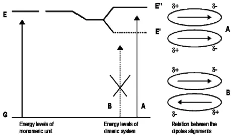
the appearance of new spectral bands due to the excitonic interactions between the cromophores (Fig. 1.21).
These changes can be explained using a vectorial picture andconsidering the interactions between the electrostatic dipole moments of the molecules. Two extreme cases exist, where the interaction is between two monomeric units having dipole moments lying along the molecular plane.
For H-aggregates two types of alignments are possible (Fig. 1.22). The “orientation mode” A corresponds to the transition at higher energy because the dipoles coupling provides for this event an alignment “in-phase”, determining repulsion between partial charges of the same sign: the resulting vectorial sum is not equal to zero.
The “orientation mode” B provides a more favourable coupling (that is at lower energy), but dipole moments cancel each other, resulting in a virtually “zero” dipole moment vector.
The ΔEtran is larger for the dimer resulting form compared to the monomer,
because the arrangement of the dipole moments of monomers within the dimeric system is energetically unfavourable.
This leads to the blue-shift of the Soret band of the aggregate compared to the one of the monomeric unit.
An opposite situation concerns J-aggregates, because the dipole moments of the monomers are parallel and lie along the line joining the molecular centres (Fig. 1.23). Again, two types of alignments are possible for the transition dipoles.
Fig. 1.23 - Orientation of dipole moments of two porphyrins forming J-aggregate. Fig. 1.22 – Orientation of dipole moments of two porphyrins forming H-aggregate.
The “aggregation mode” A corresponds to the transition at higher energy because the dipoles coupling provides for this event an alignment determining repulsion between partial charges of the same sign: the resulting vectorial sum is zero. In the orientation B this vectorial sum is not zero and coupling results energetically more favourable (that is at lower energy).
The ΔEtran is smaller for the dimer resulting form compared to the monomer,
and leads to a red-shift of the Soret band of the aggregate compared to the single unit of porphyrin.
With respect to intermediate geometries featuring by non-specific aggregates, both transitions can be partially allowed, so that the electronic spectrum is characterized by the presence of two bands simultaneously: the former at a blue-shifted wavelength and the latter at a red-blue-shifted wavelength in the aggregate relative to the monomer, or a general, less specific, broadening of the starting band resulting from the aggregation.
Because the possible implication in biological systems, aggregation process is particularly relevant when occurs in aqueous environment. However most native or synthetic porphyrins are poorly water soluble, so it is necessary to increase their affinity to water by functionalization of the macrocycle with a charged or polar group. Tetrapyrrole bearing sulfonate, phosphonate or pyridyl units, are employed to study the behaviour of porphyrin derivatives in aqueous solvent. The total charge of these molecules is pH depending, because protonation-deprotonation equilibrium process involves the inner nitrogens and the peripheral substituents. In this way their solubility may be increased or decreased, triggering the aggregation process.
One of the most important element able to promote the aggregation, is the interaction among groups with different charge. Purrello[42] showed that a positively charged matrix (polylysine) interacts with porphyrins bearing negative charged groups; once a critical porphyrin concentration has been reached, van der Waals and solvophobic forces induce their aggregation.
The normal fluorescence of the porphyrin monomers, is quenched by the aggregation process. The changing of fluorescence emission is a measure of the occurring aggregation, and it could be modulated by controlling pH.
The interaction with a matrix, is a relevant features when the matrix is a biopolymer such as DNA or RNA; in this case the owner fluorescence of the chromophore becomes a useful tool for the investigation of intracellular environment.
We have recently observed that the spontaneous deposition of ordered self-assembled porphyrin films can be obtained by functionalization of porphyrin with a peripheral cationic substituent. This group infers to the macrocycle an amphiphilic character, leading to the formation of mesoscopic ordered structures upon aggregation process[43].
Macrocycles with charged substituents are able to interact with a opposite charged surface, but they can also interact each other. The electrostatic interaction occurring between oppositely charged macrocycles, has attracted our interest because this process is involved in the formation of a structure with a well defined geometry, such that nanotube shaped[44], where the concomitant presence of electrostatic, van der Waals, hydrogen-bonding and axial coordination forces, drives the shape and the stability of the final aggregate.
Among the different tri-dimensional arrangements of molecules, leading to the formation of supramolecular system, we focused our attention on structures nanotube-like; in particular we were interested to the effect of the surface to volume ratio, because changing it, the probability of interaction with the analyte molecules increases, and consequently the sensitivity of the device.
However, ionic self-assembly is only one of the procedure developed for the construction of porphyrins nanotubes; a different method has been developed by Kojima et al.[45] (Fig. 1.24).
He choose, as building block, a saddle distorted metallo-porphyrin; the aggregation process is driven by the π-π interactions of the macrocycle and the
molecular structure is reinforced by an internal frame based on metallic oxo-cluster.
In details, dodecaphenylporhyrinato-Mo(V), having a curved surface due to both the steric hindrance of the peripheral substituents, and the presence of a high valent metal ion, has been crystallized in presence of methanol, giving an assembly with a nanotube shape.
The peculiarity, is the presence of hydrophilic environment in its inside, which facilitates inclusion of hydrophilic entities such as Mo-oxo clusters inside of hydrophobic porphyrin aggregates.
The tubular assembly is derived from intermolecular π-π interactions of alternatively inserted peripheral phenyl groups and intermolecular hydrogen bonding with tetranuclear Mo(VI)-oxo clusters: probably they could be formed and protected in accordance with the porphyrin aggregation in the hydrophilic cavity, and they could also act as templates for the tube stabilization.
A remarkable feature of this tubular structure is the formation of the hydrophilic isolated inner sphere in the hydrophobic porphyrin supramolecule.
1.5.1 Synthesis and exploitation of self-assembled porphyrinoids-based nanotube
Current methodologies in organic synthesis and supramolecular chemistry offer a wide range of opportunity to synthesize molecules endowed with suitable features for the recognition process[46]. There can be no doubt that the ability to coordinate metal ions is of primary importance to determine the sensitivity and selectivity properties of porphyrin derivatives; the role of the coordinated metal, is very important because the axially bind with volatile molecules, mimes the biological functions of these compounds. However, many factors are contemporaneously present to cooperate to bind the guest molecule: hydrogen bond, polarization, and polar interactions[47]. Because of this extensive richness of interactions, metalloporphyrins do not usually behave as receptors for molecular recognition of specific target molecules, but rather as globally selective sensors.
As recently reported by Shelnutt and co-workers[45], the ionic self-assembly of two porphyrins bearing opposite ionic charge, leads to the formation of J-aggregates, whose non-planar shape induces the formation of nanotubular structures, reaching free floating aggregates having lengths of tens of nanometers.
Porphyrins are able to arrange themselves in different ways, but aggregates nanotube-like are distinguish by the so called “cavity effect”, that means the inclusion of a guest inside the aggregate. This phenomenon causes some kind of modification in the molecular skeleton arrangement, which is the origin of variation in the optical spectra of the aggregate. These changes can be used for sensing mechanism.
The interest for that architecture is due to the enhanced sensing properties which is not possible to find in the single subunits or in other kind of arrangements. For example the presence of the inner cavity can give raise to selective endohedral inclusion of different guests, or the interaction with analytes can alter the supramolecular arrangement of the molecular nanostructure; both interaction pathways lead to dramatic changes of the porphyrin J-aggregate optical properties, which can consequently be used for sensing mechanism. We investigated the sensing properties of the porphyrin nanotubes in solution and as solid-state layers, having in mind to develop a sensor platform based on the detection of optical changes.
Indeed, the UV-vis spectroscopy provides a typical signature of porphyrin nanotube formation[43]. At the pH (2) needed for aggregation process, [TSPPorH2]4- is protonated in the inner core (two positive charges), while the
peripheral group remain in the anionic form (four negative charges), so on the whole it exists as [TSPPorH4]2- (15a).
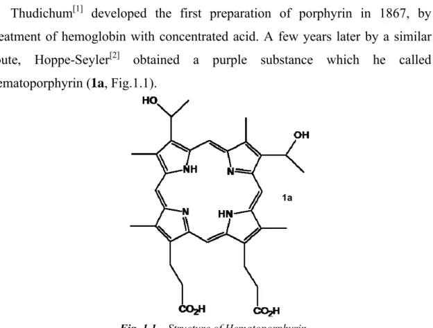
[TPyPor]SnCl2[48] is protonated at the pyridyl units (four positive charges),
while a further positive charge is given by the metal (oxidation number: +4) if one chloride ion is replaced by a water molecule. It exists as [(TPyPor)SnCl2]+4(16a) or [(TPyPor)SnCl(H2O)]5+ (Fig. 1.25).
Fig. 1.25 - Structure of porphyrins used for aggregation
In figure 1.26, the corresponding UV-Vis spectra are shown for aqueous solutions of the two constituents and for the self-assembled nanotubes.
Looking at the potential exploitation of such a nanostructures as sensing materials, we have first investigated the active role of porphyrin molecular arrangement in their sensing properties, to understand if the porphyrin nanotubes can offer additional features with respect to those of the single isolated components.
To verify this initial conjecture, we compared the effects of the modification of absorption spectra induced by the addition of different salts to solutions of precursor porphyrins and formed nanotubes.
In these conditions, in fact, we can safely exclude the influence of analyte coordination interactions with the porphyrin subunits. In acidic solution, the porphyrin free base is protonated (formation of (TSPPorH4)2-), and in this form is
not possible to coordinate any metal cations, while the anion chosen were chloride or nitrate salts, in order to reduce the influence of axial ligation
equilibria for [TPyPor]SnCl2. On the other hand, even small variation in
porphyrin J-aggregation, induced by the interaction with analytes, can be amplified by the modification of optical signatures.
Moreover, the salts we choose [Pb(NO3)2, NaCl, CdCl2, SnCl2, MnCl2, and
Cu(NO3)2], after their dissolution, do not change the pH of the solution; we need
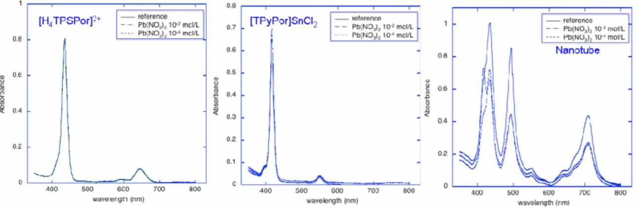
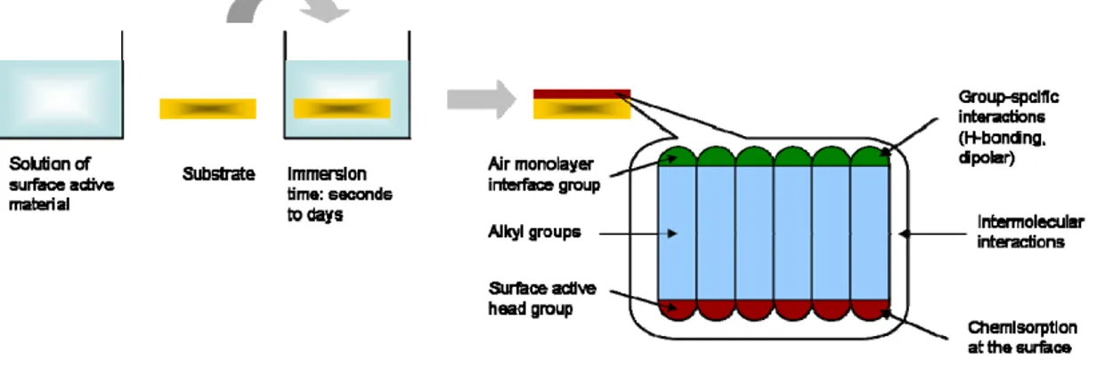
to control [H+] concentration because nanotube formation is a results of the precursors protonation, in the inner core, or at the peripheral positions, occurring at low pHs. In all cases absorbance spectra in the visible region of the two precursors did not show relevant changes, while noteworthy modification occurs in case of nanotube solutions. Figure 1.27 shows the spectra obtained in 10-2 and 10-4 mol/L Pb(NO3)2 solutions containing two single porphyrins and the
assembled structure.
Also with CdCl2, Cu(NO3)2, MnCl2, SnCl2, nanotubes spectra change,
showing a kind of interaction among the aggregate and the ions salt.
The effect may largely be attributed to the sensitivity of the forces holding together the nanotube toward the ionic strength of the solution, by shielding effect of the ionic charges, which induces a partial disassembling of the tubular aggregates. The decrease of interaction between porphyrins is indicated by the fact that the typical signals of the nanotube tend to disappear concomitantly it evolves into the spectrum of the individual precursors. Nonetheless, beside a non


![Fig. 1.24 – [Ph 12 Porphyrin]Mo-based nanotube](https://thumb-eu.123doks.com/thumbv2/123dokorg/7587275.113062/51.892.195.754.376.723/fig-ph-porphyrin-mo-based-nanotube.webp)