DOTTORATO DI RICERCA
BIOINGEGNERIA
Ciclo XX
Settore scientifico disciplinare di afferenza: ING-IND/34
Development of musculoskeletal models
for the design and the pre-clinical
validation of hip resurfacing prosthesis
Sviluppo di modelli muscolo-scheletrici
per la progettazione e valutazione
pre-clinica di protesi d’anca di rivestimento
Presentata da: Ing. MARTELLI SAULOCoordinatore Dottorato:
Chiar.mo Prof. ANGELO CAPPELLO
Relatore:
Chiar.mo Prof. LUCA CRISTOFOLINI
Co-relatori:
Ing. MARCO VICECONTI Ing. FULVIA TADDEI
CONTENT
INTRODUCTION ... 1
RIASSUNTO... 5
CHAPTER 1 ... 9
HIP RESURFACING ARTHROPLASTY: A NEW APPLICATION OF AN OLD CONCEPT ... 9
1.1 History of hip joint replacement... 10
1.2 Comparison of hip resurfacing arthroplasty and traditional THR... 13
1.3 Indications, risk factors, treatment and post operative care ... 14
1.3.1 Indications ... 14
1.3.2 Risk factors and treatment... 15
1.3.3 Postoperative care ... 16
1.4 Types of failure observed in practice ... 16
1.5 Definition of failure mode... 17
1.6 Mechanical implications on relevant biological processes ... 18
1.6.1 Bone: a living tissue ... 19
1.6.1.1 The bone matrix ... 19
1.6.1.2 Bone biology ... 20
1.6.2 Adverse biological processes for orthopaedic implants ... 24
1.6.2.1 The stress shielding effect ... 25
1.6.2.2 Osteolysis ... 26
1.6.2.3 Tissue differentiation at the bone-prosthesis interface... 28
1.7 Prediction of failure... 28
1.7.1 Predicting mechanically-related failure modes ... 29
1.7.1.1 Combined stress theory of failure ... 29
1.7.1.2 Fatigue ... 31
1.7.1.3 Fretting ... 33
1.7.1.4 Shock and impact ... 35
1.7.1.5 Wear ... 35
1.7.2.1 Adverse bone resorption and remodelling ... 36
1.7.2.2 Osteolysis ... 38
1.7.2.3 Adverse tissue differentiation at the prosthesis interface... 39
1.8 Properties of materials composing the orthopaedic implants ... 39
1.8.1 Prosthesis and cement ... 40
1.8.2 Bone ... 42
1.8.2.1 Cortical bone ... 42
1.8.2.2 Cancellous bone ... 45
1.9 Development of a protocol of analysis ... 47
CHAPTER 2 ... 57
EXPERIMENTAL VALIDATION OF THE OVERALL ACCURACY OF SUBJECT-SPECIFIC FINITE ELEMENT MODELS OF LONG BONES ... 57
2.1 Subject-specific finite element models of long bones: An in vitro evaluation of the overall accuracy ... 58
2.1.1 Materials and methods ... 60
2.1.1.1 Femur specimen ... 60
2.1.1.2 CT scanning ... 61
2.1.1.3 Experimental measurements ... 61
2.1.1.4 Subject-specific finite element model... 63
2.1.1.5 Spatial registration between model and experiment ... 65
2.1.1.6 Determination of the model accuracy ... 66
2.1.2 Results... 66
2.1.2.1 Experimental measurements ... 66
2.1.2.2 Comparison FEM vs experimental stresses ... 67
2.1.3 Discussions... 68
2.2 Accuracy of subject-specific finite element model of an implanted femur: an in-vitro study ... 71
2.2.1 Materials and methods ... 71
Finite element model... 72
2.2.2 Results... 73
2.2.3 Discussion ... 74
CHAPTER 3 ... 80
SENSITIVITY OF SUBJECT-SPECIFIC FE MODELLING TECHNIQUES TO UNCERTAINTIES ON PARAMETERS... 80
3.1 Finite-Element modelling of bones from CT data: sensitivity to geometry and material uncertainties ... 81
3.1.1 Materials and methods ... 82
3.1.1.1 Finite-Element models generation ... 84
3.1.1.2 The loads ... 84
3.1.1.3 Estimation of the random input variables ... 85
3.1.1.4 Identification of the output variables ... 87
3.1.1.5 The probabilistic design ... 87
3.1.2 Results ... 88
3.1.3 Discussion ... 89
3.2 Conclusion... 91
CHAPTER 4 ... 94
IDENTIFICATION OF THE PROTOCOL FOR THE ANALYSIS OF THE RISK OF BIOMECHANICAL FAILURE OF RESURFACED FEMURS... 94
4.1 The preclinical validation of hip resurfacing prostheses... 95
4.1.1 A general protocol for pre-clinical validation ... 96
4.1.1.1 Know your enemy ... 96
4.1.1.2 Detailed design of the validation protocols... 97
4.1.1.3 The role of numerical models in pre-clinical validation ... 97
4.1.1.4 A possible list of protocols... 98
4.1.2 Discussions... 100
4.2 In-vitro replication of spontaneous fractures of the proximal human femur ... 101
4.2.1 Identification of the most critical loading scenario: FE simulations... 102
4.2.3 Results: Most critical loading scenario ... 103
4.2.4 Discussions... 105
4.3 Conclusions ... 106
CHAPTER 5 ... 110
TESTING OF THE PRE-CLINICAL VALIDATION PROTOCOL ON A SUCCESSFUL DESIGN OF HIP RESURFACING PROSTHESIS ... 110
5.1 Combined experimental-numerical method for the pre-clinical validation of epiphyseal
prosthesis: exemplification of the method with an established design ... 111
5.1.1 Materials and methods ... 112
5.1.1.1 Bone specimens for the in-vitro study ... 112
5.1.1.2 The bone specimen for the numerical study ... 112
5.1.1.3 Failure modes associated to failure scenarios ... 113
5.1.1.4 Identification of predictive parameters ... 113
5.1.1.5 Experimental tests (intact and implanted specimen)... 115
5.1.1.6 FE models and boundary conditions... 115
5.1.1.7 Loading conditions for numerical simulations... 116
5.1.2 Results... 117
5.1.2.1 Femoral neck fracture ... 117
5.1.2.2 Prosthesis fractures ... 117
5.1.2.3 Aseptic loosening caused by cement damage ... 117
5.1.2.4 Aseptic loosening caused by fretting debris of the cement... 117
5.1.2.5 Aseptic loosening caused by lack in bone-prosthesis in-growth ... 118
5.1.2.6 Aseptic loosening caused by damage of cancellous bone... 118
5.1.2.7 Aseptic loosening caused by bone re-modelling... 118
5.1.3 Discussion ... 119
5.2 Conclusions... 121
CHAPTER 6 ... 126
DESIGN OPTIMISATION AND PRE-CLINICAL VALIDATION OF A NEW CONCEPTUAL DESIGN OF A RESURFACING PROSTHESIS ... 126
6.1 Preliminary finite element study of the first prosthesis prototype ... 127
6.1.1 Materials and methods ... 127
6.1.1.1 CT Dataset ... 127
6.1.1.2 The finite element model of the intact femur... 127
6.1.2 Results... 129
6.1.3 Discussion ... 131
6.2 Sensitivity of the risk of failure to some factors of influence ... 132
6.2.1 Roughness of the stem surface... 133
6.2.1.2 Results ... 133 6.2.2 Interdigitation ... 134 6.2.2.1 Methods ... 134 6.2.2.2 Results ... 135 6.2.3 Osteoporosis ... 135 6.2.3.1 Methods ... 136 6.2.3.2 Results ... 136 6.2.4 Discussion ... 137
6.3 Results of experimental tests on the first prototype ... 138
6.4 First step of design revision: refinement of the first prototype geometry ... 138
6.4.1 Materials and methods ... 138
6.4.2 Results ... 139
6.4.3 Discussion ... 140
6.5 Sensitivity of the risk of failure to influencing co-factors ... 141
6.5.1 Surgical errors: prosthesis mispositioning ... 141
6.5.1.1 Material and methods ... 141
6.5.1.2 Results ... 142
6.5.2 Anatomical variability: femur size ... 143
6.5.2.1 Material and methods ... 144
6.5.2.2 Results ... 145
6.5.3 Roughness of the stem surface ... 146
6.5.3.1 Materials and methods ... 146
6.5.3.2 Results ... 147
6.5.4 Discussion ... 148
6.6 Second step of design revision: geometry optimisation of small sizes ... 149
6.6.1 Materials and methods ... 149
6.6.2 Results ... 149
6.6.3 Discussion ... 150
6.7 Results of experimental tests on the optimised device... 151
6.8 Extended pre-clinical validation of the finalized design on a simulated population ... 151
6.8.1.1 Analysis of the implanted femur... 152
6.8.1.2 Comparative analysis of the implanted and the intact femur... 155
6.8.2 Results... 156
6.8.2.1 Analysis of the implanted femur... 156
6.8.2.2 Comparative analysis of the implanted and the intact femur... 158
6.8.3 Discussion ... 160
6.9 Conclusions... 161
CHAPTER 7 ... 165
MODELLING THE MUSCULOSKELETAL SYSTEM FOR PREDICTING IN-VIVO MUSCLE FORCES: THE NEW CHALLENGE ... 165
7.1 Biomechanical modelling of the musculoskeletal apparatus: status and key issues... 166
7.1.1 Biomechanical modelling of the body ... 168
7.1.1.2 Modelling strategies... 168
7.1.1.3 Possible uses of Whole-Body Models ... 170
7.1.1.4 Model identification... 170
7.1.2 Biomechanical modelling of organs ... 172
7.1.2.1 Modelling strategies... 172
7.1.2.2 Whole bones... 172
7.1.2.3 Myotendinous units... 173
7.1.2.4 Ligaments and cartilages... 174
7.1.2.5 Joint models ... 174
7.1.3 Biomechanical modelling of tissues ... 175
7.1.3.1 Some examples of tissue biomechanics modelling... 176
7.1.3.2 Models of tissue adaptation... 176
7.1.4 Multiscale and probabilistic models ... 177
7.1.5 The living human project: current challenges... 179
7.1.5.1 Community building and data repositories ... 179
7.1.5.2 Data fusion and data processing ... 180
7.1.5.3 A framework for numerical modelling ... 181
7.1.5.4 Information and scientific visualization... 182
7.1.6 Discussion ... 184
7.2 Subject-specific musculoskeletal models from medical images ... 185
7.2.1 Material and Methods... 185
7.2.2 Results and Discussion... 187
7.3 Conclusions ... 189
CONCLUSIONS... 199
ACKNOWLEDGMENTS ... 201
BIBLIOGRAPHY ... 202
APPENDIX A ... 205
A METHOD TO IMPROVE EXPERIMENTAL VALIDATION OF FINITE ELEMENT MODELS OF LONG BONES ... 205
APPENDIX B... 217
IN VITRO REPLICATION OF SPONTANEOUS FRACTURES OF THE PROXIMAL HUMAN FEMUR ... 217
APPENDIX C ... 230
ADDITIONAL RESULT PLOTS FOR CHAPTER 6... 230
APPENDIX D ... 242
The present thesis describes the results of the researches performed throughout my Ph. D. in Bioengineering. The work presented in this thesis was entirely carried out at the Laboratorio di Tecnologia Medica (LTM) of Istituti Ortopedici Rizzoli (Bologna).
The field of interest of the presented research thesis is the surgical treatment of dysfunctional hips such in case of arthritis, avascular necrosis, and other debilitating pathologies which are very severe and costly conditions for the public health. In spite of the clinical success of total hip replacement (THR) characterised by a survivorship typically higher than 90% after ten year from surgery, still THR surgeries produce strong limitations to the quality of life on operated individuals. As people live and remains active longer than in the past, the needs of more appropriate techniques in the treatment of dysfunctional joints quickly increased in the last years and it is expected to increase further. Such need pushed the industry and the research in developing new methods for the treatment of dysfunctional hips with particular attention to young and active patients.
Hip resurfacing techniques have been recently reintroduced as a mini-invasive techniques that preserve the original architecture of the hip and, mostly because of this, they seem to hold the promise of various advantages over traditional THR. The concept behind hip resurfacing is not new, it was first introduced in the 60’s and immediately abandoned because of a high failure rate, later attributed to technological limits of the medical alloys available at that time. Since late 90’s, the new CrCo medical alloys overcome the original limits giving a new spin to hip resurfacing techniques; preliminary follow-up studies are very promising.
Although many innovative designs claim to radically improve the functional outcome of traditional techniques, no hard evidences can be produced with the assessment methods currently available; thus, revision is the only reliable, although very gross, end point for assessment. This delayed assessment of the quality of orthopaedic devices is a consistent obstacle to the development of more effective designs. In fact, the lesson provided in the past by many branches of engineering is that success in designing competitive products while averting premature failure can be achieved only by evaluating all potential failure modes. To be effective in averting failure, it is essential for designer to have a good working knowledge of analytical and/or empirical techniques in predicting failure. The work developed during the three years of Ph.D. studies aimed to develop a protocol for predicting failure of hip resurfacing prosthesis. This aim was pursued by modelling the musculoskeletal system so as to provide a tool for the design and the pre-clinical validation of new devices.
The work can be divided in three principal activities: a first preliminary study in which the technique for the generation of subject specific finite element (FE) models, previously developed at the LTM, was validated either in case of intact or resurfaced femurs. The second activity,
which combines a formal analysis of failure, numerical modelling and in-vitro experiments to provide the best possible validation of a new device. The third and last part is the starting of a new research activity that holds as its primary objective the direct computing of boundary conditions trough modelling the muscular actions at the hip. It should be noted that, as it is well specified in the thesis title, the author conducted the numerical part of the work while the experimental testing activity was entirely carried out by the experimental group of LTM, and then briefly reported so as to strengthen the validity of conclusions.
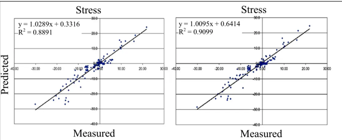
After Chapter 1, which describes failure scenarios for hip replacements, biomechanical parameters to be used in predicting failure and engineering techniques for predicting the relevant parameters, Chapter 2 describes the validation against experimental measurements of model predictions of the bone stresses and the contact mechanics at the bone-prosthesis interface. The FE meshes of an intact and of the same femur implanted were generated by a well established procedure developed in the past years at LTM, by using a set of medical images. All needed data were derived from the computerised tomography of an intact femur of a male donor of middle age which was made available from the “Banca dell’Osso” of the Istituti Ortopedici Rizzoli. After the validation of the intact model the same femur was implanted with a resurfacing prosthesis and it underwent the same validation protocol. The results of the FE simulations were compared with the measurements recorded by 13 rosette strain-gauges applied on the bone surface of the femur for five different loading conditions. The average error of predictions on bone stresses never exceeded the 10% of the measured maximum absolute value. The maximum error on prosthesis micromovement was within a micron. This results were satisfactory for the adoption of these models as a reliable tool in studying the resurfaced femur biomechanics and aligned with the current state of the art in this field.
A possible source of errors that affect the validation process are differences in boundary condition realised by the experimental set-up and the boundary conditions simulated by the model. To control these differences, a loading cell was studied to measure the location on the femoral head where the force is effectively applied so as to better model the experimental boundary conditions. As it is not of direct interest for this Ph.D. thesis, a more detailed description of the work is reported in Appendix A.
Whatever the method adopted for the generation of the FE mesh from medical imaging data and for the material properties mapping, unavoidable errors occurs in the creation of the model that influence, in an unpredictable manner, the results of the FE analysis. To understand the effects that the errors affecting the various steps of the model generation have on the FE prediction, a sensitivity FE analysis was performed. The results of the study, which are presented in Chapter
3, indicated that the uncertainty on the FE prediction is always lower than 10% of the predicted
value for the method adopted for the generation of finite element models from CT data.
The next step, described in Chapter 4, aimed to identify the protocol of analysis for evaluating all relevant failure mode for hip resurfacing prosthesis. To this scope, the definition of the protocol was carried out by the formal analysis of possible failure modes coherent with the resurfaced femur bio-mechanics and by the subsequent association of each failure mode with the more appropriated techniques of investigation.
The resulting protocol included the prediction of femoral neck fractures, which met the limit of the modelling techniques in predicting the bone mechanics in the post-elastic phase. Hence, the competence of evaluating the ultimate load to failure of implanted femurs was left to the experimental group of LTM, opening the question on which experimental set-up is the best trade off between the need to replicate physiological conditions and the need of simplicity of the set-up itself. This problem was numerically approached.
Simulations were run to identify the loading scenario which more likely produce head-neck fractures, and to determine where it is possible to simplify the experimental set-up when the head-neck region is of interest. The experimental set-up was consequently designed and clinically relevant fractures were obtained on 10 cadaveric femurs. Details of the tests conducted by the experimental group of LTM are reported in Appendix B.
So after, the protocol was ready for the analysis of resurfaced femurs. Chapter 5 describe the first application of the method to a successful design of resurfacing prosthesis developed over years. A model of an implanted femur of average size, with normal bone stock, and simulating an ideal positioning of the prosthesis during surgery was generated. Muscle forces acting on the proximal femur were modelled deriving all needed information from the literature and simulations were run for a broad range of activities of daily living. Destructive tests were conducted (by the LTM experimental group) on three pairs of cadaver specimens to measure the influence of the prosthesis implantation on the risk of neck fractures. Results of numerical simulations and of experimental test were all in a good agreement with known clinical outcomes for this class of devices. This was encouraging for its further use in studying a new prototype of prosthesis.
Chapter 6 describe the geometry optimisation process of a prototype of a new resurfacing
prosthesis. The first deterministic simulations underlined some weak aspects of the prototype geometry. These initial concerns were confirmed by destructive tests conducted by the experimental group. The subsequent optimisation loop reviewed, in two steps, both the prosthesis and the rasp geometry leading to an optimised design which didn’t rise any concern for any of the investigated risks, over a large set of operative condition the implant may face during its service life. Additional destructive tests on cadaver specimens underlined a strength increase (+15% with respect to the intact controlateral femur, +25% with respect to the first prototype of the resurfacing prosthesis) of the femoral neck implanted with the optimised design over the strength of femoral neck implanted with the first prototype.
Although this seemed to significantly improve the resurfacing prosthesis biomechanics, a full pre-clinical validation method should consider the whole population of patients the device is indicated to. To this aim, a statistical study of the risk of implant failure was carried out to extend the deterministic analysis over a larger set of conditions and taking into consideration the mutual interaction between the various input variables. Statistical input were anatomical, surgical and activity related variables. Results confirmed the overall quality of the new design although a small set of conditions were found at high risk of neck fractures. Those cases were characterised by critical combination of body weigh, bone mineral content (i.e. high osteoporosis) and activity level. A second statistical analysis compared the risk of neck fractures of the implanted and of
the intact femur, leading to the conclusion that the prosthesis insertion do not alter the “natural” risk of neck fractures of the patient.
The achieved low risk of failure associated to the biomechanical behaviour of the optimised design highlighted that either the quality of the conceptual design or the effectiveness of the adopted optimisation protocol. The optimised design was then considered appropriate for its intended function so as the resurfacing prosthesis recently started clinical trials. Nevertheless, the statistical analysis provided some critical combinations of the input parameters (i.e. related to the individual anatomy, activity and the surgery accuracy) which were found critical. These scenarios might constitute an indicative information to be used during the patient selection phase before surgery.
On the author opinion, the most important limitation of the present study is about loading conditions at the proximal femur. The analysis conducted in the present study based the loading scheme applied to the proximal femur on published data for few subjects during few activities of daily living. These boundary conditions, even significant, can hardly be considered representative of the entire range conditions the implant might face over the population the resurfacing prosthesis is indicated to.
Since experimental techniques to non-invasively measure in-vivo muscle forces are not yet available, the only feasible method to study the muscle contribution to a specific motion task is trough modelling the musculoskeletal system. To this aim a new research activity was started with a literature review on musculoskeletal models of the lower limb which is the topic of the last chapter.
Chapter 7 describe the result of the initial literature review and current challenges the LTM is
dealing with. After a first training period at the Bio-mechanical Engineering Laboratory of the Delft Technical University (Delft, The Nederland) working on a sophisticated musculoskeletal model of the shoulder, a new activity was started to develop subject-specific musculoskeletal models from medical images. In Appendix D are collected a series of activities that were object of international congresses on this topic.
La presente tesi descrive i risultati della ricerca effettuata nell'arco del Dottorato in Bioingegneria. Il titolo della ricerca è: "Sviluppo di modelli muscolo-scheletrici per la progettazione e valutazione pre-clinica di protesi d'anca di rivestimento". Il lavoro, condotto presso il Laboratorio di Tecnologia Medica (LTM) degli Istituti Ortopedici Rizzoli (Bologna), è brevemente presentato nel seguito di questo capitolo.
L’ambito in cui questa tesi si inserisce riguarda il trattamento di patologie debilitanti dell’anca come è il caso di artriti, artriti reumatoidi, necrosi avascolari, dislocazioni congenite, traumi e altre che insieme costituiscono indicazione per la ricostruzione chirurgica del giunto articolare. Tali condizioni risultano ancor oggi, e in modo crescente, un fattore di grande impatto per la salute pubblica, rappresentandone una voce di costo significativa.
Sebbene l’artroplastica d’anca, condotta con le tecniche tradizionali, abbia raggiunto percentuali di successo tipicamente superiori al 90% dopo dieci anni dall’intervento, ancora oggi i trattamenti chirurgici indicati per il trattamento delle patologie sopra elencate, comportano limitazioni importanti dello stile di vita dei pazienti operati. Tali osservazioni assumono, giorno dopo giorno, maggiore rilevanza se si considerano il continuo innalzamento dell’età media della popolazione unito con il protrarsi di stili di vita particolarmente attivi fino ad età avanzata. Questi scenari particolarmente longevi, poco realistici fino a pochi anni fa, hanno prodotto una richiesta crescente di tecniche efficienti, richiesta attesa in ulteriore crescita nei prossimi anni. Le tecniche di rivestimento d’anca come tecniche mini-invasive sembrarono, fin dai primi anni 60, promettere una migliore efficacia nel ripristino funzionale dell’articolazione quando invece, scontarono alte percentuali di fallimento a causa della mobilizzazione di entrambi gli inserti (acetabolare e femorale). Il loro successivo abbandono venne attribuito a limiti tecnologici dell’epoca ritenuti responsabili delle numerose e premature mobilizzazioni dell’impianto. Recentemente, le leghe in CrCo sviluppate per uso medico, sembrano aver superato i limiti originali cosi che le tecniche di rivestimento d’anca sono nuovamente una via percorribile e probabilmente capace di mantenere le attese iniziali. Da pochi anni reintrodotte, mostrano risultati clinici preliminari molto promettenti e in linea con le attese.
Mentre molti nuovi disegni protesici dichiarano radicali miglioramenti dell'esito funzionale rispetto ai disegni protesici tradizionali, non è oggi possibile produrne l’evidenza con i metodi di valutazione disponibili, se non attraverso studi retrospettivi. La revisione quindi continua ad essere l'unico strumento affidabile, pur se tardivo, per la valutazione della qualità dell’impianto. In aggiunta, questo approccio rudimentale ostacola lo sviluppo di impianti più efficaci attraverso l’identificazione preventiva di possibili scenari ad alto rischio di fallimento.
Lo sviluppo di prodotti ad alte prestazioni attraverso la previsione, durante la fase di progetto, di probabili scenari di fallimento in opera non è un problema nuovo. La lezione fornita in passato
da molteplici campi applicativi insegna che il successo nella progettazione di prodotti competitivi, evitando fallimenti prematuri, può essere raggiunto soltanto attraverso la valutazione di tutti i potenziali meccanismi che possono alterare le condizioni desiderate di buon funzionamento. Per essere efficace nella previsione del fallimento, è essenziale per il progettista avere una buona conoscenza delle tecniche di analisi e/o empiriche per la previsione del fallimento.
L'obiettivo del lavoro sviluppato nel corso dei tre anni di dottorato è stato sviluppare modelli muscolo-scheletrici per la progettazione e la validazione pre-clinica di protesi di rivestimento d'anca. Il lavoro può essere diviso in tre principali attività: studi preliminari, in cui la tecnica modellistica per l’analisi biomeccanica di segmenti scheletrici (precedentemente sviluppata presso il LTM) è stata validata attraverso il confronto diretto delle predizioni modellistiche con misure sperimentali. La seconda parte contiene l’attività principale, che combina l’analisi formale del fallimento per impianti di rivestimento d’anca, identifica un protocollo di analisi per finire con l’ottimizzazione geometrica e la validazione pre-clinica del comportamento biomeccanico di un nuovo disegno protesico. La terza e ultima parte rappresenta l'inizio di una nuova attività di ricerca che contiene come obiettivo primario il calcolo diretto delle condizioni al contorno all’anca attraverso la modellazione numerica del sistema muscolo-scheletrico.
Nel suo complesso l’analisi delle protesi di rivestimento ha richiesto una combinazione di analisi numeriche e prove sperimentali, spesso le une al servizio delle altre. L'autore ha condotto la parte numerica del lavoro mentre le prove sperimentali sono state condotte dal gruppo sperimentale del LTM e qui sono brevemente riportati i risultati per supportare la validità delle conclusioni. Nel seguito è descritto il contenuto dei diversi capitoli della tesi che ne svolgono il titolo, mentre sono riportate in appendice le attività svolte al contorno.
Il Capitolo 1 è un capitolo introduttivo che descrive gli aspetti storici delle tecniche di rivestimento d’anca, gli scenari di fallimento possibili, i parametri biomeccanici rilevanti per lo studio di detti scenari e le tecniche ingegneristiche usate per discriminare situazioni potenzialmente ad alto rischio di fallimento in un contesto multifunzionale.
I successivi capitoli riguardano il lavoro svolto durante il dottorato. La parte iniziale ha visto la messa a punto degli strumenti da impiegare nelle analisi successive, iniziando dalla validazione dei modelli per confronto diretto: le condizioni di prova sono state simulate definendo l’accuratezza del modello attraverso il confronto dei risultati predetti con le misure sperimentali. Poter controllare i parametri che definiscono il set-up sperimentale è un fattore chiave per un corretto confronto tra i valori modellati e quelli misurati. Con questo scopo è stata sviluppata una apposita cella di carico i grado di misurare la posizione del carico applicato rispetto al femore durante le prove sperimentali. Una descrizione meglio dettagliata dello studio è riportata in
Appendice A.
Il Capitolo 2 descrive gli studi di validazione delle capacità dei modelli nella predizione dello stato di sollecitazione dell’osso in campo elastico e della biomeccanica di interazione osso-protesi. I modelli ad elementi finiti (FE) del femore intatto e dello stesso femore operato sono stati validati attraverso il confronto diretto tra i valori di tensione predetti dai modelli e le misure
sperimentali. Il confronto è stato condotto con le misure di 13 rosette estensimetriche applicate sulla superficie dell’epifisi femorale, acquisite durante l'applicazione di diverse condizioni di carico scelte. L'errore medio sul campo di tensione è risultato minore del 10% della massima tensione dell'osso mentre, l'errore commesso sulla predizione dei movimenti osso-protesi è risultato nell'ordine del micrometro.
L’accuratezza ottenuta è stata considerata adatta per le analisi successive, molte delle quali richiederanno la nova generazione del modello per simulare condizioni differenti. Per limitare a quello presentato gli studi di validazione necessari, sono state analizzare le incertezze introdotte nei risultati dalle incertezze sui parametri di modellazione. Infatti, qualunque sia il metodo adottato per la generazione di modelli ad elementi finiti da immagini medicali e per la definizione delle proprietà dei materiali del tessuto osseo, gli inevitabili errori influenzano, in modo imprevedibile, i risultati delle analisi. Per studiare la relazione che intercorre tra le incertezze di modellazione e i risultati è stato condotto uno studio di sensitività; i risultati, presentati nel Capitolo 3, indicano che le incertezze sui parametri di modellazione producono un’incertezza nell’ordine del 10%.
Il Capitolo 4 descrive l’identificazione del protocollo di analisi di protesi di rivestimento d’anca per lo studio del rischio di fallimento associato al comportamento biomeccanico dell’impianto. Il confronto degli scenari di fallimento noti con i meccanismi che potenzialmente possono condurre agli scenari riportati ha permesso di compilare una lista analisi possibili. Tale lista comprende l’analisi dell’effetto dell’impianto sul rischio di frattura del collo del femore, evento conclusivo della fase post-elastica dell’osso. Essendo i modelli validati solo in campo elastico, la valutazione del carico a rottura del collo del femore è stata prodotta dal gruppo sperimentale del LTM attraverso schemi di carico prodotti numericamente. Le simulazioni sono state in grado di identificare la condizione fisiologica critica e di semplificare il set-up sperimentale al fine di riprodurre in laboratorio fratture clinicamente rilevanti. Lo studio riportato in Appendice B descrive l’applicazione delle condizioni di carico identificate numericamente su 10 femori di cadavere. Le fratture ottenute sono risultate essere del tipo voluto e coerenti con il tipo di fratture che si osservano nella clinica.
Il Capitolo 5 descrive la prima analisi biomeccanica di una protesi di rivestimento di successo e da anni utilizzata nella clinica. I risultati del metodo combinato numerico/sperimentale hanno evidenziato un ottimo accordo con i risultati clinici a corto e medio termine noti per questa categoria di dispositivi. Tali risultati sono stati incoraggianti e hanno costituito la base per la successiva analisi di disegno protesico.
Il Capitolo 6 descrive l’analisi, il processo di ottimizzazione della geometria e la validazione pre-clinica di un prototipo di protesi epifisearia. Dopo l’identificazione di alcuni aspetti critici del disegno iniziale i seguenti due cicli di revisione della geometria dell’impianto, supportati passo a passo dall’analisi numerica, hanno condotto ad un disegno ottimizzato. Il disegno finale non ha evidenziato rischi rilevanti per nessuno degli scenari di fallimento investigati e una seconda serie di prove distruttive ha confermato gli effetti positivi dell’ottimizzazione condotta. I test distruttivi su femori di cadavere condotti dal gruppo sperimentale del LTM hanno
evidenziato un incremento del 25% del carico ultimo a rottura degli impianti effettuati con protesi ottimizzate rispetto ad impianti realizzati con il prototipo iniziale.
La validazione pre-clinica del nuovo disegno è stata estesa per via statistica, simulando condizioni rappresentative dell’intera popolazione cui l’impianto è potenzialmente destinato. I risultati hanno confermato la qualità del nuovo disegno (che nel frattempo ha iniziato la sperimentazione clinica), sottolineando sia qualità del disegno concettuale di partenza sia l'efficacia del protocollo di ottimizzazione adottato.
Un parametro risultato particolarmente significativo nella la definizione del rischio di frattura del collo di femore è il livello di attività del paziente, analizzato in questo studio utilizzando i pochi dati pubblicati, raccolti in-vivo attraverso protesi strumentate con dispositivi telemetrici. Tali condizioni, anche se significative, non possono essere considerate rappresentative di tutta la gamma di condizioni in cui la protesi può trovarsi ad operare. Ciò ha costituito lo stimolo per l’apertura di una nuova linea di ricerca nel campo della modellazione muscolo-scheletrica dell’intero arto inferiore per la previsione delle forze muscolari, iniziata con una revisione approfondita della letteratura. Il Capitolo 7 contiene l’analisi dello stato dell’arte nell’ambito della modellazione del sistema muscolo-scheletrico e le sfide attuali che il LTM si trova ad affrontare. In Appendice D sono raccolte alcune attività svolte in questo tema e che sono state oggetto di congressi internazionali.
Hip resurfacing arthroplasty: a new application of an
old concept
Arthroplasty (literally "formation of joint") is an operative procedure of orthopaedic surgery performed, in which the arthritic or dysfunctional joint surface is replaced, modelled or re-aligned by osteotomy or other procedures. The first popular attempts in the treatment of dysfunctional joints was interpositional arthroplasty with interposition of some other tissue like skin, muscle or tendon to keep inflammatory surfaces apart or excisional arthroplasty in which the joint surface and bone was removed leaving scar tissue to fill in the gap. Other forms of arthroplasty include re-sectional arthroplasty, resurfacing arthroplasty, mold arthroplasty, cup arthroplasty, silicone replacement arthroplasty, etc. Osteotomy to restore or modify joint congruity is also an arthroplasty.
For the last 45 years the most successful and common form of arthroplasty is the surgical replacement of arthritic or destructive or necrotic joint or joint surface with prosthesis.
Hip replacement arthroplasty is the surgical replacement of all or part of the hip joint with an artificial device. Replacement of joint surfaces may be performed on both the pelvi and the femur side or only on the femur head. The first procedure is called total hip replacement (THR) while the second is called hemiarthroplasty in which only the head of femur head is replaced while the acetabular cartilage remains intact.
The purpose of this procedure is to relieve pain, to restore range of motion and to improve walking ability, thus leading to the improvement of muscle strength. A number of pathologies constitute indication for joint replacement, among others:
• osteoarthritis • rheumatoid arthritis
• avascular necrosis or osteonecrosis • congenital dislocation of the hip joint • acetabular dysplasia
• traumatized and malaligned joint • joint stiffness
Particularly relevant is failure of orthopaedic implants for which failure is a catastrophic event that may cause very high risks for the patient in relation to the complexity of the reconstructive surgery therapy. Nevertheless it constitutes a relevant part of costs of public health.
Processes that may cause failure of orthopaedics implants present particular conditions in which mechanical aspects interact with biological processes so as activated biological processes might lead to failure rather than safe conditions.
Last years have seen the resurgence of mini-invasive techniques for the complete replacement of the articular surfaces of the hip (hip resurfacing arthroplasty). On one side, the reduced impact on the hip architecture of these new devices seems to hold the promise of a substantial improvement of the quality of life of operated individuals. On the other side, the introduction of radically new designs would lead to new scenarios of failure hard to be predicted a priori.
It is clear that the understanding of the risk level of a specific application would help designers in producing new devices with a lowered risk. The present chapter addresses a method for the systematic analysis of failure of hip resurfacing devices which may be used by designers in order to achieve low risk designs of new implants.
1.1 History of hip joint replacement
Research for arthritis treatment has been trying for more than a century to successfully treat this debilitating disease. It was clear that many people required surgery to relieve pain and keep their joints mobile. Initial attempts to treat arthritic hips included arthrodesis (fusion), osteotomy, nerve division [1], and joint debridements. The goal of these early debridements was to remove arthritic spurs, calcium deposits, and irregular cartilage in an attempt to smooth the surfaces of the joint.
Indeed, there was a great search for some material which could be utilized to resurface or even replace the hip. Several proposals and trials were made including the use of muscles, fat, chromatized pig bladder, gold, magnesium and zinc. All met with failure. Surgeons and scientists were unable to find a material which was biocompatible with the body, and yet strong enough to sustain the high forces placed on the hip joint.
In 1925, a surgeon in Boston, Massachusetts, M.N. Smith-Petersen, M.D., molded a piece of glass into the shape of a hollow hemisphere which could fit over the ball of the hip joint and provide a new smooth surface for movement. The glass could not sustain the stress of walking and quickly failed. Afterward he pursued other materials including plastic and stainless steel. The use of stainless steel for surgery, where it might well resist corrosion by bodily fluids, seemed natural. During the 1940's, mold arthroplasty was "state of the art."
A dramatic improvement was made in 1936 when scientists manufactured a cobalt-chromium alloy which was almost immediately applied to orthopaedics. This new alloy was both very strong and resistant to corrosion, and has continued to be employed in various prostheses since that time. While this new metal proved to be a great success, the actual resurfacing technique was found to be less than adequate. It became clear that pain relief was not as predictable as
hoped, and hip movement remained limited for many patients. Mold arthroplasty also did not allow surgeons to treat the numerous and varied arthritic deformities of the hip.
Frederick R. Thompson of New York and Austin T. Moore of South Carolina, separately developed replacements for the entire ball of the hip. These could be used to treat hip fractures and also certain arthritis cases. This type of hip replacement, called hemiarthroplasty, only addressed the problem of the arthritic femoral head. The prosthesis consisted of a metal stem which was placed into the marrow cavity of the femur, connected in one piece with a metal ball which fit into the hip socket. While very popular in the 1950's, results remained unpredictable and arthritic destruction of the socket persisted. In addition, there was no truly effective method of securing the component to the bone. Large numbers of patients developed pain because of this loosening of the implant.
In 1938, Dr. Jean Judet and his brother, Dr. Robert Judet, of Paris, attempted to use an acrylic material to replace arthritic hip surfaces. This acrylic provided a smooth surface, but unfortunately tended to come loose. The idea did lead Dr. Edwarc J. Haboush from the Hospital for Joint Diseases in New York City to utilize a "fast setting dental acrylic" to effectively join the prosthesis to the bone starting a new era in fixation techniques.
In England, a very innovative surgeon, John Charnley, was also attempting to solve these ongoing problems. He pursued effective methods of replacing both the femoral head and acetabulum of the hip. In 1958, he addressed the eroded arthritic socket by replacing it with a polymeric implant. In order to obtain fixation to the bone he used polymethylmethacrylate from the dentists. These was the starting of a new era which is still the more common method in the treatment of dysfunctional hips; by 1961, Charnley was performing the surgery regularly with good results and the long term outcomes became very predictable.
In spite of the great improvement of clinical outcomes with the new techniques, since that time many attempts were done to reduce the amount of bone resection which is typical for traditional endomedullary stems (Fig. 1). Resurfacing arthroplasty of the hip has always been attractive conservative option in the treatment of end-stage arthritis of the hip in young and active patients. Bone conservation and non-violation of the femoral shaft make it a less invasive option. Normal load transmission and normal biomechanics hold the promise of a better preservation of the conserved bone. The life expectancy of a young patient is very likely to be more than the longevity of any artificial device, when the implant needs to be revised, resurfacing offers better revision options.
Figure 1 - On the left: a resurfaced hip. On the right: a traditional endomedullary stem.
Many different designs and material combinations were used in the past. Sir John Charnley [2] used an uncemented PTFE/PTFE arthroplasty device which had a high early failure rate. He attributed the failure to the resurfacing procedure and went on to perform metal/PTFE total hip replacement. When they also failed with osteolysis he found that PTFE wear was the problem. He also realised that a large diameter hard-on-soft bearing would give rise to excessive wear and warned against resurfacing.
In 1960s Müller used a press-fit metal-on-metal resurfacing device [3]. England [4], Japan [5, 6], the USA [7], and Germany [8]. Freeman and Furuya [4, 5] initially used PE components against metal cups but, later, they all used metal head on PE cups. Wagner tried a ceramic femoral component, which also failed to improve the result significantly. In France a metal-on-metal double cup device was developed [9] that permitted movement between the metal bearing surface and also between the outer cup and the reamed socket. The results of all these different devices appear promising in the early years, but the medium and long-term results were uniformly unsatisfactory.
It was presumed that many of these failures occurred as a result of femoral head osteonecrosis [10, 11] or stress-shielding [11, 12]. In the early period after surgery the disruption of the natural vascularisation of the femoral head was indicated as a possible source of necrosis in that region. If they survived osteonecrosis, the modification after surgery of the stress distribution under load (stress-shielding) might lead to bone resorption and consequently, to a probable mid or long term prosthesis loosening. It was predicted that high frictional torque forces from the large diameter head would be responsible of the high production of wear debris and of the subsequent
osteolysis. Cement disease was presumed in other cases responsible of stimulating osteolysis by cement debris produced by cement fretting and cementless devices were introduced. Even these did not make a marked improvement in survival. Resurfacing was thus written off as a bad concept altogether [13]. With hindsight, it is now known that many of these failures were a result of accelerate polyethylene wear from a large diameter bearing leading to osteolysis [14, 15]. In Birmingham there was 50% failure rate at 6-7 years with Wagner resurfacing. Retrieval studies on the failure showed the presence of granulomas filled with polyethylene wear particle-laden macrophages in the region of osteolysis. The femoral head outside the osteolytic regions showed a well vascularised viable trabecular bone extending right up the cement margin.
The introduction of metal-on-metal bearings was first abandoned arguing that large diameters would give rise to high frictional torque and lead to early failure [16] However in many patients with metal-on-metal endomedullary stems there was no evidence of osteolysis in spite of an extensive open bone-implant interface.
Work started towards large-diameter metal-metal resurfacings and the earlier notion that resurfacing inevitably lead to femoral head osteonecrosis was dispelled [17, 18].
Advanced in manufacturing made precision finish in terms of surface roughness and out-of-roundness possible and guaranteed a polar bearing configuration. It is now clear that such design might lead to an extremely low wear rate and advancement in tribology now lead to the understanding that large diameter have the potential to function as metal-fluid-metal joints with negligible wear [19]. Thus, it is believed that smooth surfaces and a small clearance might produce satisfactorily a low wear.
The advantage of metal-on-metal bearings must be balanced against the possible adverse effects of particulate metal debris. Some studies [20, 21] have shown increased level of metallic ions in body fluids and adjacent tissue after joint arthroplasties with metal-on-metal bearings. No studies reports evidences for linking metal-on-metal bearing with adverse biological effects even if it is known that metal ions may cause allergic hypersensitivity.
1.2 Comparison of hip resurfacing arthroplasty and traditional
THR
Resurfacing of arthritic joint surfaces with prosthetic components is accepted and widely used standard in total hip replacement. Simple resurfacing of the worn joint surfaces has less frequently been used as means of total hip arthroplasty however resurfacing has several theoretical advantages (Fig 1, left).
One obvious advantages of resurfacing is that it preserves bone on the femoral side. It is preferable to retain the femoral head and avoid using the intramedullary device that is implanted in standard hip replacements. However, resurfacing requires a more difficult surgical exposure to prepare the acetabulum without excising the femoral head and neck. For resurfacing to be truly conservative, the surgeon should not remove anymore bone from the acetabulum than would be required for THR, a goal that has been only recently addressed by companies manufacturing thinner acetabular shells.
An additional advantage is a better stress transfer to the proximal femur. This may avoid long term problems caused by stress-shielding of the proximal femur, which can occur with intramedullary fixation of traditional stems [22].
A large femoral head diameter (36 to 54 mm) is associated with lower dislocation rate than conventional prosthesis, which have smaller diameter head (22 to 32 mm in diameter). In two large recent series, no dislocations of the hip were found [23, 24]. Kinematically resurfacing implants more closely resembles the original anatomy and may have better propiception than conventional THR.
Another theoretic advantage is that revision of the femoral component, when necessary, may be easier than revision of intramedullary stem as more bone is preserved [23-26].
For balance, hip resurfacing has some disadvantages [20]. Although the reduced bone resection at the femoral side should allow maintaining unchanged the original joint centre, when it’s the case the lack of modularity of these device reduce the ability to adjust the leg length. Resurfacing is not appropriated in hips with loss of femoral head and neck bone stock or in hips with femoral cysts.
However, the second generation prosthesis has very promising outcomes, even though only preliminary. The femoral neck fracture has an incidence ranging from 0% to 4% [27].
Fracture is usually found early in series, with declining rates as the surgeon overcomes the learning curve. The cause of fracture appears to be both patient and technique related. Patient related factors include obesity, decreased bone mass and inflammatory arthritis. Intra-operative characteristic that may lead to fracture include femoral neck cysts and exposed bone found during preparation. Surgical errors include notching of the femoral neck, tilting of the prosthesis into excess varus and improper prosthetic seating. In the Australian national study (Buergy 2007) it has been shown that technical problems occurred in 85% of neck fractures.
Aseptic loosening has been found in some series [24]. Most were observed in the first group of the series and were attributed to intra-operative errors. Demographic factors that influenced loosening included an average femoral stem shaft angle of 128.3°, which differed from the rest of the patients that was in average 136°.
1.3 Indications, risk factors, treatment and post operative care
1.3.1 Indications
Total hip replacement is most commonly used to treat joint failure caused by osteoarthritis. Other indications include posttraumatic osteoarthritis, secondary osteoarthritis, and avascular necrosis of the femoral head if remaining bone stock is adequate, inflammatory arthritis if bone quality is adequate, patients with a deformity of the femur and/or internal fixation device that would make insertion of a stemmed femoral component difficult, patients with a high risk of dislocation.
Local protocols should be drawn up for criteria for referral when the symptoms impair quality of life. Referral should be based on an explicit scoring system that should be developed locally, and should take into account the extent to which the condition is causing pain, disability, sleeplessness, loss of independence, inability to undertake normal activities, reduced functional capacity or psychiatric illness.
1.3.2 Risk factors and treatment
The high majority of people who received a THR experience a better joint mobility and loss of pain. Revision is much more substantial than the primary operation and it is always a long and demanding procedure.
In the short term post-operatively, infection is a major concern. Deep infection will often require one or two stage revision surgery with an extended hospital stay and antibiotics. Recurrent dislocation is another indication for revision that might be faced either in the short or in the long term although it is more common in the early period after surgery.
In the long term, many problems relate to loosening. Inflammatory process leading osteolysis as well as the modified bone mechanics may cause bone re-sorption and the subsequent loosening or fracture.
Drawing a personalised risk indicator is not an easy task as many factors concur to the success or the failure. Anatomy, biology and the level of activity are all relevant in defining the individual risk of failure as they define condition at the bonding of the prosthesis. It is however possible to list a number of factors known to influence the implant outcomes. It is interesting to observe that managing the load carried by the prosthesis is a key factor either for the success or the success of the treatment. Known risk factor are:
1. Previous operation of the joint, in general the revision of a resurfaced femur is carried out by the insertion of a traditional endomedullary stem.
2. Surgery accuracy. Studies demonstrated that the rate of loosening is higher in hospitals that perform only small amounts of total joint operations
3. The design of the artificial joint as some models loosen more often then others.
4. The quality of bone: the more of the bone stock, the stronger the interlock will be and the longer the prosthesis will last.
5. Physical activity. Young active patients have higher rates of aseptic loosening.
6. Excessive weight. The higher is the load, the more rapid is the loosening of the implant. As revision is much more substantial that the first surgery, particular care should be taken if an incipient failure is expected. If the patients experience discomfort or even pain from their artificial joints and the diagnosis revealed an incipient aseptic loosening, the first step is usually a restricted weight bearing regime.
The loose prosthesis may find a new stable position, the discomfort and pain disappears. If the pain and other discomfort from the artificial joint increases then a revision operation becomes necessary.
The more challenging aspect of treatment is the early diagnosis of the incipient loosening. Current research in this field would find methods for the early diagnosis of the incoming failure.
1.3.3 Postoperative care
The modern trend for discharge between 3 and 5 days means that a considerable amount of care that was formerly in hospital is now in the community. Rehabilitation before operation and in hospital is also important. Post-operative rehabilitation includes exercises for stimulating the joint mobility and muscle strength. Among others strengthening exercises (seated leg extension, lying hip abduction, standing hip extension and abduction, knee bends and bridging), stretching exercises, progression of walking distance etcetera.
There is a belief that with hip resurfacing the patient can return to full participation in recreational or professional sports. Long-term results have not been studied to support this idea. Most surgeons agree that joint resurfacing allows patients to be more active than is acceptable for a standard total hip replacement (www.eorthopod.com).
1.4 Types of failure observed in practice
Hip resurfacing arthroplasty has been only recently reintroduced in the market after their first introduction in the 60s failed. Although the very high failure rate of the first generation (15-40%, Australian Orthopaedic Registry) of resurfacing devices discouraged its further use, their attractive potentialities over traditional THR sustained the development of a second generation for which recent outcomes seem very promising.
Hip resurfacing become day by day more common especially for young and active patients so as some retrospective studies are now available reporting up to six year follow-up statistics [23-25, 28-36]
Harlan reported a 94.4% survivorship (95% confidence interval, 91% to 98%) within a group of 400 patients of an average age of 48 years. He also underlined the more common complications, indicating aseptic loosening and femur neck fractures as the most important causes for failure. RIPO (Registro Italiano Protesi Ortopediche, http://ripo.cineca.it/) reported 621 cases during a follow-up of five years of a large group of different devices from different manufacturers (Table 1). Despite the high different failure rate shown by different design, the overall survivorship was (97% confidence interval, 95.1% to 98.8%) higher than what reported by Harlan. No indications are provided here about the type of dysfunctional condition that leads to revision.
Prosthesis model and brand N. % N. of failures %
BHR (Midland Medical Technologies) 326 52.5 5 1.5 BHR (Smith and Nephew) 144 23.2 1 0.7
ASR (DePuy) 30 4.8 -
-MRS (Lima) 34 5.5 3 8.8
ADEPT (Finsbury) 19 3.1 1 5.3
RECAP (Biomet) 18 2.9 2 11
CONSERVE PLUS (Wright) 17 2.7 -
-ICON (International Orthopaedics) 15 2.4 -
-MITCH TRH (Finsbury) 11 1.8 -
-DURON Hip Resurfacing (Zimmer) 7 1.1 -
-Total 621 100 12 1.9
Table 2 - Type and statistics of resurfacing devices in EmiliaRomagna(IT) in 2006 [36].
Clinical outcomes of this new type of hip replacement have been compared to traditional THR underlining that the failure rate is still slightly higher [27] for resurfacing surgeries and the type of observed failure is significantly different.
Many other authors [23-25, 28-35] studied the clinical outcomes. Although luxation, which is the major risk of complication for traditional THR [36] ,is not a major concern for resurfaced hips, the two main causes of failure are loosening and fractures of the femoral neck.
1.5 Definition of failure mode
Success in designing competitive products while averting premature mechanical failure can be consistently achieved only by recognizing and evaluating all potential failure modes [37].
To be effective in averting failure, it is essential for designer to have a good working knowledge of analytical and/or empirical techniques of predicting failure.
Engineering design is an iterative decision-making process that has as its objective the creation and optimisation of a new or improved engineering device for the fulfilment of a human need or desire. Whether a designer is creating a new device or improving an existing design, it is essential to provide the “best”, or optimum, design consistent with constraints, as time or technological limits.
The needs of improving the performance of actual design force designer to study the behaviour of materials more carefully, to better assess the nature of actual service conditions and the many modes of failure.
Failure may be defined as a change in a mechanical system that renders it incapable of satisfactorily performing its intended function.
Following the idea suggested, one might define “failure mode” as the physical process (or processes) that take place (or combine their effect) to produce failure.
It has been suggested that a systematic classification might be devised by which all possible failure modes can be predicted. Such a classification is based on defining three categories; each specific failure is then identified as a combination of one or more manifestation of failure together with one or more failure-inducing agents and failure locations.
a. Elastic deformation b. Plastic deformation c. Rupture or fracture
d. Material change (structural, chemical, nuclear and biological) 2. Failure-inducing agents
a. Force (steady, transient, cyclic and random) b. Time (very short, short, long)
c. Temperature (low, room, elevated, etcetera...) d. Active environment (chemical, nuclear, biological) 3. Location of failure
a. Body type b. Surface type
This general classification would lead to precisely describe any specific failure mode selecting appropriate category without omitting any of the three major categories. For example one might select plastic deformation from the first category, steady force and room temperature from the second category and body type from the third category. Thus, the failure mode selected could be properly described as body-type plastic deformation under steady force at room temperature. This mode is commonly called yielding.
Thanks to the specific operative condition of orthopaedic implants not all combination of categories listed above are of interest for orthopaedic implants. For example, temperature is not relevant as the body temperature is constant at 37 °C.
A further restriction of the field of analysis can be achieved when experimental trials are available for the prototype of the device under analysis or on similar designs. Thus, the analysis can be focused on a restricted set of failure modes including only those that are able to produce dysfunctional conditions coherent with what observed in practice.
1.6 Mechanical implications on relevant biological processes
The insertion of the prosthesis in the hosting bone during surgery produce an alteration of biochemical and biomechanical environments producing a number of reactions within living tissues that are not always predictable. However, before to be used in clinics materials undergo a broad and severe series of bio-compatibility tests which are not the focus of the present work. What is interesting here is to point out a group of mechano-biological implications that might promote loosening and the subsequent failure of the implant. The study of mechanical implication in biological processes is named mechanobiology.
The modification of the bone mechanics produced by THR surgery is more relevant in those bone regions close to the prosthesis and the amplitude of this variation depend mainly upon the type of prosthesis and the hosting bone characteristics, if the individual activity remains constant. Cited modifications might be big enough to stimulate precise biological processes that in different manner might produce failure. Of this class of physical phenomenon are the adverse bone re-modelling, osteolysis and the formation of a fibrotic interface at the bonding of the prosthesis.
1.6.1 Bone: a living tissue
The bone tissue is the structural material in charge of sustaining physiological loads against external and muscular forces in order to produce the desired body motion. It must change its dimensions during the life of the individual following the juvenile body growth and it is able to adapt himself optimising its structure to the particular level of activity. To this final goal, bone tissue is an extremely complicated system that serve to biological and structural functions in which some of the biological processes serve to maintaining the mechanical competences of the structure.
Following chapters evidences the complexity of the structural part in which desired mechanical properties are achieved by an appropriate mixture of a stiff mineral (hydroxyapatite) and tough organic material (collagen).
In the next two paragraphs it is described more in detail the structure of the bone matrix and the biological processes in charge of maintaining the structure efficiency over time.
1.6.1.1 The bone matrix
The two major components of mineralised bone are organic and inorganic materials. Inorganic salts makes up 76% of cortical bone, while the remaining 24% is made of organic matrix.
The fluid phase of the organic matrix is defined as the extra- cellular organic phase. It is primarily composed of protein, glycoprotein, and polysaccharide, which is secreted by osteogenic cells and surrounds them. The organic matrix consists mainly of collagen fibres (90%) embedded in an amorphous ground substance.
The amorphous ground substance is a non-collageneous cementing substance in which collagen fibres and crystals of various minerals are embedded. Although not fully characterized yet, bone ground substance is known to contain phosphoproteins, glycoproteins, and small amounts of proteoglycans, lipids and peptides. All these components play a role in the mineralization.
Bone collagen, like the collagen of tendons is composed exclusively of type I collagen. But, while collagen in bone calcifies, the type I collagen found in skin tendons does not. This difference may be due rather than to specific properties of the collagen alone to the interaction of collagen fibrils with macromolecules within the extracellular matrix. Recently, phosphoproteins have been proposed as possible regulators of mineral deposition.
Several biomechanical differences exist between the collagen of bone and that of tendons. These differences involve the post-transitional modifications of the collagen and the distribution of intermolecular cross-link. Bone collagen is less soluble, more densely packed and less hydrated. Bone collagen does not swell when exposed to dilute acids. However, how these differing properties contribute to the capacity to mineralise remains unknown.
Mineral constitutes 75% of bone weight. Around 50% of bone mineral content is in a form similar to hydroxyapatite [Ca10(PO4)6(OH)2]. In mature bone, the hydroxyapatite is present as needles, thin plates or leaves 15 to 30 Å long.
Crystals are minute and impure, containing many ions other than calcium, phosphate and the hydroxyl ions found in pure synthetic hydroxyapatite. Furthermore, there are substantial quantities of carbonate, citrate sodium, and magnesium in bone mineral. The quantity of fluoride is variable. Trace amounts of iron, zinc, copper, lead, manganese, tin, aluminium, strontium, boron and silicon have also been reported.
The hydroxyapatite crystals are regularly distributed at intervals of 600 to 700 Å along the length of the collagen fibres. Ground substance surrounds and stabilizes these crystals. This interaction between hydroxyapatite and collageneous proteins brings about the hardness and the rigidity of bone.
During growth, the amount of organic material per unit volume remains relatively constant, while the amount of water decreases and the proportion of bone mineral increases, attaining a maximum of about 65% of the fat-free dry weight of the tissue in adults. The reduction of water content results in a stiffer bone in adult with respect to the child, thus in bone structure that is less resistant to fracture loads.
1.6.1.2 Bone biology
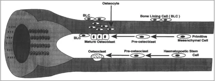
Five kinds of bone cells can usually be recognized in the growing and adult skeleton [38]: osteoprogenitor cells, osteoblasts, osteocytes, osteoclasts, bone lining cells.
Osteoprogenitor cells have the capacity of mitosis and further differentiation and specialization into mature bone cells. There are two types of osteoprogenitor cells, one giving rise to bone-forming osteoblasts, and the other giving rise to bone re-sorption osteoclasts. Both types are commonly found near the bone surface [38].
Osteoblasts are roughly cubical mononuclear cells about 15-30 microns across. Osteoblasts are bone-forming cells. They are responsible for the synthesis and deposition of non mineralised bone matrix, the osteoid [39, 40]. Osteoblasts also appear to participate in the calcification of bone, and they seem to regulate the flow of calcium and phosphate in and out of bone. The life cycle of an osteoblast involves:
- the birth from a progenitor cell;
- the differentiation and participation in elaborating matrix and calcifying units;
- either returning to the pre-osteoblast pool, transform into bone-lining cell and burial as osteocytes, or death.
Osteocytes are the principal cells of fully formed bones. They are hosted in lacunae (only one cell is ever found in a lacuna) and derive from osteoblasts that have secreted bone around them. Mature and relatively inactive osteocytes possess a cell body that has the shape of an ellipsoid, the longest axis (about 25 microns) parallel to the surrounding bony lamellae and its shortest is perpendicular to the plane of the lamellae.
Osteocytes have many cell processes that can extend from considerable distances (on the order of micron) in canaliculi. These cell processes can contact other osteocytes and bone surface cells thank to extensive microfilaments and gap junctions at the contact points. This explains how
cells can survive in such an isolated environment: ions, nutrients, waste products, and small molecules can pass through the gap junctions of adjoining cell processes; fluids can percolate through both the space between the cell and their processes and through lacunar and canalicular walls [41].
The exact functions of osteocytes are not yet clear. However, it is widely assumed that they must have an essential role in the maintenance of bone and in signalling the requirement to resorb micro-damaged bone. They may act as local sensors of the mechanical and chemical state of the bone. The average life cycle of an osteocyte varies with the metabolic activity of the bone and the likelihood that it will be remodelled, but has been estimated to be about 25 years [42, 43]. Osteoclasts are multinucleated giant cells (20-100 m or more) that are responsible for the bone re-sorption process. Active re-sorption osteoclasts are usually found in or near cavities on bone surfaces (re-sorption pits or Howship's lacunae) [44].
In the light microscope, the surface of the osteoclast adjacent to the bone has a striated appearance corresponding to an area of extensive membrane infoldings, termed the ruffled border [45]. This is a unique surface modification, which facilitates bone re-sorption. The structure and the substances concentrated at this border help the transport of materials through the membrane, while bone can be seen to dissolve beneath it. Osteoclasts lacking ruffled border are not capable of bone re-sorption.
Around the ruffled border there is a zone (clear or filamentous zone) devoid of organelles, containing actin filaments: this zone seems to be the site for the adhesion of the cell to the bone surface.
There is little information concerning the life cycle of osteoclasts in vivo. The life cycle of an osteoclast is believed to be about 10 days. Cessation of bone re-sorption is associated with migration of osteoclasts from endosteal surfaces into adjacent marrow space, where they are believed to degenerate and disintegrate.
Very flat, elongated cells with spindle-shaped nuclei, the bone-lining cells, cover most resting bone surfaces in adult skeleton. Their name is suitable to describe the cells that are apposed to inactive bone surfaces, even if it has been incorrectly used to describe any cell apposed to the bone surface [46].
Bone-lining cells serve as a barrier separating fluids percolating through the osteocyte and lacunar canalicular system from the interstitial fluids. This membrane barrier around bone may have a role in mineral homeostasis, by regulating the calcium and phosphate flows in and out the bone fluids, as well as in controlling the bone growth by maintaining a suitable environment [41].
There are now increasing evidences that the bone lining-cells are transducers for bone re-sorption and that they prepare the surface for osteoclastic re-re-sorption [39]. It is also clearer that the bone-lining cells can contribute to sense the shape of bone and its reactions to stress and strain stimuli, and to transmit these sensations as signals to the bone surface, where new bone formation or re-sorption is possible.

![Table 4 - Mean values of uniaxial static tensile properties of 5 commercial formulations of bone cement from Lewis [73]](https://thumb-eu.123doks.com/thumbv2/123dokorg/8227979.128621/52.892.113.815.154.1066/table-values-uniaxial-static-tensile-properties-commercial-formulations.webp)