ABSTRACT
Literature on intraoperative neuro monitoring (IONM) during endocrine surgery have increased over recent years. A comprehensive understanding of the role of IONM for prevention of nerve injuries is critical to maximize safety during surgery of the anterior compartment of the neck. Neuromonitoring techniques are currently considered safe technique and technology; however, albeit sporadically, have been reported some
complications and related side effects using such methods. The complications described can be related to the electrodes positioned at the larynx, at the obstruction of the endotracheal tube, the drugs used for anesthesia and the effects of electrical stimulation on nerve structures and systemic levels. This review will explore the safety issues of IONM to improve the outcomes among patients undergoing monitored thyroidectomy.
Keywords: Safety; Neural monitoring; Thyroidectomy; Endocrine Surgical Procedures
INTRODUCTION
Laryngeal nerve identification during thyroidectomy is considered the safest method to avoid alterations of the motility of vocal cords. Since when in 1938, Lahey first proposed the routine visualization method and dissection of the inferior laryngeal nerve before and during the beginning of glandular dissection, this strategy has made possible to achieve a significant reduction in the percentage of recurrent laryngeal nerve (RLN) damage (1,2).
However, the only RLN visualization, exposure and display in many situations it is not enough to assure a positive, final result with intact vocal cord mobility. In fact, an anatomical integrity does not always guarantees the functional one, and for this reason in the last 2 decades various devices for the intraoperative RLN function monitoring have been proposed, applied and affirmed (1-4).
These devices allow to stimulate physiologically and electrically the laryngeal nerves, to lead an electromyography (EMG) signal to innervated muscles. The consequence muscle activity is then converted into EMG signals acoustic and electromiographically with precise, waveform,
Review Article
Received: Nov 5, 2017 Accepted: Nov 22, 2017 Correspondence to Gianlorenzo Dionigi
Division for Endocrine and Minimally Invasive Surgery, Department of Human Pathology in Adulthood and Childhood “G. Barresi”, University Hospital G. Martino, University of Messina, Via C. Valeria 1, Messina 98125, Italy. E-mail: [email protected]
Copyright © 2018. Korean Association of Thyroid and Endocrine Surgeons; KATES This is an Open Access article distributed under the terms of the Creative Commons Attribution Non-Commercial License (https:// creativecommons.org/licenses/by-nc/4.0/). ORCID IDs
Gianlorenzo Dionigi
https://orcid.org/0000-0003-0864-6087 Author Contributions
Conceptualization: Gianlorenzo Dionigi, Vincenzo Bartolo, Antonio Giacomo Rizzo, Massimo Marullo, Valerio Fabiano, Antonina Catalfamo, Francesca Pia Pergolizzi, Antonino Cancellieri, Giuseppinella Melita; Data curation: Vincenzo Bartolo, Antonio Giacomo Rizzo, Massimo Marullo, Valerio Fabiano, Antonina Catalfamo; Formal analysis: Francesca Pia Pergolizzi, Antonino Cancellieri, Giuseppinella Melita; Project administration: Gianlorenzo Dionigi; Writing - original draft: Gianlorenzo Dionigi, Vincenzo Bartolo, Antonio Giacomo Rizzo, Massimo Marullo, Valerio Fabiano, Antonina Catalfamo, Francesca Pia Pergolizzi, Antonino Cancellieri, Giuseppinella Melita.
Gianlorenzo Dionigi , Vincenzo Bartolo, Antonio Giacomo Rizzo,
Massimo Marullo, Valerio Fabiano, Antonina Catalfamo, Francesca Pia Pergolizzi, Antonino Cancellieri, Giuseppinella Melita
Division for Endocrine and Minimally Invasive Surgery, Department of Human Pathology in Adulthood and Childhood “G. Barresi”, University Hospital G. Martino, University of Messina, Messina, Italy
Improving Safety of Neural Monitoring
in Thyroid Surgery: Educational
Considerations in Learning New
Procedure
Conflict of Interest
No potential conflict of interest relevant to this article was reported.
amplitude, and latency. The documentation of neurophysiological signals of the RLN can be therefore used for prognostic and forensic purpose (3,4).
Various techniques have been described in the literature (5). EMG can be achieved by needle electrodes inserted into the vocal cords, or the use of endotracheal surface electrodes (5). The most recent acquisition in RLN monitoring is continuous stimulation of the vagal nerve, which involves the placement of dedicated electrodes at the level of the vague nerve. Continue vagal nerve stimulation records and ensure a “real-time”, permanent monitoring of the effects of surgical manipulations on all the pathway of the RLN during all steps of intervention (6).
Neuromonitoring techniques are currently considered safe technique and technology; however, albeit sporadically, have been reported some complications and related side effects using such methods.
The complications described can be related to the electrodes positioned at the larynx, at the obstruction of the endotracheal tube (ETT), the drugs used for anesthesia and the effects of electrical stimulation on nerve structures and systemic levels.
COMPLICATIONS RELATED TO ELECTRODES
1. Recording electrodes
The positioning of the needle electrodes may be challenging and requires experience; furthermore, the electrodes can be dislocated during muscle contraction, surgical maneuvers making unstable the EMG signal.
Instead, for dedicated-prefashioned electrodes integrated into ETTs (Fig. 1) or applied externally to standard tubes, limits during the operation can depend from the migration of
Fig. 1. ETT-based surface electrode (Trivantage EMG; Medtronic Xomed Inc., Jacksonville, FL, USA). Tube is ID available in 5, 6, 7, 8, and 9 size. Is standard, non re-enforced tube. Gray area is the electrode area that pick-up EMG. Each EMG channel has an electrode placed anterior above the VF and posterior below the VF. Each length are; anterior electrode length: 25.4 mm, posterior: 30 mm, overall working length: 49 mm. Blue area is an intubation marker only. The tube will work about 5 mm beyond the blue depth marker. Tube is available in EU, USA, and Canada.
the endolaryngeal tube, i.e., displacement upward, downward, rotation or from accidental detachment (7-9).
Authors stated that the needle electrodes compared to the superficial ones, while providing more information and higher amplitudes values, can cause tearing of vocal cord or laryngeal hematoma, vocal cord laceration, infection, cuff deflation and need for reintubation, retained fractured needle segment, and accidental needle dislodgement during surgery (7,10).
Needle electrodes do typically result in larger amplitude measures but overall offer no real advantage as opposed to the surface electrodes (3).
Needle recording electrodes also only monitor unilaterally and so must be repositioned for each side (3).
In a multicenter study on 8,534 patients, 112 (1.3%) damage was found of the cuff after insertion of the needle electrodes (11). According to the authors this complication, which has not compromised the intervention itself was not associated in any case on postoperative bleeding, wound infections or increased rate of postoperative pulmonary morbidity. Damage of the cuff can easily be avoid positioning the ETT more distally (11).
However, though reusable and economically advantageous, needle electrodes are definitely used with decreasing frequency, because they represent a more invasive and more complex method for monitoring (Table 1).
Surgeon must notice that under EMG endotracheal surface electrodes, it is difficult to tell whether the change of EMG amplitude is caused by the change of contact situation between the electrodes and vocal cords or by true nerve injury. In addition, EMG signal by a continuous intraoperative neuro monitoring (C-IONM) application might be limited by the type of anesthesia, manipulation on the trachea and the vagal nerve electrode dislocation (acute signal loss=electrode dislocation).
2. Ground and return stimuli electrodes
Are usually placed in a deltoid or in presternal region (Fig. 2). The authors are unaware of local or systemic complications by electrode placement. Skin disinfection is recommended. There are rarely small subcutaneous hematomas. There are no reports of skin irritation. There is an unknown level of risk when placing electrodes over an infection (possible spreading due to muscle contractions), but cross contamination with the electrodes themselves is of greater concern. The surgeon should remember to remove the electrodes at the end of the surgery and disinfect the region again.
Table 1. Endotracheal EMG tube electrodes advantages • Availability
• Safety
• Non-invasive nature • Ease setup • Ease use
• Derive larger areas of evoked muscle potentials
• Simultaneous continuous RLN & intermitted SLN monitoring
ETT COMPLICATIONS
Oysu and Demir (12) have described 2 cases of obstruction of an EMG-ETT. The authors presented 2 cases with complication of herniation of a specially designed ETT cuff (NIM EMG-ETT; Medtronic Xomed Inc., Jacksonville, FL, USA) used for laryngeal nerve monitoring during thyroid surgery. Abrupt and total blockage of ventilation has occurred at 35th and 40th minutes of general anesthesia.
In an animal trachea, the same authors showed that overinflated cuff may herniate and block ETT tip easily (12). A 25-mm upward movement of the ETT was found as a contributing factor to cuff herniation. After overinflation, the EMG-ETT showed evidence of asymmetric cuff expansion. In the Food and Drug Administration medical device database for all other ETTs reveals 7 cases of cuff herniation. This finding cannot support the hypothesis that EMG-ETTs are more prone to cuff herniation than any other ETT.
The awareness of the possibility of EMG reinforced ETT herniation is of paramount
importance for patient health, as for any other tube adopting. In patients intubated, any event suggesting sudden ventilation blockage should be managed initially by prompt deflation of the cuff. The cuff should be inflated at the minimal necessary volume, and it should be kept in mind that the pilot balloon is not a reliable indicator of cuff pressure. The staff should pay maximum attention to the stability of endotracheal and breathing tubes (Table 2).
The manufacturer did develop appropriate structural modifications to prevent such occurrences (Figs. 1 and 3).
COMPLICATIONS RELATED TO THE TYPE OF ANESTHETIC
DRUGS
An interesting aspect of possible complications of intraoperative neuromonitoring is the type of medication to be used for the induction anesthesia.
The use of drugs that produce a neuromuscular block (neuromuscular blocking agents [NMBAs]) to facilitate endotracheal intubation can compromise the proper functioning of methodical, i.e., an EMG (Fig. 4).
For that reason, the use of such drugs is not recommended during neuromonitoring. It is essential to have full muscular activity return as soon as possible subsequent to intubation (3). Therefore, succinyl choline at 2 to 2.5 mg per kilogram or a small dose of a nondepolarizing muscle relaxant (e.g., rocuronium and atracurium at 0.5 mg/kg) may be used at intubation to allow for normal return of spontaneous respiration and resumption of normal muscle twitch activity within several minutes (3). It is important to keep in mind that a preoperatively unknown pseudocholinesterase deficiency will lead to prolonged paralysis after a depolarizing muscle relaxant such as succinyl choline and will invalidate an EMG monitoring system (3). However, the maneuvers of intubation and extubation can in itself cause vocal cord damage (14,15) (Tables 3 and 4).
Some studies argue that failure to administer of NMBA to facilitate intubation in the patient adult associates a high incidence of local effects and laryngeal lesions (16).
However, in pediatric patients' intubation without the use of these drugs is widespread practice (17).
Table 2. Monitoring tube suggested procedures for intubation • 1/2 greater ID size tube (ID size available 5–9)
• Avoid gel lubrication
• Intubation without neuromuscular blocking • Mandrel can be used
Fig. 3. The awareness of the possibility of EMG reinforced ETT herniation is of paramount importance for patient health, as for any other tube adopting. In patients intubated, any event suggesting sudden ventilation blockage should be managed initially by prompt deflation of the cuff. The cuff should be inflated at the minimal necessary volume, and it should be kept in mind that the pilot balloon is not a reliable indicator of cuff pressure. The staff should pay maximum attention to the stability of endotracheal and breathing tubes.
Actually, associating doses appropriate to propofol to an opioid, they succeed to obtain laryngeal relaxation and suppression of sufficient reflexes to get an intubation easy and without complications (18).
In a recent retrospective study, Birkholz et al. (19) compared laryngeal morbidity after thyroid surgery associated with neuromonitoring in intubated patients with or without using NMBA, without highlighting differences statistically significant between the 2 groups of patients.
RLN Pre-dissezione, destro
18/12/2009 08:37 Periodo reiez. stim.: 2.1 ms Stim 1: 2.00/2.03 mA
Soglia evento: 100 µV
Duration
676 µV
Amplitude=maximum deflection from baseline (peak to peak)
Def: time from the stimulation spike to the first evoked waveform
Latency
1 Vocale sinistro
Fig. 4. EMG profile with amplitude (green), latency (red), and duration (blue) values definition. The EMG waveform is biphasic. The graphic represents pre-dissectional right vagal nerve stimulation (V1), with amplitude value of 676 mcV. Vocal cord depolarization amplitudes are about 100–800 μV during normal awake volitional speech (Blitzer et al. Otolaryngol Head Neck Surg 2009).
EMG = electromyography; RLN = recurrent laryngeal nerve.
Table 3. Incidence of laryngeal morbidity due to intubation/extubation
Author Paper Patients Rate (%)
Proschel U Laryngorhinootologie 1993 75 73
Chilla R Laryngol Rhinol Otol 1976 100 69
Kambic V Br J Anaesth 1978 1,000 6.2
Mencke T Anesthesiology 2003 80 40–15
Mencke T Anesth Analg 2006 160 3
Mencke T Anesth Analg 2006 60 15
Peppard SB Ann Otol Rhinol Laryngol 1983 475 6
Table 4. Risk factors for laryngeal morbidity due to intubation/extubation • Cuff pressure • Size of tube • Movement of tube • Physical trauma • Duration of intubation • Gastroesophageal reflux • Mucociliary mechanism (age) • Type of anesthesia
• Cuff design • Type of surgery
For these reasons, it is usually recommended anesthesiological induction with the use of NMBA short-lived, short-acting, capable of performing function only in the initial stages of intubation and so be disposed of during the central phase of the intervention when it comes instead used monitoring (19) (Table 5).
INTRAOPERATIVE NEURO MONITORING (IONM)
MONITOR SYSTEMS
In this safety review analysis, we believe it is important to point out that it is preferable to use modern IONM monitors with both audio and qualitative, qualitative information on intraoperative EMG.
Current neural monitoring equipment can be divided into audio-only systems and systems that provide both audio and visual waveform information regarding evoked waveform (3). Audio-only systems provide substantially less information such as waveform morphology, amplitude, threshold, and latency that may provide for basic understanding of amplitude variation in normal (3). Current neural monitoring equipment can be broadly divided into audio-only systems and systems that provide both audio and visual waveform information regarding evoked waveform (3). Audio-only systems provide substantially less information such as waveform morphology, amplitude, threshold, and latency that may provide for basic understanding of amplitude variation in normal and pathologic conditions; this information may be important in surgical deliberations (3). Exact determination of loss of signal (LOS) as well as differentiation between signal and artifact may be challenging if not impossible with audio-only systems. Audio-only systems are problematic in that the EMG response to RLN stimulation cannot be quantified. and pathologic conditions; this information may be important in surgical deliberations. Exact determination of LOS as well as differentiation between signal and artifact may be challenging if not impossible with audio-only systems. Audio-only systems are problematic in that the EMG response to RLN stimulation cannot be quantified (3).
STIMULATING COMPLICATIONS ON NERVE STRUCTURES
In the literature, there is discussion about the safety of direct nerve electric stimulation and which is the optimal intensity, frequency, and amplitude (Table 6, Fig. 5).With the stimulation of the laryngeal nerves or the vagal nerve you achieve a biphasic wave; in the language normal the wave amplitude ranges from 100 to 800 μV, while amplitude obtained during monitored surgery vary by presence in the operative field of fluids or blood, of structures that cover the nerve, due to inappropriate probe-nerve contact, because of the ambient temperature or the position of the electrodes (20).
By threshold is meant the minimum electrical stimulus that can produce an EMG response. The vague nerve and larynx nerve are activated approximately at 0.3 and 0.4 mA when the nerve is dry and adequately exposed.
Table 5. Induction anesthesia drugs • Succinylcholine 2–2.5 mg per kilogram
The maximum pacing of all fibers is obtained at 0.8 mA; beyond this threshold, the greatest stimulation does not match a greater response to EMG. Therefore the threshold of stimulus to evaluate the function of the nerve is 1 mA in many cases, while 2 mA stimuli are sometimes used to map the area, for research and recurrent nerve identification (10).
A recent study by Choby et al. (21) aimed to identify the minimum threshold RLN stimulus before and after thyroidectomy, using for neuromonitoring surface contact electrodes. The results show that the average of the minimum intensity necessary to stimulate the inferior laryngeal nerve was 0.50 mA before thyroidectomy, while was 0.47 mA after removing the gland thyroid, with a mean difference between the first and the after the intervention of 0.03 mA. None of these values correlated in a statistically significant way with postoperative cordial paralysis (21).
This difference in stimulation thresholds, also confirmed in other studies is explainable theoretically with the reduction of pressure on the nerve, which can then be stimulated with lower current intensity (22).
In this regard, Wu et al. (23) developed an experimental study on pigs with vague nerve and inferior laryngeal were stimulated with currents from 0.1 to 1.0 mA (with increments of 0.1 mA) and later with 1.5, 2.0, 2.5, and 3.0 mA stimuli. The stimulus lasted 10 μs and was repeated 4 Hz for 10 minutes, with a 2 second interval every minute to record the EMG signal.
Table 6. Factors which determine possible nerve injury • Direct trauma on nerves by stimulator itself • Stimulation intensity level (milliamperes setting used) • Duration of stimulation (i.e., faticability & thermal injury) • Repetitive (I-IONM)
• Continuous, periodic (C-IONM) • Stimulation of VN (systemic safety)
I-IONM = intermitted intraoperative neural monitoring; C-IONM = continuous intraoperative neuro monitoring; VN = vagus nerve.
Fig. 5. Single use, incrementing prass stimulating probe, monopolar, standard flexible and ball tip (product No.8225490; Medtronic Xomed Inc., Jacksonville, FL, USA). Ball tip design is atraumatic for anatomical structures.
After 10 minutes signal amplitude to EMG not was significantly lower than that recorded at the beginning and the maximum amplitude was obtained with stimuli <1 mA, both for the nerve recurrent to the vague nerve (23).
The authors have therefore concluded that minimum currents (about 1 mA) are sufficient to generate the maximum response EMG and can be used to reduce the risks of nerve damage and the effects cardiopulmonary side effects (Fig. 6).
Tables 7 and 8 summarize safety studies on recurrent laryngeal, vagal nerve stimulation and stimulation in pediatric population.
Stimulation intensity (mA)
600 400 200 0 0.6 1.0 1.5 3.0 EMG amplitude (mc V) 800 1,000 1,200 2.0 2.5 0.7 0.8 0.5 0.3 0.1 0.2 0.4 RLN Vagal nerve 266 444 584 798 1,011 1,016 1,017 1,015 1,021 1,025 1,025 1,026 1,026 139 368 490 647 818 887 982 989 988 985 992 988 990
Fig. 6. EMG curve profile from Wu et al.'s study (23). ENG = electromyography; RLN = recurrent laryngeal nerve. Table 7. Safety of nerve stimulation: literature references Safety intermitted RLN stimulation
• Friedman et al. Ann Otol Rhinol Laryngol 1989 • Rea et al. Oper Tech Otolaryngol 1994 • Mermelstein et al. Laryngoscopy 1996
• Randolph et al. Otolaryngol Head Neck Surg 1996 • Rea et al. Laryngoscope 1998
Safety intermitted VN stimulation • Eisele DW. Laryngoscope 1996 • Leonetti et al. Skull Base Surg 1994 RLN = recurrent laryngeal nerve; VN = vagus nerve.
Table 8. Safety of I-IONM during thyroid surgery in childhood: literature references
• Meyer T, Hamelmann W, Timmermann W, Meyer B, Höcht B. The advantages and disadvantages of nerve monitoring during thyroid surgery in childhood. Eur J Pediatr Surg 2006;16:392–5.
• White WM, Randolph GW, Hartnick CJ, Cunningham MJ. Recurrent laryngeal nerve monitoring during thyroidectomy and related cervical procedures in the pediatric population. Arch Otolaryngol Head Neck Surg 2009;135:88–94.
• Meyer T, Höcht B. Recurrent laryngeal nerve monitoring during thyroid surgery in childhood. Eur J Pediatr Surg 2006;16:149–54.
SYSTEMIC SIDE EFFECTS OF STIMULATION
C-IONM via vagus nerve (VN) stimulation is a promising technique in thyroid surgery because it potentially avoids injury to the RLN.
The systemic side effects of stimulation aroused great interest, especially since the continuous vagal nerve monitoring was introduced (Fig. 7).
For years, vagal nerve stimulation has been used for treatment of depression and refractory epilepsy. The first application of a vagal nerve stimulation electrode for refractory epilepsy was in 1990, with more than 80,000 implants till now and a decrease rate in seizure frequency of 24%–31% (24-26).
While long-term stimuli (weeks or months) seem to cause alterations to voice and cough, short-term ones are not usually related to significant complications (24-26).
Though therapeutic vagal stimulation is generally considered a safe treatment, however, cases were described in which it determined cardiac arrhythmias and haemodynamic abnormalities (27-28).
In the 2004 publication from Ali et al. (29), the authors have described 3 cases of cardiac arrest in patients with refractory epilepsy subjected to vagal stimulator, during the perioperative period, during the initial test of electrode application. For 6–15 seconds these patients had a complete block of the atrioventricular node, spontaneously resolved and compatible with stimulation of the vagal nerve (Fig. 8) (29). Overall, according to specific letterature of vagal nerve stimulation electrodes for refractory epilepsy treatment, the rate of bradyarrhythmias is about 0.1%, usually transient and self-limiting (24-29). Authors have suggested also that sequele were due to technical malfunction of the device/probe or induction anaesthesia.
• Auricular nerve • Pharyngeal nerve • SLN
• Superior cervical cardiac branches of VN • Inferior cervical cardiac branch
• RLN
• Thoracic cardiac branches • Branches to the pulmonary plexus • Branches to the esophagus plexus • Anterior vagal trunk
• Posterior vagal trunk • Hering-Breuer reflex in alveoli
Vagal nerve branches
Fig. 7. Vagal nerve branches.
It has been hypothesized that the causes of this phenomenon may have been represented by the direct centrifugal activation of the parasympathetic system with an inhibitory effect on the atrioventricular node, or centripetal activation of afferent portions of the vague nerve (left in this cases), with effect on other central systems that in turn affect the heart rhythm. To evaluate the safety of stimulation vagal during neuromonitoring, Friedrich et al. (30) have recently conducted a study, prospective, non-randomized. They were analyzed on 22 patients the rhythm variations cardiac and immunomodulatory effects induced by vagal nerve stimulation. In this experience, despite an important increase in vagal nerve tone, no hemodynamic alterations have been observed nor have they been reduced plasma cytokine or tumor necrosis factor-α levels. Again, there were no alterations of cardiac rhythm, frequency and alteration of blood pressure. Relative parasympathetic activity increased during C-IONM via VN stimulation (30).
Moreover, the VN has essential regulatory roles in the gastric acid secretion and gastrin release. One study have investigated changes in gastric acid secretion and gastrin release during C-IONM (31). The prospective study of 58 thyroid surgery patients compared gastric acid and serum gastrin at 5 time points: 1) before skin incision, 2) after baseline calibration of C-IONM probe, 3) +20 minutes from baseline, 4) before probe removal, and 5) after extubation. Patients were excluded if they had any history of using tobacco, acid suppression medications, or drugs that affect gastric motility. Patients were also excluded if they had any history of gastroesophageal reflux symptoms, gastroesophageal reflux disease, peptic ulcer disease, helicobacter pylori infection, or chronic kidney disease (31). Non-significant differences in mean gastric pH values were observed at all time points, i.e., 1) before skin incision (2.2±0.2; P=0.50), 2) after baseline calibration of C-IONM probe (2.0±0.8; P=0.62), 3) +20 minutes from baseline (2.5±0.5; P=0.24), 4) before probe removal (2.9±0.9; P=0.52), and 5) after extubation (2.6±1.0; P=0.60). Comparisons of pH monitoring parameters revealed no significant differences in age, gender, side of C-IONM (left vs. right), sequence of C-IONM, or duration of C-IONM. Gastrin values were normal in sequential determinations and did not significantly differ at any time points. The authors, concluded that C-IONM performed via VN stimulation during total thyroidectomy in healthy patients does not influence gastrin secretion and gastric pH (31).
In conclusion intraoperative neuromonitoring of the laryngeal nerves, even with stimulation of the vagal nerve, can be considered a method safe with regard to ad complications it correlates (31-34).
SAFETY OF C-IONM ELECTRODES
The stimulating C-IONM electrodes must be configured in a manner that is protecting against dislocation and changes in their distance from the nerve during surgical manipulation within the surgical site (Fig. 9).
1 sec
1 mV
The ideal vagal electrode should meet some requirements as electrode geometry, applicability, easy removal, low stimulation current, signal stability.
Current research is in favor of closed or partially closed geometry/design C-IONM electrodes because of 1) safer implantation & extraction mode and times, 2) reduced displacement rate, 3) lower stimulation currents, 4) higher EMG amplitudes, 5) stable signal, i.e., signal stability with is a fundamental requirement for C-IONM.
One study evaluated the safety of vagal nerve dissection and analyzed the technical issues to achieve improved vagal nerve critical view of safety dissection, stimulation, and C-IONM probe placement (32). Four hundred vagal nerve dissections were analyzed considering vagal nerve diameter, mean time effort for C-IONM probe positioning, and electrode dislocation rate (32). A significant superior dislocation rate in case of: 1) when a 3-mm automatic periodic stimulating (APS) electrode size was used in a vagal nerve diameter <2 mm, 2) anterior access, and 3) vagal nerve A subtype in relation (P<0.05). No related additional local or systemic morbidity was registered in this series. There was a statistically significant positive relationship between increased diameter of vagal nerve and increased EMG amplitude (P=0.03). There was also a significant increase of amplitude between initial and final vagal nerve stimulation in uneventful cases (P=0.02).
Furthermore, one study aimed at evaluating the feasibility and safety of C-IONM in patients with second- or third-degree atrioventricular block (35). A total of 6 patients (12 nerves at risk), accounting for 0.3% of all 1,800 patients (3,049 nerves at risk) who underwent thyroid surgery during the study period, were found to have second- or third-degree atrioventricular block. All these patients maintained normal systolic and diastolic blood pressures; heart rate; and peripheral arterial oxygen saturation before, during, and after C-IONM. No clinically relevant changes in heart rate or blood pressure, cardiac arrhythmia, or other hemodynamically important events were noted despite careful monitoring of these patients. There was no interference between the biphasic waveform of the vocal muscle electromyogram and the spikes generated by the implanted cardiac pacemakers. Outcomes were uneventful with normal vocal fold and parathyroid gland function. Within the
limitations of this series and considering experimental, animal, and human data, C-IONM of the VN at ≤2 Hz seems to be reasonably safe (35). Additional research is warranted to confirm these results in larger groups of patients with advanced atrioventricular block.
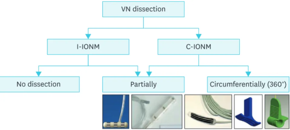
VN dissection
I-IONM
No dissection Partially Circumferentially (360°)
C-IONM
Fig. 9. Vagal nerve dissection for intermitted and continuous monitoring.
VN = vagus nerve; I-IONM = intermitted intraoperative neural monitoring; C-IONM = continuous intraoperative neuro monitoring.
Finally, surgeon should be have perfect knowledge of the VN surgical anatomy that is the medial location of the common carotid artery (CCA) and anterolateral or lateral location of the internal jugular vein (IJV) are the most common configurations (36). Thera are few cases of medial IJV position (36). Tortuosity, kinking, or coiling of the carotid arteries are frequent with advancing age. Aplasia or agenesis of the carotid artery appears in less than 0.01% of the population (36).
CONCLUSION
There is consensus that intermitted intraoperative neural monitoring (I-IONM) is safe, at 1 mA stimulation intensity set.
Sopramaximal stimulation (2–3 mA) should only be used to localize the laryngeal nerves and vagal nerve.
The surgeons and anesthesiologist should verify appropriate electrode materials and stimulation protocols, standardized settlements before and during the monitored procedure. Excellent endotracheal intubating conditions are associated with less laryngeal morbidity and these can be achieved with short-acting depolarizing NMBA or succinylcholine.
Further research is needed to investigate the local and systemic safety of C-IONM via vagal stimulation, even the current published data denied the chance to do harm. We recommend; 1) caution when dissection the vagal nerve and C-IONM probe placement (Figs. 10-12), 2) close supervision by anesthetist, and especially in 3) patients with cardiopulmonary co-morbidity.
Fig. 10. Vagal nerve identification, dissection and subsequent C-IONM probe positioning. We recommend caution when dissection the vagal nerve and C-IONM probe placement. Energy based devices should not be used near the vagal nerve for possible thermal spread injury.
Effect on learning curve must be considered when assessing the impact of IONM and C-IONM on patient safety.
Fig. 11. Positioning of C-IONM electrode. Open design electrode, requires partial VN dissection, tripolar mode of stimulation, size 54×8×0.8 mm (Dr. Langer Medical, GmbH, Waldkirch, Germany).
C-IONM = continuous intraoperative neuro monitoring; VN = vagus nerve.
Fig. 12. Positioning of C-IONM electrode. Closed design electrode, requires 360° VN dissection, monopolar mode of stimulation, 2 size 2 and 3 mm APS (Medtronic Xomed Inc., Jacksonville, FL, USA).
REFERENCES
1. Lahey FH, Hoover WB. Injuries to the recurrent laryngeal nerve in thyroid operations: their management and avoidance. Ann Surg 1938;108:545-62.
PUBMED | CROSSREF
2. Dralle H, Sekulla C, Haerting J, Timmermann W, Neumann HJ, Kruse E, et al. Risk factors of paralysis and functional outcome after recurrent laryngeal nerve monitoring in thyroid surgery. Surgery 2004;136:1310-22.
PUBMED | CROSSREF
3. Randolph GW, Dralle H, Abdullah H, Barczynski M, Bellantone R, Brauckhoff M, et al. Electrophysiologic recurrent laryngeal nerve monitoring during thyroid and parathyroid surgery: international standards guideline statement. Laryngoscope 2011;121 Suppl 1:S1-16.
PUBMED | CROSSREF
4. Dionigi G, Barczynski M, Chiang FY, Dralle H, Duran-Poveda M, Iacobone M, et al. Why monitor the recurrent laryngeal nerve in thyroid surgery? J Endocrinol Invest 2010;33:819-22.
PUBMED | CROSSREF
5. Dralle H, Sekulla C, Lorenz K, Brauckhoff M, Machens AGerman IONM Study Group. Intraoperative monitoring of the recurrent laryngeal nerve in thyroid surgery. World J Surg 2008;32:1358-66.
PUBMED | CROSSREF
6. Sanabria A, Silver CE, Suárez C, Shaha A, Khafif A, Owen RP, et al. Neuromonitoring of the laryngeal nerves in thyroid surgery: a critical appraisal of the literature. Eur Arch Otorhinolaryngol 2013;270:2383-95.
PUBMED | CROSSREF
7. Johnson S, Goldenberg D. Intraoperative monitoring of the recurrent laryngeal nerve during revision thyroid surgery. Otolaryngol Clin North Am 2008;41:1147-54.
PUBMED | CROSSREF
8. Hermann M, Hellebart C, Freissmuth M. Neuromonitoring in thyroid surgery: prospective evaluation of intraoperative electrophysiological responses for the prediction of recurrent laryngeal nerve injury. Ann Surg 2004;240:9-17.
PUBMED | CROSSREF
9. Chan WF, Lo CY. Pitfalls of intraoperative neuromonitoring for predicting postoperative recurrent laryngeal nerve function during thyroidectomy. World J Surg 2006;30:806-12.
PUBMED | CROSSREF
10. Domosławski P, Lukieńczuk T, Kaliszewski K, Sutkowski K, Wojczys R, Wojtczak B. Safety and current achievements in thyroid surgery with neuromonitoring. Adv Clin Exp Med 2013;22:125-30.
PUBMED
11. Thomusch O, Sekulla C, Machens A, Neumann HJ, Timmermann W, Dralle H. Validity of intra-operative neuromonitoring signals in thyroid surgery. Langenbecks Arch Surg 2004;389:499-503.
PUBMED | CROSSREF
12. Oysu C, Demir K. Life-threatening complication of recurrent laryngeal nerve monitoring with EMG reinforced silicone ETT. J Craniofac Surg 2011;22:2419-21.
PUBMED | CROSSREF
13. Yap SJ, Morris RW, Pybus DA. Alterations in endotracheal tube position during general anaesthesia. Anaesth Intensive Care 1994;22:586-8.
PUBMED
14. Paulsen FP, Rudert HH, Tillmann BN. New insights into the pathomechanism of postintubation arytenoid subluxation. Anesthesiology 1999;91:659-66.
PUBMED | CROSSREF
15. Echternach M, Maurer CA, Mencke T, Schilling M, Verse T, Richter B. Laryngeal complications after thyroidectomy: is it always the surgeon? Arch Surg 2009;144:149-53.
PUBMED | CROSSREF
16. Sneyd JR, O'Sullivan E. Tracheal intubation without neuromuscular blocking agents: is there any point? Br J Anaesth 2010;104:535-7.
PUBMED | CROSSREF
17. Shaikh SI, Bellagali VP. Tracheal intubation without neuromuscular block in children. Indian J Anaesth 2010;54:29-34.
PUBMED | CROSSREF
18. Erhan E, Ugur G, Alper I, Gunusen I, Ozyar B. Tracheal intubation without muscle relaxants: remifentanil or alfentanil in combination with propofol. Eur J Anaesthesiol 2003;20:37-43.
19. Birkholz T, Irouschek A, Saalfrank-Schardt C, Klein P, Schmidt J. Laryngeal morbidity after intubation with or without neuromuscular block in thyroid surgery using recurrent laryngeal nerve monitoring. Auris Nasus Larynx 2012;39:288-93.
PUBMED | CROSSREF
20. Wu CW, Dionigi G, Sun H, Liu X, Kim HY, Hsiao PJ, et al. Intraoperative neuromonitoring for the early detection and prevention of RLN traction injury in thyroid surgery: a porcine model. Surgery 2014;155:329-39.
PUBMED | CROSSREF
21. Choby G, Hollenbeak CS, Johnson S, Goldenberg D. Surface electrode recurrent laryngeal nerve monitoring during thyroid surgery: normative values. J Clin Neurophysiol 2010;27:34-7.
PUBMED | CROSSREF
22. Marcus B, Edwards B, Yoo S, Byrne A, Gupta A, Kandrevas J, et al. Recurrent laryngeal nerve monitoring in thyroid and parathyroid surgery: the University of Michigan experience. Laryngoscope 2003;113:356-61.
PUBMED | CROSSREF
23. Wu CW, Lu IC, Randolph GW, Kuo WR, Lee KW, Chen CL, et al. Investigation of optimal intensity and safety of electrical nerve stimulation during intraoperative neuromonitoring of the recurrent laryngeal nerve: a prospective porcine model. Head Neck 2010;32:1295-301.
PUBMED | CROSSREF
24. Wheless JW, Baumgartner J. Vagus nerve stimulation therapy. Drugs Today (Barc) 2004;40:501-15.
PUBMED | CROSSREF
25. Schachter SC, Wheless JW. The evolving place of vagus nerve stimulation therapy. Neurology 2002;59:S1-2.
PUBMED | CROSSREF
26. Rychlicki F, Zamponi N, Trignani R, Ricciuti RA, Iacoangeli M, Scerrati M. Vagus nerve stimulation: clinical experience in drug-resistant pediatric epileptic patients. Seizure 2006;15:483-90.
PUBMED | CROSSREF
27. Ben-Menachem E. Vagus nerve stimulation, side effects, and long-term safety. J Clin Neurophysiol 2001;18:415-8.
PUBMED | CROSSREF
28. Annegers JF, Coan SP, Hauser WA, Leestma J. Epilepsy, vagal nerve stimulation by the NCP system, all-cause mortality, and sudden, unexpected, unexplained death. Epilepsia 2000;41:549-53.
PUBMED | CROSSREF
29. Ali II, Pirzada NA, Kanjwal Y, Wannamaker B, Medhkour A, Koltz MT, et al. Complete heart block with ventricular asystole during left vagus nerve stimulation for epilepsy. Epilepsy Behav 2004;5:768-71.
PUBMED | CROSSREF
30. Friedrich C, Ulmer C, Rieber F, Kern E, Kohler A, Schymik K, et al. Safety analysis of vagal nerve stimulation for continuous nerve monitoring during thyroid surgery. Laryngoscope 2012;122:1979-87.
PUBMED | CROSSREF
31. Xiaoli L, Wu CW, Kim HY, Tian W, Chiang FY, Liu R, et al. Gastric acid secretion and gastrin release during continuous vagal neuromonitoring in thyroid surgery. Langenbecks Arch Surg 2017;402:265-72.
PUBMED | CROSSREF
32. Mangano A, Kim HY, Wu CW, Rausei S, Hui S, Xiaoli L, et al. Continuous intraoperative neuromonitoring in thyroid surgery: Safety analysis of 400 consecutive electrode probe placements with standardized procedures. Head Neck 2016;38 Suppl 1:E1568-74.
PUBMED | CROSSREF
33. Bacuzzi A, Dralle H, Randolph GW, Chiang FY, Kim HY, Barczyński M, et al. Safety of continuous intraoperative neuromonitoring (C-IONM) in thyroid surgery. World J Surg 2016;40:768-9.
PUBMED | CROSSREF
34. Dionigi G, Chiang FY, Dralle H, Boni L, Rausei S, Rovera F, et al. Safety of neural monitoring in thyroid surgery. Int J Surg 2013;11 Suppl 1:S120-6.
PUBMED | CROSSREF
35. Schneider R, Machens A, Bucher M, Raspé C, Heinroth K, Dralle H. Continuous intraoperative monitoring of vagus and recurrent laryngeal nerve function in patients with advanced atrioventricular block. Langenbecks Arch Surg 2016;401:551-6.
PUBMED | CROSSREF
36. Dionigi G, Chiang FY, Rausei S, Wu CW, Boni L, Lee KW, et al. Surgical anatomy and neurophysiology of the vagus nerve (VN) for standardised intraoperative neuromonitoring (IONM) of the inferior laryngeal nerve (ILN) during thyroidectomy. Langenbecks Arch Surg 2010;395:893-9.