UNIVERSITÀ DEGLI STUDI DI CATANIA
DOTTORATO IN BIOLOGIA UMANA E BIOINFORMATICA:
BASI CELLULARI E MOLECOLARI DEL FENOTIPO
XXVI CICLO
DIPARTIMENTO DI SCIENZE BIOMEDICHE E BIOTECNOLOGICHE
Clinical Application of S.salivarius 24SMBc in the Prevention of
Recurrent Otitis Media in Paediatric Age
Applicazione clinica di S.salivarius 24SMBc nella prevenzione
dell’otite media ricorrente in età pediatrica
TESI DI DOTTORATO
Dott.ssa Marina Scillato
Coordinatore: Chiar.mo Prof. MICHELE PURRELLO Tutor: Prof.ssa MARIA SANTAGATI
TABLE OF CONTENTS ABSTRACT (English) p.1 ABSTRACT (Italian) p.3 1. INTRODUCTION p.5 1.1 Probiotics p.5 1.2 Bacteriocins p.8 1.2.1 Bacteriocins of Gram Negative bacteria p.9
1.2.2 Bacteriocins of Gram Positive bacteria p.12
1.3 Guidelines for the Evaluation of Probiotics p.20 1.4 Oral Probiotics p.24
2. AIM OF STUDY p.34
3. ANALYSIS OF S. SALIVARIUS 24SMBc STRAIN’S GENOME SEQUENCE
p.35
4. CLINICAL TRIAL PROTOCOL OF A NASAL SPRAY FORMULATION OF S. SALIVARIUS 24SMBc
p.37
4.1 Materials and methods p.38 4.1.1 Preparation of test material p.38 4.1.2 Clinical trial p.38 4.1.3 Isolation of bacteria and culture conditions p.38 4.1.4 Test for BLIS production p.39 4.1.5 DNA extraction p.39 4.1.6 RAPD-PCR p.40
5. CLINICAL EVALUATION OF S. SALIVARIUS 24SMBc IN A PAEDIATRIC RANDOMIZED, PLACEBO-CONTROLLED, DOUBLE-BLIND TRIAL
p.44
5.1 Materials and methods p.46 5.1.1 Clinical trial p.46 5.1.2 Culture conditions p.47
5.1.3 DNA extraction p.47 5.1.4 Molecular identification of S.salivarius and
pathogenic strains
p.47
5.1.5 Real-time quantitative-polymerase chain reaction p.50
5.2 Results p.52
6. DISCUSSION p.56
1 ABSTRACT (English)
Introduction
.
Much attention has recently been devoted to the analysis of the oral microbiota to develop a bacteriotherapy focused on prevention and/or treatment of upper respiratory tract infections. The oral cavity harbours some beneficial bacterial species such as Streptococcus salivarius which is considered the predominant 'safe' colonizer, capable of fostering more balanced, health-associated oral microflora, interfering with potential pathogens. This antagonist activity is often mediated by competition for nutriments, better adhesion to target cells and release of bioactive agents such as bacteriocins. In our laboratory, we characterized one strain,S.salivarius 24SMBc, isolated from healthy children which showed excellent
inhibitory activity against S.pneumoniae and S.pyogenes and potent capacity of adhesion to HEp-2 cells.
These properties encouraged us to evaluate a possible application of S.salivarius 24SMBc as an oral probiotic for children with recurrent otitis media.
Material and methods. We sequenced the S.salivarius 24SMBc genome by pyrosequencing to verify the presence of virulent factors and to look for genes encoding bacteriocins that inhibit the growth of the pathogens previously described. Then, we included S.salivarius 24SMBc in a clinical trial protocol conducted on 17 healthy adult volunteers to evaluate its safety for the human host and its ability to colonize and persist in the upper respiratory tract. The presence of S.salivarius 24SMBc in rhinopharynx tissue was determined after different time intervals from nasal administration by molecular identification, antagonism test to evaluate BLIS production and RAPD-PCR to distinguish S.salivarius 24SMBc’s genotype from other S.salivarius strains. The following phase for the assessment of the colonization of S.salivarius 24SMBc in the upper respiratory tract of children and its efficacy to reduce the number of episodes of otitis media (OM), was realized through a paediatric randomized, placebo-controlled, double-blind trial. This study enrolled 120 “otitis prone” children and included phenotypic and molecular identification of
S.salivarius 24SMBc and pathogenic strains of OM from the biological samples.
Moreover, the level of colonization of our strain was determined by qPCR using a specific genomic target to identify S.salivarius 24SMBc.
2 Results and conclusion. Genome annotation showed that S.salivarius 24SMBc is free of streptococcal virulent factors. The results of clinical trials demonstrated the absence of adverse effects for the human host and a good capability of S.salivarius 24SMBc to colonize the human rhinopharynx tissue. Prophylactic administration of
S.salivarius 24SMB to children with a history of recurrent OM reduces episodes of
this disease as well the incidence of infection by some causative pathogens such as
S.pneumoniae and S.pyogenes. Therefore, S.salivarius 24SMBc appears a
competitive nasopharyngeal - localized strain with a good potential for use as an oral probiotic to prevent OM in paediatric subjects.
3 ABSTRACT (Italian)
Introduzione. Recentemente, lo studio della microflora orale ha suscitato molto interesse al fine di sviluppare una batterioterapia mirata alla prevenzione e/o trattamento delle infezioni delle alte vie respiratorie. La cavità orale ospita alcune specie di batteri benefici come Streptococcus salivarius che è considerato un microrganismo colonizzatore 'sicuro' e predominante in questo microhabitat, in grado di promuovere una microflora orale più equilibrata e associata allo stato di benessere, interferendo con i potenziali patogeni. Questa attività antagonista è spesso mediata dalla competizione per i nutrienti, una migliore adesione alle cellule target e il rilascio di agenti bioattivi quali le batteriocine. Nel nostro laboratorio, abbiamo caratterizzato un ceppo, S.salivarius 24SMBc, isolato da bambini sani, che ha mostrato un’eccellente attività inibitoria nei confronti di S.pneumoniae e S.pyogenes e una buona capacità di adesione alle cellule HEp-2. Queste proprietà ci hanno indotto a valutare una possibile applicazione di S.salivarius 24 SMBC come probiotico orale nei bambini con otite media ricorrente.
Materiali e metodi. Abbiamo sequenziato il genoma di S.salivarius 24SMBc mediante pyrosequencing per verificare la presenza di fattori virulenza e determinare la presenza di geni codificanti le batteriocine che inibiscono la crescita dei patogeni descritti in precedenza.
Successivamente, abbiamo incluso S.salivarius 24SMBc in un protocollo di sperimentazione clinica condotto su 17 volontari adulti sani per valutare la sicurezza di questo microrganismo nei confronti dell’ospite umano e la sua capacità di colonizzare e persistere nelle alte vie respiratorie.
La presenza di S.salivarius 24SMBc nel tessuto rinofaringeo è stata determinata dopo diversi intervalli di tempo in seguito alla somministrazione nasale del ceppo in studio, mediante identificazione molecolare, test dell’antagonismo per valutare la produzione di batteriocine e RAPD-PCR per distinguere il genotipo di S.salivarius 24SMBc da quello di altri ceppi appartenenti alla specie S.salivarius.
Lo step seguente, finalizzato alla valutazione della colonizzazione di S.salivarius 24SMBc nelle alte vie respiratorie dei bambini e della sua efficacia nel ridurre il numero di episodi di otite media, è stato realizzato attraverso uno studio pediatrico randomizzato, controllato con placebo, in doppio cieco. Questo studio ha arruolato 120 bambini con otite ricorrente ed ha previsto la ricerca di S.salivarius 24SMBc e
4 dei ceppi responsabili di otite media dai campioni biologici, mediante identificazione fenotipica e molecolare.
Inoltre, è stato determinato il livello di colonizzazione del nostro ceppo mediante qPCR, utilizzando una specifica sequenza genomica come target molecolare per identificare S.salivarius 24SMBc.
Risultati e conclusioni. La sequenza del genoma ha dimostrato che S.salivarius 24SMBc è privo dei fattori di virulenza che possono ritrovarsi nel genere
Streptococcus.
I risultati ottenuti dagli studi clinici hanno dimostrato che il nostro ceppo batterico è sicuro per l’uomo ed ha una buona capacità di colonizzare il tessuto rinofaringeo umano. La somministrazione profilattica di S.salivarius 24SMB nei bambini con una storia di otite media ricorrente, riduce gli episodi di questa malattia e anche l'incidenza di infezione da parte di alcuni agenti patogeni responsabili come
S.pneumoniae e S.pyogenes.
Pertanto, S.salivarius 24SMBc appare essere un microrganismo dalla localizzazione nasofaringea, competitivo e con un buon potenziale per un suo uso come probiotico orale nella prevenzione dell'otite media nei soggetti pediatrici.
5 1. INTRODUCTION
1.1 Probiotics
Probiotics are microorganisms, principally bacteria, which confer health benefits maintaining or restoring a host’s natural microbial flora. The use of microorganisms to promote health is very ancient and can even be traced back to classical Roman literature, where food fermented with microorganisms was used therapeutically [1]. The modern history of probiotics dates back to 1877, when Pasteur and his associate Joubert, noting suppression of anthrax bacillus growth in co-cultures with ‘common bacilli’ (probably Escherichia coli), commented that “these facts perhaps justify the highest hopes for therapeutics” [2].
The concept of probiotics evolved at the turn of the 20th century from a hypothesis
first proposed by the Ukrainian bacteriologist and Nobel Laureate Elie Metchnikoff. In 1908, working at the Pasteur Institute, Dr. Metchnikoff observed that a surprising number of people in Bulgaria lived more than 100 years. This longevity could not be attributed to the impact of modern medicine because Bulgaria, one of the poorest countries in Europe at the time, had not yet benefited from such life-extending medical advances. Dr. Metchnikoff further observed that Bulgarian peasants consumed large quantities of yogurt that contains bacteria which conferred the observed health-promoting benefits. He suggested that “the dependence of the intestinal microbes on the food makes it possible to adopt measures to modify the flora in our bodies and to replace the harmful microbes by useful microbes” [3]. The term ‘probiotics’, the antonym of the term antibiotics, was introduced in 1965 by Lilly & Stillwell as Substances produced by microorganisms which promote the growth of other microorganisms [4]. After several definitions, the final one, officially adopted by the World Health Organization, outlining the breadth and scope of probiotics as they are known today, was: “Live microorganisms, which when administered in adequate amounts, confer a health benefit on the host” [5].
In contrast, prebiotics are generally defined as not digestible food ingredients that beneficially affect the host by selectively stimulating the growth and/or activity of one or a limited number of bacterial species already established in the colon, and thus, in fact, improve host health. These prebiotics include inuline, fructo-oligosaccharides, galacto-oligosaccharides and lactulose. The concept of prebiotics essentially has the same aim as probiotics, which is to improve host health via
6 modulation of the intestinal flora, although by a different mechanism. However, there are some cases in which prebiotics may be beneficial for the probiotic, especially with regard to bifidobacteria. This is known as the symbiotic concept. Synbiotics are defined as mixtures of probiotics and prebiotics that beneficially affect the host by improving the survival and implantation of live microbial dietary supplements in the gastrointestinal tract of the host [6].
Before the advent of antibiotics, there were a series of brave attempts by physicians to give protection against diseases such as tuberculosis, anthrax and diphtheria by dosing patients with putatively innocuous commensal bacteria [7]. However, except for the treatment of minor ailments or as supplemental therapy, the application of so-called ‘bacteriotherapy’ or ‘bacterio-prophylaxis’ was largely discontinued on the spectacular discovery of antibiotics.
Indeed, in the middle of the twentieth century, antibiotics offered the promise of efficient and cheap treatment of bacterial infections and even the possibility to eliminate infectious diseases. However, within the span of a single human generation many bacterial species adapted to their antibiotic-laced ecosystems and variants flourished that are capable of resisting our most potent designer antimicrobials [8]. Thus, alternative antimicrobial approaches are being developed and probiotics have gained special interest in the last few years.
Usually, the use of antibiotics implies that the infection is already in progress otherwise probiotics, living microbes part of the natural microflora, can be used as means of prevention of diseases and maintaining of the human health [9].
Probiotic therapy or “bacteriotherapy” is based on the implantation and persistence within the normal microflora of relatively innocuous ‘effector’ bacteria, whose action can be directed at the host, the pathogens or both. Probiotics may modulate the host’s innate or acquired immune response by products like metabolites, cell wall components and DNA. The concept of “bacteriotherapy” seems to be related to bacterial interference. Mechanisms contributing to microbial interference might typically include either the greater ability of the effector bacterium to adhere to epithelial surface, blocking contact between pathogens and host cells, and to compete with others for limited space, essential for the multiplication of all microoganisms, and for nutrients from environment. Another desirable mechanical property for probiotics is their capacity to aggregate among themselves (auto-aggregation), with
7 other probiotics or with pathogens (co-aggregation). Aggregation also enables the formation of a barrier that protects the host’s epithelium from colonization by pathogens. Moreover, the ability to co-aggregate with a pathogen allows the probiotics to entrap it [10]. At last, one of the most important attributes of a ‘good’ probiotic strain is thought to be the strain’s ability to produce and be resistant to a variety of anti-competitor molecules, some relatively non-specific in their targeting (e.g. acids, hydrogen peroxide) and others (e.g. bacteriocins, and bacteriocin-like inhibitory substances (BLIS) and bacteriophages) apparently principally targeted against relatively similar bacteria [8].
8 1.2 Bacteriocins
Allelopathy is the production of chemical compounds which are toxic to other organisms but not to the producers. Microbes produce a remarkable array of substances which help them to compete in their local environments for the limited niche space and nutritional resources available, such as bacteriocins [11].
Bacteriocins were first identified in 1925, when Grazia demonstrated that E.coli V produced in a liquid media a dialyzable and heat-stable substance (later referred to colicin V) that inhibited in high dilution the growth of E.coli S. There followed a period in which a whole series of colicins produced by E.coli and closely related members of the Enterobacteriaceae were discovered [12]. In 1946, Fredericq demonstrated that colicins were proteins and that they had a limited range of activity due to the presence or absence of specific receptors on the surface of sensitive cells [13]. The study of bacteriocins of Gram-positive bacteria got off to a relatively faltering start, largely focusing on staphylococci, and with various attempts to apply similar principles of classification to those that had been established for colicins [14]. In recognition of the discovery that antibiotic substances of the colicin type may also be produced by non-coliform bacteria, the more general term “bacteriocin” was coined by Jacob et al. in 1953. Bacteriocins were specifically defined as protein antibiotics of the colicin type, i.e., molecules characterized by lethal biosynthesis, predominant intraspecies killing activity and adsorption to specific receptors on the surface of bacteriocin-sensitive cells [15].
Bacteriocins have been found in all major lineages of bacteria and, more recently, have been described as universally produced by some members of the Archaea. Indeed, the Archaea have their own distinct family of bacteriocin-like antimicrobials, known as archaeocins. According to Klaenhammer, 99% of all bacteria may make at least one bacteriocin, and the only reason we have not isolated more is that few researchers have looked for them [16].
The frequency and diversity of bacteriocin production varies greatly among bacterial populations and the dynamic interactions occurring among bacteriocin-producing, sensitive and resistant cells are likely responsible for much of this variation. However, the frequency of bacteriocinogeny and the diversity of bacteriocins produced are also determined by the habitat in which the population lives and by the genomic background of the producing strains [11].
9 Two main features distinguish the majority of bacteriocins from classical antibiotics: bacteriocins are ribosomally synthesized and have a relatively narrow killing spectrum [17]. The bacteriocin family includes a diversity of proteins in terms of size, microbial target, mode of action, release, and immunity mechanisms and can be divided into two main groups: those produced by Gram-negative and those produced by Gram-positive bacteria [11, 14].
1.2.1 Bacteriocins of Gram Negative bacteria
Gram-negative bacteria produce a wide variety of bacteriocins, which can be divided into three groups based on size: (1) large colicin-like (25–80 kDa) bacteriocins, (2) the much smaller microcins (<10 kDa) and (3) phage tail-like bacteriocins.
Colicins and Colicin-like Bacteriocins
Since their discovery, the colicins of E.coli have been the most extensively studied Gram-negative bacteriocins, and utilized as a model system for investigating the mechanisms of bacteriocin structure/function, genetic organization, ecology, and evolution [18].
Other members of the Enterobacteriaceae family also exhibit a high frequency (30– 50%) of bacteriocin production [19]. Many of these bacteriocins are similar to colicins in structure and function, and share many molecular, evolutionary and ecological features as well. They are often referred to as colicin-like bacteriocins (CLBs) and similar to colicins have narrow killing spectra which are generally restricted to closely related species. The colicin and many CLB toxin proteins are organized into three functional domains: the N-terminal translocation, the central receptor binding, and the C-terminal killing domains (Figure 1).
Figure 1: Colicin toxin.
The interaction of a colicin molecule with the target cell is initiated by the binding of the receptor-binding domain to a specific cell surface receptor located on the outer cell surface. The colicin protein is subsequently imported into the cell via the
10 translocation domain utilizing either the translocation system to move across the cell’s outer membrane to reach the inner membrane (in the case of ionophore colicins) or the cytoplasm (in the case of the nuclease colicins). The killing domain then mediates the killing of a target cell by pore formation in the cell membrane or nuclease activity. Nuclease colicins have DNase or RNase activities which degrade 16S rRNA or tRNAs. Additionally, a muraminidase function has been described for colicin M that degrades murein in the bacterial cell wall and thereby affects the cell’s structural integrity, resulting in cell lysis.
Colicin operons consist of three tightly linked genes encoding the toxin, immunity and lysis proteins, and are usually found on plasmids. The first gene encodes the toxin whose activity kills the target cells. The immunity gene encodes a protein that binds adjacent to the active site of the colicin protein and inhibits its activity by steric hindrance and electrostatic repulsion mechanisms, protecting the producer cell. In the case of ionophore colicins, the immunity gene is orientated opposite to the toxin gene while it is co-linear with toxin gene in nuclease colicins. The last gene encodes a protein (also called the bacteriocin release protein) which lyses the host cell to release the expressed bacteriocin proteins outside the cell (Figure 2).
Colicin expression is regulated by the SOS induction system and when it is triggered in cells at times of stress, colicin genes are rapidly induced to express high levels of protein. In the case of nuclease colicins, the co-linear arrangement of the immunity and colicin genes within the gene cluster results in increased co-expression of the immunity protein which will bind to newly synthesized colicins and protect the cells from its nuclease activity. In the case of pore-forming colicins, induction does not result in increased levels of immunity protein, as the immunity gene is transcribed from the other strand. Pore-forming colicins, unlike the nuclease colicins, can kill the cells only from the exterior by punching holes in the cell membrane. Therefore, it may not be necessary for the cells to increase the levels of immunity protein during a phase of rapid colicin expression.
11 Figure 2: Genetic organization of nuclease and pore-forming colicins.
The LexA-binding region is indicated by .
Colicin expression results in lysis of the producer cell, due to co-expression of a lysis protein which mediates the release of colicin into the extra-cellular environment [20]. However, experimental evidence suggests that the expression of colicin is induced in only a small fraction of the population [21]. These colicin-expressing cells eventually die but produce enough colicin to kill related, but competing, cells. Thus, a fraction of colicin-harbouring cells display altruistic behaviour by “sacrificing themselves” for the larger benefit of their clonal kin. Indeed, colicins have been implicated as a defence mechanism in competition between cells with more similar nutritional and niche requirements.
Although colicins are representative of Gram-negative bacteriocins, there are intriguing differences found within this subgroup of the bacteriocin family. E.coli encodes its colicins exclusively on plasmid replicons. The nuclease pyocins of
Pseudomonas aeruginosa, which show sequence similarity to colicins, and other, as
yet uncharacterized, are found exclusively on the chromosome. Other close relatives to the colicin family, the bacteriocins of Serratia marcescens, are found on both plasmids and chromosomes [20].
Microcins
Microcins are non-SOS-inducible low molecular weight peptides similar to the bacteriocins of Gram-positive bacteria [22]. All the microcins characterized to date are secreted from the cell, rather than being released as a consequence of cell lysis. It has also been suggested that as much as 90% of the microcins produced by a cell may be retained within the cell [23].
Phage tail-like bacteriocins
Phage tail-like bacteriocins are nuclease- and protease-resistant rod-like particles resembling a bacteriophage tail, which kill sensitive cells by depolarization of the
12 cell membrane [24]. These are multi-meric peptide assemblies, proposed to be defective phages or to have originated from phages which evolved to function as bacteriocins. For example, pyocin R2 (produced by Pseudomonas spp.) appears to be a remnant of phage P2 whereas pyocin F2 is similar to phage lambda [25].
1.2.2 Bacteriocins of Gram Positive bacteria
Bacteriocins of Gram-positive bacteria are as abundant as and even more diverse than those found in Gram-negative bacteria.
Bacteriocins produced by lactic acid bacteria (LAB), which have a long history of use in fermentation and meat and milk preservation, are the best characterized of this group [20].
In 1993, Klaenhammer attempted to put some order into the classification of the bacteriocins of LAB, by proposing four major classes [26]:
1. Class I - post-translationally modified bacteriocins, i.e., the lantibiotics, 2. Class II - small (<10 kDa) heat-stable membrane-active bacteriocins, 3. Class III - larger (>30 kDa) heat-labile bacteriocins,
4. Class IV - complex bacteriocins composed of essential lipid or carbohydrate moieties in addition to protein.
This provisional scheme was adopted by most investigators in the field but it has been reviewed by several authors such as Cotter et al. [27]. They have proposed a more radical modification to the Klaenhammer classification scheme for LAB bacteriocins, in which there are essentially only two principal categories: lantibiotics (class I) and non- lanthionine-containing bacteriocins (class II). The former class III (large heat-labile murein hydrolases) and class IV (the lipid- or carbohydrate-containing bacteriocins) are withdrawn.
Heng et al., proposed classification schema based on that of Cotter, but modified so as to be applicable to most, if not all, known bacteriocins of Gram-positive bacteria (Figure 3) [14].
13 Figure 3: Classifications of bacteriocins of Gram-positive bacteria.
Class I: The lantibiotic
The term “lantibiotic” was coined to refer to the diverse array of Gram-positive bacterial antibiotic peptides that contain the non-genetically encoded amino acids lanthionine (Lan) and/or 3-methyllanthionine (MeLan), as well as various other highly modified amino acids, commonly including the 2,3-unsaturated amino acids dehydroalanine (Dha) and dehydrobutyrine (Dhb). The lantibiotics described to date, all of which are produced exclusively by Gram-positive bacteria, are initially produced as ribosomally synthesized precursor peptides, which then undergo a series of post-translational modifications to produce the unusual amino acids that are intrinsic components of the biologically active peptides. As the family of lantibiotic molecules grew, the individual members were initially classified according to the topology of their ring structures and their biological activities [28], as either type A (elongated amphipathic structures) or type B (globular and more compact structures). In order to encompass the more recently described two-component varieties, type C lantibiotics have been proposed. Type A lantibiotics are further divided into subtypes AI and AII based on the size, charge and sequence of their leader peptides [29].
14 Class II: The Unmodified Peptide Bacteriocins
Class II represents the largest collection of bacteriocins as it essentially encompasses all of the currently described small (<10 kDa) unmodified peptide bacteriocins of Gram-positive bacteria. This class comprises over 50 members with diverse origins, ranging from genera inhabiting the oral cavity and gastrointestinal tract (of humans and other animals) to species best known for their involvement in the dairy and food industries. Class II peptides are divided into three types.
Type IIa: The Pediocin-like Peptides
The largest single collection of class II bacteriocins, consisting of over 20 members, sharing strong activity against Listeria monocytogenes [14].
Type IIb: Multi-Component Bacteriocins
Class IIb includes some bacteriocins that can require two or more peptides to effect optimal inhibitory activity. Garneau et al. proposed that two-component bacteriocins are subdivided into synergistic (S) and enhancing (E) type inhibitory agents. S-type two-component bacteriocin activities are dependent on the concerted action of both peptides, and neither component appears inhibitory on its own [30]. Conversely, for an E-type two-component bacteriocin, either each component peptide or only one peptide of the duet possesses inhibitory activity, but combination of the components results in greatly enhanced killing action toward the target species.
Type IIc: Miscellaneous Unmodified Bacteriocins
All single-peptide non-modified bacteriocins that do not fulfill the criteria of type IIa or type IIb are automatically members of type IIc.
Class III: The Large (>10 kDa) Bacteriocins
This class consists of several large antimicrobial proteins that can generally be subdivided into two distinct groups: (1) the bacteriolytic enzymes (or bacteriolysins), which facilitate the killing of sensitive strains by cell lysis, and (2) the non-lytic antimicrobial proteins.
15 Class IV: The Cyclic Bacteriocins
These inhibitory agents are ribosomally synthesized peptides, but possess a circular structure as they are post-translationally processed such that the first and last amino acids of the mature peptide are covalently bonded, corresponding to the so-called head-to-tail ligation [14].
Bacteriocins of Gram-positive bacteria differ from Gram-negative bacteriocins in two fundamental ways. First, bacteriocin production is not necessarily lethal to the producing cell. This critical difference is due to the transport mechanisms Gram-positive bacteria encode to release bacteriocin toxin. Typically, their biosynthesis is self-regulated with specifically dedicated transport mechanisms facilitating release, although some employ the sec-dependent export pathway. Second, Gram-positive bacteria have evolved bacteriocin-specific regulation, whereas bacteriocins of Gram-negative bacteria rely solely on host regulatory networks. A mechanism of auto-regulation can be observed for the production of nisin by Lactobacillus lactis. Nisin was the first bacteriocin to be isolated and approved by FDA in 1988 as a bio-preservative for a narrow range of foods, specifically to prevent the outgrowth of
Clostridium botulinum spores [20].
The nisin gene cluster contains genes encoding the nisin precursor (nisA), and proteins involved in post-translational modification of the pre-nisin (nisB and nisC), secretion of the modified precursor (nisT) and immunity of the producing L.lactis (nisIFE). Other genes constitute the nisin gene cluster, such as nisP gene that encodes for an extracellular protease involved in removal of the pre-nisin leader peptide to generate the mature nisin molecule. Moreover, in the nisin gene cluster there are genes that encode a two-component regulation system, composed of a sensor kinase (nisK) and a response regulator (nisR) (Figure 4).
The production of nisin is cell-density dependent and was revealed to be regulated at the transcriptional level. Nisin acts as AMP (antimicrobial peptide) and as a secreted peptide pheromone that induces its own biosynthesis by triggering the corresponding signal transduction system in a quorum sensing-like manner.
The ribosomally synthesized precursor (nisA) is modified and transported by a membrane-anchored multi-meric complex composed of the factors B, C and T. Modified pre-nisin processing is performed by the protease nisP. Then, the mature
16 nisin molecule, primarily, acts as AMP and producing cells are protected by the immunity system composed of the factors I, and FGE. The second role of nisin is as a peptide pheromone that is sensed by the input domain of its corresponding sensor kinase domain (KI). Subsequent phosphotransfer from the sensor kinase transmitter
domain (KT) to the receiver domain of the response regulator (RR) leads to its
activation. The output domain (RO) of the active response regulator will bind to a
specific target nis-box within the promoter present in the biosynthetic gene cluster, leading to transcriptional activation and increased production [31, 32, 33].
17 Table 1 shows bacteriocins produced by Gram negative and Gram positive bacteria known in literature [34].
18 Table 1
19 Table 1
20 1.3 Guidelines for the Evaluation of Probiotics
Over the past 20 years, there has been an increase in research on probiotic bacteria and a rapidly growing commercial interest in the use of them in food and medicine [35]. Indeed, scientific evidence continue to accumulate on the properties, functionality, and benefits of probiotics for the promotion of human health, with suggestions that they can play an important role in immunological, digestive, and respiratory functions and could have a significant effect in alleviating infectious diseases in children [36].
Therefore, the Joint FAO/WHO Expert Consultation on Evaluation of Health and Nutritional Properties of Probiotics in Food, held in Cordoba, Argentina 1-4 October, 2001 recognized the need for guidelines to set out a systematic approach for the evaluation of probiotics in food leading to the substantiation of health claims. Consequently, a Working Group was convened by the FAO/WHO in London, Ontario, April 30 and May 1, 2002 to generate guidelines and recommend criteria and methodology for assessment of probiotics and to identify and define what data need to be available to substantiate health claims accurately. A scheme outlining these guidelines is shown in figure 5 [5]. While the recommended guidelines focus particularly on intestinal probiotics, can be considered generally applicable to all probiotics [37].
21 Strain identification
It was recognized that it is necessary to know the genus and species of the probiotic strain. The current state of evidence suggests that strain identity is important to link a particular microorganism to a specific health effect as well as to enable accurate surveillance and epidemiological studies.
Speciation of the bacteria must be established using the most current, valid methodology. It is recommended that a combination of phenotypic and genetic tests be used.
Following the predevelopmental screening phase, probiotic candidate should be deposited in an internationally recognized culture collection, such as the American Type Culture Collection (ATCC) or the Deutsche Sammlung von Mikroorganismen und Zellkulturen GmbH.
Nomenclature of the bacteria must conform to the current, scientifically recognized names. Current nomenclature can be retrieved as follows:
· Approved Lists of Bacterial Names (Int. J. Syst. Bacteriol, 1980, 30:225-420) also available at http://www.bacterio.cict.fr/
· Validation Lists, published in the International Journal of Systematic and Evolutionary Microbiology (or International Journal of Systematic Bacteriology, prior to 2000) [5, 37].
Functional characterization
In vitro test are useful for functional characterization and to gain knowledge of the
mechanism of the probiotic effect. However, it was noted that currently available in
vitro tests are not fully adequate to predict functionality of probiotic microorganisms
in the human body and that probiotics for human use require substantiation of efficacy with human trials. Thus, appropriate target-specific in vitro tests that require validation with in vivo performance are recommended, for example: adherence to mucus and/or human epithelial cells and cell lines, ability to reduce pathogen adhesion to surfaces, resistance to specific environments (i.e. gastric acidity) [5].
22 Safety
An important first step in safety evaluation is a thorough search of the literature. Identification of the history of use and reports of infection resulting from the chosen species/strain should be noted. Safety is verified prior to commercial release but, in practice, it is an ongoing process and requires continual in vitro and in vivo analysis.
In vitro safety checks include: metabolic profiling to assess the production of
deleterious byproducts (i.e. D-lactate); antibiogram determination to accepted standards such as those established by the Clinical and Laboratory Standard Institute (CLSI) or European Committee on Antimicrobial Susceptibility Testing (EUCAST), to indicate antibiotic resistance; toxicity to cell lines and blood (hemolysis), and the presence of virulence factors. Many genetic techniques are available to evaluate the safety of potential probiotics, such as PCR and PFGE, but full genome sequencing allows for rapid strain identification and determination of known virulence and antibiotic resistance genes, colonization factors and genetic transfer mechanisms. Following in vitro testing, trials in animals allow for an in situ safety assessment of the probiotic and help predict potential toxicity for the human host. Typically, researchers study the effect of the probiotic analyzing changes to total body weight, individual organ weight, key biochemistry markers, urine, and blood. Human trials should be carried out using the foreseen commercial formulation and dosage levels to ensure its safety is evaluated in a “real world situation” [37, 38, 39, 40, 41].
Efficacy
The principal outcome of efficacy studies on probiotics should be proven benefits in human trials, such as statistically and biologically significant improvement in condition, symptoms, signs, well-being or quality of life; reduced risk of disease or longer time to next occurrence; or faster recovery from illness. Each should have a proven correlation with the probiotic tested.
The double blind, randomized, placebo-controlled studies measure efficacy compared with placebo (where the placebo is the food carrier devoid of the test probiotic). Sample size needs to be calculated for specific endpoints, and statistically significant differences must apply to biologically relevant outcomes.
23 It is recommended that human trials be repeated by more than one Center for confirmation of results that have to be published in peer-reviewed scientific or medical journals. Furthermore, publication of negative results is encouraged as these contribute to the evidence to support probiotic efficacy.
Effectiveness
Probiotics can be studied by comparison with standard therapy. Labeling
The following information must be given on the label:
Genus, species and strain designation. Strain designation should not mislead consumers about the functionality of the strain
Minimum viable numbers of each probiotic strain at the end of the shelf-life The suggested serving size must deliver the effective dose of probiotics
related to the health claim Health claim(s)
Proper storage conditions
24 1.4 Oral Probiotics
A variety of probiotic bacteria, such as LAB, have been targeted as potential therapeutic agents, differing in terms of their bioavailability, metabolic activity, and mode of action. Until recently, conventional probiotics have typically comprised selected bacteria obtained from intestinal sources (especially lactobacilli and bifidobacteria) and their application has almost exclusively focused on the gastrointestinal benefits [37]. However, with the more widespread acceptance of the potential for probiotic intervention to also effect health benefits for non-intestinal body sites, there has come the increased application of effector strains of species that are indigenous to alternative target tissue in order to obtain more specific and enduring benefits [41]. Moreover, the realization that much human illness can be linked either directly or indirectly to the development of oral microbiota disequilibria has diverted much contemporary probiotic research to products that are capable of fostering a healthy oral microbiota [42]. Although there have been some attempts to use conventional approved intestinal bacteria such as lactobacilli for oral cavity probiotics, it appears more likely that bacteria isolated directly from the natural oral microbiota in healthy humans will be efficacious for such purposes [43].
The microorganisms that inhabit the human oral cavity have been designated as the human oral microbiome. The term microbiome was coined by Joshua Lederberg “to signify the ecological community of commensal, symbiotic, and pathogenic microorganisms that literally share our body space and have been all but ignored as determinants of health and diseases” [44]. The human being and its microbiome together make up a “supraorganism” and the number of microbial cells within a human body exceeds the total number of human cells in the body by nearly 10 times [45]. Remarkably, these potentially overwhelming populations coexist with the host, with harmful effects occurring only if the immune status is altered or if there is a loss of control of epithelial cell sensing and discriminatory systems. It is now generally accepted that some resident commensal bacteria have been shown to provide significant benefit to the host by blocking pathogen colonization and by influencing the normal development of cell structure and the immune system [46, 47].
There are various microhabitats throughout the body that contribute to the overall microbiome, such as mouth, skin, gut, etc (Figure 6). Each microhabitat maintains a unique ecosystem with distinct atmospheric and nutritional compositions that provide
25 a setting for symbiotic interactions among the various microbes within that ecosystem and the host [42]. The Human Microbiome Project (HMP) that explores the role of the human microbiome in physiology, health, and disease through metagenomic research, states that an understanding of human health and disease is impossible without understanding the human microbiome [48].
Figure 6: Human microbiome.
Specifically, studies have shown the oral cavity’s microbiome to be a key source in the etiology of many oral and systemic diseases [49]. Indeed, the oral cavity is the primary gateway to the body and when severe cases of oral disease result in the spread of infection to other body sites, may produce systemic diseases such as cardiovascular disease or others [42]. Because the oral microbiome is vital to a body’s overall health, it is crucial to unravel its complexities to learn the mechanisms by which it maintains health or causes disease. To understand the role of the oral
26 microbiome within the oral cavity, it is important to analyze its fundamental characteristics and dynamics.
The oral cavity is a complex and heterogeneous microbial habitat. Food particles and cell debris provide some nutrients, thus contributing to the establishment of favourable conditions for microbial growth. This environment is consolidated by a constant humidity and an atmosphere that is mainly composed of expired air. Saliva contains enzymes such as lysozyme, lactoperoxidase and amylase, which may have an antibacterial action [50].
More than 700 bacterial species are present in the oral cavity and, maintaining the bacterial communities unaltered, has a significant impact on general health by either preventing or causing infections. The major genera with the largest representation in healthy oral cavities include the following: Streptococcus, Veillonella,
Granulicatella, Gaemella, Actinomyces, Corynebacterium, Rothia, Fusobacterium, Porphyromonas, Prevotella, Capnocytophaga, Neisseria, Haemophilus, Treponema, Lactobacterium, Eikenella, Leptotrichia, Peptostreptococcus, Staphylococcus, Eubacteria, and Propionibacterium [51, 52, 53].
The need for biodiversity in health may suggest that every species carries out a specific function that is required to maintain equilibrium and homeostasis within the oral cavity.
The key to oral health is an ecologically balanced and diverse microbiome that practices commensalism within itself and mutualism with its host. Commensal relationships among microbes allow them to flourish at no expense to their co-habitants and, in turn, maintain biodiversity within the oral cavity.
However, certain pathological changes within the microbial ecosystem may occur and cause a oncebeneficial microorganism to initiate disease within the oral cavity. Ecological shifts that cause pathological changes are: (1) a change in the relationships between the microbes and the host; (2) an increase in relative abundance; and (3) acquisition of virulence factors. In disease, microbes alter their relationship with their host from mutualistic to parasitic and with other microbes from commensal to opportunistic. Pathogens will grow with disregard of their co-habiting bacteria, and any beneficial bacteria will not be able to inhibit the disease manifestation [42, 52, 54].
27 The members of the genus Streptococcus, in particular nonpathogenic streptococci, are the most abundant bacterial species at the oropharyngeal level, and they have been proposed to exert an important role in the protection against pathogenic agents, which cause inflammation and infections [8]. It is well known that the first studies of oral probiotics can be traced to the use of oral alpha-haemolytic streptococci, isolated from the human pharynx, with inhibitory activity against potential pathogens of the upper airways. Roos at al. investigated the effect of alpha-haemolytic streptococci administered as a nasal spray containing two Streptococcus sanguinis, two
Streptococcus mitis and one Streptococcus oralis, on the incidence of OM in
otitis-prone children. The results of this double-blind randomized, placebo-controlled trial showed that the nasal spray was effective in reducing the incidence of OM. Based on these findings, Tano et al. tested a nasal spray containing alpha-haemolytic streptococci but there was no significant change in the nasopharyngeal microbiota or the number of OM episodes compared to the control group when the spray was used without a prior appropriate antibiotic treatment [55, 56, 57]. However, the three species utilized in the formulation are recognized as potential pathogens, for example
Streptococcus mitis has been associated with lung infection and abscess formation,
and both Streptococcus oralis and Streptococcus sanguinis have been implicated in bacterial endocarditis [37].
Other strains of streptococci from the human oral cavity and belonging to commensal species known to have extremely low pathogenic potential are now being developed. In this regard, a key species is Streptococcus salivarius, which has been investigated for its role in the prevention of upper respiratory tract diseases.
S.salivarius is a lactic acid bacterium that is mainly encountered in the mouths of
human beings. It is the first commensal bacterium that appears in the oral cavity of newborns where it colonizes the upper respiratory tract [51] and persists there as a predominant member of the native microbiota throughout the life of its human host [58]. S.salivarius has an exclusive and intimate association with humans that are its sole natural host. Since it has no other known reservoir in nature and its survival time elsewhere is short, the inevitable source of S.salivarius for the newborn baby is the saliva of its closest early contacts. Predictably, the mother will have the major contributing role towards her baby’s pioneer oral microbiota, and indeed, it seems
28 that over the first few days of life the baby’s S.salivarius population progressively changes to more closely resemble that of the mother.
According to several studies, large populations of S.salivarius efficiently adhere to the oral epithelial cells, especially the papillary surface of the tongue that is a strategically location to carry out a population surveillance and modulation role within the oral microbiota [59, 60]. The presence of an adhesion system such as pili, fibrils, saliva-binding proteins and host-cell-binding proteins, together with its high competition rate, helps this species to stay in the human mouth (Figure 7) [61].
Figure 7: Scanning electron micrograph showing the attachment of S.salivarius K12 to microspikes on Hep-2 cells through pilus-like appendages [60].
S.salivarius is an oral streptococcal species that is not known to have any disease
associations in healthy humans [62]. Indeed, there have been occasional reports of infections involving S.salivarius, though their occurrence (even in adverse medical conditions) is extremely low [63, 64].
S.salivarius, is still generally classified as a risk group 2 organism in Europe [37] and
has not been granted a positive QPS status by European Food Safety Authority (EFSA). ‘Qualified Presumption of Safety’ is a safety assessment system based on four parameters: establishing the identity, body of knowledge, possible pathogenicity and end use of the microorganism. However, a microorganism might not be approved by EFSA but may have gained a GRAS (Generally Recognized as Safe) status by the Food and Drug Administration (FDA) in the USA that indicates the use of the product without any demonstrable harm to consumers. This is the case for
Streptococcus salivarius K12 which has been approved as a food ingredient and has
been commercialized in both Australia and New Zealand for several years as an oral probiotic (BLISK12TM Throat Guard). Its safety has recently been assessed in a
29 clinical trial which shows that the intake of this bacterium is well tolerated by humans. Moreover, Streptococcus salivarius is closely related to Streptococcus
thermophilus, a benign organism used in the manufacture of yogurt, which has both
the QPS and the GRAS status [10, 65, 66, 41, 67]. Many comparative genomic studies regarding taxonomy and phylogeny among dairy streptococci have demonstrated that Streptococcus spp. are clustered in two main groups, one comprising S.macedonicus, and S.bovis species and the other S.thermophilus and
S.salivarius: the species in each group show strong similarities in the DNA sequence
of the ribosomal locus [67, 68].
Many strains of S.salivarius are producers of bacteriocin-like inhibitory substances (BLIS) that are diverse in their activity spectra and are thought to play an important role in both stabilizing the oral microbiota and preventing overgrowth (or infection) by potential pathogens [60].
Table 2 shows S.salivarius bacteriocins [60].
Table 2: BLIS produced by strains of S. salivarius.
Salivaricin A (SalA) was the first completely characterized S.salivarius BLlS. It is a type AII lantibiotic, produced by members of the species S.salivarius, S.pyogenes,
30 each of these peptides is capable of inducing production of any one of the salivaricin A subtypes [69].
In S.salivarius the genetic locus comprises eight open reading frames designated
salABCTXYKR. The first gene is salA encoding the precursor lantibiotic peptide,
downstream of it, the salB and salC genes are predicted to encode peptides involved in post-translational modification. The next two genes, salT and salX, encode for the SalTX protein complex located within the cell membrane that carries out cleavage and export of modified SalA. Then, there is salY encoding a protein associated with self-protection against SalA while salKR genes, situated at the distal end of the locus, encode products forming a two-component sensor kinase-response regulator system. Production of this lantibiotic is auto regulated, like the production of nisin, previously described. An interesting finding was that SalA production by one streptococcal species may be induced by sensing of the homologous peptide from another streptococcal species. Indeed, the SalA peptide sensing system apparently does not discriminate between SalA and SalA1 produced by S.salivarius and S.
pyogenes, respectively. The ability of SalA1-producing S.pyogenes strains to respond
to SalA from S.salivarius (and vice versa) could provide a selective mechanism for co-colonization of the mucosal epithelium by pathogen and commensal cell populations. For example, SalA1 produced by rapidly multiplying S.pyogenes cells might stimulate production of SalA by S.salivarius strains, leading to modulation of the number of S.pyogenes cells [72]. It represents a model of regulated coexistence of streptococcal populations in the oral microbial community structure.
Many advantageous and different characteristics make S.salivarius strains promising candidates for the development of oral probiotics and several of these microorganisms have been studied for their bacterial interference against human infections regarding the oral cavity and/or upper respiratory tract (Figure 8). Moreover, different studies on S.salivarius show that strains of this genus are potential probiotics for all ages as they may alleviate many diseases that generally tend to manifest during human life (Figure 9).
The pioneering studies on S.salivarius strains affected S.salivarius TOVE-R (R for rough colony morphology) which demonstrated to colonize rat dental plaque
31 reducing populations of S.mutans and S.sobrinus that are commonly implicated in the etiology of dental caries [73, 74].
The first S.salivarius specifically selected for its potential to interfere with the colonization of the upper respiratory tract by S.pyogenes was strain S.salivarius K58. Its antagonist activity was due to the production of the bacteriocin enocin whose action appeared to involve interference with pantothenate utilization, inhibiting the growth of organisms requiring exogenous pantothenate such as S.pyogenes [55]. Another S.salivarius strain, well-known in scientific literature, that was selected initially for its antagonist activity against S.pyogenes was S.salivarius K12. It harbours two lantibiotics, salivaricin A and salivaricin B that are responsible for the inhibitory growth of the principal causative agent of streptococcal pharyngitis. Further studies showed additional features of S.salivarius K12, such as anti-inflammatory effect, good adhesion to epithelial cells, inhibitory spectrum encompassing some of the key Gram-negative anaerobes that have been implicated in halitosis, and a protective effect against Candida albicans. Since 2001, strain K12 has been used as an oral probiotic and marketed internationally by the New Zealand company BLIS Technologies Ltd. [70, 75, 76, 77].
Similar to S.salivarius TOVE-R, S.salivarius M18 is a strain that inhibits S.mutans and S.sobrinus, moreover, its antagonist activity affects other bacterial species implicated in diseases of the upper respiratory tract, through production of four salivaricins: A2, 9, MPS, and M. Furthermore, S.salivarius M18 inhibits the expression of pro-inflammatory cytokines IL-6 and -8 [62, 78].
Another interesting strain of S.salivarius is S.salivarius ST3 which has the ability to inhibit S.pyogenes, to reduce the levels of pro-inflammatory cytokines IL-6 and -8 and has a good adhesion to epithelial cells [75].
32 Figure 8: Influence of S.salivarius strains in the oral cavity.
Figure 9: Diseases that may be alleviated by S.salivarius probiotics and the ages at which they generally tend to manifest.
I characterized with my research group a strain of S.salivarius, specifically selected for its potential to interfere with the colonization of upper respiratory tract (URT) pathogens: S.salivarius 24SMBc.
33
S.salivarius 24SMBc was selected by screening of 81α-hemolytic streptococci
isolated from 62 nasal and/orpharyngeal swabs of healthy children and identified by currently available phenotypical and molecular techniques. This strain is susceptible to a variety of commonly utilized antibiotics for URT infections treatment and has no hemolytic or harmful enzymatic activity. In addition, it also lacks the main streptococcal virulence genes encoding for streptolysin S, mitogenic exotoxin Z, pyrogenic toxin B, fibronectin-binding protein, serum opacity factor, and exotoxin type C, and G, that can be found in the Streptococcus genus.
S.salivarius 24SMBc was assessed through deferred antagonism test for antagonist
activity against representative strains of URT infections including OM pathogens. It showed an inhibitory spectrum for S.pneumoniae when tested on Columbia agar base supplemented with 5% horse blood and 0.1% CaCo3, and S.pyogenes when tested on
TSYCa.
In particular, S.salivarius 24SMBc inhibits S.pneumoniae strains that include different serotypes, such as 19A, responsible for cases of pediatric meningitis in Sicily, and one S.pyogenes group that is represented by some serotypes such as M1 that is very virulent and very diffused in Italy, and involved in severe infections in children and adults.
Furthermore, it does not interfere with other S.salivarius strains, thus it can coexist with other “friendly bacteria” that colonize the host oral cavity.
This strain demonstrated a good capability to adhere to epithelial cells, indeed, the cells of S. salivarius 24SMBc remained attached to the in vitro HEp-2 monolayer test.
These characteristics led my research group to patent (Pat. num: WO 2011/125086) and register as DSM 23307 our S.salivarius 24SMBc strain [38, 79].
34 2. AIM OF STUDY
During my PhD carried out at the Microbial Molecular Antibiotic Resistance (MMAR) laboratory my research line focused on further analysis of S.salivarius 24SMBc strain, finalized to its application as an oral probiotic for children with recurrent OM. Indeed, this microorganism showed a good in vitro antagonist activity against some pathogens of OM, in particular S.pneumoniae and S.pyogenes.
The first phase was the analysis of the S.salivarius 24SMBc genome in order to verify the presence of virulent factors and look for genes encoding bacteriocins that determine the capability of our strain to interfere with the growth of the pathogens previously described.
To realize our aim, focused on the use of S.salivarius 24SMBc strain as an oral probiotic, we followed the standard guidelines for the evaluation of probiotics, recommended by the FAO and WHO [5].
Thus, we assessed the safety and human tolerance of S.salivarius 24SMBc including it in a clinical trial protocol conducted on healthy adult volunteers to verify the lack of adverse events. Moreover, in this study we determined the adherence to human epithelial cells in vivo analysing its colonization and persistence in the human upper respiratory tract.
The following phase focused on efficacy of S.salivarius 24SMBc and was realized through a paediatric randomized, placebo-controlled, double-blind trial that involved children with recurrent OM. This clinical study was finalized for the assessment of persistence and the level of colonization of S.salivarius 24SMBc in the upper
respiratory tract of these children and its efficacy to prevent or reduce the presence of causative bacteria of OM.
35 3. ANALYSIS OF S. SALIVARIUS 24SMBc STRAIN’S GENOME
SEQUENCE
The genome of S.salivarius 24SMBc strain was sequenced by Pyrosequencing 454. The chromosome reads were assembled into 376 contigs. The estimated length of the chromosome is 1,893,903 bp with a GC content of 40%.
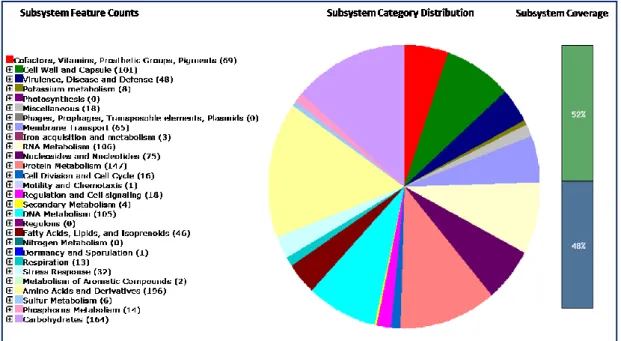
Genome annotation was performed using RAST [80]. S.salivarius 24SMBc was found to be free of streptococcal virulent factors (Figure 10) and this assessment suggested that this microorganism is safe for use as a probiotic.
Figure 10: Organism Overview for Streptococcus salivarius 24SMBc
Sequencing the genome of S.salivarius 24SMBc, we detected a blp (bacteriocin-like protein) locus that resembles the blp locus that has been described in S.pneumoniae and S. thermophilus [81, 82].
The blp locus of S.thermophilus, when fully functional, is organized in independent transcription units coding specific functions related to bacteriocin production:
blpABC, encoding an ABC-transporter (blpA), a transport accessory protein (blpB),
and a peptide pheromone (blpC); blpRH, encoding a histidine kinase (blpH) and a response regulator (blpR). The other operons are: blpD-orf2, blpU-orf3 and blpE-F, encoding bacteriocin precursors and proteins involved in immunity, and blpG-X, whose function is unknown (Figure 11).
36 Figure 11: The blp locus of S.thermophilus
This locus contains all the genetic information required for the production of bacteriocin and is regulated at the transcriptional level by a Quorum Sensing mechanism in which the mature form(s) of the induction factor blpC trigger(s) the expression of the bacteriocin and immunity genes through the blpR-blpH TCS (Two Component System). The mechanism of regulation by cell density implies that there is a basal level of secretion of IF (induction factor) and that a critical concentration of IF triggers its auto-induction, resulting in the amplification of the response.
The ABC transporter recognizes the N-termini of both the pheromone and the bacteriocins and transports these peptides across the cytoplasmatic membrane, concurrent with cleavage at a conserved double-glycine motif. Cleaved extracellular blpC can then bind the sensor kinase, blpH. This interaction results in the activation of blpR and upregulation of the entire gene cluster via binding to consensus sequences within each promoter [81, 82].
Sequence analysis of S.salivarius 24SMBc blp locus of 4.2 kb in size showed the presence of a specific sequence of 258 bp missing in other strains of S.pneumoniae,
S.thermophilus and S.salivarius previously described (Figure 12). Therefore, this
sequence was used to discriminate our microorganism from other strains of
S.salivarius.
37 4. CLINICAL TRIAL PROTOCOL OF A NASAL SPRAY FORMULATION
OF S. SALIVARIUS 24SMBc
S.salivarius 24SMBc, thanks to its significant probiotic characteristics, was included
in a clinical trial protocol conducted on healthy adult volunteers to evaluate its safety and ability to colonize and persist in the human upper respiratory tract.
The study enrolled 17 subjects that were treated with the nasal spray formulation of
S.salivarius 24SMBc following a 6-days course of cefixoral. This antibiotic
treatment was necessary to effect a temporary reduction in the levels of native oral bacterial populations in order to facilitate subsequent colonization by S.salivarius 24SMBc.
The presence of S.salivarius 24SMBc was determined after 2h, 4h, 24h and 7 days from nasal spray administration, collecting rhino-pharyngeal swabs and plating them for each time determination onto Columbia Agar Base and Mitis Salivarius agar. Furthermore, rhino-pharyngeal swabs were obtained just prior to the antibiotic treatment to evaluate the pre-existent microbiota in the upper respiratory tract of volunteers and after the antibiotic treatment to verify the permanence of antibiotic-resistant bacteria.
Each α-haemolitic streptococcal colony isolated on Mitis Salivarius agar that showed a typical aspect of a S.salivarius colony was analyzed by antagonism test to evaluate BLIS production and RAPD-PCR to distinguish S.salivarius 24SMBc from other
S.salivarius strains through genotype profiling.
The levels of colonization by S.salivarius 24SMBc were estimated by calculating the proportion of samples containing S.salivarius colonies with the same characteristics of the strain under study, and all the samples collected from volunteers enrolled in this study.
38 4.1 Materials and methods
4.1.1 Preparation of test material
The S.salivarius 24SMBc strain was formulated for a nasal spray device containing not less than 1x10⁹ CFU/ml. The cell counts were obtained just prior to
commencement and at completion of the study. The product is manufactured by DMG, Rome, Italy.
4.1.2 Clinical trial
The clinical trial involved 17 health subjects, males and females (aged 18-54 years), enrolled in the area of Catania, Italy. This research was carried out during routine clinical practice, following international guidelines and in line with the principles outlined in the Declaration of Helsinki, such that approval from local ethics boards was not required. Exclusion criteria considered were: pregnancy and breast feeding, morpho-functional disorders of the nasal passages and nasal airflow, inflammatory hypertrophic vasomotor diseases. Moreover, the clinical trial excluded patients with diabetes, cystic fibrosis, gastroesophageal reflux, chronic renal failure, recurrent or relapsing inflammation of the upper respiratory tract, mucosal atrophy and impaired mucociliary clearance deficit, hypersensitivity to cephalosporins and subjects treated with immunosuppressants and antibiotics.
The nasal spray was administered 3 times daily for 3 days after an antibiotic treatment with cefixoral (400 mg daily) for 6 days. Rhino-pharyngeal swabs were collected from the volunteers before and later the antibiotic treatment and after 2h, 4h, 24h and 7 days following the nasal spray administration. Then, the biological samples were sent to our MMAR laboratory to be tested for their content of
S.salivarius 24SMBc.
4.1.3 Isolation of bacteria and culture conditions
The rhino-pharyngeal swabs were plated directly onto Columbia Agar Base (Oxoid, Basingstoke, UK), plus 5% horse blood to determine a total microflora population and Mitis Salivarius agar (Difco Laboratories), a selective medium for streptococci to isolate viridans strains. Cultures were incubated overnight at 37 °C in 5% CO2 in air atmosphere.


![Figure 7: Scanning electron micrograph showing the attachment of S.salivarius K12 to microspikes on Hep-2 cells through pilus-like appendages [60]](https://thumb-eu.123doks.com/thumbv2/123dokorg/4478155.32236/31.892.356.601.404.611/figure-scanning-electron-micrograph-attachment-salivarius-microspikes-appendages.webp)
![Table 2 shows S.salivarius bacteriocins [60].](https://thumb-eu.123doks.com/thumbv2/123dokorg/4478155.32236/32.892.305.650.586.939/table-shows-s-salivarius-bacteriocins.webp)