ISSN 1948-5182 (online)
World Journal of
Hepatology
Contents
Monthly Volume 10 Number 10 October 27, 2018
EDITORIAL
639 European Association for the Study of the Liver and French hepatitis C recent guidelines: The paradigm shift
Loustaud-Ratti V, Debette-Gratien M, Carrier P REVIEW
645 Aberrant expression of alternative isoforms of transcription factors in hepatocellular carcinoma Krivtsova O, Makarova A, Lazarevich N
662 Complements are involved in alcoholic fatty liver disease, hepatitis and fibrosis Lin CJ, Hu ZG, Yuan GD, Lei B, He SQ
MINIREVIEWS
670 Era of direct acting anti-viral agents for the treatment of hepatitis C Ahmed M
685 Nutritional support in chronic liver disease and cirrhotics Shergill R, Syed W, Rizvi SA, Singh I
695 Anthropometric indicators of visceral adiposity as predictors of non-alcoholic fatty liver disease: A review Almeida NS, Rocha R, Cotrim HP, Daltro C
702 Dental pulp cell bank as a possible future source of individual hepatocytes Ohkoshi S, Hirono H, Nakahara T, Ishikawa H
708 Adiponectin as a novel biomarker for liver fibrosis Udomsinprasert W, Honsawek S, Poovorawan Y
ORIGINAL ARTICLE
Basic Study
719 Experimental bio-artificial liver: Importance of the architectural design on ammonia detoxification performance
Pizarro MD, Mamprin ME, Daurelio LD, Rodriguez JV, Mediavilla MG
Retrospective Cohort Study
731 Spleen stiffness mirrors changes in portal hypertension after successful interferon-free therapy in chronic-hepatitis C virus patients
Contents
Monthly Volume 10 Number 10 October 27, 2018
EVIDENCE-BASED MEDICINE
743 Comparison of hepatitis C virus testing recommendations in high-income countries Irvin R, Ward K, Agee T, Nelson NP, Vellozzi C, Thomas DL, Millman AJ
SYSTEMATIC REVIEW
752 Thrombosis prophylaxis in pediatric liver transplantation: A systematic review Nacoti M, Ruggeri GM, Colombo G, Bonanomi E, Lussana F
META-ANALYSIS
761 Liver transplantation and atrial fibrillation: A meta-analysis
Chokesuwattanaskul R, Thongprayoon C, Bathini T, Ungprasert P, Sharma K, Wijarnpreecha K, Pachariyanon P, Cheungpasitporn W CASE REPORT 772 Proton beam therapy in apneic oxygenation treatment of an unresectable hepatocellular carcinoma: A case report and review of literature Lin YL 780 Neuroendocrine tumor incidentally detected during living donor hepatectomy: A case report and review of literature
Contents
Volume 10 Number 10 October 27, 2018
World Journal of Hepatology
EDITORS FOR
THIS ISSUE
Responsible Assistant Editor: Xiang Li Responsible Science Editor: Ying Dou
Responsible Electronic Editor: Han Song Proofing Editorial Office Director: Jin-Lei Wang
Proofing Editor-in-Chief: Lian-Sheng Ma
NAME OF JOURNAL
World Journal of Hepatology
ISSN ISSN 1948-5182 (online) LAUNCH DATE October 31, 2009 FREQUENCY Monthly EDITORIAL BOARD MEMBERS
All editorial board members resources online at http:// www.wjgnet.com/1948-5182/editorialboard.htm EDITORIAL OFFICE
Jin-Lei Wang, Director
World Journal of Hepatology
Baishideng Publishing Group Inc 7901 Stoneridge Drive, Suite 501,
Pleasanton, CA 94588, USA Telephone: +1-925-2238242 Fax: +1-925-2238243
E-mail: [email protected] Help Desk: http://www.f6publishing.com/helpdesk http://www.wjgnet.com
PUBLISHER
Baishideng Publishing Group Inc 7901 Stoneridge Drive, Suite 501, Pleasanton, CA 94588, USA Telephone: +1-925-2238242 Fax: +1-925-2238243 E-mail: [email protected]
Help Desk: http://www.f6publishing.com/helpdesk http://www.wjgnet.com
PUBLICATION DATE October 27, 2018
COPYRIGHT
© 2018 Baishideng Publishing Group Inc. Articles pub-lished by this Open Access journal are distributed under the terms of the Creative Commons Attribution Non-commercial License, which permits use, distribution, and reproduction in any medium, provided the original work is properly cited, the use is non commercial and is otherwise in compliance with the license.
SPECIAL STATEMENT
All articles published in journals owned by the Baishideng Publishing Group (BPG) represent the views and opinions of their authors, and not the views, opinions or policies of the BPG, except where other-wise explicitly indicated.
INSTRUCTIONS TO AUTHORS http://www.wjgnet.com/bpg/gerinfo/204 ONLINE SUBMISSION
http://www.f6publishing.com
ABOUT COVER
AIM AND SCOPE
INDEXING/ABSTRACTING
Editorial Board Member of World Journal of Hepatology, Seren Ozenirler, MD,
Professor, Gazi University Department of Gastroenterology and Hepatology, Besevler, Ankara 06510, Turkey
World Journal of Hepatology (World J Hepatol, WJH, online ISSN 1948-5182, DOI: 10.4254), is a peer-reviewed open access academic journal that aims to guide clinical practice and improve diagnostic and therapeutic skills of clinicians.
WJH covers topics concerning liver biology/pathology, cirrhosis and its complications, liver fibrosis, liver failure, portal hypertension, hepatitis B and C and inflammatory disorders, steatohepatitis and metabolic liver disease, hepatocellular carcinoma, biliary tract disease, autoimmune disease, cholestatic and biliary disease, transplantation, genetics, epidemiology, microbiology, molecular and cell biology, nutrition, geriatric and pediatric hepatology, diagnosis and screening, endoscopy, imaging, and advanced technology. Priority publication will be given to articles concerning diagnosis and treatment of hepatology diseases. The following aspects are covered: Clinical diagnosis, laboratory diagnosis, differential diagnosis, imaging tests, pathological diagnosis, molecular biological diagnosis, immunological diagnosis, genetic diagnosis, functional diagnostics, and physical diagnosis; and comprehensive therapy, drug therapy, surgical therapy, interventional treatment, minimally invasive therapy, and robot-assisted therapy.
We encourage authors to submit their manuscripts to WJH. We will give priority to manuscripts that are supported by major national and international foundations and those that are of great basic and clinical significance.
World Journal of Hepatology (WJH) is now abstracted and indexed in PubMed, PubMed Central, Emerging Sources Citation Index (Web of Science), Scopus, China National Knowledge Infrastructure (CNKI), and Superstar Journals Database.
Federico Ravaioli, Antonio Colecchia, Elton Dajti, Giovanni Marasco, Luigina Vanessa Alemanni, Mariarosa Tamè, Francesco Azzaroli, Stefano Brillanti, Giuseppe Mazzella, Davide Festi, Gastroenterology Unit, Sant’Orsola-Malpighi University Hospital, Department of Medical and Surgical Sciences (DIMEC), University of Bologna, Bologna 40138, Italy Antonio Colecchia, Unit of Gastroenterology, Borgo Trento University Hospital, Verona 37100, Italy
ORCID number: Federico Ravaioli (0000-0002-1142-8585); Antonio Colecchia (0000-0002-8384-801X); Elton Dajti (0000- 0003-2905-1146); Giovanni Marasco (0000-0001-7167-8773); Luigina Vanessa Alemanni (0000-0003-3013-7772); Mariarosa Tamè (0000-0002-6299-216X); Francesco Azzaroli (0000-0003- 3675-8545); Stefano Brillanti (0000-0003-4181-795X); Giuseppe Mazzella (0000-0001-8656-8112); Davide Festi (0000-0001-9534 -1745).
Author contributions: Ravaioli F, Dajti E, Marasco G and Alemanni V collected data, analysed data, wrote the manuscript, approved the final manuscript; Tamè M, Azzaroli F, Brillanti S and Mazzella G analysed data and contributed to the drafting and final approval of the manuscript; Colecchia A, Mazzella G and Festi D provided overall oversight of the study, analysed data and contributed to the drafting and final approval of the manuscript. Institutional review board statement: This study was approved by the National Institutional Review Board of the Italian Medicines Agency Committee. Local IRB [Institutional Ethics Committee of Sant’Orsola-Malpighi University Hospital (Bologna, Italy)] approval was authorized.
Informed consent statement: Patients were not required to give informed consent to the study because the analysis used anonymous data that were obtained after each patient agreed to treatment by written consent.
Conflict of interest statement: The authors disclose no conflicts.
STROBE statement: The guidelines of the STROBE statement have been adopted and a fulfilled version of the checklist has been attached with the submission of the manuscript.
Open-Access: This article is an open-access article which was selected by an in-house editor and fully peer-reviewed by external reviewers. It is distributed in accordance with the Creative Commons Attribution Non Commercial (CC BY-NC 4.0) license, which permits others to distribute, remix, adapt, build upon this work non-commercially, and license their derivative works on different terms, provided the original work is properly cited and the use is non-commercial. See: http://creativecommons.org/ licenses/by-nc/4.0/
Manuscript source: Invited manuscript
Correspondence to: Antonio Colecchia, MD, Unit of Gastroenterology, Borgo Trento University Hospital, Verona 37100, Italy. [email protected]
Telephone: +39-335-5876834 Fax: +39-51-2144111 Received: July 3, 2018
Peer-review started: July 3, 2018 First decision: July 24, 2018 Revised: July 27, 2018 Accepted: August 12, 2018 Article in press: August 13, 2018 Published online: October 27, 2018
Abstract
AIM
To investigate changes in spleen stiffness measur-ements (SSMs) and other non-invasive tests (NITs) after treatment with direct-acting antivirals (DAAs) and identify predictors of SSM change after sustained
Submit a Manuscript: http://www.f6publishing.com DOI: 10.4254/wjh.v10.i10.731
World J Hepatol 2018 October 27; 10(10): 731-742
ISSN 1948-5182 (online)
ORIGINAL ARTICLE
Spleen stiffness mirrors changes in portal hypertension
after successful interferon-free therapy in chronic-hepatitis
C virus patients
Retrospective Cohort Study
Federico Ravaioli, Antonio Colecchia, Elton Dajti, Giovanni Marasco, Luigina Vanessa Alemanni, Mariarosa
Tamè, Francesco Azzaroli, Stefano Brillanti, Giuseppe Mazzella, Davide Festi
virological response (SVR). METHODS
We retrospectively analysed 146 advanced-chronic liver disease (ACLD) patients treated with DAA with available paired SSM at baseline and SVR24. Liver stiffness (LSM), spleen diameter (SD), platelet count (PLT) and liver stiffness-spleen diameter to platelet ratio score(LSPS)
were also investigated. LSM ≥ 21 kPa was used as a
cut-off to rule-in clinically significant portal hypertension (CSPH). SSM reduction > 20% from baseline was defined as significant.
RESULTS
SSM significantly decreased at SVR24, in both patients with and without CSPH; in 44.8% of cases, SSM reduction was > 20%. LSPS significantly improved in the entire cohort at SVR24; SD and PLT changed significantly only in patients without CSPH. LSM significantly decreased in 65.7% of patients and also in 2/3 patients in whom SSM did not decrease. The independent predictor of decreased SSM was median relative change of LSM. CSPH persisted in 54.4% patients after SVR. Delta LSM and baseline SSM were independent factors associated with CSPH persistence.
CONCLUSION
SSM and other NITs significantly decrease after SVR, although differently according to the patient’s clinical condition. SSM faithfully reflects changes in portal hypertension and could represent a useful NIT for the follow-up of these patients.
Key words: Clinically significant portal hypertension;
Spleen stiffness measurement; Advanced chronic liver disease; Direct-acting antivirals; Portal hypertension; Hepatitis C; Non-invasive test
© The Author(s) 2018. Published by Baishideng Publishing
Group Inc. All rights reserved.
Core tip: Liver stiffness measurement (LSM) and spleen
stiffness measurement (SSM) are widely validated surrogates of portal hypertension (PH) and its complica-tions. Their role in the assessment of therapy response, such as treatment with direct-acting antivirals (DAAs) of hepatitis C virus patients, is still under investigation. We demonstrated in a large cohort that not only LSM, but also SSM, is reduced six months after successful DAA therapy. As opposed to LSM, SSM directly reflects PH and is less influenced by the immediate reduction of liver necro-inflammation. We believe that SSM could represent a helpful tool for the clinician in the follow-up of these patients.
Ravaioli F, Colecchia A, Dajti E, Marasco G, Alemanni LV, Tamè M, Azzaroli F, Brillanti S, Mazzella G, Festi D. Spleen stiffness mirrors changes in portal hypertension after successful interferon-free therapy in chronic-hepatitis C virus patients. World J Hepatol 2018; 10(10): 731-742 Available from: URL: http://www.
Ravaioli F et al. Spleen stiffness measurement and PH after DAAs
wjgnet.com/1948-5182/full/v10/i10/731.htm DOI: http://dx.doi. org/10.4254/wjh.v10.i10.731
INTRODUCTION
Chronic hepatitis C virus (HCV) infection represents one of the major causes of liver disease and is a lea ding cause of liver transplantation[1,2]. Recently, the
introduction of the highly effective interferonfree direct acting antivirals (DAAs) has enormously increased the number of patients who have achieved sustained virologi cal response (SVR), even in patients with liver cirrhosis[3–5].
Studies, mostly from the interferon era, have shown that achieving SVR improves liver function[6,7], liver
histology[8] and overall clinical outcomes[9]. However, the
real impact of SVR in the DAA era, in terms of changes in portal hypertension (PH) and risk of decompensation on immediate followup, is not completely known, especially in patients with advanced chronic liver diseases (ACLD). PH is a progressive condition that represents a key point in the natural history of liver diseases[10]; therefore, its
assessment by the hepatic venous pressure gradient (HVPG) measurement is fundamental in ACLD patients[11– 14]. Indeed, the development of clinically significant portal
hypertension (CSPH) in patients with compensated ACLD (cACLD)[11] is highly associated with the risk of clinical
decompensation events (ascites, variceal bleeding, jaundice and hepatic encephalopathy)[10].
To date, several studies have demonstrated a signi ficant reduction in HVPG (> 10%-20%) after achieving SVR, both after interferonbased[15–17] and DAAbased
regimens[18–21]. Although the HVPG measurement is the
gold standard to assess PH[11], it remains an invasive
method[22] and its use is still limited only to highly spe
cialized centres[12]; thus, its repeated measurements
during the followup would hardly be applicable.
Consequently, many noninvasive tests (NITs) in the last decade, including liver and spleen stiffness measurements (LSM and SSM) as well as liver stiffness spleen diameter to platelet ratio score (LSPS), have been developed and validated to accurately assess PH degree and its complications[11,22–29]. In fact, the
Baveno Ⅵ Consensus recently recommended that LSM values of 10 kPa should rule out cACLD patients, and values of 2025 kPa should accurately identify CSPH in patients with viral hepatitis[11]. However, to date,
few studies have evaluated the role of NITs in the PH assessment of SVR patients after DAA treatment, and their role in the followup. Even if most studies agree on the fact that LSM rapidly decreases after virus eradication[18,19,30–32], controversial data have emerged
regarding the changes of SSM after SVR[30–32].
MATERIALS AND METHODS
Aims of the study
DAA treatment on PH, evaluated by spleen stiffness changes as a mirror of PH; (2) as well as those of other NITs, after HCVDAA treatment; moreover, we aimed to (3) identify the presence of predictors of the SSM changes in SVR patients after DAA therapy.
Study design and population
This is a retrospective analysis of prospectively collected data of HCVrelated cACLD patients treated with DAAs between January 2015 and September 2017 at our department, with valid measurements of LSM and SSM by transient elastography (TE) at baseline (BL) and at six mo after the end of DAA treatment (SVR24).
According to the Baveno Ⅵ Criteria[11], values of
LSM > 10 kPa at TE were considered suggestive of having cACLD; LSM cutoff ≥ 21 kPa was used to rule in CSPH, as previously described[33,34]. At baseline,
laboratory values, Model for endstage liver disease (MELD) and ChildTurcotte Pugh (CTP) scores were also reported for each patient.
We excluded patients who (1) had incomplete response to surgical resection or locoregional ablation of previous HCC; (2) developed HCC during antiviral treatment; (3) developed variceal bleeding and/or endoscopic banding legation (EBL) during the study period; and (4) initiated or changed the dosage of nonselective betablockers (NSBB) or had portal vein thrombosis, transjugular intrahepatic portosystemic shunt (TIPS) and noncirrhotic PH. A subgroup of the patients who did not achieve SVR were separately investigated.
Antiviral treatment
Eligibility for treatment of HCV patients with DAAs was assessed following the priority criteria established in the protocol approved by the Italian Medicines Agency committee. The choice of DAA and treatment duration (12 or 24 wk) was based on viral genotype and stage of disease, according to the guidelines available at the time of enrollment[35]. SVR was defined as undetectable
HCVRNA using realtime PCR, with a detection limit of 15 IU/mL at the 12wk posttreatment followup visit.
NITs for PH assessments
LSM values were assessed by expert operators using the FibroScan® apparatus with “M” probe (Echosens®,
Paris, France) after overnight fasting and after a com plete abdominal US examination. LSM values were obtained as previously reported[16]
, and the reliability criteria considered were according to the last EFSUMB Guidelines and Recommendations on the Clinical Use of Ultrasound Elastography[36]. SSM was assessed on the
same day as LSM assessment, with the same probe utilized to perform LSM using the FibroScan® apparatus,
as previously described[24]. Since no specific literature
is present, we translated data from HVPG experience[11]
and defined significant SSM as a reduction > 20% from BL. LSPS was calculated as liver stiffness × (spleen
diameter (SD)/platelet count)[37]. SD was considered to
be the bipolar diameter of the spleen as assessed by ultrasound.
Statistical analysis
Categorical data are expressed as numbers (percen tages) and continuous variables as medians (IQR or range). For group comparison, the MannWhitney U test was used for continuous variables and the chi2 test for
categorical variables. Group comparisons among NITs at BL and SVR24 were evaluated with Friedman’s non parametric test, and Bonferronicorrected alphas were used for post hoc pairwise comparison. Demographic, clinical, functional and elastometric variables were evaluated with univariate and multivariate Logistic Regression models in order to assess the predictive factors associated with PH improvement as assessed by SSM. After evaluation of multicollinearity, variables with a pvalue < 0.10 upon univariate analysis were included in several multivariate Logistic Regression models with stepwise backward procedures. Prevalence of esophageal varices (EV) was not included in the multivariate analysis due to the limited number of patients with available EGD data (within 6 mo from TE assessment). The estimated odds ratios with their 95% confidence intervals, LR chi2 and Area under ROC
Curve were presented. For each multivariable logistic regression, the model discrimination and calibration were reported together with Akaike information crite rion and Bayesian information criterion measures for comparing maximum likelihood models. Only pvalues < 0.05 were considered statistically significant. The statistical analysis was conducted using Stata/SE (Ver sion 14.0; Stata Corp, Texas, United States).
Ethics
The DAAs treatment protocol was approved by the National Institutional Review Board (IRB) of the Italian Medicines Agency committee. Local IRB [Institutional Ethics Committee of Sant’OrsolaMalpighi University Hospital (Bologna, Italy)] approval was authorized.
RESULTS
Patients characteristics
One hundredninetyseven cACLD patients treated with DAAs and with available valid baseline LS and SS measurements were evaluated. The following patients were excluded: two (1%) had HCC occurrence and three (1.5%) presented with active HCC, one (0.5%) underwent EBL during the study period, four (2%) had previous EBL, two (1%) patients presented with complete portal vein thrombosis, one (0.5%) required an increase in NSBB dosage and one (0.5%) had previous TIPS placement. An additional 37 (18.8%) patients were excluded: 22 (out of 197, 11.2%) due to lack of follow-up and 15 (out of 197, 7.6%) due to unfeasible SSM at followup. Accordingly, a total of 134 Ravaioli F et al. Spleen stiffness measurement and PH after DAAs
patients with paired LSM and SSM at BL and SVR24 were included in the final analysis; 12 (6%) patients who did not achieve SVR were analysed separately (Figure 1).
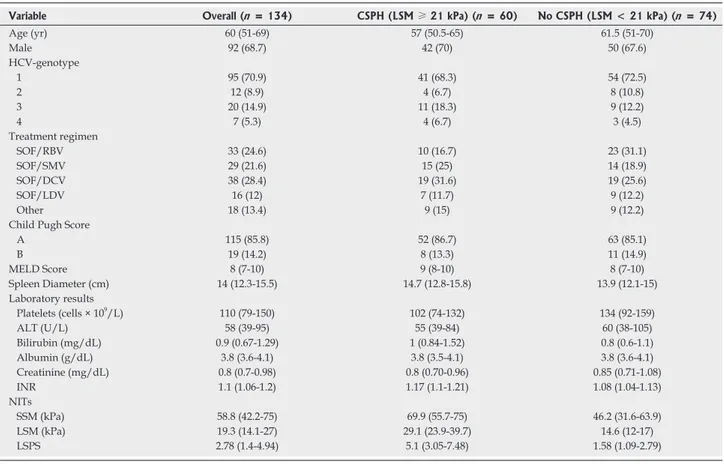
Table 1 depicts the baseline characteristics of the study cohort. Regarding main NITs, the median values at BL were LSM 19.3 kPa (14.127 kPa) and SSM 58.8 kPa (42.275 kPa). In a subanalysis, patients with CSPH (LSM ≥ 21 kPa) differed significantly for MELD score, platelet count, total serum bilirubin, INR, SSM, LSM, SSM and LSPS.
Changes in SSM and LSM after SVR
In the patients who achieved SVR, the median SSM significantly decreased from 58.8 kPa to 38.2 kPa (p = 0.001), with a median delta change in SSM of – 12.3%. The decrease in SSM was statistically significant in both groups, CSPH and not (Figure 2A); the median delta SSM was higher in patients without CSPH at baseline when compared to patients with CSPH (-20.4% vs -4.7%), although this difference did not reach statistical significance. A decrease in SSM values was found in 92 (68.7%) patients, of whom 73 (54.5%) had a decrease > 10% and 60 (44.8%) > 20% (Table 2 and Figure 3A). LSM values also decreased after SVR, with respective median values of 19.3 kPa and 13.8 kPa at baseline and SVR24 (p < 0.0001). The median delta LSM was -30% with similar
changes in both groups; LSM decreased in 114 (85.1%) patients, of whom 88 (65.7%) had a decrease of > 20% (Table 2 and Figure 3A).
A LSM decrease was found in almost all patients in whom SSM decreased (95.3%). On the other hand, LSM significantly decreased (p = 0.022) in 2/3 of the patients in whom SSM did not decrease, with a median delta LSM of -28.3%. (Figure 3B).
Changes in other NITs after SVR
The median spleen diameter (SD) at baseline and SVR24 were 14 cm and 13.2 cm, respectively. Althou gh the reduction was not statistically significant in the overall population, it reached significance in the subgroup of patients without CSPH. The increase of PLT (from 110 109/L to 130 109/L) did not reach stati
stical significance in the entire cohort either, but only in patients without CSPH (Figure 2B). Moreover, median LSPS differed significantly between baseline (2.78) and SVR (1.34), and in both subgroups as well.
Non-SVR patients
Twelve patients did not achieve SVR in our cohort. Baseline characteristics did not statistically differ from the patients included in the final analysis. In particular, in nonSVR patients, an LSM decrease (23.2 kPa at BL vs 21.6 kPa at FU24), an SSM increase (45.6 kPa at BL vs 57.8 kPa at FU24) and a PLT decrease (128 ×
Figure 1 Flowchart of study design. DAA: Direct-acting antiviral; EBL: Endoscopic band ligation; HCC: Hepatocellular carcinoma; HCV: Hepatitis C virus; LSM:
Liver stiffness measurement; NSBB: Non-selective beta-blocker; SSM: Spleen stiffness measurement; SVR: Sustained virological response; TIPS: Transjugular intrahepatic portosystemic shunt.
HCV-patients treated with DAAs regimens with LSM > 10 kPa and available SSM at baseline n = 197
Patients with HCC occurrence during study period or active previous HCC (n = 5) Patients who developed variceal bleeding or underwent EBL during study period (n = 1) Patients with previous EBL, NSBB initiation/change of dosage, portal vein thrombosis, TIPS or non-cirrhotic portal hypertension (n = 8)
Patients lost at follow-up (n = 22)
Patients with unfeasible SSM at follow-up visit (n = 15)
Included patients with paired LSM and SSM before and 6 mo after SVR
n =134
Included patients who did not achieve SVR
n =12 Ravaioli F et al. Spleen stiffness measurement and PH after DAAs
109/L at BL vs 100 × 109/L at FU24) were observed;
none of these changes reached statistical significance (Supplementary Table 1).
Predictors of significant SSM Decrease (> 20%)
Table 3 shows the differences observed between patients who had an SSM decrease > 20% and those who did not. In the entire cohort, patients with sig nificant SSM reduction differed in the prevalence of EV, MELD score, albumin levels, as well as baseline SSM, LSPS values and LSMrelated variables. In multivariate analysis, relative LSM changes remained as the only independent predictor of an SSM decrease > 20%. Furthermore, predictors of an SSM decrease > 20% (Supplementary Table 2) were investigated among patients with CSPH at baseline. Once again, a higher prevalence of EV, higher creatinine levels, lower LSM values at SVR24 and higher delta LSM were observed among patients with an SSM decrease > 20%. In multivariate analysis, higher serum creatinine levels and delta LSM > 20% were the predictors of a significant SSM decrease.
Changes of CSPH state after SVR
Figure 4 shows that 60 (44.8%) patients presented
with CSPH at baseline, defined as an LSM ≥ 21 kPa. After a 6 mo followup, none of the 74 patients without CSPH at baseline progressed to CSPH. In patients with CSPH, 46.7% of them had reduced LSM under the CSPH threshold after treatment. Supplementary Table 3 shows the predictors of CSPH persistence after DAA treatment.
DISCUSSION
The main aim of our study was to evaluate PH changes assessed by noninvasive methods after successful viral eradication in patients treated with DAAs. Our data show that SSM and LSM significantly decrease after SVR, according to the baseline clinical patient condition.
The IFNfree regimens are highly effective, allowing to treat and achieve SVR in patients who also have ACLD[4,38]. However, the individual clinical benefit in
these patients is still under debate, especially in terms of changes in PH and CSPHdriven complications[39–41].
While results from the interferon era might not nece ssarily be translatable to DAA regimens[21], recent stu
dies have also unanimously demonstrated that HVPG significantly decreases after SVR[18–21]. Although many
Qualitative data were expressed as number and percentage (%); quantitative data were expressed as median (25%-75% quantiles). ALT: Alanine aminotransferase; AST: Aspartate aminotransferase; CSPH: Clinically significant portal hypertension; DCV: Daclatasvir; HRV: High risk varices; INR: International normalized ratio; LDV: Ledipasvir; LSM: Liver stiffness measurement; LSPS: Liver stiffness to spleen/platelet score; MELD: Model for end-stage liver disease; NITs: Non-invasive tests; RBV: Ribavirin; SMV: Simeprevir; SOF: Sofosbuvir; SVR: Sustained virological response; SSM: Spleen stiffness measurement.
Variable Overall (n = 134) CSPH (LSM ≥ 21 kPa) (n = 60) No CSPH (LSM < 21 kPa) (n = 74)
Age (yr) 60 (51-69) 57 (50.5-65) 61.5 (51-70) Male 92 (68.7) 42 (70) 50 (67.6) HCV-genotype 1 95 (70.9) 41 (68.3) 54 (72.5) 2 12 (8.9) 4 (6.7) 8 (10.8) 3 20 (14.9) 11 (18.3) 9 (12.2) 4 7 (5.3) 4 (6.7) 3 (4.5) Treatment regimen SOF/RBV 33 (24.6) 10 (16.7) 23 (31.1) SOF/SMV 29 (21.6) 15 (25) 14 (18.9) SOF/DCV 38 (28.4) 19 (31.6) 19 (25.6) SOF/LDV 16 (12) 7 (11.7) 9 (12.2) Other 18 (13.4) 9 (15) 9 (12.2)
Child Pugh Score
A 115 (85.8) 52 (86.7) 63 (85.1) B 19 (14.2) 8 (13.3) 11 (14.9) MELD Score 8 (7-10) 9 (8-10) 8 (7-10) Spleen Diameter (cm) 14 (12.3-15.5) 14.7 (12.8-15.8) 13.9 (12.1-15) Laboratory results Platelets (cells × 109/L) 110 (79-150) 102 (74-132) 134 (92-159) ALT (U/L) 58 (39-95) 55 (39-84) 60 (38-105) Bilirubin (mg/dL) 0.9 (0.67-1.29) 1 (0.84-1.52) 0.8 (0.6-1.1) Albumin (g/dL) 3.8 (3.6-4.1) 3.8 (3.5-4.1) 3.8 (3.6-4.1) Creatinine (mg/dL) 0.8 (0.7-0.98) 0.8 (0.70-0.96) 0.85 (0.71-1.08) INR 1.1 (1.06-1.2) 1.17 (1.1-1.21) 1.08 (1.04-1.13) NITs SSM (kPa) 58.8 (42.2-75) 69.9 (55.7-75) 46.2 (31.6-63.9) LSM (kPa) 19.3 (14.1-27) 29.1 (23.9-39.7) 14.6 (12-17) LSPS 2.78 (1.4-4.94) 5.1 (3.05-7.48) 1.58 (1.09-2.79)
Table 1 Baseline characteristics of included patients
110 × 109/L 130 × 10 9/L n.s. 0 50 100 150 200 250 300 Baseline SVR24 Platelet count (PLT) 134 151 101 102 p = 0.035 n.s. BL SVR24 BL SVR24 Without CSPH With CSPH 0 50 100 150 200 250 300
B
p < 0.0001 19.3 kPa 13.8 kPa 0 15 30 45 60 75 Baseline SVR24 14.6 kPa 9.8 kPa 21.6 kPa 29.1 kPa p < 0.0001 p < 0.0001 BL SVR24 BL SVR24 Without CSPH With CSPH Liver stiffness (LSM) 0 15 30 45 60 75A
p = 0.001 58.8 kPa 38.2 kPa 0 15 30 45 60 75 Baseline SVR24 46.2 kPa 34.5 kPa 61.1 kPa 69.9 kPa p = 0.0004 p = 0.007 BL SVR24 BL SVR24 Without CSPH With CSPH Spleen stiffness (SSM) 0 15 30 45 60 75 14 cm 13.2 cm n.s. 5 10 15 20 25 Baseline SVR24 Spleen diameter (SD) 13.9 cm 12.9 cm 14.7 cm 14 cm p = 0.05 n.s. BL SVR24 BL SVR24 Without CSPH With CSPH 5 10 15 20 25studies have shown that LSM rapidly decreases after DAA treatment[42,43], not much is known about the
changes of PH surrogate NITs, such as SSM and LSPS, after viral eradication. In fact, NITs have yet to be validated in SVR patients, and their role in the clinical followup is still to be determined.
The main finding of this study is that SSM signi ficantly changes after 24 wk of SVR in patients with cACLD, with a median relative change of -12.3% (Table 2). To our knowledge, only two complete papers[30,32]
and one letter to the editor[39] have investigated the
changes in SSM after SVR, with opposing results. In fact, only in the study by Pons et al[32] SSM was found
to rapidly decrease at only 4 wk after therapy initiation in 41 patients, with no ulterior significant changes until 48 wk of followup; the other studies concluded that SSM did not significantly decrease at SVR24[30,32].
In our study that analyzed a large cohort of cACLD patients, we demonstrated that SSM significantly decreased after DAA treatment. These results confirm previous studies in which PH was assessed by paired HVPG measurements[18–21]. Moreover, our study is the
first to assess and demonstrate the improvement of
LSPS, another accurate surrogate of PH, after SVR24. Moreover, in the eight patients who did not achieve SVR, SSM and other NITs did not significantly differ during followup measurements (Supplementary Table 1).
We classified patients with and without CSPH accor-ding to a LSM cutoff of 21 kPa[33,34]. Interestingly, the
relative changes in SSM and LSM performed differently in patients with and without CSPH. In fact, while the median delta LSM in patients with and without CSPH was very similar (-28.3% vs -30.8%), the reduction of SSM was much more evident in patients without CSPH (-20.4% vs -4.7%). This last result is consistent with the relative HVPG changes described by Mandorfer et al[18]. Moreover, the other surrogates of PH, including
the platelet and spleen diameter, significantly chan ged only when split by CSPH presence. Regarding the different changes of NITs in patients with and without CSPH, we could speculate that this behaviour can reflect the different stages of underlying PH pathogenic mechanisms. Indeed, determinants of portal pressure affecting SSM, such as intrahepatic resistance and liver necro-inflammation[44], improve in both subgroups.
However, in CSPH, other major actors of PH, such as
Figure 2 Non-invasive tests changes after sustained viral response by clinically significant portal hypertension presence. A: LSM and SSM changes; B: PLT,
SD, LSPS changes. CSPH: Clinically significant portal hypertension; LSM: Liver stiffness measurement; SSM: Spleen stiffness measurement; PLT: Platelet count; SD: Spleen diameter; LSPS: Liver stiffness-to-spleen diameter-to-platelet count ratio score.
2.78 1.34 p = 0.0004 0 1 2 3 4 5 6 7 8 9 10 1 1 12 13 Baseline SVR24
Liver stiffness to spleen diameter-to-platelet ratio (LSPS) 1.58 0.89 3.26 5.1 p < 0.0001 p = 0.003 BL SVR24 BL SVR24 Without CSPH With CSPH 0 1 2 3 4 5 6 7 8 9 10 1 1 12 13
extrahepatic hemodynamic factors[34] and spleen stru
ctural changes[45], might not ameliorate in the short
term followup (6 mo after SVR). This hypothesis could explain why we found a less prominent SSM decrease (-4.7% vs -20.4%), even when liver necro-inflammation reduction as assessed by delta LSM (-28.3% vs -30.8%) was the same.
SSM reduction was present in 68.7% of patients after 6 mo of followup. We found that the only inde pendent predictor of a significant PH improvement, as reflected by a SSM decrease > 20%, was the relative change in LSM (Table 3), confirming previous studies with HVPG[18,21]. However, when we assessed
PH improvement as reflected by SSM in our study, as PH surrogate, and by HVPG in the study by Lens et
al[21], we noticed similar proportions of patients with a
significant response (> 20%) when comparing SSM and HVPG (38.3% vs 39.8%, respectively), but not LSM and HVPG (66.7% vs 39.8%, respectively) (Figure 3A). Even if a correlation between HVPG and SSM changes after DAA treatment has not been demonstrated to date, our data may suggest that an SSM reduction > 20% could be a more accurate non-invasive predictor of a significant HVPG reduction[11].
A statement in the Baveno Ⅵ consensus was that the main therapeutic goal in patients with mild PH (69 mmHg) is to prevent CSPH development[11]. In
our cohort, none of the patients who achieved SVR progressed to CSPH. More challenging, however, is the concept of assessment of CSPH presence/absence after SVR due to its clinical implications, since there is not sufficient evidence showing that the cut-offs after DAAs are the same as the ones used in the pretreatment phase[23,46]. However, promising data documented
that a LSM of 2025 kPa could be an accurate cutoff to rulein CSPH after DAA therapy[21]. Accordingly, we
also investigated CSPH persistence after SVR (Figure 4). Using these cut-offs, we found that 53% of the patients with CSPH at baseline presented CSPH at SVR24. In multivariate analysis, higher baseline values of SSM (indicating a more severe PH) and lower LSM relative changes were found to be predictors of CSPH persistence (Supplementary Table 3). These results are in line with another study[21] in which higher BL
HVPG and relative LSM changes were predictors of CSPH persistence after DAA treatment.
All of the above results seem to reflect the different dynamics in LSM and SSM changes after achieving SVR. LSM consensually decreased in almost all patients with SSM reduction (95.2%), while the opposite was not found to be true. In fact, LSM significantly decreased, with a median delta -28.3%, in 2/3 of the patients in whom no SSM reduction was found. This result empha sizes the fact that LSM is heavily influenced by the reduction of liver necro-inflammation[44]
after SVR, and that changes in LSM might not be the most adequate predictors of PH changes in this context. On the other hand, a SSM decrease > 20% could identify patients who significantly clinically benefit from viral eradication.
Figure 3 Spleen and liver stiffness measurement decreases after sustained viral response (A), and liver stiffness measurement decreases in patients without spleen stiffness measurement improvements (B). BL: Baseline; CSPH:
Clinically significant portal hypertension; LSM: Liver stiffness measurement; SSM: Spleen stiffness measurement; SVR: Sustained virological response.
CSPH: Clinically significant portal hypertension; LSM: Liver stiffness measurement; PLT: Platelet count; SSM: Spleen stiffness measurement.
Table 2 Liver and Spleen stiffness measurement decreases after sustained viral response
Variable Overall (n = 134) CSPH (LSM ≥ 21 kPa) (n = 60) No CSPH (LSM < 21 kPa) (n = 74) Relative SSM decrease (%) 12.3 (0-36.3) 4.7 (0-32.5) 20.4 (0-39.7) Overall SSM decrease 92 (68.7) 40 (66.7) 52 (70.3) > 10% 73 (54.5) 31 (51.7) 42 (56.8) > 20% 60 (44.8) 23 (38.3) 37 (50) Relative LSM decrease (%) 30 (13.5-42.4) 28.3 (11.4-41.9) 30.8 (13.9-42.4) Overall LSM decrease 114 (85.1) 51 (85) 63 (85.1) > 10% 108 (80.6) 48 (80) 60 (81.1) > 20% 88 (65.7) 40 (66.7) 48 (64.9) PLT Increase (%) 12.4 (-10.1 to 29.6) 5.5 (-15.6 to 25.9) 17.4 (-0.67 to 35.6) Overall SSM decrease (68.7%) 100% 90% 80% 70% 60% 50% 40% 30% 20% 10% 0% Overall LSM decrease (85.1%) SSM LSM
Increase No change Decrease > 20% Decrease > 10% Any decrease Relative changes of SSM and LSM after SVR in CSPH patients
40 31 23 (38.3%) 7 (11.7%) 13 (21.6%) 51 48 40 (66.7%) 9 (15%)
A
SSM decrease w/out LSM decrease No SSM and LSM decrease LSM decrease w/out SSM decrease SSM and LSM decrease 3% 12% 25% 60%B
Agency committee eligibility criteria for the treatment of HCV patients with DAAs, and (2) the absence of a gold standard reference for PH assessment. However, accor ding to the Baveno Ⅵ consensus[11]
, we could consider NITs, in addition to LSM, to be good surrogates of invasive methods, such as liver biopsy and HVPG. The time of followup was too short to fully correlate SSM changes with clinical outcomes after viral eradication, such as events of decompensation after SVR[48]. As in previous studies that
include SSM, the upper limit of 75 kPa for SSM affects the possibility to detect changes in patients with severe PH[49,50]; in fact, both BL and SVR24 values were 75 kPa
in seven (5.2%) patients.
In conclusion, SSM could be an accurate and use ful NIT for the followup of patients after SVR, as it faithfully reflects changes in PH better than other NITs, including LSM. Further prospective studies are required in order to confirm the accuracy and usefulness of SSM and other NITs in the followup of patients with ACLD and its correlation with clinical outcomes.
ARTICLE HIgHLIgHTS
Research background
The long-term benefits of achieving sustained virological response (SVR) in cirrhotic patients are still to be established. Non-invasive tests (NITs), such
Figure 4 Clinically significant portal hypertension presence, according to Baveno Ⅵ (liver stiffness measurement ≥ 21 kPa) at baseline and after
SVR24. CSPH: Clinically significant portal hypertension; LSM: Liver stiffness
measurement; SSM: Spleen stiffness measurement; SVR: Sustained virological response.
Qualitative data were expressed as number and percentage (%); quantitative data were expressed as median (25%-75% quantiles). AIC: Akaike information criterion; ALT: Alanine aminotransferase; AUROC: Area under curve ROC; AST: Aspartate aminotransferase; BIC: Bayesian information criterion; CSPH: Clinically significant portal hypertension; DCV: Daclatasvir; HRV: High risk varices; INR: International normalized ratio; LDV: Ledipasvir; LR: Like-hood ratio; LSM: Liver stiffness measurement; LSPS: Liver stiffness to spleen/platelet score; MELD: Model for end-stage liver disease; NITs: Non-invasive tests; RBV: Ribavirin; SMV: Simeprevir; SOF: Sofosbuvir; SVR: Sustained virological response; SSM: Spleen stiffness measurement.
Table 3 Univariate and multivariate analysis of factors associated with a SSM decrease > 20% Entire Population (n = 134) Variable Univariate analysis Multivariate analysis LR Chi-2 = 16.48 AIC = 171.8 AUROC = 0.6821 BIC = 177.6 SSM Decrease >
20% (n = 60) No SSM Decrease > 20% (n = 74) OR (95%CI) P value OR (95%CI) P value Age (yr) 62 (52-69) 56 (50-68) 1.005 (0.975-1.037) 0.727
Sex (male) 21 (28.4) 21 (35) 1.359 (0.653-2.828) 0.412 Presence of varices (n = 67) (yes) 9 (34.2) 24 (82.8) 0.110 (0.031-0.388) 0.001 Spleen diameter (cm) 13.6 (11.65-15.15) 14.5 (13-16) 0.800 (0.660-0.970) 0.023 Child Pugh Score 5 (5-6) 5 (5-6) 0.885 (0.601-1.303) 0.535 Child Pugh Score B (yes) 8 (13.3) 11 (14.9) 0.881 (0.330-2.352) 0.801 MELD score 8 (7-10) 9 (8-10) 0.786 (0.648-0.954) 0.015 MELD > 10 12 (20) 30 (40.5) 0.367 (0.167-0.804) 0.012 AST (U/L) 54.5 (38-85) 56 (35.5-87) 0.996 (0.987-1.004) 0.322 ALT (U/L) 62 (37-105) 53 (40-90) 1.002 (0.995-1.008) 0.620 ALT ≥ 2 × ULN at BL 59.5 (37.5-101) 54 (40-91.5) 1.326 (0.597-2.944) 0.489 INR 1.09 (1.05-1.17) 1.12 (1.09-1.21) 0.127 (0.001-0.551) 0.023 Bilirubin (mg/dl) 0.85 (0.65-1.16) 1.02 (0.71-1.52) 0.903 (0.535-1.525) 0.703 Albumin (g/dl) 3.8 (3.52-4.12) 3.78 (3.52-4.12) 2.096 (0.959-4.581) 0.063 Creatinine (mg/d) 0.8 (0.7-1) 0.81 (0.69-0.93) 0.327 (0.674-1.585) 0.165 Platelet count (10^9/L) 118 (92-154) 91 (74-137) 1.002 (0.996-1.007) 0.579 LSM BL (kPa) 18 (14.6-25.7) 21.1 (14-38.5) 0.988 (0.962-1.015) 0.391 LSM SVR24 (kPa) 12.4 (9.4-18) 17.5 (10.4-32.4) 0.944 (0.908-0.981) 0.004 SSM BL (kPa) 60.4 (45.7-70.7) 53.2 (37.4-75) 1.012 (0.992-1.032) 0.225 LSPS BL 2.17 (1.33-3.77) 4.15 (1.65-6.26) 0.817 (0.684-0.975) 0.025 LSM decrease (Delta, %) 33 (18.1–44.6) 19.4 (0–31.3) 0.0332 (0.005-0.225) < 0.0001 0.0332 (0.005-0.225) < 0.0001 LSM decrease > 10% (yes) 54 (90) 54 (73) 3.333 (1.242-8.946) 0.017 LSM decrease > 20% (yes) 47 (78.3) 41 (55.4) 2.910 (1.352-6.262) 0.006 100% 90% 80% 70% 60% 50% 40% 30% 20% 10% 0% 60 74 28 32 74 CSPH (LSM ≥ 21 kPa) Baseline SVR No CSPH CSPH CSPH (SVR) No CSPH (SVR)
When looking at the bigger picture, SSM could represent a feasible tool to monitor therapy response and assess its benefit. This is also supported by a recent study by Buechter et al[47] that investigated LSM and SSM
changes after TIPS placement.
The present study has some limitations: (1) its retro spective nature, even though SSM and LSM were pros pectively collected according to the Italian Medicines
ARTICLE HIgHLIgHTS
as liver (LSM) and especially spleen stiffness (SSM), are widely validated in hepatology as portal hypertension (PH) surrogates. However, their use in SVR patients and their changes after virus eradication is still under discussion.
Research motivation
Many studies have reported rapid LSM decrease after achieving SVR. However, only a few have investigated changes in SSM in such patients, with contrasting results. Given that there is a decrease in SSM after therapy, it means that SSM could be exploited to assess changes in PH and PH-driven complication after achieving SVR.
Research objectives
The main objective of the study was to investigate changes in PH after success-ful eradication of HCV infection, as reflected by its non-invasive assessment by SSM and other NITs.
Research methods
This is a retrospective study of prospectively collected data. Patients with available paired SSM assessment at baseline and 6 mo after end-of-therapy (SVR24) were included in the study.
Research results
Our main result is that a significant SSM decrease at SVR24 was demonstrated in a large cohort of 134 patients. This is the first study that also reveals a decrease in LSPS after SVR. SSM reduction differed according to the patient’s clinical condition, especially when divided by the presence of clinically signi-ficant PH. An LSM decrease of > 20% was evident in the majority of patients, and also in patients in whom no SSM reduction was present. This finding likely reflects the reduction in liver necro-inflammation rather than PH improvement.
Research conclusions
PH, reflected by NITs, improves after achieving SVR in cirrhotic patients. SSM is a direct surrogate of PH and less influenced by liver necro-inflammation, as opposed to LSM. Its decrease (> 20%) could help the clinician to stratify the risk for PH-related complication after DAA therapy.
Research perspectives
Future prospective studies should investigate whether changes in SSM are predictive of clinical decompensation or other complications of cirrhosis after viral eradication. SSM could become a helpful and accurate method to assess therapy response and the risk of complications.
REFERENCES
1 Hajarizadeh B, Grebely J, Dore GJ. Epidemiology and natural history of HCV infection. Nat Rev Gastroenterol Hepatol 2013; 10: 553-562 [PMID: 23817321 DOI: 10.1038/nrgastro.2013.107] 2 Millman AJ, Nelson NP, Vellozzi C. Hepatitis C: Review of the
Epidemiology, Clinical Care, and Continued Challenges in the Direct Acting Antiviral Era. Curr Epidemiol Rep 2017; 4: 174-185 [PMID: 28785531 DOI: 10.1007/s40471-017-0108-x]
3 Manns M, Samuel D, Gane EJ, Mutimer D, McCaughan G, Buti M,
Prieto M, Calleja JL, Peck-Radosavljevic M, Müllhaupt B, Agarwal K, Angus P, Yoshida EM, Colombo M, Rizzetto M, Dvory-Sobol H, Denning J, Arterburn S, Pang PS, Brainard D, McHutchison JG, Dufour JF, Van Vlierberghe H, van Hoek B, Forns X; SOLAR-2 investigators. Ledipasvir and sofosbuvir plus ribavirin in patients with genotype 1 or 4 hepatitis C virus infection and advanced liver disease: a multicentre, open-label, randomised, phase 2 trial. Lancet
Infect Dis 2016; 16: 685-697 [PMID: 26907736 DOI: 10.1016/
S1473-3099(16)00052-9]
4 Ferenci P, Kozbial K, Mandorfer M, Hofer H. HCV targeting of patients with cirrhosis. J Hepatol 2015; 63: 1015-1022 [PMID: 26100497 DOI: 10.1016/j.jhep.2015.06.003]
5 Babatin MA, Alghamdi AS, Albenmousa A, Alaseeri A, Aljarodi M, Albiladi H, Alsahafi A, Almugharbal M, Alothmani HS, Sanai FM, Bzeizi KI. Efficacy and Safety of Simeprevir or Daclatasvir
in Combination With Sofosbuvir for the Treatment of Hepatitis C Genotype 4 Infection. J Clin Gastroenterol 2018; 52: 452-457 [PMID: 28767462 DOI: 10.1097/MCG.0000000000000896] 6 Charlton M, Everson GT, Flamm SL, Kumar P, Landis C, Brown RS
Jr, Fried MW, Terrault NA, O’Leary JG, Vargas HE, Kuo A, Schiff E, Sulkowski MS, Gilroy R, Watt KD, Brown K, Kwo P, Pungpapong S, Korenblat KM, Muir AJ, Teperman L, Fontana RJ, Denning J, Arterburn S, Dvory-Sobol H, Brandt-Sarif T, Pang PS, McHutchison JG, Reddy KR, Afdhal N; SOLAR-1 Investigators. Ledipasvir and Sofosbuvir Plus Ribavirin for Treatment of HCV Infection in Patients With Advanced Liver Disease. Gastroenterology 2015; 149: 649-659 [PMID: 25985734 DOI: 10.1053/j.gastro.2015.05.010] 7 Deterding K, Höner Zu Siederdissen C, Port K, Solbach P, Sollik
L, Kirschner J, Mix C, Cornberg J, Worzala D, Mix H, Manns MP, Cornberg M, Wedemeyer H. Improvement of liver function parameters in advanced HCV-associated liver cirrhosis by IFN-free antiviral therapies. Aliment Pharmacol Ther 2015; 42: 889-901 [PMID: 26250762 DOI: 10.1111/apt.13343]
8 Cammà C, Di Bona D, Schepis F, Heathcote EJ, Zeuzem S, Pockros PJ, Marcellin P, Balart L, Alberti A, Craxì A. Effect of peginterferon alfa-2a on liver histology in chronic hepatitis C: a meta-analysis of individual patient data. Hepatology 2004; 39: 333-342 [PMID: 14767986 DOI: 10.1002/hep.20073]
9 van der Meer AJ, Veldt BJ, Feld JJ, Wedemeyer H, Dufour JF, Lammert F, Duarte-Rojo A, Heathcote EJ, Manns MP, Kuske L, Zeuzem S, Hofmann WP, de Knegt RJ, Hansen BE, Janssen HL. Association between sustained virological response and all-cause mortality among patients with chronic hepatitis C and advanced hepatic fibrosis. JAMA 2012; 308: 2584-2593 [PMID: 23268517 DOI: 10.1001/jama.2012.144878]
10 D’Amico G, Garcia-Tsao G, Pagliaro L. Natural history and prog-nostic indicators of survival in cirrhosis: a systematic review of 118 studies. J Hepatol 2006; 44: 217-231 [PMID: 16298014 DOI: 10.1016/j.jhep.2005.10.013]
11 de Franchis R; Baveno Ⅵ Faculty. Expanding consensus in portal hypertension: Report of the Baveno Ⅵ Consensus Workshop: Stratifying risk and individualizing care for portal hypertension.
J Hepatol 2015; 63: 743-752 [PMID: 26047908 DOI: 10.1016/
j.jhep.2015.05.022]
12 Groszmann RJ, Wongcharatrawee S. The hepatic venous pressure gradient: anything worth doing should be done right. Hepatology 2004; 39: 280-282 [PMID: 14767976 DOI: 10.1002/hep.20062] 13 Abraldes JG, Tarantino I, Turnes J, Garcia-Pagan JC, Rodés J,
Bosch J. Hemodynamic response to pharmacological treatment of portal hypertension and long-term prognosis of cirrhosis.
Hepatology 2003; 37: 902-908 [PMID: 12668985 DOI: 10.1053/
jhep.2003.50133]
14 Bosch J, Abraldes JG, Berzigotti A, García-Pagan JC. The clinical use of HVPG measurements in chronic liver disease. Nat Rev
Gastroenterol Hepatol 2009; 6: 573-582 [PMID: 19724251 DOI:
10.1038/nrgastro.2009.149]
15 Rincon D, Ripoll C, Lo Iacono O, Salcedo M, Catalina MV, Alvarez E, Nuñez O, Matilla AM, Clemente G, Bañares R. Antiviral therapy decreases hepatic venous pressure gradient in patients with chronic hepatitis C and advanced fibrosis. Am J
Gastroenterol 2006; 101: 2269-2274 [PMID: 17032192 DOI:
10.1111/j.1572-0241.2006.00743.x]
16 Roberts S, Gordon A, McLean C, Pedersen J, Bowden S, Thomson K, Angus P. Effect of sustained viral response on hepatic venous pressure gradient in hepatitis C-related cirrhosis. Clin
Gastroenterol Hepatol 2007; 5: 932-937 [PMID: 17544878 DOI:
10.1016/j.cgh.2007.02.022]
17 Reiberger T, Payer BA, Ferlitsch A, Sieghart W, Breitenecker F, Aichelburg MC, Schmied B, Rieger A, Trauner M, Peck-Radosavljevic M; Vienna Hepatic Hemodynamic Lab and Vienna HIV & Liver Study Group. A prospective evaluation of pulmonary, systemic and hepatic haemodynamics in HIV-HCV-coinfected patients before and after antiviral therapy with pegylated interferon and ribavirin. Antivir Ther 2012; 17: 1327-1334 [PMID: 22948263 DOI: 10.3851/IMP2349]
18 Mandorfer M, Kozbial K, Schwabl P, Freissmuth C, Schwarzer R, Stern R, Chromy D, Stättermayer AF, Reiberger T, Beinhardt S, Sieghart W, Trauner M, Hofer H, Ferlitsch A, Ferenci P, Peck-Radosavljevic M. Sustained virologic response to interferon-free therapies ameliorates HCV-induced portal hypertension. J
Hepatol 2016; 65: 692-699 [PMID: 27242316 DOI: 10.1016/
j.jhep.2016.05.027]
19 Mauro E, Crespo G, Montironi C, Londoño MC, Hernández-Gea V, Ruiz P, Sastre L, Lombardo J, Mariño Z, Díaz A, Colmenero J, Rimola A, Garcia-Pagán JC, Brunet M, Forns X, Navasa M. Portal pressure and liver stiffness measurements in the prediction of fibrosis regression after sustained virological response in recurrent hepatitis C. Hepatology 2018; 67: 1683-1694 [PMID: 28960366 DOI: 10.1002/hep.29557]
20 Afdhal N, Everson GT, Calleja JL, McCaughan GW, Bosch J, Brainard DM, McHutchison JG, De-Oertel S, An D, Charlton M, Reddy KR, Asselah T, Gane E, Curry MP, Forns X. Effect of viral suppression on hepatic venous pressure gradient in hepatitis C with cirrhosis and portal hypertension. J Viral Hepat 2017; 24: 823-831 [PMID: 28295923 DOI: 10.1111/jvh.12706]
21 Lens S, Alvarado-Tapias E, Mariño Z, Londoño MC, LLop E, Martinez J, Fortea JI, Ibañez L, Ariza X, Baiges A, Gallego A, Bañares R, Puente A, Albillos A, Calleja JL, Torras X, Hernández-Gea V, Bosch J, Villanueva C, Forns X, García-Pagán JC. Effects of All-Oral Anti-Viral Therapy on HVPG and Systemic Hemodynamics in Patients With Hepatitis C Virus-Associated Cirrhosis. Gastroenterology 2017; 153: 1273-1283.e1 [PMID: 28734831 DOI: 10.1053/j.gastro.2017.07.016]
22 Berzigotti A. Non-invasive evaluation of portal hypertension using ultrasound elastography. J Hepatol 2017; 67: 399-411 [PMID: 28223101 DOI: 10.1016/j.jhep.2017.02.003]
23 European Association for Study of Liver; Asociacion Latino-americana para el Estudio del Higado. EASL-ALEH Clinical Practice Guidelines: Non-invasive tests for evaluation of liver disease severity and prognosis. J Hepatol 2015; 63: 237-264 [PMID: 25911335 DOI: 10.1016/j.jhep.2015.04.006]
24 Colecchia A, Montrone L, Scaioli E, Bacchi-Reggiani ML, Colli A, Casazza G, Schiumerini R, Turco L, Di Biase AR, Mazzella G, Marzi L, Arena U, Pinzani M, Festi D. Measurement of spleen stiffness to evaluate portal hypertension and the presence of esophageal varices in patients with HCV-related cirrhosis.
Gastroenterology 2012; 143: 646-654 [PMID: 22643348 DOI:
10.1053/j.gastro.2012.05.035]
25 Ma X, Wang L, Wu H, Feng Y, Han X, Bu H, Zhu Q. Spleen Stiffness Is Superior to Liver Stiffness for Predicting Esophageal Varices in Chronic Liver Disease: A Meta-Analysis. PLoS One 2016; 11: e0165786 [PMID: 27829057 DOI: 10.1371/journal.pone.0165786] 26 Abraldes JG, Bureau C, Stefanescu H, Augustin S, Ney M,
Blasco H, Procopet B, Bosch J, Genesca J, Berzigotti A; Anticipate Investigators. Noninvasive tools and risk of clinically significant portal hypertension and varices in compensated cirrhosis: The “Anticipate” study. Hepatology 2016; 64: 2173-2184 [PMID: 27639071 DOI: 10.1002/hep.28824]
27 Voutilainen S, Kivisaari R, Lohi J, Jalanko H, Pakarinen MP. A Prospective Comparison of Noninvasive Methods in the Assess-ment of Liver Fibrosis and Esophageal Varices in Pediatric Chronic Liver Diseases. J Clin Gastroenterol 2016; 50: 658-663 [PMID: 27105175 DOI: 10.1097/MCG.0000000000000532]
28 Chon YE, Jung ES, Park JY, Kim DY, Ahn SH, Han KH, Chon CY, Jung KS, Kim SU. The accuracy of noninvasive methods in predicting the development of hepatocellular carcinoma and hepatic decompensation in patients with chronic hepatitis B. J Clin
Gastroenterol 2012; 46: 518-525 [PMID: 22688146 DOI: 10.1097/
MCG.0b013e31825079f1]
29 Colecchia A, Ravaioli F, Marasco G, Colli A, Dajti E, Di Biase AR, Bacchi Reggiani ML, Berzigotti A, Pinzani M, Festi D. A combined model based on spleen stiffness measurement and Baveno Ⅵ criteria to rule out high-risk varices in advanced chronic liver disease. J Hepatol 2018; 69: 308-317 [PMID: 29729368 DOI: 10.1016/j.jhep.2018.04.023]
30 Knop V, Hoppe D, Welzel T, Vermehren J, Herrmann E, Vermehren A, Friedrich-Rust M, Sarrazin C, Zeuzem S, Welker MW. Regression of fibrosis and portal hypertension in HCV-associated cirrhosis and sustained virologic response after interferon-free antiviral therapy. J Viral Hepat 2016; 23: 994-1002 [PMID: 27500382 DOI: 10.1111/jvh.12578]
31 Verlinden W, Francque S, Michielsen P, Vanwolleghem T. Successful antiviral treatment of chronic hepatitis C leads to a rapid decline of liver stiffness without an early effect on spleen stiffness. Hepatology 2016; 64: 1809-1810 [PMID: 27118145 DOI: 10.1002/hep.28610]
32 Pons M, Santos B, Simón-Talero M, Ventura-Cots M, Riveiro-Barciela M, Esteban R, Augustin S, Genescà J. Rapid liver and spleen stiffness improvement in compensated advanced chronic liver disease patients treated with oral antivirals. Therap Adv
Gastroenterol 2017; 10: 619-629 [PMID: 28835776 DOI: 10.1177/
1756283X17715198]
33 Llop E, Berzigotti A, Reig M, Erice E, Reverter E, Seijo S, Abraldes JG, Bruix J, Bosch J, García-Pagan JC. Assessment of portal hypertension by transient elastography in patients with compensated cirrhosis and potentially resectable liver tumors.
J Hepatol 2012; 56: 103-108 [PMID: 21827733 DOI: 10.1016/
j.jhep.2011.06.027]
34 Vizzutti F, Arena U, Romanelli RG, Rega L, Foschi M, Colagrande S, Petrarca A, Moscarella S, Belli G, Zignego AL, Marra F, Laffi G, Pinzani M. Liver stiffness measurement predicts severe portal hypertension in patients with HCV-related cirrhosis.
Hepatology 2007; 45: 1290-1297 [PMID: 17464971 DOI: 10.1002/
hep.21665]
35 European Association for Study of Liver. EASL Clinical Practice Guidelines: management of hepatitis C virus infection.
J Hepatol 2014; 60: 392-420 [PMID: 24331294 DOI: 10.1016/
j.jhep.2013.11.003]
36 Dietrich CF, Bamber J, Berzigotti A, Bota S, Cantisani V, Castera L, Cosgrove D, Ferraioli G, Friedrich-Rust M, Gilja OH, Goertz RS, Karlas T, de Knegt R, de Ledinghen V, Piscaglia F, Procopet B, Saftoiu A, Sidhu PS, Sporea I, Thiele M. EFSUMB Guidelines and Recommendations on the Clinical Use of Liver Ultrasound Elastography, Update 2017 (Long Version). Ultraschall Med 2017;
38: e16-e47 [PMID: 28407655 DOI: 10.1055/s-0043-103952]
37 Kim BK, Han KH, Park JY, Ahn SH, Kim JK, Paik YH, Lee KS, Chon CY, Kim DY. A liver stiffness measurement-based, noninvasive prediction model for high-risk esophageal varices in B-viral liver cirrhosis. Am J Gastroenterol 2010; 105: 1382-1390 [PMID: 20087336 DOI: 10.1038/ajg.2009.750]
38 Chayama K, Suzuki F, Karino Y, Kawakami Y, Sato K, Atarashi T, Naganuma A, Watanabe T, Eguchi Y, Yoshiji H, Seike M, Takei Y, Kato K, Alves K, Burroughs M, Redman R, Pugatch DL, Pilot-Matias TJ, Krishnan P, Oberoi RK, Xie W, Kumada H. Efficacy and safety of glecaprevir/pibrentasvir in Japanese patients with chronic genotype 1 hepatitis C virus infection with and without cirrhosis. J Gastroenterol 2018; 53: 557-565 [PMID: 28948366 DOI: 10.1007/s00535-017-1391-5]
39 Sack J, Garcia-Tsao G. Variceal Hemorrhage in a Patient With Hepatitis C Virus Cirrhosis in Whom Liver Synthetic Function had Normalized After Viral Elimination. Hepatology 2016; 63: 1733-1735 [PMID: 26806550 DOI: 10.1002/hep.28470]
40 Dailey F, Ayoub WS. Hepatitis C Virus Therapy for Decompensated and Posttransplant Patients. J Clin Gastroenterol 2017; 51: 215-222 [PMID: 28178089 DOI: 10.1097/MCG.0000000000000701] 41 Saadi T, Khoury J. Is There a Relationship Between Treatment With
Direct Antiviral Agents for HCV Infection and the Development of Malignancies? J Clin Gastroenterol 2018; 52: 353-359 [PMID: 28590324 DOI: 10.1097/MCG.0000000000000853]
42 Singh S, Facciorusso A, Loomba R, Falck-Ytter YT. Magnitude and Kinetics of Decrease in Liver Stiffness After Antiviral Therapy in Patients With Chronic Hepatitis C: A Systematic Review and Meta-analysis. Clin Gastroenterol Hepatol 2018; 16: 27-38.e4 [PMID: 28479504 DOI: 10.1016/j.cgh.2017.04.038]
43 Facciorusso A, Del Prete V, Turco A, Buccino RV, Nacchiero MC, Ravaioli F et al. Spleen stiffness measurement and PH after DAAs
Muscatiello N. Long-term liver stiffness assessment in hepatitis C virus patients undergoing antiviral therapy: Results from a 5-year cohort study. J Gastroenterol Hepatol 2018; 33: 942-949 [PMID: 28976021 DOI: 10.1111/jgh.14008]
44 Pinzani M. Liver Fibrosis in the Post-HCV Era. Semin Liver Dis 2015; 35: 157-165 [PMID: 25974901 DOI: 10.1055/s-0035-1550056] 45 Mejias M, Garcia-Pras E, Gallego J, Mendez R, Bosch J, Fernandez M. Relevance of the mTOR signaling pathway in the pathophysiology of splenomegaly in rats with chronic portal hypertension. J Hepatol 2010; 52: 529-539 [PMID: 20206401 DOI: 10.1016/j.jhep.2010.01.004]
46 Castera L. Non-invasive tests for liver fibrosis progression and regression. J Hepatol 2016; 64: 232-233 [PMID: 26603523 DOI: 10.1016/j.jhep.2015.10.011]
47 Buechter M, Manka P, Theysohn JM, Reinboldt M, Canbay A, Kahraman A. Spleen stiffness is positively correlated with HVPG
and decreases significantly after TIPS implantation. Dig Liver Dis 2018; 50: 54-60 [PMID: 29102174 DOI: 10.1016/j.dld.2017.09.138] 48 Colecchia A, Colli A, Casazza G, Mandolesi D, Schiumerini
R, Reggiani LB, Marasco G, Taddia M, Lisotti A, Mazzella G, Di Biase AR, Golfieri R, Pinzani M, Festi D. Spleen stiffness measurement can predict clinical complications in compensated HCV-related cirrhosis: a prospective study. J Hepatol 2014; 60: 1158-1164 [PMID: 24607624 DOI: 10.1016/j.jhep.2014.02.024] 49 Ravaioli F, Montagnani M, Lisotti A, Festi D, Mazzella G, Azzaroli
F. Noninvasive Assessment of Portal Hypertension in Advanced Chronic Liver Disease: An Update. Gastroenterol Res Pract 2018;
2018: 4202091 [PMID: 29977287 DOI: 10.1155/2018/4202091]
50 Colecchia A, Ravaioli F, Marasco G, Festi D. Spleen Stiffness by Ultrasound Elastography. In: Diagnostic Methods for Cirrhosis and Portal Hypertension. Cham: Springer International Publishing; 2018: 113-137 [DOI: 10.1007/978-3-319-72628-1_8]
P- Reviewer: Ferraioli G, Furuichi Y, Kahraman A S- Editor: Gong ZM L- Editor: Filipodia E- Editor: Song H