Università degli Studi di
Ferrara
DOTTORATO DI RICERCA IN
"FARMACOLOGIA E ONCOLOGIA MOLECOLARE"
CICLO XXVI
COORDINATORE Prof. Antonio Cuneo
Endogenous kynurenic acid modulates extracellular glutamate and GABA levels in the rat prefrontal cortex and striatum: in vivo microdialysis studies
Settore Scientifico Disciplinare BIO/14
Dottorando Tutore
Dott. Beggiato Sarah Prof. Ferraro Luca
__________________________ ______________________
Ai miei genitori e a mio nonno!!! I hope u forgive me for not being able to have come say goodbye to you…
“Never Give Up, Just Follow Your Dreams....”
INDEX
1. INTRODUCTION Pag. 1
1.1. SCHIZOPHRENIA Pag. 3
1.1.1. Cognitive deficits in schizophrenia and
alterations in the prefrontal cortex Pag. 4
1.1.1.1. Impaired cortical interneurons: relevance for cognitive dysfunctions
in schizophrenia Pag. 4
1.1.1.2. Deficits in neurons projecting to the
prefrontal cortex Pag. 7
1.2. BASAL GANGLIA Pag. 9
1.2.1. Corticostriatal projections and synaptic
targets Pag. 11
1.3. KYNURENINE PATHWAY AND NEUROACTIVE
KYNURENINES Pag. 12
1.3.1. Enzymes involved in the kynurenine pathway Pag. 15
1.3.2. Targets of kynurenic acid Pag. 16
1.3.2.1. Nicotinic receptor Pag. 17
1.3.2.2. Influences of nAChRs on cortical functions:
relevance for cognition Pag. 18
1.3.2.3. nAChRs and schizophrenia Pag. 19
1.3.2.4. N-methyl-D-aspartate receptor Pag. 21
1.3.2.5. Kynurenic acid, α7nAChRs and NMDARs Pag. 22 1.3.3. Kynurenic acid metabolism and regulation in
the brain Pag. 23
1.3.4. Kynurenic acid and neurotransmission Pag. 24 1.3.5. Possible relevance of astrocyte-derived kynurenic
acid Pag. 26
1.3.6. Kynurenine pathway and brain dysfunctions Pag. 27
1.3.7. Kynurenic acid and schizophrenia Pag. 29
3. MATERIAL AND METHODS Pag. 33
3.1. MICRODIALYSIS Pag. 33
3.2. ANIMALS Pag. 35
3.3. MICRODIALYSIS EXPERIMENTS Pag. 36
3.3.1. Surgery Pag. 36
3.3.2. Experimental protocol Pag. 36
3.3.3. Glutamate and GABA analysis Pag. 37
3.4. MATERIALS Pag. 38
3.4.1. Data management and statistical analysis Pag. 38
4. RESULTS Pag. 39
4.1. STRIATUM Pag. 39
4.1.1. Basal extracellular GABA and glutamate levels Pag. 39 4.1.2. Effects of intrastriatal KYNA perfusion Pag. 39 4.1.3. Effects of intrastriatal 7-Cl-KYNA perfusion Pag. 42 4.1.4. Effects of intrastriatal galantamine perfusion Pag. 45 4.1.5. Effects of intrastriatal ESBA perfusion Pag. 48 4.1.6. Effects of intrastriatal perfusion with TTX in
combination with ESBA Pag. 51
4.2. PREFRONTAL CORTEX Pag. 56
4.2.1. Basal extracellular GABA and glutamate levels Pag. 56 4.2.2. Effects of intra-PFC KYNA perfusion Pag. 56 4.2.3. Effects of intra-PFC 7-Cl-KYNA perfusion Pag. 59 4.2.4. Effects of intra-PFC galantamine perfusion Pag. 62 4.2.5. Effects of intra-PFC ESBA perfusion Pag. 65 4.2.6. Effects of intra-PFC CNQX perfusion Pag. 68
5. DISCUSSION Pag. 70
5.1 Striatum Pag. 71
5.2 Prefrontal cortex Pag. 79
5.3. Conclusion Pag. 82
ABBREVIATIONS
α7nAChR: α7 nicotinic acetylcholine receptor;
AMPAR: α-amino-3-hydroxy-5-methyl-4-isoxazolepropionic acid receptor; CNS: central nervous system;
CSF: cerebrospinal fluid;
ESBA: (S)-4-ethylsul-fonylbenzoylalanine; GABA: γ-aminobutyric acid;
3-HANA: 3-hydroxyanthranilic acid; 3-HK: 3-hydroxy-L-kynurenine; IDO: indoleamine 2,3-dioxygenase; KAT: kynurenine aminotransferase; KMO: kynurenine 3-monoxygenase; KP: kynurenine pathway;
KYNA: kynurenic acid;
NMDAR: N-methyl-D-aspartate receptor; PFC: prefrontal cortex;
PV: parvalbumin; QUIN: quinolinic acid; SN: substantia nigra; SZ: schizophrenia;
1
1. INTRODUCTION
“We believe that interventions aimed specifically at reducing the level of kynurenic acid in the brain are a promising strategy for cognitive improvement in both healthy individuals and in those suffering from a variety of brain diseases ranging from schizophrenia to Alzheimer’s disease,” Dr. R. Schwarcz.
Prof. Robert Schwarcz
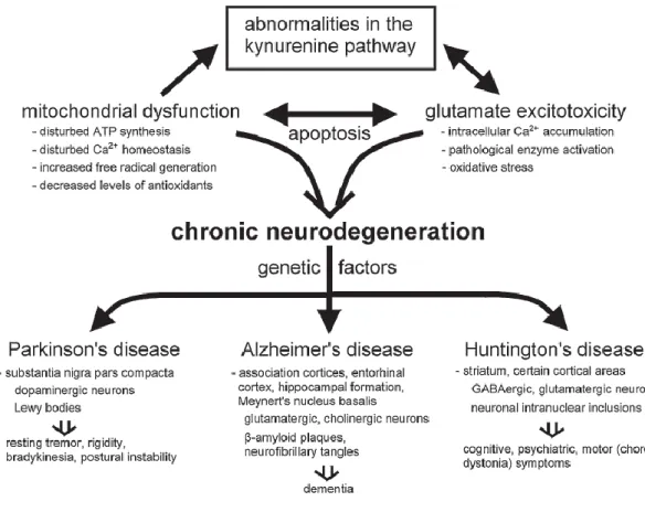
Kynurenic acid (KYNA) was discovered in 1853 by the German chemist Justus von Liebig in dog urine. It was the firstly identified component of the family of “kynurenines”, a group of metabolically related compounds which are derived from the essential amino acid tryptophan. In mammals, the diet represents the main source of tryptophan and the amino acid is metabolized via the so-called kynurenine pathway (KP). Recently, it has been demonstrated that a dysfunction of this pathway is associated with neurodegenerative and other neurological disorders, including psychiatric diseases such as schizophrenia (SZ, Figure 1). In particular, it has been demonstrated that fluctuations in the levels of kynurenines showed discrete effects on the nervous system and immune system (Schwarcz,
2
2004). Specifically, KYNA levels are elevated in the prefrontal cortex (PFC) of individuals with SZ, and normalization of KYNA concentrations in the brain has been suggested as an effective treatment strategy for alleviating cognitive deficits in this mental illness.
In this dissertation, the effects of KYNA on extracellular GABA and glutamate levels in the rat striatum and prefrontal cortex (PFC), will be described. Although the obtained results might be also involved in the neuroprotective action of KYNA, their possible relevance in the ethiopathogenesis of SZ is emphasized.
Figure 1. Kynurenine dysfunction and the common pathways in chronic neurodegenerative disorders (Zadori et al., 2009).
3 1.1. SCHIZOPHRENIA
SZ is amongst the most common of the severe mental illnesses and one of the top ten causes of global disease burden in adults. It is a chronic psychotic disorder with a lifetime prevalence of about 0.7%, primarily affecting adults, and having a peak age of onset in the early twenties in men, and three or four years later in women. Based on converging evidence from a number of research disciplines, it has been generally accepted that both genetic and environmental factors play a significant role in the etiopathogenesis of SZ. However, the exact nature of these two main etiological factors, their pattern of interaction, and their pathogenic mechanisms are poorly understood, despite extensive neurobiological, clinical, genetic, and epidemiological research.
SZ is formally characterized by three distinct categories of symptoms:
positive symptoms or psychotic symptoms refer to hallucinations and delusions, abnormality in the form of thoughts and also atypical psychomotor activity;
negative symptoms like asociality, lack of motivation and abnormality in social interaction;
cognitive symptoms, including disturbances in selective attention, working memory and executive functions.
Even though the positive symptoms are usually the presenting and the most noticeable clinical features of SZ, cognitive deficits are now recognized as a core domain of the disease for a number of reasons (Green, 1996; Gold, 2004; Keefe, 2007). First, it is well demonstrated that cognitive deficits are relatively stable over time and are also independent from psychotic symptoms. Importantly, cognitive dysfunctions have been found throughout the lifetime of the SZ individuals. Furthermore, it has been shown that patient unaffected relatives have similar cognitive deficits, even if milder. Finally, the severity of these deficits is more predictive than the positive symptoms (Green, 1996).
4
1.1.1. Cognitive deficits in schizophrenia and alterations in the prefrontal cortex
It is well established that SZ-related cognitive deficits primarily affect central functions that depend, at least in part, on the integrity of the PFC such as working memory, attention and cognitive flexibility (Floresco et al., 2009; Goto et al., 2010; Volk and Lewis, 2010).
The following part of this paragraph focuses on the main components of PFC neuronal circuitry and their possible roles in the SZ cognitive dysfunctions.
Pyramidal neurons: constitute about 75% of neurons in the cortex. They provide an axon projecting to other brain regions and utilize the excitatory neurotransmitter glutamate acting on the α-amino-3-hydroxy-5-methyl-4-isoxazolepropionic acid receptors (AMPARs) and the N-methyl-D-aspartate receptors (NMDARs) located on their dendritic spines. Several studies have suggested a reduction in the number of excitatory inputs in SZ, even though it has not been yet elucidated the molecular mechanism(s) underlying these structural abnormalities (Krystal et al., 1994).
Interneurons: represent approximately 25% of cortical neurons and they use the inhibitory neurotransmitter γ-aminobutyric acid (GABA). They project locally by an axon and regulate the activity of the pyramidal neurons. Post-mortem studies on the brains of SZ individuals have revealed a reduced level of mRNA encoding GAD67, the enzyme responsible for the majority of GABA synthesis, in a subset of cortical interneurons (Hashimoto et al., 2008).
1.1.1.1. Impaired cortical interneurons: relevance for cognitive dysfunctions in schizophrenia
In the last few years emerged that GABA and glutamate neurotransmitters are critically involved in the pathophysiology of SZ (Coyle, 2004; Lewis et al., 2005; O’Donnell, 2012). In fact, a proper balance between the two amino acid transmitters is necessary for the appropriate activity of the PFC and, especially, adequate cognitive functions (Lewis and Moghaddam, 2006).
5
Several preclinical and clinical studies have shown that local cortical GABA interneurons are critical elements in SZ pathophysiology (Coyle, 2004; Homayoun and Moghaddam, 2006). For example, recent evidence indicates that changes in fast-spiking interneurons are observed blocking the NMDARs (Behrens et al., 2007) and it has also been observed that mice yielded with several signs of a disinhibited cortex after knocking out NMDRs from the calcium-binding protein parvalbumin (PV) interneurons (Belforte et al., 2010), usually predominant in the basket and chandelier subclasses of interneurons (Figure 2).
It has also been suggested that chandelier interneurons may be preferentially involved in the pathophysiology of SZ (Lewis, 1998). Several of the chandelier GABA neurons furnish inhibitory synapses very close to the site action potential generation in pyramidal cells. In this way chandelier interneurons are able to strongly control the excitatory output of the pyramidal neurons that play a pivotal role in working memory and cognitive processes which are altered in SZ (Goldman-Rakic, 1995; Lewis and Anderson, 1995). Furthermore, it has been hypothesized that dysfunctions in the axon terminals of the chandelier GABA neurons could lead to the alterations in GABA neurotransmission observed in SZ patients (Lewis, 1998).
6
Figure 2. Schematic representation of some cortical circuit alterations found in SZ patients and in animal models of this disorder (Marin, 2012).
Finally, recent studies reported an increased expression of GABAA receptor α2
subunits in SZ patients. It has been suggested that the pre- and postsynaptic receptor changes are compensatory responses to deficient GABA release from chandelier neurons (Lewis et al., 2005; Gonzalez-Burgos and Lewis, 2008).
7
1.1.1.2. Deficits in neurons projecting to the prefrontal cortex
It is important to note that inputs from dopaminergic neurons located in the ventral mesencephalon (Figure 3) modulate the activity of both pyramidal and GABA neurons. Thus it is also possible that dysfunctions in dopaminergic signaling contribute to working memory deficits in SZ individuals (Goldman-Rakic et al., 2004). For example it seems that dopaminergic innervation of the PFC is decreased in SZ patients, as indicated by lower levels of expression of tyrosine hydroxylase, the rate limiting enzyme in dopamine synthesis and along with a reduction in the dopamine transporter (Akil et al., 1999). Furthermore, since it has been observed that the inhibition of the dopamine-degrading enzyme catechol-O-methyl transferase (COMT) significantly increases dopamine levels in rats PFC, the availability of extracellular dopamine in the PFC might be reduced in SZ (Tunbridge et al., 2004). It is thought that the hypoactivation that occurs in the PFC may be followed by a compensatory hyperactivation of directly adjacent regions, reproducing a dysfunction in the capacity to engage the functional networks subserving executive functions (Barch, 2005; Glahn et al., 2005; Ragland et al., 2007).
Specifically, numerous studies have revealed marked dysregulations in PFC neurotransmitter systems, involving cholinergic (Mathew et al., 2007; Scarr et al., 2009), glutamatergic (Bauer et al., 2008; Volk and Lewis, 2010), dopaminergic (Guillin et al, 2007) and GABAergic transmission (Volk and Lewis, 2010; Gonzalez-Burgos et al., 2010). In addition, there is a longstanding hypothesis sustaining that the pathophysiology of SZ is associated to dysfunction of selective corticostriatal circuits (Kleist, 1960). Several studies have reported the presence of structural abnormalities in the basal ganglia of SZ individuals (Buchsbaum, 1990) and also neurochemical imbalances in the corticostriatal circuits (Carlsson and Carlsson, 1990).
8
Figure 3. Schematic illustration of functional deficits in PFC circuitry in SZ (Lewis and Sweet, 2009).
9 1.2. BASAL GANGLIA
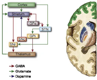
The basal ganglia are a group of subcortical nuclei, where the major input nucleus is represented by the striatum, which is made up of the caudate and putamen. The basal ganglia are traditionally involved in the control of the motor movement, but also in a variety of associative, cognitive and mnemonic functions (Saint-Cyr et al., 1995; Graybiel, 1998; Lalonde and Botez-Marquard, 1997; Yin and Knowlton, 2006; Simpson et al., 2010). The major input to the striatum is derived from the cortex, while the globus pallidus, the subthalamic nucleus and the ventral tegmental area represent the main output nuclei of the basal ganglia. Furthermore, these output structures project to the principal target of the basal ganglia, the thalamus. Finally the thalamus, complete a loop involving the cortex, the striatum and the thalamus itself, projecting back to the cortex (Figure 4) (Alexander et al., 1986).
Figure 4. Simplified picture of the basal ganglia circuitry.
On the basis of the classical view of basal ganglia circuitry, it is reported that the corticostriatal projections are processed within the striatum and then the “transformed signal” is transmitted to the output nuclei of the basal ganglia (Bolam et al., 2000). Thus, basal ganglia are primarily involved in action selection, that is, the decision of which of several possible behaviors to execute at a given time.
10
Dysfunctions in basal ganglia activity play a central role in a number of neurological conditions, including several movement disorders, such as Parkinson's and Huntington's diseases. In fact, these two neurodegenerative pathologies specifically target the basal ganglia. The most important neuropathological alteration in Parkinson's disease is the loss of dopaminergic neurons of the substantia nigra (SN), which causes a severe depletion of striatal dopamine. At variance, the pathological hallmark of Huntington's disease is a massive atrophy of the striatum due to the degeneration of striatal projection neurons, striatal interneurons being relatively spared in the disease.
Studies conducted in humans and non-human primates have demonstrated that the striatum plays a crucial role in more complex forms of behaviours, including decision making and executive function, in addition to modulating motor control and motor learning (Graybiel, 2008). Importantly, it has been shown that lesions performed in the dorsomedial striatum influence spatial working memory in the same way that lesions of the PFC do (Mair et al., 2002; Voorn et al., 2004). Furthermore, the role of the striatum in attentional processes and cognitive control has also been suggested in humans. The measure of the attentional set shifting, in fact, activates the caudate nucleus in healthy human individuals (Rogers et al., 2000). So it has been suggested that working memory and other executive functions are mediated by both the PFC and striatum, working in concert (Simpson et al., 2010).
Particularly, the striatal subregions are involved in the learning processes through at least three distinct circuits:
a circuit involving associative cortical regions like medial PFC connections to the dorsomedial striatum/caudate;
sensory and motor cortex circuitry, connected to the dorsolateral striatum/putamen;
limbic circuitry (i.e. limbic cortex, basolateral amygdala and hippocampus) connections to the ventral striatum/nucleus accumbens (Yin and Knowlton, 2006; Yin et al., 2008).
Several studies with brain damaged patients have reported that, by measuring implicit learning using the serial reaction time test, cognitive performance critically
11
depends on the neostriatum function. In fact patients with damage in the medial temporal lobe were able to learn, while patients with damage in the basal ganglia were cognitively impaired (Knowlton et al., 1994; 1996).
1.2.1. Corticostriatal projections and synaptic targets
It is well known that cortical signals are transmitted within the basal ganglia through the direct and indirect pathways (Albin et al., 1989; DeLong, 1990). Briefly, in the direct pathway cerebral cortical input to the striatum activates the inhibitory projection neurons in the striatum and then the information is transmitted directly to the output nuclei. In the indirect pathway the information coming from the cortex are then transmitted to the output nuclei via the globus pallidus and the subthalamic nucleus (Shink et al., 1996).
The striatum contains a single class of projections neurons, known as medium spiny neurons (MSNs), constituting the 90-95% of the total population of the striatal neurons (Kemp and Powell, 1971), utilizing GABA as their main neurotransmitter. Furthermore, MSNs receive GABAergic synaptic input from striatal cholinergic interneurons, less numerous than MSNs, but able to form extensive connections within the striatum (Tepper et al., 2004).
An equal amount of MSNs belongs to the direct and indirect pathways (Gerfen et al., 2004), in which MSNs express dopamine receptors. It is well known that striatal dopamine released by nigro-striatal neurons, exerts tonic modulation on either MSNs or glutamatergic terminals mainly coming from the motor areas of the cerebral cortex and from the thalamus (Cepeda et al., 1993; Flores Hernandez et al., 1997; Bamford et al., 2004; Centonze et al., 2004; Yin and Lovinger, 2006). The other striatal neurons can be subdivided into three groups: burst-firing GABAergic interneurons, also expressing somatostatin, neuropeptide Y and nitric oxide; GABAergic interneurons expressing PV and cholinergic interneurons (Dani et al., 2007). Several studies have demonstrated that the cortex sends the major inputs to PV-positive GABA interneurons (Lapper et al., 1992; Bennett and Bolam, 1994).
The activation of corticostriatal neurons is followed by the activation of the AMPARs and NMDARs, mainly localized within the synapse (Bernard et al., 1997; Kawaguchi, 1997; Bernard and Bolam, 1998), leading then to the neuronal
12
depolarization (Wilson, 1993; Kita, 1996). Noteworthy, several studies reported that the PV-positive GABA interneurons receive cortical input and are the principal mediators of inhibition in the striatum, providing a feed-forward inhibition of cortical information to MSNs. Even though the number of PV-positive GABA interneurons is low, they are in a position to strongly control the activity of neurons, giving rise to the output of the striatum, by the direct or indirect pathway (Bolam et al., 2000).
1.3. KYNURENINE PATHWAY AND NEUROACTIVE KYNURENINES
The discovery of the endogenous compound KYNA, more than 150 years ago and the successive understanding of its biosynthesis and chemical structure led to a series of discoveries that turned out to have significant implications for the neurosciences. In fact, it was successively demonstrated that KYNA represented the first of the "kynurenines", a group of metabolically related compounds derived from the essential amino acid tryptophan. Biochemical studies during the first part of the 20th century elaborated the enzymatic steps linking the individual members
of the so-called KP, which was found to account for >90% of peripheral tryptophan metabolism in mammals.
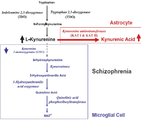
The KP started from tryptophan oxidative ring opening and subsequent degradation of the reaction compound. The final product of this metabolic cascade (Figure 5) is the NAD+ coenzyme, which plays a crucial role in many fundamental biological processes (i.e. redox reactions necessary for mitochondrial function).
13
Figure 5. Tryptophan degradation via KP (Wonodi and Schwarcz, 2010).
This catabolic cascade originates the so-called “kynurenines” deriving directly or indirectly from L-kynurenine (L-KYN), the major primary degradation product of tryptophan. Although the majority of the kynurenines harbor neuroactive properties, the triad of KYNA, quinolinic acid (QUIN) and 3-hydroxy-L-kynurenine (3-HK) is traditionally referred to as ‘the neuroactive kynurenines’.
KYNA, one of these metabolites, has long been recognized as a competitive, broad-spectrum antagonist of glutamate receptors. At high, non-physiological, micromolar concentrations, KYNA inhibits NMDARs, kainate receptors and AMPARs (Perkins and Stone, 1982). The action of KYNA on AMPARs has been demonstrated to be dual in a dose-dependent manner, evoking facilitation at lower concentrations, and exerting neuroinhibition at higher concentrations (µM–mM) (Prescott et al., 2006; Rozsa et al., 2008). It has been shown that high concentrations of KYNA display anticonvulsant properties and provide an excellent protection against excitotoxic injury (Foster et al., 1984).
14
It is well known that at much lower (nM) concentrations than those necessary to bind AMPARs, KYNA acts as an antagonist of the glycine allosteric site on the NMDAR and as a non-competitive antagonist of the α7 nicotinic acetylcholine receptor (α7nAChR) (Hilmas et al., 2001), both of which are critically involved in physiological processes underlying learning, memory and other manifestations of synaptic plasticity (Hilmas et al., 2001; MacDonald et al., 2006; Albuquerque et al., 2009).
In the past few years additional targets of KYNA have been identified, such as the former orphan G protein-coupled receptor (Wang et al., 2006) and the aryl hydrocarbon receptor, a nuclear receptor able to recognize aromatic hydrocarbons and 2,3,7,8-tetrachlorodibezo-p-dioxin (DiNatale et al., 2010). Although it has been recently proposed that KYNA is the endogenous ligand for GPR35 (Berlinguer-Palmini et al., 2013), only the α7nAChR as so far been verified as a “bona fide” KYNA receptor in the brain (Hilmas et al., 2001; Grilli et al., 2006; Wu et al., 2010). Other studies have also demonstrated antioxidant properties of KYNA, linked to its ability to scavenge hydroxyl superoxide anion and other free radicals (Lugo-Huiltron et al., 2011).
3-HK, another product of L-KYN degradation and a biological precursor of QUIN is present in the brain at nanomolar concentrations and can generate free radicals and elevate the oxidative stress levels, causing neuronal damage (Okuda et al., 1998). Furthermore, it has also been observed after neuronal exposition to both 3-HK and QUIN a considerable potentiation of excitotoxicity (Guidetti and Schwarcz, 1999). So far no physiological role of 3-HK in the brain has been established and it seems likely that this metabolite does not interact with a specific recognition site (Eastman and Guilarte, 1989).
QUIN possesses excitatory properties due to its capability to directly and selectively stimulate NMDARs, as demonstrated by using selective NMDAR antagonists. In 1978, Lapin observed convulsions after an intracerebroventricular injection of QUIN in mice. This provided the first indication that KP metabolites exert central effects (Stone and Perkins, 1981).
As shown in figure 5, the tryptophan metabolism also includes other metabolites, like 3-hydroxyanthranilic acid (3-HANA) and anthranilic acid; however, until now, direct effects of these metabolites on neuronal activity (Stone, 1993) have not
15
been demonstrated. Though there are only few examples showing that several kynurenines exhibit pro- and anti-oxidant activities in vitro (Giles et al., 2003).
1.3.1. Enzymes involved in the kynurenine pathway
As illustrated in Figure 5, the KP is initiated by the convertion of tryptophan to N-formylkynurenine by indoleamine dioxygenase (IDO) and tryptophan 2,3-dioxygenase (TDO). N-formylkynurenine is then degraded to the pivotal metabolite kynurenine by formamidase. Several studies have reported that the levels of these enzymes in the brain are normally lower than in the peripheral organs. Kynurenine undergoes irreversible transamination to form KYNA by four kynurenine aminotransferases (KATs). So far it has been established that at least two of these enzymes are present in the brain. In the rat brain, KAT I, occurring in a cytosolic and mitochondrial form, could have a major function in cortical maturation. This enzyme is, in fact, selectively expressed in neurons in the perinatal period (Csillik et al., 2002). In the mouse, instead, KAT II is the major enzyme that catalyzes KYNA formation (Yu et al., 2004). Nowadays KAT II is thought to be the main biosynthetic enzyme of KYNA in the mammalian brain (Guidetti et al., 2007). A potent and selective inhibitor of KAT II, (S)-4-ethylsul-fonylbenzoylalanine (ESBA; Pellicciari et al., 2006; Figure 6), has been synthetized.
Figure 6. (S)-4-ethylsul-fonylbenzoylalanine hydrochloride.
Biological assays conducted in vitro showed that ESBA inhibits KAT II obtained from partially purified rat liver with an IC50 value of 6.1 mM. In vivo studies support
16
these results, demonstrating that the administration of ESBA lowers the extracellular levels of KYNA in the rat striatum (Amori et al., 2009).
A second branch of the KP is characterized by the kynurenine 3-monoxygenase (KMO) and kynureninase, responsible for the synthesis of 3-HK and anthranilic acid, respectively (Chiarugi et al., 1995). Anthranilic acid is then converted to α-amino-ω-carboxymuconic acid semialdehyde that under physiologically conditions spontaneously rearranges to form QUIN. Finally, QUIN is metabolized to nicotinic acid mononucleotide subsequently degraded to the end product NAD+ (Braidy et al., 2011).
1.3.2. Targets of kynurenic acid
As previously reported, it is generally assumed that KYNA modifies neuronal function because it antagonizes the glycine site of the NMDARs and/or the neuronal α7nAChRs. However, whether the basal levels of KYNA found in brain extracellular spaces are sufficient to interact with these targets is not fully cleared, especially for NMDARs. Another reported target for KYNA is GPR35, an orphan receptor negatively coupled to Gi proteins. GPR35 is expressed both in neurons and other cells (including glia, macrophages and monocytes). KYNA affinity for GPR35 in native systems has not been clarified but current data in modified expression systems suggest that it is quite low. However, it has been proposed that by interacting with GPR35, KYNA may decrease glutamate release in brain and pro-inflammatory cytokines release in cell lines. The inhibition of inflammatory mediator release from both glia and macrophages may explain why KYNA has analgesic effects in inflammatory models (Moroni et al., 2012). Finally, KYNA has been reported as an agonist of aryl hydrocarbon receptor, a nuclear protein involved in the regulation of gene transcription and able to cause immunosuppression after binding with dioxin. Thus, KYNA has receptors in the nervous and the immune systems and may play interesting regulatory roles in cell function. However the roles of GPR35 and aryl hydrocarbon receptor in mediating endogenous KYNA effects remain to be elaborated. Thus, in the following paragraphs the general aspects of α7nAChRs and NMDARs as KYNA molecular targets will be briefly described.
17
1.3.2.1. Nicotinic receptor
Nicotinic acetylcholine receptors (nAChRs) belong to the superfamily of ligand-gated ion channels and on the basis of their subtypes along with their dendritic, somal, axonal, presynaptic and postsynaptic localization, play a different role in the central nervous system (CNS). For example, it is well established that presynaptically and preterminally located nAChRs increase neurotransmitter release, while postsynaptic and non-synaptic nAChRs generally mediate excitation (Dani and Bertrand, 2007).
nAChRs are constituted from five transmembrane subunits, arranged symmetrically around a central pore. Each subunit comprises four transmembrane domains with both the N- and C-terminus located extracellularly; there are two acetylcholine (ACh) binding sites per receptor (Figure 7).
Figure 7. A schematic representation of the nAChR structure (Karling, 2002).
The structural features of the ligand binding site and the specific amino acid interactions establish the pharmacological properties of a specific nAChR subtype along with the specific amino acid interactions determining conformational transitions (Jensen et al., 2005).
In addition, there are certain ligands that modulate conformational transitions and functions by binding to the subunits or to the interfaces between subunits at positions different from the agonist binding site, leading to the phenomena called
18
as allosteric modulation. It is well known that positive modulators potentiate nAChRs function, while negative modulators inhibit nAChRs function. Notably, the α7nAChRs, one of the well described KYNA target, are good candidate for allosteric modulators because of their unique structural organization, low sensitivity to acetylcholine and fast kinetics. Among the positive allosteric modulators, it is well known that galantamine, physostigmine and codeine, effectively increase α7nAChRs activation at subsaturating agonist concentrations (Pereira et al., 2002).
The most studied nicotinic role in CNS regards the modulation of neurotransmitter release by presynaptic nAChRs. It is well demonstrated that the activation of nAChRs is generally followed by an increase of the release of several neurotrasmitters (McGehee and Role, 1995; Albuquerque et al., 1997; Jones et al., 1999; Sher et al., 2004). For example, exogenous application of nicotinic agonists cause an augmentation in the release of glutamate, GABA and acetylcholine, as well as dopamine and serotonin, while nicotinic antagonists often diminish the release of these neurotransmitters. Particular functional studies revealed that in the rat hippocampus the α7 subunit is at nearly every synapse in the CA1 stratum radiatum (Fabian-Fine et al., 2001); in addition, this subunit was also found on both glutamatergic and GABAergic presynaptic terminals.
The nicotine-induced increase of neurotransmitter release is a consequence of the presynaptic nAChRs activation that initiates a direct and indirect intracellular calcium signal (McGehee and Role, 1995; Wonnacott et al., 1997). Thus, the activity of the presynaptic nAChRs increases intraterminal calcium and contributes to the increased neurotransmitter release. In addition, nicotinic stimulation, increasing glutamate release, promotes synaptic plasticity (Wonnacott et al., 1997; Ge and Dani, 2005).
It has been found that nicotinic receptors are also distributed to preterminal, axonal, dendritic and somatic locations (Lena et al., 1993; Zarei et al., 1999).
1.3.2.2. Influences of nAChRs on cortical functions: relevance for cognition
Wide areas of the brain are innervated by diffuse cholinergic afferents. Cholinergic innervation to the cortex originates from neurons located within basal forebrain nuclei (Wolf, 1991). In addition, a high number of small varicosities that contain
19
synaptic vesicles does not make synaptic contacts with adjacent neurons, thus producing volume transmission where acetylcholine and choline reach locations distant from the release site (Descarries et al., 1997; Vizi, 2000).
On the basis of their localizations, nAChRs are supposed to modulate cortical activity in a multiple way. For example, it has been observed that activation of nAChRs on distal apical dendrites causes a depolarization of the cell and promotes action potential firing. On the other hand, if the activation occurs in the proximal dendrites nearer to the cell soma, it causes a reduction of the membrane impedance and a shunt of the signals incoming from the apical tuft (Dani et al., 2007).
It is well known that nAChRs are implicated in a wide range of neuronal diseases and mental illnesses (Lena and Changeaux, 1998; Picciotto and Zoli, 2002; Dani and Harris, 2005). Several studies have reported that in Alzheimer’s disease, Parkinson’s disease and Down syndrome nAChRs are reduced or nicotinic mechanisms are impaired, thus contributing to cognitive dysfunctions (Picciotto and Zoli, 2002; Perry et al., 2000). Notably, it has to take into account that the variable age-related decline of cholinergic transmission and decrease in nAChRs numbers influenced the results.
It has been observed that, generally, nicotinic agonists improve certain forms of memory and nicotinic antagonists and/or cholinergic lesions impair memory. It has been reported in animal studies that acute or chronic nicotine administration, ameliorates working memory. In addition, studies performed in humans using nicotinic agonists have demonstrated an improvement in learning and memory (Levin et al., 2006). Importantly, the responses to nicotinic agonists are highly dose-dependent and those compounds do not have effects at the lowest concentration tested (Dani et al., 2007).
1.3.2.3. nAChRs and schizophrenia
Preclinical and clinical studies provided evidence linking SZ to a deficiency of α7nAChRs-mediated neurotransmission (Deutsch et al., 2005; Martin and Freedman, 2007; Jones et al., 2011). It has been observed that SZ individuals and their biological relatives show a deficient gating of P50 auditory event-related potential and the locus on chromosome 15, which codes the gene for the
20
α7nAChRs subunit (i.e. CHRNA7) (Freedman et al., 1997). Specifically, P50 gating alludes to a paradigm in which two acoustic clicks are given about 500 msec apart, resulting in a reduced P50 response to the second stimulus (Green, 2006).
Furthermore, post-mortem studies examining the brains of SZ individuals have also reported a reduction of the α7nAChR protein levels in the dentate gyrus and CA3 region of the hippocampus along with a degree of global cognitive deficits in these individuals (Martin-Ruiz et al., 2003). In addition, recent studies have also identified significant connections between brain areas of α7nAChRs expression, their impact on other neurotransmitters and how these changes might be linked to the dopamine and glutamate theory of SZ.
Several findings have reported that α7nAChRs are found on midbrain dopamine cells bodies of the ventral tegmental area, subthalamic nuclei, presynaptically on dopamine terminal regions in the striatum, nucleus accumbens and PFC. Moreover α7nAChRs are located on glutamate and GABA neurons that project into dopaminergic regions and terminals (Quik et al., 2000; Dani and Bertrand, 2007; Machaalani et al., 2010). Additionally, dopamine release can be enhanced in several brain regions such as the striatum, ventral tegmental area, nucleus accumbens and PFC, as a result of the α7nAChR activation (Sydserff et al., 2009; Thomsen et al., 2010; Castner et al., 2011). Other studies also reported that α7nAChR activation releases GABA from GABAergic interneurons (Albuquerque et al., 1998; Frazier et al., 1998) and then GABA, acting on GABAB receptors,
leads to a reduction of striatal glutamate release that may, in turn, results in the increased dopamine release (Bencherif et al., 2012). Moreover, it has recently been reported that α7nAChRs modulate glutamate release in the PFC of rats. Notably, kynurenine (the precursor of KYNA) and α-bungarotoxin are able to block choline-induced stimulation of glutamate release (Kondardsson-Geuken et al., 2010).
It has been recognized that interactions between α7nAChRs and NMDARs have been implicated in cognitive deficits observed in SZ (Timofeeva and Levin, 2011). In a recent paper it has also been hypothesized that interactions between α7nAChRs and NMDARs modulate cortical glutamatergic development. In addition, an altered α7nAChR-mediated control of synaptic NMDARs, due to the
21
loss of the α7nAChRs, may produce permanent changes contributing to cortical impairment present in SZ patients (Lin et al., 2014).
1.3.2.4. N-methyl-D-aspartate receptor
Several studies have shown that hypofunction of the NMDARs has implications in the pathophysiology of SZ. NMDARs are heteromeric protein complexes constituted of at least one NR1 along with several combinations of the NR2 and/or NR3 subunits (Cull-Candy et al., 2001).
Regarding the activation of the NMDARs, it is well known that magnesium blocks the pore of the NMDAR channel, at resting membrane potential. Moreover the NMDAR includes a glutamate recognition site on the NR2 subunit and a glycine or D-serine modulatory site on the NR1 subunit (Johnson and Ascher, 1987; Clements and Westbrook, 1991). In order to activate the NMDAR, it is fundamental that the D-serine/glycine site has to be occupied by glutamate (Figure 8) (Clements and Westbrook, 1991). KYNA acts as an antagonist of the glycine allosteric site on the NMDAR.
22
Notably, NMDAR has the property of being both voltage-dependent and ligand-gated, giving thus the ability to NMDAR to act as a coincidence detector for presynaptic activity and post-synaptic activity. When the NMDAR is activated, the calcium influx is permitted through the channel, stimulating intracellular signaling cascades that can then influence synaptic plasticity, like long-term potentiation (LTP), underlying several types of learning and memory formation (Nicoll, 2003).
1.3.2.5. Kynurenic acid, α7nAChRs and NMDARs
Since endogenous KYNA is able to inhibit both the NMDARs and the α7nAChRs, it is reasonable to hypothesize that it might also be involved in cognitive functions. In addition, as described above, both of these receptors are known to be critical for physiological processes involving synaptic plasticity and more specifically memory and learning (MacDonald et al., 2006; Albuquerque et al., 2009). Thus, fluctuations in KYNA levels could influence these and related phenomena. Several studies reported that increases in brain KYNA levels, induced by the administration of kynurenine, lead to cognitive impairments in experimental animals, thus supporting this concept (see below). For example, increased KYNA levels disrupt prepulse inhibition and auditory sensory gating (Shepard et al., 2003; Erhardt et al., 2004) and also cause deficits in contextual learning and memory (Chess and Bucci, 2006; Chess et al, 2007). Thus, dysfunction of α7nAChRs and NMDARs in SZ may be induced or exacerbated by disruptions in the KP. These deleterious effects of KYNA are therefore very interesting in view of the fact that SZ patients present increased KYNA levels in brain and cerebrospinal fluid (CSF) (Erhardt et al., 2001; Schwarcz et al., 2001; Miller et al., 2006; Sathyasaikumar et al., 2011).
23
1.3.3. Kynurenic acid metabolism and regulation in the brain
In order to provide additional information about the cellular localization of KYNA and QUIN in cerebral tissues, several immunocytochemical and lesion studies have been performed. The results clearly indicate that glial cells, rather than neurons, harbor the enzymatic machinery for the biosynthesis of brain kynurenines. Furthermore, all enzymes involved in the KP are primary contained in astrocytes and microglial cells (Guidetti et al., 1995; Heyes et al., 1996; Schwarcz et al., 1996; Guillemin et al., 2001). In the mammalian brain, the metabolites of the KP are normally present in the nanomolar to micromolar concentration range (Turski et al., 1988; Baran and Schwarcz, 1990; Smythe et al., 2003). The cerebral KP is mainly driven by the pivotal metabolite blood-born kynurenine, which readily enters the brain from the circulation (Figure 9) using the large neutral amino acid transporter (Fukui et al., 1991). Once in the brain, kynurenine is taken up by astrocytes and presumably microglial cells (Speciale and Schwarcz, 1990).
Notably, KYNA synthesis occurs in the astrocytes, as they do not appear to contain kynureninase. Newly produced KYNA is then rapidly released into the extracellular milieu where it has access to neuronal α7nAChRs and NMDARs. On the contrary, very poor KAT activity is present in the microglial cells and, therefore, the QUIN branch of the pathway is favored in these elements (Guillemin et al., 2001).
Because of their polar nature and as so far there are no evidence of efficacious transport processes, KYNA and QUIN very poorly cross the blood-brain barrier (Fukui et al., 1991) and, thus, they must be locally formed within the brain to exert their central effects (Schwarcz et al., 2012).
The intracellular kynurenine concentration determines the enzymatic KYNA formation. Furthermore, the release of KNYA is also influenced by several factors. For example, it has been demonstrated that KYNA formation, in the presence of depolarizing stimuli such as high extracellular K+ concentration, glutamate receptor agonists or altered cellular energy metabolism, is reduced and hence extracellular KYNA levels. It is noteworthy that in neuron-depleted brain tissue these effects are not observed, suggesting that the glial KYNA synthesis in the brain is controlled by neuronal activity (Gramsbergen et al., 1997). Some studies have also demonstrated that the systemic administration of dopaminergic (Rassoulpour et al., 1998) or cholinergic (Hilmas et al., 2001) agents induced extracellular
24
fluctuations in KYNA levels in the brain, but not in the periphery. It has also been observed that several of these effects are lost in absence of neurons. Thus KYNA production in, and release from, astrocytes depend on neuronal signals (Gramsbergen et al., 1997).
Figure 9. Dynamics of the cerebral kynurenine pathway in the brain (Schwarcz et al., 2012).
1.3.4. Kynurenic acid and neurotransmission
As shortly mentioned in the previous paragraphs, it appears that SZ is a heterogeneous disease, also characterized by various neurotransmitter imbalances. Namely, corticostriatal neuron loops greatly involved in cognitive skills are functionally impaired. Thus, it becomes relevant that the reported disruption of the KP of tryptophan degradation, in particular the alterations in KYNA levels may cause an exacerbation of the neurotransmitter specific dysregulations observed in SZ (Erhardt et al., 2001; Schwarcz et al., 2001 Miller et al., 2006; Wonodi et al., 2011). For example, it has been demonstrated that extracellular dopamine levels in rat striatum are decreased after intrastriatal infusion of nanomolar concentrations of KYNA. This effect is mediated by α7nAChRs located on the glutamatergic nerve terminals, rather than the NMDARs, since the local perfusion
25
of KYNA in combination with choline, the selective α7nAChRs agonist, resulted in a prevention of the decrease in extracellular levels of dopamine induced by KYNA (Rassoulpour et al., 2005). In addition, the same study revealed that after treatment with a non-specific astrocyte poisoning leading to an interference with KYNA synthesis, extracellular dopamine levels in the striatum were increased. Thus, dopaminergic tone is regulated by the fluctuations in the astrocytic production of KYNA. Similarly, KYNA induce a reduction in striatal glutamate, first targeting α7nAChRs on glutamatergic nerve terminals. In addition, these results are in line with other studies showing that low concentrations of galantamine, a positive allosteric modulator of α7nAChRs (Santos et al., 2002; Samochocki et al., 2003), prevent the KYNA-induced reduction of extracellular dopamine levels, in the same way of the choline, the direct α7nAChRs agonist (Rassoulpour et al., 2005).
As cognitive deficits affecting SZ individuals involve several central areas, the effects of KYNA have also been evaluated in other brain regions such as PFC and hippocampus. It has been shown that KYNA at nanomolar concentrations induced a reduction in the extracellular glutamate levels, not only in the striatum, but also in the rat PFC (Carpenedo et al., 2001; Rassoulpour et al., 2005; Grilli et al., 2006; Wu et al., 2007, 2008). On the other hand, reductions in the endogenous KYNA synthesis induced an increase in the extracellular levels of glutamate (Wu et al., 2006; 2008) suggesting that KYNA bi-directionally modulate glutamatergic transmission by endogenously formed KYNA. In addition, other studies reported that endogenous KYNA is also able to modulate basal levels of PFC acetylcholine in the rat cortex. Notably, the perfusion of ESBA (Pellicciari et al., 2006), the selective inhibitor of the astrocyte enzyme KAT II which catalyses the KYNA formation from its bioprecursor L-KYN, reduced cortical KYNA levels and increased cortical acetylcholine levels (Zmarowski et al., 2009). Furthermore, the functional effects of nanomolar KYNA concentrations can be replicated by specific α7nAChR ligands (i.e. methylcaconitine, α-bungarotoxin) (Rassoulpour et al., 2005; Lopes et al., 2007). On the contrary, these effects are not observed using selective antagonist of the glycineB site of the NMDARs (Hilmas et al., 2001).
26
1.3.5. Possible relevance of astrocyte-derived kynurenic acid
Since astrocytes are responsible for the de novo synthesis of KYNA from L-kynurenine in the brain, these cells deserve consideration as modulators of α7nAChRs function and consequently also cholinergic transmission (Zmarowski et al., 2009).
For much of the past century, astrocytes were considered just as a subtype of glial cell that contacts synapses and the vasculature, providing metabolic support to the neurons. Recent experimental evidence suggests that in addition to being essential supporters of neuronal function, astrocytes also closely interact with neurons and participate in the regulation of synaptic neurotransmission. It has been proposed that synapses are tripartite, where the astrocytic process is associated with the presynaptic and postsynaptic elements of the synapses. In addition, it has also been demonstrated that astrocytes possess a large array of neurotransmitter receptors (Araque et al., 1999; Halassa et al., 2007) and they can also release chemical transmitters such as, among others, glutamate, GABA and glycine. In addition, impairments of the bi-directional communication between astrocytes and neurons, lead to an abnormal neurotransmission and information processing. Thus, it has been hypothesized that the astrocytes could also be involved in various manifestations of brain dysfunctions (Kettenmann and Ransom, 2004; Tian et al., 2005). Importantly, astrocytes may also regulate synaptic communication through volume transmission, the diffusion through the brain extracellular fluid of neurotransmitters released at points that may be remote from the target cells with the resulting activation of extrasynaptic receptors (Agnati and Fuxe, 2000).
Biochemical and immunocytochemical studies, demonstrated that astrocytes are the major source of KYNA in the mammalian brain (Ceresoli-Borroni, et al., 1999; Rejdak et al., 2003; Melendez-Ferro et al., 2005). Thus, in the brain, changes in extracellular KYNA concentrations reflect similar fluctuations in intracellular KYNA formation, determined by intra-astrocytic control mechanisms (Schwarcz and Pellicciari, 2002). These regulatory events are likely to influence the neuromodulatory effects of astrocyte-derived KYNA in the normal and diseased human brain. For example, it has been reported that fluctuations in the astrocytic production of KYNA, through indirect local actions possibly involving volume transmission, inversely regulate striatal dopaminergic tone. Such a mechanism
27
may profoundly influence dopamine function under physiological and pathological conditions (Wu et al., 2007). From a pathological point of view, it has been proposed (see above) that astrocytes lack kynurenine-hydroxylase so that large amounts of kynurenine KYNA are produced, while minor amounts of QUIN are normally synthesised. This suggests that astrocytes alone are neuroprotective by minimising QUIN production and maximising synthesis of KYNA. However, kynurenine added to macrophages led to significant production of QUIN. Thus, in the presence of macrophages and/or microglia, astrocytes could be neurotoxic by producing large concentrations of kynurenine that can be metabolised by neighbouring monocytic cells to QUIN (Guillemin et al., 2001).
1.3.6. Kynurenine pathway and brain dysfunctions
Kynurenines have become recognized as key players in the mechanism leading to neuronal damage, neurodegenerative pathologies and neuroimmunological disorders (Table 1). As already reported, a link between endogenous kynurenines and excitotoxic phenomena has been suggested (Vecsei et al., 2012). In addition, it is now generally accepted that a dysfunction of the KP metabolism may have untoward effects on brain functions (Schwarcz et al., 2012). However, the exact nature of the cellular and molecular events underlying these effects is not yet definitively established. Among other hypotheses, it has been proposed that some pathologies of the CNS could be due to an imbalance of KYNA, QUIN and 3-HK alone or in concert (Schwarcz and Pellicciari, 2002; Schwarcz et al., 2012; Szalardy et al., 2012; Vecsei et al., 2012). Furthermore, several experimental animal studies have also investigated the possible secondary role of the KP, as mediator of central dysfunctional states. Thus, it has been demonstrated that in presence of pathogenic events, kynurenine brain levels are often abnormal (Stone, 2002).
28
Table 1. Correlation between neuroactive kynurenines and central nervous system pathologies (Schwarcz and Pellicciari, 2002).
For example, it has been observed that substantial elevations of 3-HK and QUIN levels occur in response to the activation of glial cells or when macrophages infiltrate the brain during immunological compromises. Since the KAT activity is very low in microglia, the increases in 3-HK and QUIN come with relatively modest changes in KYNA formation. Furthermore, has not so far been consistently seen a reduction in the brain levels of neuroactive kynurenines after injurious events. This could be explained by the fact that if localized neuroactive kynurenine level reductions take place, they are probably obscured by the upregulation of kynurenine synthesis in reactive glial cells (Schwarcz and Pellicciari, 2001).
It is noteworthy that the fundamental basis of the KP metabolism characterized in rodents and other mammals might be also applied to humans. Numerous post-mortem studies have investigated about the disposition of kynurenines in the human brain along with analysis in serum and CSF. These studies revealed that almost always diseases affecting the human brain shown KP-related changes that parallel those seen in relevant animal models (Stone, 2001).
However, not all disease-linked changes in cerebral kynurenine metabolism in humans can be easily associated to animal studies. For example, so far, it is not clear why in the end-stage of Huntington’s disease KYNA levels are markedly reduced (Beal et al., 1990) or why in SZ KYNA levels are elevated in the cortex (Schwarcz et al., 2001).
29 1.3.7. Kynurenic acid and schizophrenia
Clinical studies have reported that KYNA concentrations were significantly increased in PFC and CSF of SZ patients (Erhardt et al., 2001; Schwarcz et al., 2001). Furthermore, it has also been demonstrated that the upregulation of KYNA levels in SZ was associated with elevations in the tissue levels of L-kynurenine, the KYNA’s immediate bioprecursor (Figure 8) (Schwarcz et al., 2001).
It was originally thought that the high levels of L-kynurenine seen in SZ patients could be a direct consequence of its increased synthesis catalyzed by either TDO or IDO, based on the evidence that both of them are dysregulated in inflammatory and immune sensitization events. However, recent studies have demonstrated that even though the expression of TDO gene is significantly increased in SZ patients, these dysregulations seem to be exacerbated in individuals with additional risk genes. Furthermore, brain IDO gene expression seems to be normal in individuals affected by SZ. Thus, more recently a different mechanism possibly involved in the observed increased KYNA levels in SZ patients has been proposed. In particular, it has been suggested that a reduction in KMO activity in the periphery and/or in the brain of SZ subjects could lead to abnormally high cortical concentrations of L-kynurenine and, subsequently, to high KYNA levels. In fact, KMO, the enzyme converting L-kynurenine to 3-HK, shows much higher activity in peripheral tissues than in the brain. Thus, it seems likely that under certain physiological conditions or during pathological events, an increase of L-kynurenine levels might overstep the catabolic capacity of KMO, thus leading to brain KYNA formation (Figure 10; Wonodi and Schwarcz, 2010).
30
Figure 10. Kynurenine pathway in schizophrenia (Wonodi and Schwarcz, 2010).
The high concentrations of brain KYNA levels in the PFC of SZ individuals, may be clinically relevant since it is well established that hypofunction of both NMDAR and the α7nAChR are implicated in SZ pathophysiology and, in particular, in the cognitive deficits associated with the disease.
31
2. AIM
Dysregulations of the KP pathway of tryptophan degradation, causing hyper- or hypofunction of active metabolites, have been associated with several CNS disorders, as widely described in the Introduction. In particular KYNA, a neuroactive metabolite of the KP, has received in the last decades considerable attention from neurobiologists and neuroscientists. The reasons of this interest derive from the observations that even relatively low fluctuations in brain KYNA levels have pronounced consequences, influencing neurotransmission and affecting cognitive and motor behaviors in experimental animals (Schwarcz et al., 2012). Consciousness of these bi-directional effects has led to the hypothesis that endogenous KYNA is a significant neuromodulator in the mammalian brain. Notably, this role likely extends to pathological situations, which frequently present with significant changes in brain KYNA levels. Examples include catastrophic neurodegenerative disorders (e.g. Huntington’s disease and Alzheimer’s disease) as well as psychiatric diseases (e.g. SZ) (Schwarcz et al., 2012; Szalardy et al., 2012). Therefore, understanding the nature and characteristics of the remarkable neuromodulatory properties of KYNA have implications for brain physiology and pathology alike.
Previous studies demonstrated that increased KYNA concentrations reduce extracellular dopamine and glutamate levels in distinct brain regions such as the striatum, the PFC and the hippocampus (Carpenedo et al., 2001; Rassoulpour et al., 2005; Grilli et al., 2006; Wu et al., 2010; Banerjee et al., 2012) and these effects have been associated to KYNA-induced neuroprotection or CNS dysfunctions. On the contrary, no studies previously investigated the possibility that endogenous KYNA might also influence the in vivo levels of GABA, the major inhibitory neurotransmitter in the mammalian brain. As such an effect may have significant physiological consequences and may also have implications for pathological events, in the present study we acutely manipulated KYNA levels and assessed extracellular GABA concentrations over time through microdialysis studies in the striatum and the PFC of awake, freely moving rats. These two brain regions were chosen because they play essential roles in cognitive processes and
32
the striatum is also involved in various neuropathological disturbances, such as Huntington’s and Parkinson’s diseases in which dysfunctions in KP have been largely documented. Finally, as a previous electrophysiology study demonstrated that α7nAChR-dependent glutamatergic transmission is more sensitive to KYNA than α7nAChR-dependent-GABAergic signaling in hippocampal cell cultures (Banerjee et al., 2012; Albuquerque and Schwarcz, 2013), the present research included parallel determinations of extracellular GABA and glutamate levels both in the rat striatum and PFC.
33
3. MATERIAL AND METHODS
3.1. MICRODIALYSIS
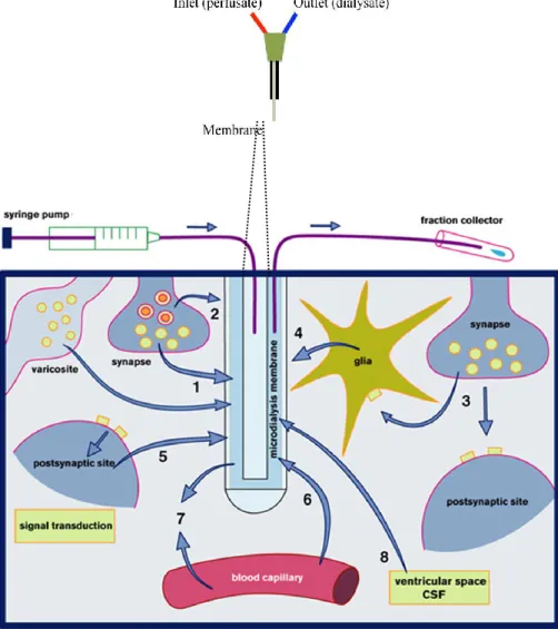
Cerebral microdialysis is a well-established laboratory tool which permits the monitoring of neurotransmitters and other biological molecules from interstitial fluids. This method is widely used for sampling and quantifying neurotransmitters, neuropeptides, and hormones in the brain and periphery. Depending on the availability of an appropriate analytical assay, virtually any soluble molecule in the interstitial space fluid can be measured by microdialysis. In addition, microdialysis is being increasingly used as a bedside monitor to provide on-line analysis of brain tissue biochemistry during neurointensive care.
The microdialysis concept is based on the biological tissue implantation of a catheter consisting of a fine double lumen probe, lined at its tip with a semi-permeable dialysis membrane. The probe is then perfused via an inlet tube with fluid isotonic to the tissue interstitium. The perfusate passes along the membrane before exiting via outlet tubing into a collecting chamber (Figure 11). Diffusion drives the passage of molecules across the membrane along their concentration gradient. Molecules at high concentration in the brain extracellular fluid pass into the perfusate with minimum passage of water and, as the perfusate flows and is removed at a constant rate, the concentration gradient is maintained. It is usually state that the microdialysis probe therefore acts an artificial blood capillary and the concentration of substrate in the collected fluid (microdialysate) will depend in part on the balance between substrate delivery to, and uptake/excretion from, the extracellular fluid (Figure 11). This simple concept provides a powerful technique with many potential applications in which any molecule small enough to pass across the membrane can be sampled.
Microdialysis is also commonly used for the continuous and local administration of exogenous compounds (reverse microdialysis). In this case, the compound of interest is dissolved into the perfusion medium.
34
Figure 11. Schematic picture illustrating the principle of microdialysis sampling.
As previously reported, microdialysis probes are constituted of a length of tubular dialysis membrane through which a solution is constantly perfused. The dialysis membrane is semipermeable and permits free transport of some but not all solutes. Permeability is typically limited to compounds with molecular masses ˂20,000 Da. A variety of membranes are available which differ in pore size and material used. The length of the membrane is chosen based on the dimension of the brain area under examination. In this study we used 2 mm membrane length probes.
It has been clearly demonstrated that the concentration of a given molecule in the dialysate will be lower than its real concentration in the brain extracellular fluid unless there is total equilibration across the dialysis membrane. The proportion of
35
the true concentration collected in the dialysate is termed the relative recovery and is dependent on membrane pore size, membrane area, rate of flow of perfusate and diffusion speed of the substance.
In vivo microdialysis is not without limitations. While suitable for the real-time measurement of endogenous substances, animal studies have shown that prolonged probe placement results in edema in the surrounding region and infiltration of the membrane by inflammatory cells, rendering the results obtained in such a situation unreliable. This problem has been surmounted in the closed technique in which the modification of the probe is such that it is inserted into a relatively superficial “bolt” which may be locked upon removal of the probe. This technique of repeated probe insertion has been employed in animal studies and has not been shown to be associated with any increase in gliosis in the region of probe placement. Thus, the aspect of this technique that ensures minimal trauma to the cerebral parenchyma may be preserved along with the benefit of continuous, serial sampling. Another proposed benefit to the technique of intermittent sampling as opposed to the use of a continuous indwelling probe is the decreased risk of infection.
3.2. ANIMALS
Adult male Sprague-Dawley rats (225-250 g, Harlan Italy S.r.l., Udine, Italy) were used. The animals, housed in cages in groups of five animals in a temperature- and relative humidity- controlled environment with a regular 12 hour light/dark cycle, had free access to food and water. Following delivery, the animals were allowed to adapt to the environment for at least one week before the experiment commenced.
Experiments were carried out in strict accordance with the European Communities Council Directive (86/609/EEC) and the Guidelines released by the Italian Ministry of Health (D.L. 116/92) and (D.L. 111/94-B). A formal approval to conduct the experiments described was obtained by the local Ethics Committee (University of Ferrara, Italy). Efforts were made to minimize the number of animals used and to reduce pain and discomfort. The ARRIVE guidelines for reporting research (http://www.nc3rs.org.uk/ARRIVE) were followed throughout the design and execution stage of all experiments.
36 3.3. MICRODIALYSIS EXPERIMENTS
3.3.1. Surgery
On the day of surgery, the animals, kept under isoflurane anaesthesia (1.5% mixture of isoflurane and air), were mounted in a David Kopf stereotaxic frame (Tujunga, CA, USA) with the upper incisor bar set at -2.5 mm below the interaural line. After exposing the skull and drilling a burr hole, a guide cannula (CMA12; CMA Microdialysis, Solna, Sweden) was positioned on top of the striatum (stereotaxic coordinates = AP: 1 mm anterior to bregma, L: 2.5 mm from the midline, V: 3.5 mm below the dura) or in the medial PFC (AP: 3.2 mm anterior to bregma, L: 0.6 mm from the midline; V:2.0 mm below the dura; Paxinos & Watson, 1986) and secured to the skull with anchor screws and acrylic dental cement. After surgery, the animals were housed individually in microdialysis chambers.
3.3.2. Experimental protocol
On the day after surgery, a microdialysis probe of concentric design (CMA12; molecular weight cutoff: 20 kD; outer diameter 0.5 mm; length of dialysing membrane 2 mm; Alfatech S.p.A., Genova, Italy) was inserted through the guide cannula, extending throughout the striatum or the PFC. The probe was connected to a microperfusion pump (CMA 100; Carnegie Medicin, Stockholm, Sweden) set to a speed of 1 µl/min and perfused with Ringer solution containing (in mM): NaCl, 144; KCl, 4.8; MgSO4, 1.2; CaCl2, 1.7; pH 6.7 (Figure 12). The probe was employed both for intrastriatal or intracortical perfusion with the selected compounds and the recovery of perfusate samples for measurement of local glutamate and GABA levels. Perfusates were collected every 30 min. After three stable basal glutamate and GABA values were obtained, KYNA (30-1000 nM), galantamine (5 µM), ESBA (1 mM, 5 mM) 7-Cl-KYNA (100 nM) and/or CNQX (100 µM) were locally perfused for 2 h (four samples). Subsequently, perfusion with Ringer solution continued for another 2.5 h (5 samples). In some experiments performed in the striatum, tetrodotoxin (TTX; 2 µM) was locally perfused 3.5 h prior to the addition of KYNA, and perfusion continued until the end of the experiment.
37
Following each experiment, the brain was removed from the skull, and the position of the dialysis probe was verified using 30 µm-thick coronal cryostat sections. Only those animals in which the probe was correctly located were included in this study.
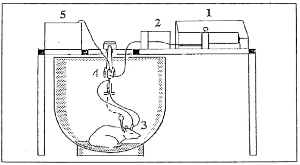
Figure 12. Schematic representation of the experimental protocol. In 1 is illustrated the microinfusion pump that is connected to a syringe selector as shown in 2, perfusing in the probe represented by 3. In 4 is illustrated a swivel, to permit a free movement of the animal and 5 represent the fractional collector.
3.3.3. Glutamate and GABA analysis
Glutamate and GABA levels in the dialysate were measured by HPLC coupled to fluorimetric detection. Briefly, 25 μl samples were pipetted into glass microvials and placed in a temperature-controlled (4°C) Triathlon autosampler (Spark Holland, Emmen, The Netherlands). Thirty μl of o-phthaldialdehyde/mercaptoethanol reagent were added to each sample, and 30 μl of the mixture were injected onto a Chromsep analytical column (3 mm inner diameter, 10 cm length; Chrompack, Middelburg, The Netherlands). The column was eluted at a flow rate of 0.48 ml/min (Beckman125 pump; Beckman Instruments, Fullerton, CA, USA) with a mobile phase containing 0.1 M sodium acetate, 10% methanol and 2.2% tetrahydrofuran (pH 6.5). Glutamate and GABA were detected by means of a Jasco fluorescence spectrophotometer FP-2020
38
Plus (Jasco, Tokyo, Japan). The retention times of glutamate and GABA were ~3.5 and ~ 15.0 min, respectively.
3.4. MATERIALS
KYNA, 7-Cl-KYNA, CNQX, TTX and galantamine were obtained from Sigma Chemical Co. (St. Louis, MO, USA). For administration by reverse microdialysis, KYNA, 7-Cl-KYNA, CNQX, and galantamine were dissolved in Ringer solution and delivered through the probe. TTX was dissolved in water at a concentration of 1 mM and stored at -80°C. Upon thawing, the toxin was diluted in Ringer solution on the day of the experiment. ESBA, kindly provided by R. Pellicciari (University of Perugia, Italy), was synthesized and used in microdialysis experiments as described (Pellicciari et al., 2006). All other chemicals were of the highest commercially available purity.
3.4.1. Data management and statistical analysis
Data were not adjusted for recovery from the microdialysis probe. Results from individual time points are reported as percentages of the mean of the last three baseline samples before treatment commenced. For experiments involving KYNA and TTX, results from individual time points are reported as percentages of the mean of the three samples prior to KYNA perfusion. Data are expressed as the mean ± SEM, and only the statistical significance of the peak effects (maximal responses) is shown in the Figures. In addition, the area created by the curve (AUC), reflecting the duration of the effect, was determined for each animal. Area values (overall effects) were calculated as percentages of changes in baseline value over time by using the trapezoidal rule. Statistical analysis was carried out by analysis of variance (ANOVA) followed by the Newman-Keuls test for multiple comparisons.