www.impactjournals.com/oncotarget/ Oncotarget, Advance Publications 2017
Bone marrow micro-environment is a crucial player for
myelomagenesis and disease progression
Patrizia Mondello
1,2,3, Salvatore Cuzzocrea
2, Michele Navarra
2and Michael Mian
4,51 Department of Human Pathology, University of Messina, Messina, Italy
2 Department of Chemical, Biological, Pharmaceutical and Environmental Sciences, University of Messina, Messina, Italy 3 Lymphoma Service, Memorial Sloan Kettering Cancer Center, New York, NY, USA
4 Department of Hematology and Center of Bone Marrow Transplantation, Hospital of Bolzano, Bolzano/Bozen, Italy 5 Department of Internal Medicine V, Hematology & Oncology, Medical University Innsbruck, Innsbruck, Austria
Correspondence to: Patrizia Mondello, email: [email protected]
Keywords: multiple myeloma, micro-environment, therapeutic opportunities, osteoclast activation, angiogenesis Received: July 18, 2016 Accepted: January 05, 2017 Published: January 12, 2017
ABSTRACT
Despite the advent of many therapeutic agents, such as bortezomib and lenalidomide that have significantly improved the overall survival, multiple myeloma remains an incurable disease. Failure to cure is multifactorial and can be attributed to the underlying genetic heterogeneity of the cancer and to the surrounding micro-environment. Understanding the mutual interaction between myeloma cells and micro-environment may lead to the development of novel treatment strategies able to eradicate this disease. In this review we discuss the principal molecules involved in the micro-environment network in multiple myeloma and the currently available therapies targeting them.
INTRODUCTION
Multiple myeloma (MM) is a malignant
lymphoproliferative disorder deriving from a clonal
plasma cell dyscrasia with the ability to produce
monoclonal immunoglobulins in most cases. The
incidence of the disease has been estimated to be 26,850
new cases in 2015 in the United States. Disease occurrence
increases with age, affecting mostly people in the sixth
and seventh decade of life with a predominance in males
and Afro-Americans [1]. Clinical presentation at onset is
highly variable ranging from a completely asymptomatic
disease without evidence of end-organ damage to an acute
life threatening condition.
Despite intensive research, the etiology of MM is
largely unknown. Possible risk factors include exposure
to ionizing radiation, chemicals such as benzene,
asbestos, lucite and antigens. Also genetic factors play a
major role [2]. Transformation of a normal plasma cell
into a neoplastic cell is a multistep process [3] due to
genetic and molecular events [4] as well as to important
and irreversible alterations in the bone marrow
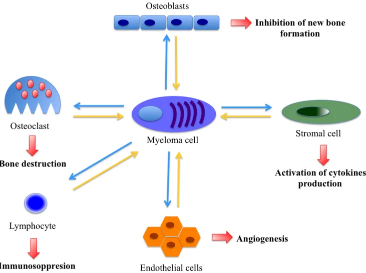
micro-environment [5, 6]. The myeloma cell is immersed in
the bone marrow micro-environment where it mutually
interacts with stromal cells (BMSC), osteoblasts,
osteoclasts, lymphocytes and endothelial cells [6] (Figure
1). Indeed, culture of plasma cells is only possible in the
presence of BMSC [6].
A process of major importance is the interaction
between myeloma cells and BMSC through adhesion
molecules, such as VLA-4 and LFA-1. These interactions
trigger a complex cytokine network between cancer
cells and micro-environment, ultimately impacting
further development and prognosis of the disease itself.
Another system interaction involves C-X-C chemokine
ligand (CXCL)-12, expressed by BMSC, and its receptor
C-X-C chemokine receptor (CXCR)-4, located on MM
cells, which is involved in motility and cytoskeletal
rearrangements [6]. Moreover, CXCL-12 up-regulates
transiently VLA-4, further influencing cellular adhesion
of myeloma cells to the BMSC and cytokines secretion
[7]. However, adhesion molecules are not the only players
in this complex game: pathogenesis of MM also depends
on the presence of growth factors [8, 9] that are usually
produced by BMSC to regulate activity of
lympho-hemopoietic cells [10, 11].
In the present review, we focus on the pathogenetic
mechanisms involving the bone marrow
micro-environment and promoting myeloma.
INTERLEUKINE 6 (IL-6)
IL-6 is produced by mononuclear phagocytes,
endothelial cells, fibroblasts and many other cell types as
a response to IL-1 and tumor necrosis factor (TNF). This
molecule also stimulates the secretion of the proteins of
the acute phase immune response, such as the protein or
mannose-binding fibrinogen by the liver cells. IL-6 acts
as a growth factor for activated B-cells and differentiation
towards the plasma cell line and has multiple effects on
hematopoietic and other cells [12]. It is closely involved in
the pathogenesis of MM: 1) IL-6 induces in vitro growth
of fresh cells isolated from myeloma patients; 2) The
myeloma cells spontaneously produce IL-6 and express the
corresponding receptor; 3) antibodies against IL-6 inhibit
the growth of myeloma cells; 4) treatment of myeloma
patients with antibodies against IL-6 has shown anti-tumor
effect [9, 13, 14]; 5) retinoic acid induces apoptosis in
myeloma cells by down-regulation expression of the IL-6
receptor [15]. Preliminary data suggests that the secretion
of IL-6 is regulated by plasmoblast cytokines, such as
TNF-alpha and transforming growth factor (TGF)-beta
[9].
Enhancing sensitivity of the myeloma cell to IL-6
contributes to the growth and expansion of the neoplastic
cells, as is the case with the soluble receptors for IL-6
(sIL-6R) [16]. These receptors derive from cleavage of the
receptor itself or from alternative splicing mechanisms of
the respective RNA [17, 18]. sIL-6R is present in the serum
and urine of healthy individuals [9], but it is significantly
elevated in MM patients [16, 19-22]. Therefore, the
significance of sIL -6R is controversial. Unlike other
Authors [16, 20, 22], Ohtani et al. [19] observed a good
correlation between sIL-6R levels and tumor burden. This
is in line with the observation that elevated serum levels
of IL-6 as well as its soluble receptor are able to predict a
poor prognosis and to reflect the level of disease activity
[16, 23, 24], whereas the decrease of these parameters is
associated with a good response to treatment [20].
Interaction between MM cell and BMSCs stimulates
IL-6 secretion [25]. Originally identified as a regulator of
normal B-cell differentiation, IL-6 has shown to promote
Figure 1: Interplay between various micro-environmental cells promoting angiogenesis and proliferation in multiple
myeloma.
myeloma cell proliferation and protect cells from apoptosis
[26]. After co-culture with BMSCs, MM cells increased
levels of phosphorylated AKT and ERK [27, 28], cyclin
D2, CDK4, and Bcl-XL, and decreased cleaved Caspase-
3 and PARP [29], which are important signaling pathways
involved in proliferation and apoptosis of MM cells.
Furthermore, IL-6 contributes to the dysfunction
of immunosystem. In MM patients dendritic cells
(DCs) presented a lower expression of HLA-DR, CD40
and CD80 antigens, and impaired activation of T-cell
proliferation compared with controls. These DCs were
unable to present the specific tumor antigen to autologous
T cells [30]. Hwang et al demonstrated that the IL-6Rα
knockdown-DC vaccine significantly enhances the
frequency of tumor-specific CD8+ producing effector
molecules such as IFN-γ, TNF-α, FasL, perforin, and
granzyme B, and generates more memory T cells, resulting
in prolonged survival [31].
Finally, IL-6 is also produced by osteoclasts. These
cells produce high levels of IL-6 when grown in
co-culture with MM cells, resulting in further increase of cell
proliferation and inhibition of apoptosis [32, 33]. Although
its precise role is still under debate, IL-6 released by
osteoclasts seems to increase MM tumor burden, and
enhance bone destruction since it increases production of
IL-17 by T-cells. Increased IL-17 secretion by these bone
marrow T cells results in up-regulation of RANKL and
increases osteoclast formation [34]. Expression of the
IL-17 receptor on MM plasma cells leads to IL-IL-17 mediated
growth of plasma cells [35].
Overall, therapies against IL-6 would be a rational
target. Siltuximab is a monoclonal antibody (Ab) against
IL-6, which has demonstrated to have a good safety
profile (NCT01484275) [36]. Therefore, it was evaluated
in transplant-ineligible MM patients in combination with
bortezomib, melphalan, and prednisone (VMP). However,
Siltuximab did not demonstrate an improvement in terms
of complete response rate when compared to the VMP
alone [37]. Since IL-6 is involved in early
myeloma-stroma interaction and survival of neoplastic cells, IL-6
Figure 2: Bone remodelling in multiple myeloma. Myeloma cells also directly promote osteoclast formation via the endogenous
expression of RANKL, and downregulation of the RANKL decoy receptor (OPG).therapy at earlier stages of the disease might be more
beneficial. Therefore, Siltuximab is currently under
investigation in patients with asymptomatic disease
(NCT01484275).
OSTEOCLAST ACTIVATION
Osteoclast activation contributes to the decrease
of bone matrix and ultimately to osteolysis. In normal
conditions, osteoclast differentiation and activation
are controlled by stromal cell/osteoblast through the
Receptor Activator of NF-κB Ligand (
RANK-L
)/RANK/
osteoprotegerin (OPG) system [38-41]. Osteocytes are the
major source of the osteoclastogenic cytokine RANK-L
[42, 43]. The specific receptor of this protein, RANK, is
located on osteoclast progenitors and its stimulation leads
to differentiation into mature and active osteoclasts [40,
41, 44]. RANK-L activity is physiologically countered
by interferon-gamma (INFγ), which regulates osteoclast
formation by preventing excessive bone resorption
[39-41].
In MM, RANK expression is dysregulated in
osteoclast precursor cells [45]. Myeloma cells also
directly promote osteoclast formation via the endogenous
expression of RANK-L [45], TNF-α, and macrophage
inflammatory protein 1 alpha (MIP-1α) [46-50].
Myeloma cells further contribute to osteoclastogenesis by
downregulating expression of the RANK-L decoy receptor
(OPG) [51, 52]. (Figure 2) This inhibitory effect is
secondary to the interaction of myeloma cells and BMSC
through VLA4 and VCAM1 [47, 53]. Preclinical studies
have supported the importance of the RANK-L/OPG in the
pathogenesis of bone lesions in MM. In xenograft model,
treatment with either OPG or inhibitor of the receptor
RANK inhibited bone lysis and reduced the osteoclasts
number [54]. Furthermore, inhibition of this receptor
resulted in decreasing of tumor burden and paraprotein
levels in MM. Elevated levels of IL-6 induce RANK-L
expression also in lymphocytes and decrease INFγ
production [55], leading to bone resorption. Ultimately,
there is an imbalance of osteoclast homeostasis in favor of
RANK-L and to the detriment of OPG and INFγ, resulting
in osteoclast activation, bone resorption and myeloma cell
survival [56].
Therefore, bisphosphonates and monoclonal
antibodies binding RANK-L are commonly used to limit
osteoclast activity [57, 58]. These therapies significantly
delay skeletal-related events such as pathological fracture
and therefore reduce disease-associated morbidity [59,
60], and prolong survival [61-63].
Osteoblast-mediated bone formation/mineralization
is significantly reduced in MM patients, contributing
to bone destruction. The factors involved in osteoblast
suppression have been widely described within the
Wnt pathway and myeloma-derived factors [64]. Many
therapeutic agents that increase bone formation or
block agonists in myeloma patients are currently under
investigation. These include potent osteogenic factors
such as Wnt, Dickkopf Wnt signaling pathway 1
(DKK-1), fibroblast growth factor (FGF) 23, and heparanase
[65-67]. In xenograft model, the DKK-1 inhibitor
BHQ880 increased osteoblast number and trabecular
bone volume [68]. Similarly, using this drug combined
with bisphosphonate resulted in a trend towards increased
bone mineral density in relapsed MM [69]. Other phase I/
II clinical trials examining its efficacy are completed with
pending reports (NCT01302886, NCT00741377, and
NCT01337752).
CD28
CD8+ T cells recognize and eliminate cells infected
by intracellular pathogens [70] as well as neoplastic cells
[71]. The T-cell receptor (TCR) identifies such cells by
interaction with MHC-I-receptor and the bound peptide
antigen presented on the surface of antigen-presenting
cells (APCs), namely DCs and macrophages/monocytes
[72]. However, the stimulation via TCR alone is not
sufficient to activate the CD8+ T cells [73-75] since a
co-stimulatory signal is necessary [75]. One of the best
characterized co-stimulus is the interaction of CD28
of the T cell and CD86 or CD80 of the APC [72, 73].
Persistent antigenic stimulation or repetitive stimulation/
proliferation rounds lead to progressively down-regulation
of CD28 expression and accumulation of highly antigen
experienced CD8+ CD28 T cells [72, 74]. The decrease
of CD28 is associated with the increased expression of
CD57 (HNK, Leu-7, L-2) [76, 77], a terminally sulphated
carbohydrate-determinant found on various surface
glycoproteins, proteoglycans and glycolipids on subsets
of natural killer (NK) cells, T cells, and others [72, 78, 79].
High levels of CD8+ CD28- (CD8+ CD57+) T-cell
population are associated with malignancy. Increased
number of these T cells was found both in the
micro-enviroment and peripheral blood of patients with solid
tumors [71, 80-83] and hematologic malignancies
[84-88]. Patients affected by melanoma showed the expanded
CD8+ CD28- T cells with high perforin expression,
suggesting an active immune response against the
tumor [81]. Conversely, lung cancer patients presented
expanded CD8+ CD28- T cells with significant levels of
FOXP3+, which were attributed to the immunosuppressive
component of the antitumour immune response
[82]. Probably the predominance of cytotoxic versus
immunosuppressive CD8+ CD28- T cell population differs
in various types of cancer or even between individual
patients with the same oncological disease.
CD38
CD38 is a multifunctional cell surface protein that
has receptor [89] as well as enzyme functions [90, 91]
with an important role in cell signaling. This protein is
found at high levels on normal plasma cells as well as
in hematological malignancies [92-98]. CD38 levels are
even higher in malignant plasma cells [99], which is why
it is an attractive therapeutic target for MM. Various
anti-CD38 antibodies have been developed and up to now
available preclinical and clinical data are very promising.
The so far best investigated antibody is Daratumumab.
This IgG1 antibody binds a unique epitope present on
the CD38 molecule leading to complement-dependent
cytotoxicity [100]. Furthermore, Daratumumab decreases
the CD38+ immunosopressive cellular population,
increases T-helper and cytotoxic T-cells population, and
enhances TCR clonality [101]. In vivo daratumumab
alone showed substantial tumor cell lysis. This effect
markedly increased, when the antibody was combined
with anti-myeloma drugs, such as lenalidomide and
bortezomib [102, 103]. Its clinical efficacy was evaluated
in the CASTOR and Pollux trial [104, 105]. The former
included nearly 500 relapsed or refractory multiple
myeloma patients, each of whom underwent eight cycles
of a standard two-drugs induction regimen (bortezomib
and dexamethasone). Afterwards they were randomized
to receive daratumumab maintenance therapy or not. In
interim analysis, daratumumab combination reduced the
risk of cancer progression by 70%, and doubled both
rates of very good partial response from 29% to 59% and
complete response from 9% to 19% with a manageable
toxicity profile [104]. Also in the POLLUX trial, in
which 569 relapsed or refractory patients were treated
with daratumumab in combination with lenalidomide and
dexamethasone, the addition of the antibody significantly
prolonged progression-free survival (PFS) (p < 0.0001)
and reduced the risk of disease progression of 63% [105].
Based on these promising results, Daratumumab was
approved by the U.S. Food and Drug Administration as
the first monoclonal antibody for the treatment of multiple
myeloma.
Currently, there are ongoing phase 3 studies,
which include daratumumab in combination with either
lenalidomide or bortezomib-based regimens in newly
diagnosed MM (NCT02252172, NCT02195479, and
NCT02541383). An additional phase 1 study is also
investigating the subcutaneous delivery of daratumumab
in association with recombinant human hyaluronidase in
patients with relapsed/refractory MM (NCT02519452).
PD-1 AND PD-L1
Tumors can escape immunosurveillance
by expression of molecules that inhibit antitumor
immunoresponse, such as programmed cell death ligand 1
(PD-L1) [106]. Although PD-L1 expression has not been
observed in normal epithelial cells, it is highly expressed
on many solid tumors [107]. PD-1 is a cell surface
receptor
of the immunoglobulin superfamily and is expressed on
T
cells,
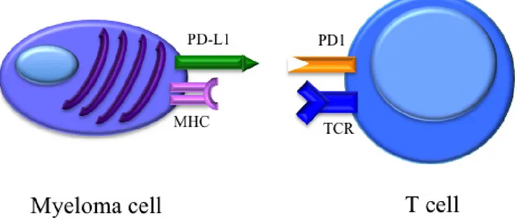
B cells, and NK cells [108]. Within the tumor, cells
of the micro-environment express PD-L1, leading to T cell
anergy upon cellular contact. (Figure 3) Furthermore, T
cells produce INFγ, which upregulates PD-L1 expression
on tumor and infiltrating immune cells, forming a
feedback loop that generates a PD-1 signal maintaining
immunosuppression [109, 110]. Although not detected
on normal plasma cells, myeloma cell lines and primary
myeloma cells up-regulate PD-L1, while its ligand PD-1
is found on a proportion of T-cells in myeloma patients
Figure 3: Binding of PD-1 on the T cell with tumor-associated PD-L1 results in downregulation of T-cell effector
functions.
[111-115]. The highest PD-L1 expression levels were
detected in relapsed/refractory MM and correlated with
tumor burden and poor treatment response [107].
Anti-PD-L1 antibodies inhibit dendritic cells and
myeloid-derived suppressor cells enhancing the cytolytic activity
of NK cells against MM cell [114]. Phase I studies using
checkpoint inhibitors reported disappointing results,
achieving stable disease as best response [116, 117].
Nevertheless, combination treatments including these
checkpoint inhibitors could lead to an increased anti-MM
host immunity and therefore better clinical responses.
Clinical trials investigating the combination of anti-PD-L1
treatment with either immunomodulatory agents, such as
lenalidomide (NCT02077959), myeloma vaccines (e.g.,
NCT01067287), or other T-cell co-inhibitor molecules
such as cytotoxic T-lymphocyte-associate protein 4
(NCT01592370), which modulate MM-host immune
responses, are ongoing.
ANGIOGENESIS AND ANGIOGENIC
FACTORS
In 1999 Vacca et al described an increased
neovascularization of the bone marrow stroma in patients
affected by MM [118]. The grade of this neovascularization
seems to increase during the evolution from monoclonal
gammopathy of undetermined significance (MGUS) to
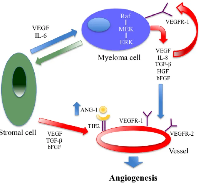
MM [119]. This process is triggered by neoangiogenetic
factors, which are produced from the BMSC, [such
as vascular endothelial growth factor (VEGF), basic
fibroblasr growth factor (b-FGF) and TGF-β, and from
the myeloma cells themselves (such as VEGF, IL-8 and
TGF-β). (Figure 4)
Figure 4: Autocrine and paracrine VEGF-mediated pathways in multiple myeloma: both are important for tumor
angiogenesis and growth. A close relationship between VEGF and IL-6 has been found in the paracrine pathways.
Vascular endothelial growth factor (VEGF)
The VEGF is the major pro-angiogenic molecule.
It has a key role in regulating physiological as well as
pathological angiogenesis [120, 121]. Up to now, four
genes structurally related to VEGF have been identified.
Among their known products, there are placenta growth
factors (PGF) [122-124], VEGF-a, VEGF-b [124],
VEGF-c [125],VEGF-d [126] and VEGF-e [126]. The
VEGF-a isoform is the most important VEGF molecule
contributing to MM since its receptor, VEGFR-2, is highly
expressed on plasma cells and endothelial cells in MM
[127, 128]. After stimulation of VEGFR-2,
mitogen-activated protein (MAP)-kinase is phosphorylated,
resulting in an enhanced cell proliferation. Probably there
is a paracrine mechanism behind the VEGF-induced
stimulation of the endothelium: the plasma cell secretes
VEGF-a, which binds to the VEGFR-2 on angiopoietic
cells, ultimately activating a proliferative signal.
VEGF-a is also an important communication
signal between stromal and myeloma cells, inducing the
microenvironment to produce the same VEGF, but also
other proangiogenic factors, such as FGF-β and TGF-β
[129]. Furthermore, VEGF-a has an autocrine effect
on the neoplastic cell, mediated by VEGFR-2 and the
intracellular transmission pathway RAF-1/MEK-1, which
would strengthen further angiogenic stimulus [129].
In tumors, hypoxia may be a consequence of
the distance of the growing tumor cells from existent
capillaries or inefficiency of the new vessels [130, 131].
Therefore, the expression of VEGF and its receptor is
induced in angiopoietic cells in order to avoid cell death.
Apart from the stimulation of neovascularization and
the increase of production of cytokines and proteolytic
enzymes contributing to neo-angiogenesis, VEGF leads
to upregulation of some proto-oncogenes typically found
in MM, such as V-ras, K-ras, V-raf, Src and Fos [132].
Indeed, inhibition of VEGFR-2 and VEGF-a reduces
myeloma clone proliferation. Patients with MM have
low levels of semaforina III, which has a physiological
antiangiogenic effect, balancing the action of VEGF
[133]. Microvascular density of the bone marrow assessed
with anti CD34 and DC15 (which are antigens present
on the endothelium) is increased in patients with MM
compared to healthy controls and also seems to correlate
with the stage of disease. Of note, although bone marrow
vascularization appears to be a prognostic marker, it
does not change after therapy [134]. Another important
question is related to the measurable levels of the signaling
proteins as well as their receptors. For example, Dales et al
have demonstrated an increased expression of VEGF-R1
in diseases such as breast cancer, a disease in which
VEGF-R1 seems to be a negative prognostic marker with
a high risk of metastases and relapses [135] and the use
of antibodies against VEGF-R1 determines a powerful
inhibition of neoplastic cell growth [136].
Overall, drugs targeting mechanisms involving
the various VEGF forms are promising targets for new
treatment approaches. For example, Bevacizumab targets
and blocks VEGF and VEGF’s binding to its receptor on
the vascular endothelium [137] and has demonstrated
to be effective when used alone, and in combination
with radiation in earlier preclinical studies [137, 138].
It is currently being studied clinically in many solid and
blood tumors including primary systemic amyloidosis
and MM [139, 140]. NCI’s Cancer Therapy Evaluation
Program sponsored a phase II study of bevacizumab plus
thalidomide in MM. The study was closed early due to
poor accrual. Combination therapy, in this limited sample,
yielded similar results to single agent thalidomide. The
small number of patients prevents correlation of VEGF or
VEGFR1/VEGFR2 expression with response [141].
Angiopoietins (Ang)
Angs, consisting of 4 structurally related proteins,
termed Ang-1, 2, and 4, are ligands for the
vascular-specific tyrosine kinase TIE-2 receptor located on
endothelial cells. Ang-1 and Ang-4 behave as activating
ligands for Tie-2, whereas Ang-2 and Ang-3 function
as competitive antagonists. When bound to Tie-2,
Ang-1 stimulates maturation and stabilization of the vascular
wall [142, 143]. In contrast, Ang-2 antagonizes Tie-2
binding and induces vessel destabilization, leading to
the angiogenic sprouting [144]. In MM 1 and
Ang-2, along with VEGF, possess an important role in the
initiation of tumor angiogenesis [145]. This is further
confirmed by the observation that tumor angiogenesis
can be prevented using both antibodies against Ang-1 and
VEGF-a, while neutralizing only either VEGF-a or
Ang-1 reduces it partially [Ang-146]. Ang-2 serum levels, alone
or in ratio with Ang-1, are importantly prognostic for
response to therapy and mainly for survival [147]. These
data highlight the role of the angiopoietins pathway in the
biology of MM, which could be an interesting target for
new anti-myeloma agents.
Platelet-derived growth factor -beta (PDGF-β)
PDGF-beta is able to up-regulate the expression of
the protein c-MYC, thus reducing the sensibility of cancer
cells to melphalan treatment. Additionally,
melphalan-resistant patients have an overexpression of c-MYC and
higher levels of PDGF-β [148]. The latter correlated
with disease burden (estimated by Durie-Salmon staging
system, levels of IL-6 and beta-2-microglobulin) [149,
150]. Therefore, compounds targeting this mechanism
could improve MM treatment. PTK787/ZK22258 (PTK/
ZK) is a potent tyrosine kinase inhibitor of both vascular
endothelial growth factor receptor 1 (VEGF-R1) and
VEGF-R2, and also inhibits the tyrosine kinase activity
of PDGFR-β, Flt-4, c-kit and c-fms, although with less
potency [133]. PTK/ZK inhibits endothelial cell migration
and proliferation without cytotoxic or antiproliferative
effects on cells that do not express VEGF receptors
[133]. Oral administration of PTK/ZK at a dose of
25-100 mg/kg/day decreases tumor growth in human cancer
xenografts [133, 151]. Overall, PTK/ZK inhibits multiple
essential signaling pathways involved in proliferation
and fibrogenesis [152], which is why this molecule could
improve the efficacy of existing MM treatments.
Fibroblast growth factor beta
FGF-beta is able to trigger neovascularization
in MM bone marrow [153]. In the altered
micro-environment, the myeloma cells appear to be the main
source of FGF-beta, which is why their expression levels
could correlate with disease burden and therefore could be
an important prognostic parameter for MM [154]. In fact,
the concentration of FGF-beta is considerably increased in
plasma cell lysates in the bone marrow and in peripheral
blood of myeloma patients [154].
Apart from the pro-angiogenic effects of this growth
factor, paracrine interactions between tumor cells and
BMSC were observed: stimulation of stromal cells with
FGF-beta induces an increase of IL-6 secretion. This
process can be completely abrogated by the administration
of antibodies against FGF-beta. On the other hand,
stimulation with IL-6 increases the expression and
secretion of FGF-beta by the myeloma cell lines [155].
Matrix metallopeptidase (MMP)-1, MMP-2 and
MMP-9
The expression of these metal proteinases is
considerably increased in myeloma plasma cells as well
as cells of the tumor environment, such as macrophages,
fibroblasts and osteoblasts [156, 157]. Up to now their
role in the context of tumor angiogenesis is unclear, but
they probably are mediators for VEGF and
PDGF-beta-dependent mechanisms. Indeed, in vivo studies in murine
models indicate that zoledronic acid is able to inhibit the
expression of MMP-9 of macrophage origin and finally
neoangiogenesis [158].
Increased levels of the MMP-1 has been associated
with an enhanced capacity to degrade collagen I [159]. In
MMP-1 transgenic mice, the synthesis of the latent form
of collagenase leads to extracellular matrix degradation
[160]. MMP-2 instead directly modulates tumor invasion
[161], favoring the spreading of myeloma cells. The
mechanisms responsible for the upregulation of MMP
activity remain to be elucidated.
Overall, the most important mechanisms in MM
are the production of MMP-9, activation of MMP-2 and
induction of MMP-1 by the neoplastic cells, since they are
major players for bone resorption and tumor spreading.
Some inhibitors of MMPs are now available. In arthritis,
these inhibitors lead to a dramatic suppression of cartilage
and bone destruction [162]. In cancer, the MMP inhibitor
Batimastat was effective in a variety of murine tumor
metastatic models and was successfully used in patients
with malignant ascites [163] and malignant pleural
effusion [164].
OTHERS FACTORS
Granulocyte colony stimulating factor (G-CSF)
G-CSF is a hematopoietic growth factor with several
similarities to IL-6 [165, 166]. Both IL-6 and G-CSF
induce the activation of NF-IL-6, which is involved in
the synthesis of the IL-6 [167]. In vivo G-CSF appears to
be a potent growth factor for myeloma cells [9].
Despite
their well-known physiological functions, G-CSF and IL-6
could modulate neutrophils in bone marrow, altering the
activation potential of signaling pathways in neutrophils,
especially that of STAT3. Co-stimulation with G-CSF
and IL-6 induced a higher level of phospho-STAT3 in
neutrophils, stimulating angiogenesis and tumor growth
[167].
IL-10
IL-10 is another important growth factor
contributing to the pathogenesis of MM. IL-10 has been
proven to inhibit various immune functions, such as
macrophage activation, cytokine production, and antigen
presentation [168], while it induces both plasma cell
proliferation and angiogenesis in MM [169]. IL-10 is also
involved in initiating and promoting other malignancies
[170].
High levels of IL-10 secreted by T regulatory cells
or produced from myeloma cells can modulate antitumor
host immune response, including the abrogation of DCs
function [171, 172]. Furthermore, IL-10 seems to induce
chemo-resistance [173, 174]. In particular, the serum
concentration of IL-10 has been found much higher in
MM patients than in normal healthy people, correlating
with poor prognosis [175] and poor treatment response
[175].
CONCLUSIONS
The herein presented data provide evidence that the
bone marrow environment is a very important player in
various biological processes of myeloma development.
The involved mechanisms are very complex and not
only limited to osteoblast and osteoclast dysfunction, but
include pathologic immune processes as well as growth
factor dysregulations. Thus, these findings are the basis for
innovative treatment approaches. Clinical trials designed
to target immune and stromal components of the
micro-environment are ongoing and, in combination with other
anti-myeloma agents, should get us closer to our ultimate
objective of eradicating the disease.
CONFLICTS OF INTEREST
The authors have no conflicts of interest to disclose
that could be perceived as prejudicing the impartiality of
the research reported.
GRANT SUPPORT
This article was not supported by any grant from
any funding agency in the public, commercial or
not-for-profit sector.
REFERENCES
1. Siegel RL, Miller KD, Jemal A. Cancer statistics, 2016. CA Cancer J Clin. 2015; 66:7-30.
2. Sergentanis TN, Zagouri F, Tsilimidos G, Tsagianni A, Tseliou M, Dimopoulos MA, Psaltopoulou T. Risk Factors for Multiple Myeloma: A Systematic Review of Meta-Analyses. Clin Lymphoma Myeloma Leuk. 2015; 15: 563– 77.e1–3.
3. Kuehl WM, Bergsagel PL. Molecular pathogenesis of multiple myeloma and its premalignant precursor. J Clin Invest. 2012; 122: 3456–63.
4. Chapman MA, Lawrence MS, Keats JJ, Cibulskis K, Sougnez C, Schinzel AC, Harview CL, Brunet J-P, Ahmann GJ, Adli M, Anderson KC, Ardlie KG, Auclair D, et al. Initial genome sequencing and analysis of multiple myeloma. Nature. 2011; 471: 467–72.
5. Podar K, Richardson PG, Hideshima T, Chauhan D, Anderson KC. The malignant clone and the bone-marrow environment. Best Pract Res Clin Haematol. 2007; 20: 597– 612.
6. Podar K, Chauhan D, Anderson KC. Bone marrow microenvironment and the identification of new targets for myeloma therapy. Leukemia. 2009; 23: 10–24.
7. Hideshima T, Bergsagel PL, Kuehl WM, Anderson KC. Advances in biology of multiple myeloma: clinical applications. Blood. 2004; 104: 607–18.
8. Klein B, Bataille R. Cytokine network in human multiple myeloma. Hematol Oncol Clin North Am. 1992; 6: 273–84. 9. Klein B. Cytokine, cytokine receptors, transduction
signals, and oncogenes in human multiple myeloma. Semin Hematol. 1995; 32: 4–19.
10. Clark SC, Kamen R. The human hematopoietic colony-stimulating factors. Science. 1987; 236: 1229–37. 11. Sieff CA. Biology and clinical aspects of the hematopoietic
growth factors. Annu Rev Med. 1990; 41: 483–96. 12. Akira S, Taga T, Kishimoto T. Interleukin-6 in biology and
medicine. Adv Immunol. 1993; 54: 1–78.
13. Klein B, Zhang XG, Jourdan M, Content J, Houssiau F, Aarden L, Piechaczyk M, Bataille R. Paracrine rather than autocrine regulation of myeloma-cell growth and differentiation by interleukin-6. Blood. 1989; 73: 517–26. 14. Zhang XG, Bataille R, Widjenes J, Klein B. Interleukin-6
dependence of advanced malignant plasma cell dyscrasias. Cancer. 1992; 69: 1373–6.
15. Levy Y, Labaume S, Colombel M, Brouet JC. Retinoic acid modulates the in vivo and in vitro growth of IL-6 autocrine human myeloma cell lines via induction of apoptosis. Clin Exp Immunol. 1996; 104: 167–72.
16. Gaillard JP, Bataille R, Brailly H, Zuber C, Yasukawa K, Attal M, Maruo N, Taga T, Kishimoto T, Klein B. Increased and highly stable levels of functional soluble interleukin-6 receptor in sera of patients with monoclonal gammopathy. Eur J Immunol. 1993; 23: 820–4.
17. Lust JA, Donovan KA, Kline MP, Greipp PR, Kyle RA, Maihle NJ. Isolation of an mRNA encoding a soluble form of the human interleukin-6 receptor. Cytokine. 1992; 4: 96–100.
18. Müllberg J, Dittrich E, Graeve L, Gerhartz C, Yasukawa K, Taga T, Kishimoto T, Heinrich PC, Rose-John S. Differential shedding of the two subunits of the interleukin-6 receptor. FEBS Lett. 1993; 332: 174–8. 19. Ohtani K, Ninomiya H, Hasegawa Y, Kobayashi T, Kojima
H, Nagasawa T, Abe T. Clinical significance of elevated soluble interleukin-6 receptor levels in the sera of patients with plasma cell dyscrasias. Br J Haematol. 1995; 91: 116– 20.
20. Kyrtsonis MC, Dedoussis G, Zervas C, Perifanis V, Baxevanis C, Stamatelou M, Maniatis A. Soluble interleukin-6 receptor (sIL-6R), a new prognostic factor in multiple myeloma. Br J Haematol. 1996; 93: 398–400. 21. Papadaki H, Kyriakou D, Foudoulakis A, Markidou F,
Alexandrakis M, Eliopoulos GD. Serum levels of soluble IL-6 receptor in multiple myeloma as indicator of disease activity. Acta Haematol. 1997; 97: 191–5.
22. Stasi R, Brunetti M, Parma A, Di Giulio C, Terzoli E, Pagano A. The prognostic value of soluble interleukin-6 receptor in patients with multiple myeloma. Cancer. 1998; 82: 1860–6.
23. Filella X, Blade J, Guillermo AL, Molina R, Rozman C, Ballesta AM. Cytokines (IL-6, TNF-alpha, IL-1alpha) and soluble interleukin-2 receptor as serum tumor markers in multiple myeloma. Cancer Detect Prev. 1996; 20: 52–6. 24. Merlini G, Perfetti V, Gobbi PG, Quaglini S, Franciotta
DM, Marinone G, Ascari E. Acute phase proteins and prognosis in multiple myeloma. Br J Haematol. 1993; 83: 595–601.
25. Lauta VM. Interleukin-6 and the network of several cytokines in multiple myeloma: an overview of clinical and
experimental data. Cytokine. 2001; 16: 79–86.
26. Yoshizaki K, Nakagawa T, Fukunaga K, Tseng LT, Yamamura Y, Kishimoto T. Isolation and characterization of B cell differentiation factor (BCDF) secreted from a human B lymphoblastoid cell line. J Immunol. 1984; 132: 2948–54.
27. Tu Y, Gardner A, Lichtenstein A. The phosphatidylinositol 3-kinase/AKT kinase pathway in multiple myeloma plasma cells: roles in cytokine-dependent survival and proliferative responses. Cancer Res. 2000; 60: 6763–70.
28. Ogata A, Chauhan D, Teoh G, Treon SP, Urashima M, Schlossman RL, Anderson KC. IL-6 triggers cell growth via the Ras-dependent mitogen-activated protein kinase cascade. J Immunol. 1997; 159: 2212–21.
29. Xu S, Menu E, De Becker A, Van Camp B, Vanderkerken K, Van Riet I. Bone marrow-derived mesenchymal stromal cells are attracted by multiple myeloma cell-produced chemokine CCL25 and favor myeloma cell growth in vitro and in vivo. Stem Cells. 2012; 30: 266–79.
30. Ratta M, Fagnoni F, Curti A, Vescovini R, Sansoni P, Oliviero B, Fogli M, Ferri E, Della Cuna GR, Tura S, Baccarani M, Lemoli RM. Dendritic cells are functionally defective in multiple myeloma: the role of interleukin-6. Blood. 2002; 100: 230–7.
31. Hwang W, Jung K, Jeon Y, Yun S, Kim TW, Choi I. Knockdown of the interleukin-6 receptor alpha chain of dendritic cell vaccines enhances the therapeutic potential against IL-6 producing tumors. Vaccine. 2010; 29: 34–44. 32. Abe M, Hiura K, Wilde J, Shioyasono A, Moriyama K,
Hashimoto T, Kido S, Oshima T, Shibata H, Ozaki S, Inoue D, Matsumoto T. Osteoclasts enhance myeloma cell growth and survival via cell-cell contact: a vicious cycle between bone destruction and myeloma expansion. Blood. 2004; 104: 2484–91.
33. Anderson KC, Jones RM, Morimoto C, Leavitt P, Barut BA. Response patterns of purified myeloma cells to hematopoietic growth factors. Blood. 1989; 73: 1915–24. 34. Noonan K, Marchionni L, Anderson J, Pardoll D,
Roodman GD, Borrello I. A novel role of IL-17-producing lymphocytes in mediating lytic bone disease in multiple myeloma. Blood. 2010; 116: 3554–63.
35. Prabhala RH, Pelluru D, Fulciniti M, Prabhala HK, Nanjappa P, Song W, Pai C, Amin S, Tai Y-T, Richardson PG, Ghobrial IM, Treon SP, Daley JF, et al. Elevated IL-17 produced by THIL-17 cells promotes myeloma cell growth and inhibits immune function in multiple myeloma. Blood. 2010; 115: 5385–92.
36. Kurzrock R, Voorhees PM, Casper C, Furman RR, Fayad L, Lonial S, Borghaei H, Jagannath S, Sokol L, Usmani SZ, van de Velde H, Qin X, Puchalski TA, et al. A phase I, open-label study of siltuximab, an anti-IL-6 monoclonal antibody, in patients with B-cell non-Hodgkin lymphoma, multiple myeloma, or Castleman disease. Clin Cancer Res. 2013; 19: 3659–70.
37. San-Miguel J, Bladé J, Shpilberg O, Grosicki S, Maloisel F, Min C-K, Polo Zarzuela M, Robak T, Prasad SVSS, Tee Goh Y, Laubach J, Spencer A, Mateos M-V, et al. Phase 2 randomized study of bortezomib-melphalan-prednisone with or without siltuximab (anti-IL-6) in multiple myeloma. Blood. 2014; 123: 4136–42.
38. Anderson DM, Maraskovsky E, Billingsley WL, Dougall WC, Tometsko ME, Roux ER, Teepe MC, DuBose RF, Cosman D, Galibert L. A homologue of the TNF receptor and its ligand enhance T-cell growth and dendritic-cell function. Nature. 1997; 390: 175–9.
39. Lacey DL, Timms E, Tan HL, Kelley MJ, Dunstan CR, Burgess T, Elliott R, Colombero A, Elliott G, Scully S, Hsu H, Sullivan J, Hawkins N, et al. Osteoprotegerin ligand is a cytokine that regulates osteoclast differentiation and activation. Cell. 1998; 93: 165–76.
40. Hsu H, Lacey DL, Dunstan CR, Solovyev I, Colombero A, Timms E, Tan HL, Elliott G, Kelley MJ, Sarosi I, Wang L, Xia XZ, Elliott R, et al. Tumor necrosis factor receptor family member RANK mediates osteoclast differentiation and activation induced by osteoprotegerin ligand. Proc Natl Acad Sci U S A. 1999; 96: 3540–5.
41. Simonet WS, Lacey DL, Dunstan CR, Kelley M, Chang MS, Lüthy R, Nguyen HQ, Wooden S, Bennett L, Boone T, Shimamoto G, DeRose M, Elliott R, et al. Osteoprotegerin: a novel secreted protein involved in the regulation of bone density. Cell. 1997; 89: 309–19.
42. Nakashima T, Hayashi M, Fukunaga T, Kurata K, Oh-Hora M, Feng JQ, Bonewald LF, Kodama T, Wutz A, Wagner EF, Penninger JM, Takayanagi H. Evidence for osteocyte regulation of bone homeostasis through RANKL expression. Nat Med. 2011; 17: 1231–4.
43. Xiong J, Onal M, Jilka RL, Weinstein RS, Manolagas SC, O’Brien CA. Matrix-embedded cells control osteoclast formation. Nat Med. 2011; 17: 1235–41.
44. Fuller K, Wong B, Fox S, Choi Y, Chambers TJ. TRANCE is necessary and sufficient for osteoblast-mediated activation of bone resorption in osteoclasts. J Exp Med. 1998; 188: 997–1001.
45. Colombo M, Thümmler K, Mirandola L, Garavelli S, Todoerti K, Apicella L, Lazzari E, Lancellotti M, Platonova N, Akbar M, Chiriva-Internati M, Soutar R, Neri A, et al. Notch signaling drives multiple myeloma induced osteoclastogenesis. Oncotarget. 2014; 5: 10393–406. 46. Raisz LG, Luben RA, Mundy GR, Dietrich JW, Horton JE,
Trummel CL. Effect of osteoclast activating factor from human leukocytes on bone metabolism. J Clin Invest. 1975; 56: 408–13.
47. Pearse RN, Sordillo EM, Yaccoby S, Wong BR, Liau DF, Colman N, Michaeli J, Epstein J, Choi Y. Multiple myeloma disrupts the TRANCE/ osteoprotegerin cytokine axis to trigger bone destruction and promote tumor progression. Proc Natl Acad Sci U S A. 2001; 98: 11581–6.
GD. Macrophage inflammatory protein-1alpha is an osteoclastogenic factor in myeloma that is independent of receptor activator of nuclear factor kappaB ligand. Blood. 2001; 97: 3349–53.
49. Farrugia AN, Atkins GJ, To LB, Pan B, Horvath N, Kostakis P, Findlay DM, Bardy P, Zannettino ACW. Receptor activator of nuclear factor-kappaB ligand expression by human myeloma cells mediates osteoclast formation in vitro and correlates with bone destruction in vivo. Cancer Res. 2003; 63: 5438–45.
50. Heider U, Langelotz C, Jakob C, Zavrski I, Fleissner C, Eucker J, Possinger K, Hofbauer LC, Sezer O. Expression of receptor activator of nuclear factor kappaB ligand on bone marrow plasma cells correlates with osteolytic bone disease in patients with multiple myeloma. Clin Cancer Res. 2003; 9: 1436–40.
51. Giuliani N, Bataille R, Mancini C, Lazzaretti M, Barillé S. Myeloma cells induce imbalance in the osteoprotegerin/ osteoprotegerin ligand system in the human bone marrow environment. Blood. 2001; 98: 3527–33.
52. Seidel C, Hjertner Ø, Abildgaard N, Heickendorff L, Hjorth M, Westin J, Nielsen JL, Hjorth-Hansen H, Waage A, Sundan A, Børset M. Serum osteoprotegerin levels are reduced in patients with multiple myeloma with lytic bone disease. Blood. 2001; 98: 2269–71.
53. Roux S, Meignin V, Quillard J, Meduri G, Guiochon-Mantel A, Fermand J-P, Milgrom E, Mariette X. RANK (receptor activator of nuclear factor-kappaB) and RANKL expression in multiple myeloma. Br J Haematol. 2002; 117: 86–92.
54. Croucher PI, Shipman CM, Lippitt J, Perry M, Asosingh K, Hijzen A, Brabbs AC, van Beek EJ, Holen I, Skerry TM, Dunstan CR, Russell GR, Van Camp B, et al. Osteoprotegerin inhibits the development of osteolytic bone disease in multiple myeloma. Blood. 2001; 98: 3534–40. 55. Bataille R, Jourdan M, Zhang XG, Klein B. Serum levels
of interleukin 6, a potent myeloma cell growth factor, as a reflect of disease severity in plasma cell dyscrasias. J Clin Invest. 1989; 84: 2008–11.
56. Viereck V, Emons G, Lauck V, Frosch K-H, Blaschke S, Gründker C, Hofbauer LC. Bisphosphonates pamidronate and zoledronic acid stimulate osteoprotegerin production by primary human osteoblasts. Biochem Biophys Res Commun. 2002; 291: 680–6.
57. Rogers MJ. From molds and macrophages to mevalonate: a decade of progress in understanding the molecular mode of action of bisphosphonates. Calcif Tissue Int. 2004; 75: 451–61.
58. Lipton A, Fizazi K, Stopeck AT, Henry DH, Brown JE, Yardley DA, Richardson GE, Siena S, Maroto P, Clemens M, Bilynskyy B, Charu V, Beuzeboc P, et al. Superiority of denosumab to zoledronic acid for prevention of skeletal-related events: a combined analysis of 3 pivotal, randomised, phase 3 trials. Eur J Cancer. 2012; 48: 3082– 92.
59. Berenson JR, Lichtenstein A, Porter L, Dimopoulos MA, Bordoni R, George S, Lipton A, Keller A, Ballester O, Kovacs M, Blacklock H, Bell R, Simeone JF, et al. Long-term pamidronate treatment of advanced multiple myeloma patients reduces skeletal events. Myeloma Aredia Study Group. J Clin Oncol. 1998; 16: 593–602.
60. Henry D, Vadhan-Raj S, Hirsh V, von Moos R, Hungria V, Costa L, Woll PJ, Scagliotti G, Smith G, Feng A, Jun S, Dansey R, Yeh H. Delaying skeletal-related events in a randomized phase 3 study of denosumab versus zoledronic acid in patients with advanced cancer: an analysis of data from patients with solid tumors. Support Care Cancer. 2014; 22: 679–87.
61. McCloskey E V, Dunn JA, Kanis JA, MacLennan IC, Drayson MT. Long-term follow-up of a prospective, double-blind, placebo-controlled randomized trial of clodronate in multiple myeloma. Br J Haematol [Internet]. 2001; 113: 1035–43.
62. Henry DH, Costa L, Goldwasser F, Hirsh V, Hungria V, Prausova J, Scagliotti GV, Sleeboom H, Spencer A, Vadhan-Raj S, von Moos R, Willenbacher W, Woll PJ, et al. Randomized, double-blind study of denosumab versus zoledronic acid in the treatment of bone metastases in patients with advanced cancer (excluding breast and prostate cancer) or multiple myeloma. J Clin Oncol. 2011; 29: 1125–32.
63. Wang X, Yan X, Li Y. A meta-analysis of the antitumor effect and safety of bisphosphonates in the treatment of multiple myeloma. Int J Clin Exp Med [Internet]. e-Century Publishing Corporation; 2015; 8: 6743–54.
64. Roodman GD. Osteoblast function in myeloma. Bone. 2011; 48: 135–40.
65. Kim J, Denu RA, Dollar BA, Escalante LE, Kuether JP, Callander NS, Asimakopoulos F, Hematti P. Macrophages and mesenchymal stromal cells support survival and proliferation of multiple myeloma cells. Br J Haematol. 2012; 158: 336–46.
66. Fukumoto S. FGF23-FGF Receptor/Klotho Pathway as a New Drug Target for Disorders of Bone and Mineral Metabolism. Calcif Tissue Int. 2016; 98: 334–40.
67. Ruan J, Trotter TN, Nan L, Luo R, Javed A, Sanderson RD, Suva LJ, Yang Y. Heparanase inhibits osteoblastogenesis and shifts bone marrow progenitor cell fate in myeloma bone disease. Bone. 2013; 57: 10–7.
68. Fulciniti M, Tassone P, Hideshima T, Vallet S, Nanjappa P, Ettenberg SA, Shen Z, Patel N, Tai Y-T, Chauhan D, Mitsiades C, Prabhala R, Raje N, et al. Anti-DKK1 mAb (BHQ880) as a potential therapeutic agent for multiple myeloma. Blood. 2009; 114: 371–9.
69. Iyer SP, Beck JT, Stewart AK, Shah J, Kelly KR, Isaacs R, Bilic S, Sen S, Munshi NC. A Phase IB multicentre dose-determination study of BHQ880 in combination with anti-myeloma therapy and zoledronic acid in patients with relapsed or refractory multiple myeloma and prior skeletal-related events. Br J Haematol. 2014; 167: 366–75.
70. Nagata T, Koide Y. Induction of Specific CD8 T Cells against Intracellular Bacteria by CD8 T-Cell-Oriented Immunization Approaches. J Biomed Biotechnol. 2010; 2010: 764542.
71. Tsukishiro T, Donnenberg AD, Whiteside TL. Rapid turnover of the CD8(+)CD28(-) T-cell subset of effector cells in the circulation of patients with head and neck cancer. Cancer Immunol Immunother. 2003; 52: 599–607. 72. Arosa FA. CD8+CD28- T cells: certainties and uncertainties
of a prevalent human T-cell subset. Immunol Cell Biol. 2002; 80: 1–13.
73. Bernard A, Lamy And L, Alberti I. The two-signal model of T-cell activation after 30 years. Transplantation. 2002; 73: S31–5.
74. Vallejo AN. CD28 extinction in human T cells: altered functions and the program of T-cell senescence. Immunol Rev. 2005; 205: 158–69.
75. Boesteanu AC, Katsikis PD. Memory T cells need CD28 costimulation to remember. Semin Immunol. 2009; 21: 69–77.
76. Merino J, Martínez-González MA, Rubio M, Inogés S, Sánchez-Ibarrola A, Subirá ML. Progressive decrease of CD8high+ CD28+ CD57- cells with ageing. Clin Exp Immunol. 1998; 112: 48–51.
77. Bandrés E, Merino J, Vázquez B, Inogés S, Moreno C, Subirá ML, Sánchez-Ibarrola A. The increase of IFN-gamma production through aging correlates with the expanded CD8(+high)CD28(-)CD57(+) subpopulation. Clin Immunol. 2000; 96: 230–5.
78. Focosi D, Bestagno M, Burrone O, Petrini M. CD57+ T lymphocytes and functional immune deficiency. J Leukoc Biol. 2010; 87: 107–16.
79. Uusitalo M, Kivelä T. The HNK-1 carbohydrate epitope in the eye: basic science and functional implications. Prog Retin Eye Res. 2001; 20: 1–28.
80. Filaci G, Fenoglio D, Fravega M, Ansaldo G, Borgonovo G, Traverso P, Villaggio B, Ferrera A, Kunkl A, Rizzi M, Ferrera F, Balestra P, Ghio M, et al. CD8+ CD28- T regulatory lymphocytes inhibiting T cell proliferative and cytotoxic functions infiltrate human cancers. J Immunol. 2007; 179: 4323–34.
81. Casado JG, Soto R, DelaRosa O, Peralbo E, del Carmen Muñoz-Villanueva M, Rioja L, Peña J, Solana R, Tarazona R. CD8 T cells expressing NK associated receptors are increased in melanoma patients and display an effector phenotype. Cancer Immunol Immunother. 2005; 54: 1162– 71.
82. Meloni F, Morosini M, Solari N, Passadore I, Nascimbene C, Novo M, Ferrari M, Cosentino M, Marino F, Pozzi E, Fietta AM. Foxp3 expressing CD4+ CD25+ and CD8+CD28- T regulatory cells in the peripheral blood of patients with lung cancer and pleural mesothelioma. Hum Immunol.2006; 67: 1–12.
83. Characiejus D, Pasukoniene V, Jonusauskaite R,
Azlauskaite N, Aleknavicius E, Mauricas M, Otter W Den. Peripheral blood CD8highCD57+ lymphocyte levels may predict outcome in melanoma patients treated with adjuvant interferon-alpha. Anticancer Res.; 28: 1139–42.
84. Urbaniak-Kujda D, Kapelko-Słowik K, Wołowiec D, Dybko J, Hałoń A, Jaźwiec B, Maj J, Jankowska-Konsur A, Kuliczkowski K. Increased percentage of CD8+CD28- suppressor lymphocytes in peripheral blood and skin infiltrates correlates with advanced disease in patients with cutaneous T-cell lymphomas. Postepy Hig Med Dosw (Online). 2009; 63: 355–9.
85. Frassanito MA, Silvestris F, Cafforio P, Dammacco F. CD8+/CD57 cells and apoptosis suppress T-cell functions in multiple myeloma. Br J Haematol. 1998; 100: 469–77. 86. Focosi D, Petrini M. CD57 expression on lymphoma
microenvironment as a new prognostic marker related to immune dysfunction. J Clin Oncol. 2007; 25: 1289–91; author reply 1291–2.
87. Serrano D, Monteiro J, Allen SL, Kolitz J, Schulman P, Lichtman SM, Buchbinder A, Vinciguerra VP, Chiorazzi N, Gregersen PK. Clonal expansion within the CD4+CD57+ and CD8+CD57+ T cell subsets in chronic lymphocytic leukemia. J Immunol. 1997; 158: 1482–9.
88. Vidriales MB, Orfao A, López-Berges MC, González M, Hernandez JM, Ciudad J, López A, Moro MJ, Martínez M, San Miguel JF. Lymphoid subsets in acute myeloid leukemias: increased number of cells with NK phenotype and normal T-cell distribution. Ann Hematol. 1993; 67: 217–22.
89. Dianzani U, Funaro A, DiFranco D, Garbarino G, Bragardo M, Redoglia V, Buonfiglio D, De Monte LB, Pileri A, Malavasi F. Interaction between endothelium and CD4+CD45RA+ lymphocytes. Role of the human CD38 molecule. J Immunol. 1994; 153: 952–9.
90. Funaro A, Ferrero E, Mehta K, Malavasi F. Schematic portrait of human CD38 and related molecules. Chem Immunol. 2000; 75: 256–73.
91. Howard M, Grimaldi JC, Bazan JF, Lund FE, Santos-Argumedo L, Parkhouse RM, Walseth TF, Lee HC. Formation and hydrolysis of cyclic ADP-ribose catalyzed by lymphocyte antigen CD38. Science. 1993; 262: 1056–9. 92. Fernàndez JE, Deaglio S, Donati D, Beusan IS, Corno F, Aranega A, Forni M, Falini B, Malavasi F. Analysis of the distribution of human CD38 and of its ligand CD31 in normal tissues. J Biol Regul Homeost Agents. 1998; 12: 81–91.
93. Cesano A, Visonneau S, Deaglio S, Malavasi F, Santoli D. Role of CD38 and its ligand in the regulation of MHC-nonrestricted cytotoxic T cells. J Immunol. 1998; 160: 1106–15.
94. Konoplev S, Medeiros LJ, Bueso-Ramos CE, Jorgensen JL, Lin P. Immunophenotypic profile of lymphoplasmacytic lymphoma/Waldenström macroglobulinemia. Am J Clin Pathol. 2005; 124: 414–20.
95. Parry-Jones N, Matutes E, Morilla R, Brito-Babapulle V, Wotherspoon A, Swansbury GJ, Catovsky D. Cytogenetic abnormalities additional to t(11;14) correlate with clinical features in leukaemic presentation of mantle cell lymphoma, and may influence prognosis: a study of 60 cases by FISH. Br J Haematol. 2007]; 137: 117–24.
96. Keyhani A, Huh YO, Jendiroba D, Pagliaro L, Cortez J, Pierce S, Pearlman M, Estey E, Kantarjian H, Freireich EJ. Increased CD38 expression is associated with favorable prognosis in adult acute leukemia. Leuk Res. 2000; 24: 153–9.
97. Marinov J, Koubek K, Starý J. Immunophenotypic significance of the "lymphoid" CD38 antigen in myeloid blood malignancies. Neoplasma. 1993; 40: 355–8. 98. Wang L, Wang H, Li P, Lu Y, Xia Z, Huang H, Zhang Y. CD38 expression predicts poor prognosis and might be a potential therapy target in extranodal NK/T cell lymphoma, nasal type. Ann Hematol. 2015; 94: 1381–8.
99. Lin P, Owens R, Tricot G, Wilson CS. Flow cytometric immunophenotypic analysis of 306 cases of multiple myeloma. Am J Clin Pathol. 2004; 121: 482–8.
100. de Weers M, Tai Y-T, van der Veer MS, Bakker JM, Vink T, Jacobs DCH, Oomen LA, Peipp M, Valerius T, Slootstra JW, Mutis T, Bleeker WK, Anderson KC, et al. Daratumumab, a novel therapeutic human CD38 monoclonal antibody, induces killing of multiple myeloma and other hematological tumors. J Immunol. 2011; 186: 1840–8.
101. Krejcik J, Casneuf T, Nijhof IS, Verbist B, Bald J, Plesner T, Syed K, Liu K, van de Donk NWCJ, Weiss BM, Ahmadi T, Lokhorst HM, Mutis T, et al. Daratumumab depletes CD38+ immune regulatory cells, promotes T-cell expansion, and skews T-cell repertoire in multiple myeloma. Blood. 2016; 128: 384–94.
102. van der Veer MS, de Weers M, van Kessel B, Bakker JM, Wittebol S, Parren PWHI, Lokhorst HM, Mutis T. Towards effective immunotherapy of myeloma: enhanced elimination of myeloma cells by combination of lenalidomide with the human CD38 monoclonal antibody daratumumab. Haematologica. 2011; 96: 284–90.
103. van der Veer MS, de Weers M, van Kessel B, Bakker JM, Wittebol S, Parren PWHI, Lokhorst HM, Mutis T. The therapeutic human CD38 antibody daratumumab improves the anti-myeloma effect of newly emerging multi-drug therapies. Blood Cancer J. 2011; 1: e41.
104. Palumbo A, Chanan-Khan A, Weisel K, Nooka AK, Masszi T, Beksac M, Spicka I, Hungria V, Munder M, Mateos M V, Mark TM, Qi M, Schecter J, et al. Daratumumab, Bortezomib, and Dexamethasone for Multiple Myeloma. N Engl J Med. 2016; 375: 754–66.
105. Dimopoulos MA, Oriol A, Nahi H, San-Miguel J, Bahlis NJ, Usmani SZ, Rabin N, Orlowski RZ, Komarnicki M, Suzuki K, Plesner T, Yoon S-S, Ben Yehuda D, et al. Daratumumab, Lenalidomide, and Dexamethasone for Multiple Myeloma. N Engl J Med. 2016; 375: 1319–31.
106. Freeman GJ, Long AJ, Iwai Y, Bourque K, Chernova T, Nishimura H, Fitz LJ, Malenkovich N, Okazaki T, Byrne MC, Horton HF, Fouser L, Carter L, et al. Engagement of the PD-1 immunoinhibitory receptor by a novel B7 family member leads to negative regulation of lymphocyte activation. J Exp Med. 2000; 192: 1027–34.
107. Dong H, Strome SE, Salomao DR, Tamura H, Hirano F, Flies DB, Roche PC, Lu J, Zhu G, Tamada K, Lennon VA, Celis E, Chen L. Tumor-associated B7-H1 promotes T-cell apoptosis: a potential mechanism of immune evasion. Nat Med. 2002; 8: 793–800.
108. Parry R V, Chemnitz JM, Frauwirth KA, Lanfranco AR, Braunstein I, Kobayashi S V, Linsley PS, Thompson CB, Riley JL. CTLA-4 and PD-1 receptors inhibit T-cell activation by distinct mechanisms. Mol Cell Biol. 2005; 25: 9543–53.
109. Spranger S, Spaapen RM, Zha Y, Williams J, Meng Y, Ha TT, Gajewski TF. Up-regulation of PD-L1, IDO, and T(regs) in the melanoma tumor microenvironment is driven by CD8(+) T cells. Sci Transl Med. 2013; 5: 200ra116. 110. Taube JM, Klein A, Brahmer JR, Xu H, Pan X, Kim JH,
Chen L, Pardoll DM, Topalian SL, Anders RA. Association of PD-1, PD-1 ligands, and other features of the tumor immune microenvironment with response to anti-PD-1 therapy. Clin Cancer Res. 2014; 20: 5064–74.
111. Liu J, Hamrouni A, Wolowiec D, Coiteux V, Kuliczkowski K, Hetuin D, Saudemont A, Quesnel B. Plasma cells from multiple myeloma patients express B7-H1 (PD-L1) and increase expression after stimulation with IFN-{gamma} and TLR ligands via a MyD88-, TRAF6-, and MEK-dependent pathway. Blood. 2007; 110: 296–304.
112. Tamura H, Ishibashi M, Yamashita T, Tanosaki S, Okuyama N, Kondo A, Hyodo H, Shinya E, Takahashi H, Dong H, Tamada K, Chen L, Dan K, et al. Marrow stromal cells induce B7-H1 expression on myeloma cells, generating aggressive characteristics in multiple myeloma. Leukemia. 2013; 27: 464–72.
113. Paiva B, Azpilikueta A, Puig N, Ocio EM, Sharma R, Oyajobi BO, Labiano S, San-Segundo L, Rodriguez A, Aires-Mejia I, Rodriguez I, Escalante F, de Coca AG, et al. PD-L1/PD-1 presence in the tumor microenvironment and activity of PD-1 blockade in multiple myeloma. Leukemia. 2015; 29: 2110–3.
114. Ray A, Das DS, Song Y, Richardson P, Munshi NC, Chauhan D, Anderson KC. Targeting PD1-PDL1 immune checkpoint in plasmacytoid dendritic cell interactions with T cells, natural killer cells and multiple myeloma cells. Leukemia. 2015;29:1441-4.
115. Sponaas A-M, Moharrami NN, Feyzi E, Standal T, Holth Rustad E, Waage A, Sundan A. PDL1 Expression on Plasma and Dendritic Cells in Myeloma Bone Marrow Suggests Benefit of Targeted anti PD1-PDL1 Therapy. PLoS One. 2015; 10: e0139867.
116. Lesokhin AM, Ansell SM, Armand P, Scott EC, Halwani A, Gutierrez M, Millenson MM, Cohen AD, Schuster
SJ, Lebovic D, Dhodapkar M, Avigan D, Chapuy B, et al. Nivolumab in Patients With Relapsed or Refractory Hematologic Malignancy: Preliminary Results of a Phase Ib Study. J Clin Oncol. 2016; 34: 2698–704.
117. Berger R, Rotem-Yehudar R, Slama G, Landes S, Kneller A, Leiba M, Koren-Michowitz M, Shimoni A, Nagler A. Phase I safety and pharmacokinetic study of CT-011, a humanized antibody interacting with PD-1, in patients with advanced hematologic malignancies. Clin Cancer Res. 2008; 14: 3044–51.
118. Vacca A, Ribatti D, Presta M, Minischetti M, Iurlaro M, Ria R, Albini A, Bussolino F, Dammacco F. Bone marrow neovascularization, plasma cell angiogenic potential, and matrix metalloproteinase-2 secretion parallel progression of human multiple myeloma. Blood. 1999; 93: 3064–73. 119. Zingone A, Kuehl WM. Pathogenesis of monoclonal
gammopathy of undetermined significance and progression to multiple myeloma. Semin Hematol. 2011; 48: 4–12. 120. Conn G, Bayne ML, Soderman DD, Kwok PW, Sullivan
KA, Palisi TM, Hope DA, Thomas KA. Amino acid and cDNA sequences of a vascular endothelial cell mitogen that is homologous to platelet-derived growth factor. Proc Natl Acad Sci U S A. 1990; 87: 2628–32.
121. Senger DR, Connolly DT, Van de Water L, Feder J, Dvorak HF. Purification and NH2-terminal amino acid sequence of guinea pig tumor-secreted vascular permeability factor. Cancer Res. 1990; 50: 1774–8.
122. Maglione D, Guerriero V, Viglietto G, Delli-Bovi P, Persico MG. Isolation of a human placenta cDNA coding for a protein related to the vascular permeability factor. Proc Natl Acad Sci U S A. 1991; 88: 9267–71.
123. Maglione D, Guerriero V, Viglietto G, Ferraro MG, Aprelikova O, Alitalo K, Del Vecchio S, Lei KJ, Chou JY, Persico MG. Two alternative mRNAs coding for the angiogenic factor, placenta growth factor (PlGF), are transcribed from a single gene of chromosome 14. Oncogene. 1993; 8: 925–31.
124. Olofsson B, Pajusola K, Kaipainen A, von Euler G, Joukov V, Saksela O, Orpana A, Pettersson RF, Alitalo K, Eriksson U. Vascular endothelial growth factor B, a novel growth factor for endothelial cells. Proc Natl Acad Sci U S A. 1996; 93: 2576–81.
125. Joukov V, Pajusola K, Kaipainen A, Chilov D, Lahtinen I, Kukk E, Saksela O, Kalkkinen N, Alitalo K. A novel vascular endothelial growth factor, VEGF-C, is a ligand for the Flt4 (VEGFR-3) and KDR (VEGFR-2) receptor tyrosine kinases. EMBO J. 1996; 15: 290–8.
126. Achen MG, Jeltsch M, Kukk E, Mäkinen T, Vitali A, Wilks AF, Alitalo K, Stacker SA. Vascular endothelial growth factor D (VEGF-D) is a ligand for the tyrosine kinases VEGF receptor 2 (Flk1) and VEGF receptor 3 (Flt4). Proc Natl Acad Sci U S A. 1998; 95: 548–53.
127. Abu-Jawdeh GM, Faix JD, Niloff J, Tognazzi K, Manseau E, Dvorak HF, Brown LF. Strong expression of vascular
permeability factor (vascular endothelial growth factor) and its receptors in ovarian borderline and malignant neoplasms. Lab Invest. 1996; 74: 1105–15.
128. Davis-Smyth T, Chen H, Park J, Presta LG, Ferrara N. The second immunoglobulin-like domain of the VEGF tyrosine kinase receptor Flt-1 determines ligand binding and may initiate a signal transduction cascade. EMBO J. 1996; 15: 4919–27.
129. Browder TM, Dunbar CE, Nienhuis AW. Private and public autocrine loops in neoplastic cells. Cancer Cells. 1989; 1: 9–17.
130. Wang GL, Semenza GL. Purification and characterization of hypoxia-inducible factor 1. J Biol Chem. 1995; 270: 1230–7.
131. Iliopoulos O, Levy AP, Jiang C, Kaelin WG, Goldberg MA. Negative regulation of hypoxia-inducible genes by the von Hippel-Lindau protein. Proc Natl Acad Sci U S A. 1996; 93: 10595–9.
132. Palumbo AP, Pileri A, Dianzani U, Massaia M, Boccadoro M, Calabretta B. Altered expression of growth-regulated protooncogenes in human malignant plasma cells. Cancer Res. 1989; 49: 4701–4.
133. Wood JM, Bold G, Buchdunger E, Cozens R, Ferrari S, Frei J, Hofmann F, Mestan J, Mett H, O’Reilly T, Persohn E, Rösel J, Schnell C, et al. PTK787/ZK 222584, a novel and potent inhibitor of vascular endothelial growth factor receptor tyrosine kinases, impairs vascular endothelial growth factor-induced responses and tumor growth after oral administration. Cancer Res. 2000; 60: 2178–89. 134. Pruneri G, Ponzoni M, Ferreri AJM, Decarli N, Tresoldi M,
Raggi F, Baldessari C, Freschi M, Baldini L, Goldaniga M, Neri A, Carboni N, Bertolini F, et al. Microvessel density, a surrogate marker of angiogenesis, is significantly related to survival in multiple myeloma patients. Br J Haematol. 2002; 118: 817–20.
135. Dales J-P, Garcia S, Bonnier P, Duffaud F, Carpentier S, Djemli A, Ramuz O, Andrac L, Lavaut M, Allasia C, Charpin C. [Prognostic significance of VEGF receptors, VEGFR-1 (Flt-1) and VEGFR-2 (KDR/Flk-1) in breast carcinoma]. Ann Pathol. 2003; 23: 297–305.
136. Wu Y, Hooper AT, Zhong Z, Witte L, Bohlen P, Rafii S, Hicklin DJ. The vascular endothelial growth factor receptor (VEGFR-1) supports growth and survival of human breast carcinoma. Int J cancer. 2006; 119: 1519–29.
137. Ferrara N, Hillan KJ, Novotny W. Bevacizumab (Avastin), a humanized anti-VEGF monoclonal antibody for cancer therapy. Biochem Biophys Res Commun. 2005; 333: 328– 35.
138. Gorski DH, Beckett MA, Jaskowiak NT, Calvin DP, Mauceri HJ, Salloum RM, Seetharam S, Koons A, Hari DM, Kufe DW, Weichselbaum RR. Blockage of the vascular endothelial growth factor stress response increases the antitumor effects of ionizing radiation. Cancer Res. 1999; 59: 3374–8.
139. Hoyer RJ, Leung N, Witzig TE, Lacy MQ. Treatment of diuretic refractory pleural effusions with bevacizumab in four patients with primary systemic amyloidosis. Am J Hematol. 2007; 82: 409–13.
140. Goldman B. For investigational targeted drugs, combination trials pose challenges. J Natl Cancer Inst. 2003; 95: 1744–6. 141. Somlo G, Lashkari A, Bellamy W, Zimmerman TM,
Tuscano JM, O’Donnell MR, Mohrbacher AF, Forman SJ, Frankel P, Chen HX, Doroshow JH, Gandara DR. Phase II randomized trial of bevacizumab versus bevacizumab and thalidomide for relapsed/refractory multiple myeloma: a California Cancer Consortium trial. Br J Haematol. 2011; 154: 533–5.
142. Yancopoulos GD, Davis S, Gale NW, Rudge JS, Wiegand SJ, Holash J. Vascular-specific growth factors and blood vessel formation. Nature. 2000; 407: 242–8.
143. Thurston G. Complementary actions of VEGF and angiopoietin-1 on blood vessel growth and leakage. J Anat. 2002; 200: 575–80.
144. Scharpfenecker M, Fiedler U, Reiss Y, Augustin HG. The Tie-2 ligand angiopoietin-2 destabilizes quiescent endothelium through an internal autocrine loop mechanism. J Cell Sci. 2005; 118: 771–80.
145. Nakayama T, Yao L, Tosato G. Mast cell-derived angiopoietin-1 plays a critical role in the growth of plasma cell tumors. J Clin Invest. 2004; 114: 1317–25.
146. Giuliani N, Colla S, Lazzaretti M, Sala R, Roti G, Mancini C, Bonomini S, Lunghi P, Hojden M, Genestreti G, Svaldi M, Coser P, Fattori PP, et al. Proangiogenic properties of human myeloma cells: production of angiopoietin-1 and its potential relationship to myeloma-induced angiogenesis. Blood. 2003; 102: 638–45.
147. Pappa CA, Alexandrakis MG, Boula A, Thanasia A, Konsolas I, Alegakis A, Tsirakis G. Prognostic impact of angiopoietin-2 in multiple myeloma. J Cancer Res Clin Oncol. 2014; 140: 1801–5.
148. Greco C, D’Agnano I, Vitelli G, Vona R, Marino M, Mottolese M, Zuppi C, Capoluongo E, Ameglio F. c-MYC deregulation is involved in melphalan resistance of multiple myeloma: role of PDGF-BB. Int J Immunopathol Pharmacol. 2006; 19: 67–79.
149. Dong X, Han ZC, Yang R. Angiogenesis and antiangiogenic therapy in hematologic malignancies. Crit Rev Oncol Hematol. 2007; 62: 105–18.
150. Tsirakis G, Pappa CA, Kanellou P, Stratinaki MA, Xekalou A, Psarakis FE, Sakellaris G, Alegakis A, Stathopoulos EN, Alexandrakis MG. Role of platelet-derived growth factor-AB in tumour growth and angiogenesis in relation with other angiogenic cytokines in multiple myeloma. Hematol Oncol. 2012; 30: 131–6.
151. Liu Y, Poon RT, Li Q, Kok TW, Lau C, Fan ST. Both antiangiogenesis- and angiogenesis-independent effects are responsible for hepatocellular carcinoma growth arrest by tyrosine kinase inhibitor PTK787/ZK222584. Cancer Res.
2005; 65: 3691–9.
152. Liu Y, Wen XM, Lui ELH, Friedman SL, Cui W, Ho NPS, Li L, Ye T, Fan ST, Zhang H. Therapeutic targeting of the PDGF and TGF-beta-signaling pathways in hepatic stellate cells by PTK787/ZK22258. Lab Invest. 2009; 89: 1152–60. 153. Otsuki T, Yamada O, Yata K, Sakaguchi H, Kurebayashi J, Nakazawa N, Taniwaki M, Yawata Y, Ueki A. Expression of fibroblast growth factor and FGF-receptor family genes in human myeloma cells, including lines possessing t(4;14) (q16.3;q32. 3) and FGFR3 translocation. Int J Oncol. 1999; 15: 1205–12.
154. Rajkumar S V, Leong T, Roche PC, Fonseca R, Dispenzieri A, Lacy MQ, Lust JA, Witzig TE, Kyle RA, Gertz MA, Greipp PR. Prognostic value of bone marrow angiogenesis in multiple myeloma. Clin Cancer Res. 2000; 6: 3111–6. 155. Bisping G, Leo R, Wenning D, Dankbar B, Padró T, Kropff
M, Scheffold C, Kröger M, Mesters RM, Berdel WE, Kienast J. Paracrine interactions of basic fibroblast growth factor and interleukin-6 in multiple myeloma. Blood. 2003; 101: 2775–83.
156. MacNaul KL, Chartrain N, Lark M, Tocci MJ, Hutchinson NI. Discoordinate expression of stromelysin, collagenase, and tissue inhibitor of metalloproteinases-1 in rheumatoid human synovial fibroblasts. Synergistic effects of interleukin-1 and tumor necrosis factor-alpha on stromelysin expression. J Biol Chem. 1990; 265: 17238–45. 157. Rifas L, Fausto A, Scott MJ, Avioli L V, Welgus
HG. Expression of metalloproteinases and tissue inhibitors of metalloproteinases in human osteoblast-like cells: differentiation is associated with repression of metalloproteinase biosynthesis. Endocrinology. 1994; 134: 213–21.
158. Bergers G, Song S, Meyer-Morse N, Bergsland E, Hanahan D. Benefits of targeting both pericytes and endothelial cells in the tumor vasculature with kinase inhibitors. J Clin Invest. 2003; 111: 1287–95.
159. Barillé S, Akhoundi C, Collette M, Mellerin MP, Rapp MJ, Harousseau JL, Bataille R, Amiot M. Metalloproteinases in multiple myeloma: production of matrix metalloproteinase-9 (9), activation of pro2, and induction of MMP-1 by myeloma cells. Blood. MMP-1997; 90: MMP-1649–55.
160. D’Armiento J, Dalal SS, Okada Y, Berg RA, Chada K. Collagenase expression in the lungs of transgenic mice causes pulmonary emphysema. Cell. 1992; 71: 955–61. 161. Ray JM, Stetler-Stevenson WG. Gelatinase A activity
directly modulates melanoma cell adhesion and spreading. EMBO J. 1995; 14: 908–17.
162. Conway JG, Wakefield JA, Brown RH, Marron BE, Sekut L, Stimpson SA, McElroy A, Menius JA, Jeffreys JJ, Clark RL. Inhibition of cartilage and bone destruction in adjuvant arthritis in the rat by a matrix metalloproteinase inhibitor. J Exp Med. 1995; 182: 449–57.
163. Wojtowicz-Praga S, Low J, Marshall J, Ness E, Dickson R, Barter J, Sale M, McCann P, Moore J, Cole A, Hawkins MJ.