Many theories have been debated to explain the occurrence of SAH-induced acute cerebral ischemia. It is widely accepted that most of the complications following SAH can be attributed to lumi-nal narrowing of the major intracranial vessels. This mechanism, however, can not account for the diffuse brain ischemia, brain edema, blood–brain barrier (BBB) dysfunction and altered cerebrovascular reactivity that are observed frequently following SAH (3). Therefore, other mech-anisms, such as cerebral micro circulatory dysfunction, may be implicated as con-tributory causes in the pathogenic cascade after SAH. Acute cerebral ischemia has been partially related to a decrease in cerebral perfusion pressure (CPP) (4). However, experimental and clinical stud-ies have shown that CPP does not usually reach the point of perfusion arrest (5,6), suggesting that the decrease in CPP can-not fully explain the SAH-induced acute cerebral ischemia. Furthermore, it has the bleeding and a delayed vasospasm
that occurs more than 48 h later (1,2). Although many studies have extensively identified the delayed vasospasm as the major complication in patients affected by aneurysmal SAH, the physiopatho-logical and clinical importance of acute vasoconstriction, a phenomenon well documented in experimental settings (1), remains to be elucidated in humans. In this regard, considerable evidence has accumulated suggesting that immediate vasoconstriction produces the acute cere-bral ischemia that typically follows SAH. INTrODUCTION
Cerebral vasoconstriction following aneurysmal subarachnoid hemorrhage (SAH) can produce cerebral ischemia, neu rological disability and premature death. To date, there are no truly effec-tive treatments for this condition, and numerous experimental and clinical studies have been performed in the poten tial development of effective ther-apeutic strategies. It is well known that cerebral arteries respond to SAH with a biphasic contraction: an acute vasocon-striction that begins minutes following
a Feasible Ingredient for a successful Medical recipe
Giovanni Grasso,
1Giovanni Tomasello,
2Marcello Noto,
3Concetta Alafaci,
4and Francesco Cappello
21Neurosurgical Clinic, Department of Experimental Biomedicine and Clinical Neurosciences, 2Section of Anatomy, Department of
Experimental Biomedicine and Clinical Neurosciences, and Euro-Mediterranean Institute of Science and Technology (IEMEST), Palermo, Italy, 3AOU Policlinico of Palermo, Italy; and 4Department of Neurosurgery, University of Messina, Messina, Italy
Subarachnoid hemorrhage (SAH) following aneurysm bleeding accounts for 6% to 8% of all cerebrovascular accidents. Although an aneurysm can be effectively managed by surgery or endovascular therapy, delayed cerebral ischemia is diagnosed in a high percentage of patients resulting in significant morbidity and mortality. Cerebral vasospasm occurs in more than half of all patients after aneurysm rupture and is recognized as the leading cause of delayed cerebral ischemia after SAH. Hemody-namic strategies and endovascular procedures may be considered for the treatment of cerebral vasospasm. In recent years, the mechanisms contributing to the development of vasospasm, abnormal reactivity of cerebral arteries and cerebral ischemia following SAH, have been investigated intensively. A number of pathological processes have been identified in the pathogenesis of vasospasm, including endothelial injury, smooth muscle cell contraction from spasmogenic substances produced by the sub-arachnoid blood clots, changes in vascular responsiveness and inflammatory response of the vascular endothelium. To date, the current therapeutic interventions remain ineffective as they are limited to the manipulation of systemic blood pressure, variation of blood volume and viscosity and control of arterial carbon dioxide tension. In this scenario, the hormone erythropoietin (EPO) has been found to exert neuroprotective action during experimental SAH when its recombinant form (rHuEPO) is administered systemically. However, recent translation of experimental data into clinical trials has suggested an unclear role of recombinant human EPO in the setting of SAH. In this context, the aim of the current review is to present current evidence on the potential role of EPO in cerebrovascular dysfunction following aneurysmal subarachnoid hemorrhage.
Online address: http://www.molmed.org doi: 10.2119/molmed.2015.00177
Address correspondence to Giovanni Grasso, Clinica Neurochirurgica, Policlinico Uni-versitario di Palermo, Via del Vespro 129, 90100 Palermo, Italy. Phone: +39-0916554299; Fax: +39-091-6554130; E-mail: [email protected].
Submitted July 29, 2015; Accepted for publication November 16, 2015; Published Online (www.molmed.org) November 16, 2015.
human life (17). Recent studies have shown that astrocytes and neurons express both EPO and EPOR (16,18,19). Microglia, although not a source of EPO, produce EPOR mRNA (20) and synthe-size EPO protein (21).
Further, hypoxia induces EPO express-ion in cultured astrocytes (16,22) and rodent and primate brain (15,16,23). The knowledge that cells of the nervous system express EPO and its receptor sup-port a function of EPO as an autocrine– paracrine factor outside the bone marrow.
A crucial mechanism by which EPO exerts neuroprotective actions is via the inhibition of apoptosis in the tissue adja-cent to a lesion (24). Further studies have demonstrated that EPO additionally can modulate NO synthesis in the vascular endothelium (25), promote neurotrans-mitter release (26,27) and counteract the BBB dysfunction that is induced by vas-cular endothelial growth factor (VEGF) (28) or inflammation (29).
The emerging understanding is that EPO protects and repairs tissue dam-age that result from the activation of an innate inflammatory response. Primary triggers of the various molecular path-ways of the innate inflammatory response are proinflammatory cytokines and the hypoxia- inducible factor (HIF). The latter is a protein that initiates the synthesis of genes encoding for EPO, VEGF, glucose transporters and glycolytic enzymes (30).
The tissue-protective molecular path-ways that are triggered by EPO have simi larities to, as well as differences from, those activated during erythropoiesis.
It has been proposed that a different receptor than EPOR specifically medi-ates tissue protection (31). In this regard, the hematopoietic and tissue-protective acti vities could be separated occurring the hormonal and neuroprotective actions of EPO via a different signaling systems (32). Based on this information, engineered molecules have been devel-oped that mediate tissue protection but do not bind to erythroid progenitors, thereby dissociating the biology of EPO- mediated cytoprotection from that of erythropoiesis (32,33).
prised of an extracellular and intracellular domain. A single EPO molecule binds to two assembled receptor units on the cell surface, and tyrosines located in the intracellular domain are consequently phosphorylated. This process triggers an intracellular signaling cascade that regulates gene expression in the nucleus, which in turn controls cell survival, pro-liferation and differentiation (13).
The binding of EPO to EPOR is not the only signaling pathway of relevance in EPO function, since the common recep-tor (βCR), a shared receprecep-tor subunit of interleukin-3 (IL-3), IL-5 and granulocyte macrophage colony stimulation factor (GM-CSF) receptors, also mediates sev-eral nonhematopoietic effects of EPO in various types of cells by forming a heter-oreceptor with EPOR subunits (33,53–55).
At the beginning, EPO was well known for the function in maintaining tissue oxy genation by regulating the number of erythrocytes in a negative-feedback control system that operates between the kidney and the bone marrow. Tissue hypoxia causes a 50-fold or more increase of the EPO level in the serum. EPO is required for erythroid development, allow ing matu-ration of erythroid precursors by inhibiting programmed cell death. The antiapoptotic activity is transduced by a signaling pathway involving the phos phorylation of Janus tyrosine kinase 2 (JAK2). JAK2 in turns activates secondary signaling molecules such as signal transducer and activator of tran-scription (STAT), Ras–mitogen-activated protein kinase (MAPK) and phosphatidy-linositol 3-kinase (PI3K). In erythroid pro-genitor cells, this mechanism results in the upregulation of antiapoptotic proteins of the B-cell leukemia/lymphoma 2 (BCL2) family, such as BCL-XL (14).
The discovery that EPO has biological functions apart from regulating erythro-poiesis was unexpected and is supported by numerous studies. It has been pointed out that EPO and EPOR are localized in brain areas and spinal cord (15,16). Immu-nohistochemical studies have demon-strated that EPO and its receptors have a particular distribution in the developing human brain since the first stages of been suggested that acute ischemia
follow-ing SAH can be asso ciated with a sudden and long-lasting decrease in cerebral blood flow (7). It has been pointed out that alteration in the nitric oxide (NO) vasodilatory system may play a role in acute cerebral vaso constriction and isch-emia following aneu rysmal SAH (8).
Hence, neuroprotection may be an eff ective strategy to counteract the dam-age affecting neurons and glial cells after aneurysmal SAH. Neuroprotective drugs potentially could block the cellular, bio-chemical and metabolic processes that ulti-mately lead to brain injury. Accordingly, an ideal addition to the current SAH treatment armamentarium would be a well-tolerated neuroprotective agent with the ability to reduce arterial vaso-spasm and the delayed ischemic neuro-logic deficit. Thus far, a few candidate molecules have been investigated and, among these, the hormone erythropoie-tin (EPO) has shown promising results. ErYTHrOPOIETIN
Erythropoietin is present in all the ver-tebrates and is a 165-amino acid (~30 kDa) glycoprotein and a member of the type I cytokine superfamily. It is the primary hormone that regulates the differenti-ation and proliferdifferenti-ation of immature eryth-roid cells (9). This cytokine was cloned in 1985 (10) and rapidly adapted into clinical practice. Subsequently, its recom-binant form (rHuEPO) has significantly improved the management of anemia in chronic renal failure and has greatly improved the quality of life for dialysis patients. In the fetus, EPO is produced in the liver until late gestation and, there-after, a switch is initiated gradually from the liver to the kidney, which is com-pleted after birth (11). In the adult, the kidney is the predominant source of EPO, with about 10–15% being generated by the liver and possibly other organs (11). During erythropoiesis, EPO acts by binding to its receptor (EPOR) to stimu-late the proliferation, differentiation and maturation of erythroid progenitor cells (12). EPOR, a member of the cytokine- receptor type I superfamily and is
com-and ischemic neuronal damage (52). The main responsible agent in causing endothelial impairment appears to be either oxyhemoglobin or bilirubin (49), with oxyhemoglobin proposed as able to scavenge NO, inactivate guanylate cyclase (GC) or increase production of oxygen radicals.
It has been reported that βCR may play an integrative role in the EPO signaling- mediated activation of eNOS in ECs through a Src/JAK2/Akt pathway (56). Taking the classic EPOR and βCR interac-tion collectively, substantial evidence has shown that EPO has several protective effects on the vascular system through the NO system, although the detailed molec-ular mechanisms underlying such inter-action remain to be determined (42,57,58). In this regard, EPO has shown to promote EC angiogenesis by an eNOS-dependent mechanism (59–61). Furthermore, EPO protects cardiac myocytes against isch-emia-induced injury and apoptosis by increasing eNOS activation or protein expression (62,63). In an isolated rat heart ischemia-reperfusion model, pretreatment with EPO provides cardioprotection that is dependent on NO (64).
It has been demonstrated that EPO promotes proliferation, migration and tube formation in ECs (41,42). Blocking βCR and EPO-induced activation of Src/ JAK2/Akt signaling pathway inhibited the EPO-induced increase in prolifer-ation, migration and tube formprolifer-ation, which indicates the critical role of βCR in EPO-mediated beneficial effects on ECs (56). In experimental intracerebral hemor-rhage, EPO reduced inflammation around the hematoma and acti vated eNOS, lead-ing to improved perfusion (65).
NO also has an important role as a neuroprotectant in traumatic brain injury (TBI). In an experimental model of TBI, administration of l-arginine increased NO levels in cerebral microdialyzate, restored CBF to near preinjury levels and reduced the contusion volume (66). Pretreatment with EPO administration resulted in a significant CBF recovery in the penumbra within 2 h after the injury by a NO-dependent mechanism (67). understood (43). Figure 1 illustrates the
main intracellular EPO signaling path-ways associated with neuroprotection.
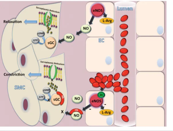
Endothelium-derived NO is one of the main regulators of the vessel tone. The activity of endothelial NO synthase (eNOS) promotes NO production in endothelial cells (ECs) (44). eNOS is regulated by a complex signaling network including kinase-dependent signaling pathways such as PI3K/Akt, Src and JAK2 in res-ponse to different stimuli (45-47).
Under normal conditions, NO pro-vides vasodilation of the microcircula-tion and maintenance of normal vascular tone, antithrombotic effects, prevention of excess platelet adhesion and aggrega-tion, inhibition of endothelial apoptosis, as well as vascular smooth muscle cell hyperplasia. NO released from ECs dif-fuses to adjacent smooth muscle cells and activates soluble guanylate cyclase (GC). GC, in turn, produces cyclic gua-nosine monophosphate (cGMP), which activates intracellular calcium pumps sequestering free Ca2+ into sarcoplasmic
reticulum and relaxing smooth muscle cells (48). Dysregulation of eNOS has been reported in several vascular dis-eases, including aneurismal SAH. Fol-lowing hemorrhage, hemoglobin binds NO, thus reducing its availability (49). Accordingly, scavenging of NO results in a decrease of GC activity, which, in return, causes vasoconstriction. Further-more, hemoglobin has shown to directly inactivate GC by oxidization, thus leading to reduced production of cGMP (50). Furthermore, after SAH, increased oxidative stress can uncouple eNOS via Zn2+ thiolate oxidation, or, theoretically,
by tetrahydrobiopterin depletion or oxi-dation, resulting in a paradoxical release of superoxide anion radical, further exacerbating oxidative stress and micro-vascular damage (51, Figure 2).
To date, endothelial mechanisms are considered to be the main contributors to induced vasospasm (52). It has been reported that NO level decreases acutely within 10 min after SAH both in exper-imental models and in humans, leading cerebrovascular relaxation impairment ErYTHrOPOIETIN sIGNaLING
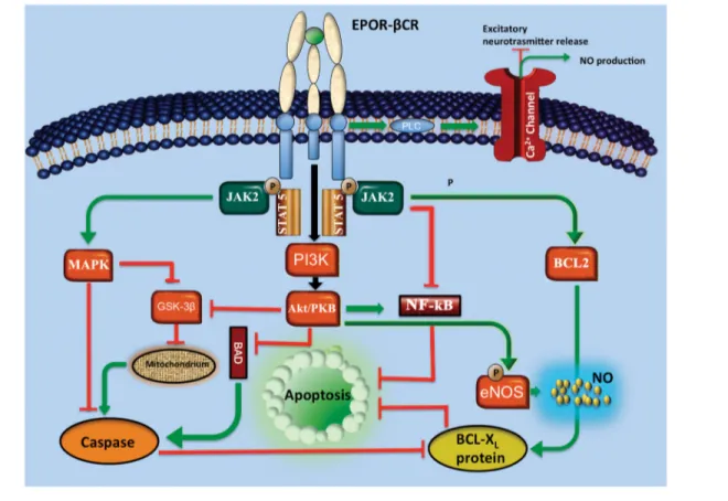
PaTHWaYs IN THE NErVOUs sYsTEM The mechanism by which rHuEPO acts in the central nervous system across the BBB remains a matter of controversy. EPO exerts its neuroprotective effects by act-ing through several signalact-ing pathways. In the bone marrow, the EPO binding to its receptor results in phosphorylation of JAK2. Upon JAK2 phosphorylation and activation, the signaling propagates through the MAPK or the protein kinase B (PKB/Akt)–nuclear factor-κB (NF-κB) pathways (34). In erythroid cells, MAPK activation promotes the accumulation of the antiapoptotic protein BCL-XL by the inhibition of caspases. Also, EPO appears to prevent apoptotic injury through an Akt-dependent mechanism (35).
Substantial evidence also indicates that EPO mediates protective effects by maintaining normal vascular autoregu-lation. This has clinical relevance in pa-thologies such as stroke, traumatic brain injury (TBI), spinal cord injury (SCI) and aneurismal SAH. The principal mech-anism of this protective mechmech-anism appears to depend upon NO signaling via increased endothelial NO synthase (25), which mediates vascular relaxation and blood flow preservation. This obser-vation could explain the remarkable effi-cacy of EPO in reversing the vascular spasm that accompanies subarachnoid hemorrhage (36–38) and spinal cord com-pression (39,40), which, in turn, reduces tissue damage. Moreover, recent studies reported that EPO promotes neovascu-larization through an Akt-dependent activation of eNOS in endothelial pro-genitor cells (EPCs) (41,42). Thus, the ability of EPO to counteract cerebral vaso-spasm may be related to a direct effect on the vascular endothelium rather than a direct action on cerebral parenchyma. The recognized ability of EPO to activate protein kinase B (Akt) and subsequent phosphorylation of eNOS in endothelial cells suggests that increased formation of NO could be an important mecha-nism underlying the therapeutic effect of EPO, although the definitive interac-tion between EPO and NO is not fully
injury produced in several experimental models of brain insult. In this regard, many studies have shown a neuroprotec-tive effect of EPO in models of cerebral ischemia (21,71–75), concussive brain injury (76,77), experimental autoimmune encephalomyelitis (78) and kainate- induced seizures (79). In these experimen-tal studies, EPO has been administered both intrathecally or systemically, pro-voking controversy regarding the ability to cross the BBB. It has been reported that systemic administration of rHuEPO, following experimental SAH, reduces the mortality rate, improves functional Further studies are necessary to better
understand the potential roles exerted by EPO and its derivatives in the plasticity and tissue protection of the nervous system. Although the exact mechanisms underlying the interaction between EPO and NO system are unclear, overall these findings have implications for treat ment of brain injury following SAH.
FrOM BENCH TO BEDsIDE
During the past several years, interest has focused on the efficacy of recombi-nant human EPO (rHuEPO) as a neuro-protective agent against neurological Previous investigations have
demon-strated an acute decrease in cerebral NO levels after SAH (8) and a significant improvement of NO system activity after administration of EPO (27,68,69), suggesting that EPO could act against vasospasm by enhancing the endothelial release of NO during the early stage of SAH. In experimental SAH, gene trans-fer of EPO following intracisternal blood injection increased the phosphorylation of Akt and eNOS, resulting in increased NO production in the basilar arteries, suggesting the beneficial effect of EPO during SAH (70).
Figure 1. Main EPO signaling pathways. Following the EPO binding with the complex EPO receptor (EPOR) and β common receptor (βCR), Janus tyrosine kinase 2 (JAK2) is activated. At this time, a secondary signaling pathway involves mitogen-activated protein kinase (MAPK), phosphatidylinositol 3-kinase (PI3K) and nuclear factor-κB (NF-κB). Signal transduction and activator of transcription 5 (STAT5) can also be triggered. These processes allow an increase in antiapoptotic proteins of the B-cell leukemia/lymphoma (BCL) family. Some of the pathways act directly, while others indirectly by inhibiting the activity of a group of enzymes called caspases. These are key mediators of apoptosis and are responsible for the degradation of antiapoptotic proteins. EPO also inhibits glycogen synthase kinase 3β (GSK3β), which, in turn, prevents the mitochondrial permeability transition pore, a key factor of cell death, through caspase activation. Finally, EPO modulates the activity of calcium channels through phospholipase C (PLC), thus reducing the release of excit-atory neurotransmitters and increasing the production of nitric oxide (NO). Moreover, recent studies reported that EPO promotes neo-vascularization through an Akt-dependent activation of eNOS in endothelial progenitor cells. The recognized ability of EPO to activate protein kinase B (Akt) and subsequent phosphorylation of eNOS in endothelial cells suggests that increased formation of NO could be an important mechanism underlying the therapeutic effect of EPO.
important finding provided by this study was the observation that the concentra-tion of EPO in the cerebrospinal fluid (CSF), assessed before euthanization of the animals, was significantly higher in the rHuEPO-treated animals than in the other groups, suggesting for the first time that systemically-administered EPO by crossing the BBB can result in increased EPO concentrations within the CSF. In a subsequent study, rabbits were given intraperitoneal injections of rHuEPO (1,000 IU/kg) starting 5 min after the induc-tion of SAH and repeated every 8 h for 72 h (37). The authors aimed to investigate the ability of exogenous administered EPO to exert a vascular effect and, in partic-ular, to counteract the spastic response of the cerebral arteries during SAH. By test performed at 24, 48 and 72 h after
SAH showed an increase in locomotor activity at 72 h in the placebo-treated group, while no increase in locomotor activity was observed in rabbits treated with rHuEPO.
Subsequently, the efficacy of rHuEPO has been evaluated on acute cerebral isch-emia following experimental SAH in a rabbit model (36,80). Histological anal-ysis performed 24 h following injury docu mented a reduction in brain isch-emic damage in animals given rHuEPO (1,000 IU/kg). In particular, analysis of cortical neurons showed that the EPO- treated rabbits presented with a significant decrease in the amount of necrotic neu-rons compared with the untreated and placebo-treated animals (80). Another outcome, and prevents brain ischemic
damage (37,38,80–88). Given these find-ings, and according to reports that have demonstrated that EPO enhances the NO system activity (27,68,69), and neuropro-tective effects on cerebral cortical neurons from N-methyl-d-aspartate receptor- mediated glutamate toxicity (71), we fur-ther investigated the potential protective effects of rHuEPO in a rabbit model of SAH. Briefly, after experimental SAH has been induced by intracisternal blood injec-tion, the results showed an improvement in functional recovery and mortality rate following systemic rHuEPO administra-tion (1,000 IU/kg) for 72 h post-SAH (81). Interestingly, all EPO-treated animals survived, while 42.9% of placebo-treated animals died within 72 h. An open-field
Figure 2. Main metabolic pathways involving NO in SAH. In endothelial cell (EC), NO is produced by NO synthase (eNOS) action. The soluble NO released from endothelial cells diffuses to adjacent smooth muscle cell (SMC) and activates soluble guanylate cyclase (GC). GC, in turn, produces cyclic guanosine monophosphate (cGMP), which activates intracellular calcium pumps thus sequestering free Ca2+ into intracellular sarcoplasmic reticulum and relaxing SMC. Following aneurysm bleeding hemorrhage, hemoglobin (Hb) binds
NO thus reducing its availability. Hence, scavenging of NO results in a decrease of GC activity, which in return causes vasoconstriction. Furthermore, Hb directly inactivates GC by oxidization, thus leading to reduced production of cGMP. Finally, increased oxidative stress uncouples eNOS via Zn2+ thiolate oxidation resulting in a release of superoxide anion radical, further exacerbating oxidative stress and
Doppler ultrasonography. Secondary end-points were incidence of delayed isch-emic deficits and outcome at discharge and at 6 months. As result, although no differences were demonstrated in the incidence of vasospasm and adverse events between the two groups, patients receiving EPO had a decreased incidence of severe vasospasm, reduced DIDs, a shortened duration of impaired autoreg-ulation and more favorable outcome at discharge.
In spite of some limitations, includ-ing a small number of cases, a sinclud-ingle EPO dose, a single center and a lack of scheduled computed tomographic scan examinations, the study demonstrates in humans what was already observed in experimental studies (88). EPO admi-nistration can be effective in limiting cerebral vasospasm and ischemia after aneurysmal SAH.
The interesting features observed in experimental and clinical studies suggest new therapeutic strategies. Some issues, however, should be considered. The first concern is the safety of recombinant human EPO administration in the setting of SAH. It should be considered that the current information about the safety of this drug in humans comes from its use in anemic patients. Translating such information from anemic therapy to SAH-affected patients can be critical, since many pieces of information regard-ing the interaction and influence between EPO and physiologic variables, as well as with common therapy administered to patients with SAH, are unknown.
Second, several lines of evidence sug-gest that chronic EPO administration can produce hypertension, hypertensive encephalopathy, accelerated atheroscle-rosis, seizures and thrombotic/vascu-lar events (82). In a model of embolic stroke in rats, EPO (5,000 U/kg) in combination with tissue plasminogen activator (tPA) exacerbated tPA-induced brain hemorrhage without reduction of ischemic brain damage when adminis-tered 6 h after stroke by upregulating matrix metalloproteinase-9, NF-κB, and interleukin-1 receptor-associated such as bcl-2 and bcl-xL, were
upregu-lated after injections of rhEPO. Recently, the effect of EPO and darbepoetin-α (DA), a novel EPO-derived agent with an exten-ded circulatory half-life and an increased
in vivo biological activity greater than EPO, were assessed in a rabbit model of SAH (92). Both erythropoietin and darbepoetin α treatments were found to attenuate cerebral vasospasm and pro-vide neuroprotection after SAH.
Based on the experimental evidence suggesting an efficacious EPO-based ther-apy in SAH (86), clinical trials blossomed, however, with uncertain results (93,94). In this regard, the first clinical trial was ter-minated preliminarily, with inconclusive results, because of a lower than expected inclusion rate (93). Seventy- three patients were randomized to treat ment with EPO (500 IU/kg/day for three days) or placebo. The primary end point was Glasgow Out come Score at six months. Surrogate measures of secondary ischemia, that is, transcranial Doppler (TCD) flow velocity, sym pto matic vasospasm, cerebral meta-bolism and jugular venous oximetry, bio chemical markers of brain damage and blood–brain barrier integrity were assessed. The study failed in assessing the primary endpoint due to the limited sample size. Furthermore, except for an increased EPO concentration in the CSF of the EPO-treated group, there were no statistically significant group differences in the primary or secondary outcome measures.
A recent Phase II, proof-of-concept trial tested the hypothesis that acute sys-temic EPO therapy in patients following aneurismal SAH can reduce cerebral vaso spasm and shorten impaired auto-regulation as primary endpoints, which subsequently decrease delayed ischemic deficits (DIDs) and improve clinical out come as secondary endpoints (94). Within 72 h following aneurysm bleed-ing, 80 patients were randomized to receive intravenous EPO (30,000 U) or placebo every 48 h for a total of 90,000 U. Primary endpoints were the incidence, duration and severity of vasospasm and impaired autoregulation on transcranial morphometric analysis of the basilar
artery, the authors observed that the administration of rHuEPO significantly reduced the vasoconstriction in SAH-plus-rHuEPO-treated animals compared with other animals that underwent SAH. Patho logical findings also showed that EPO attenuated SAH-induced brain injury. Further evidence for the bene ficial effect in the setting of SAH has also been provided by an experimen-tal SAH model where a single dose of EPO (400 IU/kg) given subcutaneously prevented SAH-induced impairment of CBF autoregulation (89). Further evidence confirmed this observation (90). An addi-tional experiment has confirmed the neu-roprotective properties exerted by EPO by measuring the S-100 protein concentration in CSF of SAH-injured rabbits (38). The findings of this study indicated that high levels of S-100 protein correlated with mortality rate, neurological outcome, and ischemic brain damage. Animals treated with rHuEPO were found to have signi-ficantly lower levels of S-100 protein in their CSF, no deaths, favorable neurolog-ical outcome and significant protection against brain ischemic damage.
In a recent experimental study, recom-binant adenoviral vectors (109 plaque-
forming units per animal) encoding genes for human EPO (AdEPO), and β-galacto-sidase were injected immediately after injection of autologous arterial blood into the cisterna magna of rabbits, resulting in significant reversal of arterial vasospasm (70). Subsequent experimental studies have tried to provide new insight into the mechanisms underlying EPO-mediated neuroprotection. In particular, Cheng and collaborators (91) investigated whether rhEPO administration influenced the endo-thelial cell apoptosis in the basilar artery after SAH in the rabbit. They also inves-tigated the modulation of rhEPO on the activity of JAK2 and STAT3 as part of the signaling in apoptotic mechanisms. As a result, they found that administration of rhEPO could activate JAK2 and STAT3 in the basilar artery and decrease the apop-tosis index of endothelial cells following SAH. Moreover, the antiapoptotic genes,
6. Dorsch N, Branston NM, Symon L, Jakubowski J. (1989) Intracranial pressure changes following primate subarachnoid haemorrhage. Neurol. Res. 11:201–4.
7. Bederson JB, et al. (1998) Acute vasoconstriction after subarachnoid hemorrhage. Neurosurgery. 42:352–60; discussion 360–2.
8. Sehba FA, Schwartz AY, Chereshnev I, Bederson JB. (2000) Acute decrease in cerebral nitric oxide levels after subarachnoid hemorrhage. J. Cereb. Blood Flow
Metab. 20:604–611.
9. Jelkmann W. (1994) Biology of erythropoietin.
Clin. Investig. 72:S3–10.
10. Jacobs K, et al. (1985) Isolation and characteri-zation of genomic and cDNA clones of human erythropoietin. Nature. 313:806–10.
11. Jelkmann W. (1992) Erythropoietin: structure, control of production, and function. Physiol. Rev. 72:449–89.
12. Rosenlof K, Fyhrquist F, Pekonen F. (1987) Re-ceptors for recombinant erythropoietin in human bone marrow cells. Scand. J. Clin. Lab. Invest. 47:823–7.
13. Ohashi H, Maruyama K, Liu YC, Yoshimura A. (1994) Ligand-induced activation of chimeric recep tors between the erythropoietin receptor and receptor tyrosine kinases. Proc. Natl. Acad.
Sci. U. S. A.91:158–62.
14. Fisher JW. (2003) Erythropoietin: physiology and pharmacology update. Exp. Biol. Med. (Maywood). 228:1–14.
15. Digicaylioglu M, et al. (1995) Localization of spe-cific erythropoietin binding sites in defined areas of the mouse brain. Proc. Natl. Acad. Sci. U. S. A. 92:3717–20.
16. Masuda S, et al. (1994) A novel site of erythro-poietin production. Oxygen-dependent pro-duction in cultured rat astrocytes. J. Biol. Chem. 269:19488–93.
17. Juul SE, Yachnis AT, Rojiani AM, Christensen RD. (1999) Immunohistochemical localization of erythropoietin and its receptor in the developing human brain. Pediatr. Dev. Pathol. 2:148–58. 18. Chin K, et al. (2000) Production and processing of
erythropoietin receptor transcripts in brain. Brain
Res. Mol. Brain Res. 81:29–42.
19. Grasso G, et al. (2005) Erythropoietin and eryth-ropoietin receptor expression after experimental spinal cord injury encourages therapy by exog-enous erythropoietin. Neurosurgery. 56:821–7; discussion 821–7.
20. Nagai A, et al. (2001) Erythropoietin and eryth-ropoietin receptors in human CNS neurons, astrocytes, microglia, and oligodendrocytes grown in culture. J. Neuropathol. Exp. Neurol. 60:386–92.
21. Bernaudin M, et al. (1999) A potential role for erythropoietin in focal permanent cerebral isch-emia in mice. J. Cereb. Blood Flow Metab. 19: 643–51.
22. Marti HH, et al. (1996) Erythropoietin gene ex-pression in human, monkey and murine brain.
Eur. J. Neurosci. 8:666–76.
CONCLUsION
To date, all phase III trials using neu-roprotective drugs have failed in demon-strating efficacy, thus suggesting that great optimism can lead to premature clinical trials driven by wishful thinking instead of detailed scientific evidence.
Further studies must be tailored and performed to assess the safety of EPO in the setting of this delicate clinical application. Optimal tolerated dosages, therapeutic time window and duration of therapy must be clearly identified. Furthermore, since increased blood vis-cosity and thro mbotic events appear to be the major com plications for chronic EPO administration, new EPO-derived drugs without erythropoietic effects (32,97) developed and experimentally tested with efficacy at present, should be further investigated to tailor successful future clinical trials.
aCKNOWLEDGMENTs
The study has been supported by FFR 2012 of the University of Palermo, Italy. DIsCLOsUrE
The authors declare that they have no competing interests as defined by Mole
cular Medicine, or other interests that might be perceived to influence the results and discussion reported in this paper.
rEFErENCEs
1. Jackowski A, Crockard A, Burnstock G, Russell RR, Kristek F. (1990) The time course of intra-cranial pathophysiological changes following experimental subarachnoid haemorrhage in the rat. J. Cereb. Blood Flow Metab. 10:835–49. 2. Delgado TJ, Brismar J, Svendgaard NA. (1985)
Subarachnoid haemorrhage in the rat: angiogra-phy and fluorescence microscopy of the major cerebral arteries. Stroke. 16:595–602.
3. da Costa L, et al. (2015) Impaired cerebrovascular reactivity in the early phase of subarachnoid hemorrhage in good clinical grade patients does not predict vasospasm. Acta Neurochir.
Suppl.120:249–53.
4. Schmidt JM, et al. (2011) Cerebral perfusion pressure thresholds for brain tissue hypoxia and metabolic crisis after poor-grade subarachnoid hemorrhage. Stroke. 42:1351–6.
5. McCormick PW, McCormick J, Zabramski JM, Spetzler RF. (1994) Hemodynamics of subarach-noid hemorrhage arrest. J. Neurosurg. 80:710–5.
kinase-1 (95). In the recent prospective, randomized, double-blind, placebo- controlled trial, the safety and efficacy of a single intravenous bolus of epoetin alfa (60,000 U) in patients with acute ST-segment elevation myocardial infar-ction (STEMI) was evaluated (96). In the efficacy cohort, EPO administration within 4 h of percutaneous coronary intervention did not reduce infarct size and was associated with higher rates of adverse cardiovascular events among older patients.
Although in the SAH clinical studies so far reported, no adverse effects during the EPO treatment have been observed, the short-term treatment and low EPO dosage used in these pioneering studies should be considered. In preclinical stud-ies, recombinant human EPO treatment at a dose of 1,000 IU/kg administered every 8 h was effective in reducing cere-bral vasospasm and cerecere-bral ischemia, and in significantly improving neurolog-ical performance. The dosage used in the clinical setting is the lowest dose con-sidered effective following SAH. It can be argued that the uncertain results from the first clinical trial (93) and the weak findings of the second clinical study (94) can find answer in the low dosage used and fre quency of treatment. Accordingly, it is widely accepted that vasospasm and cerebral ischemia after aneurysmal SAH follow a different time course between humans and animals. In experimental settings, EPO has been found to be eff-ective at a dosage starting from 400 IU to 1,000 IU/kg with a duration of 24 to 72 h (37).
Although in experimental studies and some clinical reports, an early, short-lived phase of vasospasm occurring im-mediately after SAH has been observed, the subsequent phase that is prolonged or chronic, noted on an angiogram in 40–70% of patients in the second week after hemorrhage, appears to be most important clinically. Accordingly, to achieve stronger effects, there is a ratio-nale for starting and continuing neuro-protection for at least 14 d following the onset of SAH.
54. Arcasoy MO. (2008) The non-haematopoietic bi-ological effects of erythropoietin. Br. J. Haematol. 141:14–31.
55. Sautina L, et al. (2010) Induction of nitric oxide by erythropoietin is mediated by the {beta} common receptor and requires interaction with VEGF receptor 2. Blood. 115:896–905.
56. Su KH, et al. (2011) beta Common receptor integrates the erythropoietin signaling in activa-tion of endothelial nitric oxide synthase. J. Cell.
Physiol. 226:3330–9.
57. Carlini RG, et al. (1999) Effect of recombinant human erythropoietin on endothelial cell apopto-sis. Kidney Int. 55:546–53.
58. Urao N, et al. (2006) Erythropoietin-mobilized endothelial progenitors enhance reendotheliali-zation via Akt-endothelial nitric oxide synthase activation and prevent neointimal hyperplasia.
Circ. Res. 98:1405–13.
59. Lacombe C, Mayeux P. (1999) The molecular bi-ology of erythropoietin. Nephrol. Dial. Transplant. 14 Suppl 2:22–8.
60. d’Uscio LV, et al. (2007) Essential role of endo-thelial nitric oxide synthase in vascular effects of erythropoietin. Hypertension. 49:1142–8. 61. Santhanam AV, d’Uscio LV, Peterson TE,
Katusic ZS. (2008) Activation of endothelial nitric oxide synthase is critical for erythropoietin- induced mobilization of progenitor cells. Peptides. 29:1451–5.
62. Fiordaliso F, et al. (2005) A nonerythropoietic derivative of erythropoietin protects the myo-cardium from ischemia-reperfusion injury. Proc.
Natl. Acad. Sci. U. S. A. 102:2046–51. 63. Burger D, et al. (2006) Erythropoietin protects
cardiomyocytes from apoptosis via upregulation of endothelial nitric oxide synthase. Cardiovasc.
Res. 72:51–9.
64. Gao E, et al. (2007) Darbepoetin alfa, a long-acting erythropoietin analog, offers novel and delayed cardioprotection for the ischemic heart. Am.
J. Physiol. Heart Circ Physiol 293:H60–8. 65. Lee ST, et al. (2006) Erythropoietin reduces
peri-hematomal inflammation and cell death with eNOS and STAT3 activations in experimental in-tracerebral hemorrhage. J. Neurochem. 96:1728–39. 66. Cherian L, Chacko G, Goodman JC, Robertson CS. (1999) Cerebral hemodynamic effects of phen-ylephrine and L-arginine after cortical impact injury. Crit. Care Med. 27:2512–2517.
67. Cherian L, Goodman JC, Robertson C. (2011) Improved cerebrovascular function and reduced histological damage with darbepoietin alfa ad-ministration after cortical impact injury in rats.
J. Pharmacol. Exp. Ther. 337:451–6.
68. Banerjee D, Rodriguez M, Nag M, Adamson JW. (2000) Exposure of endothelial cells to recombi-nant human erythropoietin induces nitric oxide synthase activity. Kidney Int. 57:1895–1904. 69. Migliori M, et al. (1999) Nitric oxide-dependent
renal vasodilatation is not altered in rat with rHuEpo-induced hypertension. Kidney Blood
Press Res. 22:140–5. 39. Grasso G, et al. (2006) Amelioration of spinal
cord compressive injury by pharmacological pre-conditioning with erythropoietin and a noneryth-ropoietic erythropoietin derivative. J. Neurosurg.
Spine. 4:310–8.
40. Gorio A, et al. (2002) Recombinant human erythropoietin counteracts secondary injury and markedly enhances neurological recovery from experimental spinal cord trauma. Proc. Natl.
Acad. Sci. U. S. A. 99:9450–5.
41. Bahlmann FH, et al. (2004) Erythropoietin regu-lates endothelial progenitor cells. Blood. 103: 921–6.
42. d’Uscio LV, Katusic ZS. (2008) Erythropoietin increases endothelial biosynthesis of tetrahydro-biopterin by activation of protein kinase B alpha/ Akt1. Hypertension. 52:93–9.
43. Genc S, Koroglu TF, Genc K. (2004) Erythropoi-etin and the nervous system. Brain Res. 1000: 19–31.
44. Sessa WC. (2004) eNOS at a glance. J. Cell Sci. 117:2427–9.
45. Cai H, Davis ME, Drummond GR, Harrison DG. (2001) Induction of endothelial NO synthase by hydrogen peroxide via a Ca(2+)/calmodulin- dependent protein kinase II/janus kinase 2-dependent pathway. Arterioscler. Thromb. Vasc.
Biol. 21:1571–6.
46. Duval M, Le Boeuf F, Huot J, Gratton JP. (2007) Src-mediated phosphorylation of Hsp90 in response to vascular endothelial growth factor (VEGF) is required for VEGF receptor-2 signal-ing to endothelial NO synthase. Mol. Biol. Cell. 18:4659–68.
47. Merla R, et al. (2007) The central role of adenos-ine in statin-induced ERK1/2, Akt, and eNOS phosphorylation. Am. J. Physiol. Heart Circ.
Physiol. 293:H1918–28.
48. Ignarro LJ. (1990) Biosynthesis and metabolism of endothelium-derived nitric oxide. Annu. Rev.
Pharmacol. Toxicol. 30:535–60.
49. Pluta RM, Afshar JK, Boock RJ, Oldfield EH. (1998) Temporal changes in perivascular concen-trations of oxyhemoglobin, deoxyhemoglobin, and methemoglobin after subarachnoid hemor-rhage. J. Neurosurg. 88:557–61.
50. Kim P, Schini VB, Sundt TM Jr., Vanhoutte PM. (1992) Reduced production of cGMP underlies the loss of endothelium-dependent relaxations in the canine basilar artery after subarachnoid hem-orrhage. Circ. Res. 70:248–56.
51. Sabri M, Ai J, Lass E, D’Abbondanza J, Macdonald RL. (2013) Genetic elimination of eNOS reduces secondary complications of exper-imental subarachnoid hemorrhage. J. Cereb. Blood
Flow Metab. 33:1008–14.
52. Sehba FA, Bederson JB. (2011) Nitric oxide in early brain injury after subarachnoid hemor-rhage. Acta Neurochir. Suppl. 110:99–103. 53. Brines M, et al. (2004) Erythropoietin mediates
tissue protection through an erythropoietin and common beta-subunit heteroreceptor. Proc. Natl.
Acad. Sci. U. S. A. 101:14907–12. 23. Tan CC, Eckardt KU, Firth JD, Ratcliffe PJ. (1992)
Feedback modulation of renal and hepatic eryth-ropoietin mRNA in response to graded anemia and hypoxia. Am. J. Physiol.263: F474–81. 24. Siren AL, et al. (2001) Erythropoietin prevents
neu-ronal apoptosis after cerebral ischemia and meta-bolic stress. Proc. Natl. Acad. Sci. U. S. A. 98:4044–9. 25. Beleslin-Cokic BB, et al. (2011) Erythropoietin
and hypoxia increase erythropoietin receptor and nitric oxide levels in lung microvascular endo-thelial cells. Cytokine. 54:129–135.
26. Kawakami M, Iwasaki S, Sato K, Takahashi M. (2000) Erythropoietin inhibits calcium-induced neurotransmitter release from clonal neuronal cells. Biochem. Biophys. Res. Commun. 279:293–7. 27. Koshimura K, Murakami Y, Sohmiya M, Tanaka J,
Kato Y. (1999) Effects of erythropoietin on neuro-nal activity. J. Neurochem. 72:2565–72.
28. Martinez-Estrada OM, et al. (2003) Erythropoietin protects the in vitro blood-brain barrier against VEGF-induced permeability. Eur J. Neurosci. 18:2538–2544.
29. Li W, et al. (2004) Beneficial effect of erythropoi-etin on experimental allergic encephalomyelitis.
Ann. Neurol. 56:767–77.
30. Franke K, Gassmann M, Wielockx B. (2013) Erythrocytosis: the HIF pathway in control. Blood 122:1122–8.
31. Brines M, Cerami A. (2008) Erythropoietin- mediated tissue protection: reducing collateral damage from the primary injury response. J. Intern.
Med. 264:405–32.
32. Leist M, et al. (2004) Derivatives of erythropoietin that are tissue protective but not erythropoietic.
Science. 305:239–42.
33. Brines M, et al. (2008) Nonerythropoietic, tissue- protective peptides derived from the tertiary structure of erythropoietin. Proc. Natl. Acad. Sci.
U. S. A. 105:10925–30.
34. Digicaylioglu M, Lipton SA. (2001) Erythropoietin- mediated neuroprotection involves cross-talk between Jak2 and NF-kappaB signalling cas-cades. Nature. 412:641–7.
35. Bao H, et al. (1999) Protein kinase B (c-Akt), phosphatidylinositol 3-kinase, and STAT5 are activated by erythropoietin (EPO) in HCD57 erythroid cells but are constitutively active in an EPO-independent, apoptosis-resistant subclone (HCD57-SREI cells). Blood. 93:3757–73. 36. Grasso G. (2001) Neuroprotective effect of
re-combinant human erythropoietin in experimen-tal subarachnoid hemorrhage. J. Neurosurg. Sci 45:7–14.
37. Grasso G, et al. (2002) Beneficial effects of sys-temic administration of recombinant human erythropoietin in rabbits subjected to subarach-noid hemorrhage. Proc. Natl. Acad. Sci. U. S. A. 99:5627–31.
38. Grasso G, et al. (2002) Does administration of recombinant human erythropoietin attenuate the increase of S-100 protein observed in cerebrospi-nal fluid after experimental subarachnoid hemor-rhage? J. Neurosurg. 96:565–70.
86. Grasso G, Sfacteria A. (2010) Erythropoietin and subarachnoid hemorrhage. J. Neurosurg. 112:699–700. 87. Grasso G, et al. (2007) The role of erythropoietin
in neuroprotection: therapeutic perspectives.
Drug News Perspect. 20:315–20.
88. Grasso G, Tomasello F. (2012) Erythropoietin for subarachnoid hemorrhage: is there a reason for hope? World Neurosurg. 77:46–8.
89. Springborg JB, et al. (2002) A single subcutane-ous bolus of erythropoietin normalizes cerebral blood flow autoregulation after subarachnoid haemorrhage in rats. Br. J. Pharmacol. 135:823–9. 90. Murphy AM, Xenocostas A, Pakkiri P, Lee TY.
(2008) Hemodynamic effects of recombinant human erythropoietin on the central nervous system after subarachnoid hemorrhage: reduc-tion of microcirculatory impairment and func-tional deficits in a rabbit model. J. Neurosurg. 109:1155–64.
91. Chen G, Zhang S, Shi J, Ai J, Hang C. (2009) Effects of recombinant human erythropoietin (rhEPO) on JAK2/STAT3 pathway and endo-thelial apoptosis in the rabbit basilar artery after subarachnoid hemorrhage. Cytokine. 45:162–8. 92. Kertmen H, et al. (2014) The comparative effects
of recombinant human erythropoietin and dar-bepoetin-alpha on cerebral vasospasm following experimental subarachnoid hemorrhage in the rabbit. Acta Neurochir. (Wien). 156:951–62. 93. Springborg JB, et al. (2007) Erythropoietin in
pa-tients with aneurysmal subarachnoid haemorrhage: a double blind randomised clinical trial. Acta Neu
rochir. (Wien). 149:1089–101; discussion 1101. 94. Tseng MY, et al. (2009) Acute systemic
erythropoi-etin therapy to reduce delayed ischemic deficits following aneurysmal subarachnoid hemorrhage: a Phase II randomized, double-blind, placebo- controlled trial. Clinical article. J. Neurosurg. 111: 171–80.
95. Jia L, Chopp M, Zhang L, Lu M, Zhang Z. (2010) Erythropoietin in combination of tissue plasmin-ogen activator exacerbates brain hemorrhage when treatment is initiated 6 hours after stroke.
Stroke. 41:2071–6.
96. Najjar SS, et al. (2011) Intravenous erythropoietin in patients with ST-segment elevation myocar-dial infarction: REVEAL: a randomized con-trolled trial. JAMA. 305:1863–72.
97. Brines M, et al. (2014) ARA 290, a nonerythropoi-etic peptide engineered from erythropoietin, improves metabolic control and neuropathic symptoms in patients with type 2 diabetes. Mol.
Med. 20:658–66.
Cite this article as: Grasso G, Tomasello G, Noto M, Alafaci C, Cappello F. (2015) Erythropoietin for the treatment of subarachnoid hemorrhage: a feasible ingredient for a successful medical recipe.
Mol. Med. 21:979–87. 70. Santhanam AV, et al. (2005) Role of endothelial
NO synthase phosphorylation in cerebrovascular protective effect of recombinant erythropoietin during subarachnoid hemorrhage-induced cere-bral vasospasm. Stroke. 36:2731–7.
71. Morishita E, Masuda S, Nagao M, Yasuda Y, Sasaki R. (1997) Erythropoietin receptor is ex-pressed in rat hippocampal and cerebral cortical neurons, and erythropoietin prevents in vitro glutamate- induced neuronal death. Neuroscience. 76:105–16.
72. Sadamoto Y, et al. (1998) Erythropoietin prevents place navigation disability and cortical infarction in rats with permanent occlusion of the middle cerebral artery. Biochem. Biophys. Res. Commun. 253:26–32.
73. Sakanaka M, et al. (1998) In vivo evidence that erythropoietin protects neurons from ischemic damage. Proc. Natl. Acad. Sci. U. S. A.95:4635–40. 74. Wang R, et al. (2014) Intra-artery infusion of
recombinant human erythropoietin reduces blood-brain barrier disruption in rats following cerebral ischemia and reperfusion. Int. J. Neuro
sci. 125:693–702.
75. Dang S, et al. (2011) Neuroprotection by local in-tra-arterial infusion of erythropoietin after focal cerebral ischemia in rats. Neurol. Res. 33:520–8. 76. Wang L, et al. (2015) Recombinant human
eryth-ropoietin improves the neurofunctional recovery of rats following traumatic brain injury via an in-crease in circulating endothelial progenitor cells.
Transl. Stroke Res. 6:50–9.
77. Grasso G, et al. (2007) Neuroprotection by eryth-ropoietin administration after experimental trau-matic brain injury. Brain Res. 1182:99–105. 78. Cervellini I, Ghezzi P, Mengozzi M. (2013)
Ther-apeutic efficacy of erythropoietin in experimental autoimmune encephalomyelitis in mice, a model of multiple sclerosis. Methods Mol. Biol. 982:163–73. 79. Brines ML, et al. (2000) Erythropoietin crosses
the blood-brain barrier to protect against experi-mental brain injury. Proc. Natl. Acad. Sci. U. S. A. 97:10526–31.
80. Alafaci C, et al. (2000) Effect of recombinant human erythropoietin on cerebral ischemia fol-lowing experimental subarachnoid hemorrhage.
Eur. J. Pharmacol. 406:219–25.
81. Buemi M, et al. (2000) In vivo evidence that eryth-ropoietin has a neuroprotective effect during sub arachnoid hemorrhage. Eur. J. Pharmacol. 392:31–4.
82. Grasso G. (2003) Erythropoiesis and neuroprotection: two sides of the same coin? Lancet Neurol. 2:332. 83. Grasso G. (2004) An overview of new
pharmaco-logical treatments for cerebrovascular dysfunction after experimental subarachnoid hemorrhage.
Brain Res. Brain Res. Rev. 44:49–63.
84. Grasso G. (2006) Erythropoietin: a new paradigm for neuroprotection. J. Neurosurg. Anesthesiol 18:91–2. 85. Grasso G, Buemi M, Giambartino F. (2015) The
role of erythropoietin in aneurysmal subarach-noid haemorrhage: from bench to bedside. Acta