1
UNIVERSITÀ POLITECNICA DELLE MARCHE
FACOLTÀ DI MEDICINA E CHIRURGIA
Corso di Dottorato di Ricerca in Salute dell’Uomo
XXXI Ciclo
Direct and indirect evidence of brown adipose tissue in adult
patients from Yakutia
Dottoranda: Docente tutor:
Agrafena Efremova Prof. Saverio Cinti
Contents
1 Summary 2
2 Introduction 4
2.1. Climatic and geographical conditions of Yakutia (Siberia) 4
2.2. Human adaptation in the North 5
2.3. Brown adipose tissue in human 12
2.4. Anatomical features of brown and beige adipocytes 13
2.5. Developmental origins and plasticity of brown and beige adipocytes
15
3 Material and methods 22
3.1 Gene expression 22
3.1.1. Experimental design 22
3.1.2. RNA isolation 23
3.1.3. PCR analysis 23
3.2. Morphology 25
3.2.1. Subjects (human adipose tissue) 25
3.2.2. Light microscopy 26 3.2.3. Immunohistochemistry 26 3.2.4. Morphometry 27 3.2.5. Statistical analysis 28 3.2.6. Ethics statement 28 4 Results 29
4.1. Gene expression of peripheral blood mononuclear cells is affected by cold.
29
4.2. Direct evidence of brown adipose tissue in adult humans from Siberia
34
5 Discussion 38
Summary
After the re-discovery of brown adipose tissue (BAT) in humans, there is increasing interest in the study of induction of this thermogenic tissue as a basis to combat obesity and related complications (Cinti S., 2018). Cold exposure is one of the strongest stimuli able to activate BAT and to induce the appearance of brown-like adipocytes in white fat depots (browning process). We analyzed the potential of peripheral blood mononuclear cells (PBMCs) to reflect BAT activity based on previous studies that gave promising results in mice. BAT studies in humans require invasive techniques such as biopsies of adipose tissue or the use of techniques such as positron emission tomography, which implies the use of radioactive isotopes. Thus, it would be of interest to have a readily available source of biomarkers in mononuclear cells of peripheral blood as it was shown in experimental procedures with mice (Garcia-Ruiz et al., 2009; Díaz-Rúa et al., 2015). Our data have shown that gene expression in a subset of blood cells, PBMCs reflect certain features of brown adipose tissue response to cold exposure. PBMCs are able to express 80% of the genome, including tissue-specific transcripts (Liew CC. et al., 2006). Although, these cells did not express the key BAT thermogenic gene UCP1, they were able to express other brown markers such as Cidea, Hoxc9, Prdm16, Cpt1a and Slc27a1 in peripheral blood of Siberian people. To our knowledge, there are no previous reports on the study of browning effects of cold exposure on PBMC gene expression in humans. The data presented here demonstrate that these cells can reflect some of the cold response changes in gene expression usually found in fat. The most relevant data was the significant increase of Cidea mRNA expression observed in cold-exposed Siberian group. We also performed UCP1 and TH immunohistochemistry on autoptic biopsies from periaortic and perirenal adipose depots of patients from Siberia. Our results showed that most of the depots observed, clearly showed UCP1-positive islands both in outdoor and in indoor working patients. Comparing the two groups, we observed that outdoor workers had much more brown adipose tissue and more intensely stained for the functional protein UCP1 than indoor workers. The results showed also that there is a positive correlation between the presence of multilocular adipocytes and the density of positive TH fibers. The
possibility of using an easily obtainable biological material, such as PBMCs, to perform BAT studies, opens new and interesting possibilities for analyzing the relevance of this tissue and of WAT browning on energy homeostasis and body weight control in humans by using non-invasive approaches. Furthermore, our data show for the first time the presence of relevant amount of BAT in adult humans living in Siberia with a positive correlation between multilocular adipocytes and noradrenergic parenchymal nerve fibers
2. Introduction
2.1. Climatic and geographical conditions of Yakutia (Siberia).
Yakutia (Sakha Republic, Russian Federation) is the coldest country in the whole Northern Hemisphere, showing annual thermal amplitudes over 100 °C and its entire range covered by permafrost.
By its complex of climatic and geographical factors affecting the human body, Yakutia is a quite harsh land for living.
The Sakha Republic (Yakutia), in the Northeast of Russia, occupies a vast area of about 3.1 million km2, which is 18% of the entire territory of the Russian
Federation. Over 40% of Yakutia lies beyond the Arctic Circle. The terrain of Yakutia is very complicated – from mountain ranges with peaks over 3000 m to the lowlands, slightly elevated above the sea level. 42% of the territory is lowland, in which the Arctic desert zone and tundra stand out. Openness to the Arctic and remoteness from the Atlantic Ocean create special climatic conditions, most important of which is the extreme continentality, manifested in a very low winter (up to -60°C) and high summer temperatures (up to +39º С) with an annual temperature difference of more than 100°C (Izyumenko S.A. et.al., 1982; Maximov G.N., 2003). By its small amount of precipitation over the most part of the territory (below 200 mm), the Republic is close to the steppe and semi desert areas of Central Asian, with about 50% of precipitation falling as snow (Makarov AA, 1989). The permafrost thickness ranges from 120 to 1500 m (in the Central Yakutia – 250-300 m). Its depth in summer straight thawing depending on geographical location, soil and vegetation cover, as well as the terrain features varies from 0.1 m in the north to 3 m in the south. The great extent of the territory of the Republic from the North to the South determines the uneven intake of solar radiation throughout the year. Beyond the Arctic Circle, the polar night reigns for three months (November-January), and it is the midnight sun for summer. Powerful heliomagnetic perturbations specific to high latitudes result from the exposure by the Earth's
magnetic field of surface layer of biosphere in the Polar Regions from direct cosmic radiation (Khansulin V.I., 2009).
2.2. Human adaptation in the North
Human existence in extreme climatic and geographical conditions is associated with adaptation. Panin L.E. (2006), summarizing a series of studies, notes that in high latitudes under the influence of harsh environmental factors, the human body moves to a new level of homeostasis, characterized by greater use of fats and less use of carbohydrates for energy needs. The role of proteins in energy metabolism significantly increases the need of water and fat-soluble vitamins. Highlighting the role of nutrition in metabolic processes in the indigenous population of the North, L.E. Panin writes that although food of the aborigines contains a large amount of proteins, fats and much less carbohydrates, their bodies easily cope with protein and lipid loads. Furthermore, the author concludes that the switch of energy metabolism in the aborigines of the North from the carbohydrate type to the fat is associated with the use of not endogenous fat, but exogenous food. The presence of a large amount of unsaturated fatty acids in edible fat provides a high rate of lipid oxidation. All this creates favorable conditions for lipid metabolism and defines a lower cholesterol level (Panin L.E., 1987). In contrast to the data obtained by L.E. Panin (1979), other researchers (Bobrov N.I., 1979; Krivoshapkin V.G., 2001) note that along with prolongation of periods of residence in the North, the concentration of cholesterol and general lipids in human blood increases significantly and the level of lipoproteins even tends to decrease. In the studies of T.I. Kochan (2008), it was found that along with the increased lipid metabolism, in course of adaptation of the body to the harsh conditions of the polar climate, glycolysis intensifies. Some inconsistencies of various contingents on an unbalanced diet influence of seasonal factors. Similar evidences of the protein-lipid orientation of metabolism of the indigenous people is reported in other studies (Khaltaev N.G.,1979; Alexeyeva T. I. 1989; Agadzhanyan N., Ermakova N., 1998; Wlosinski P. E., 1999; Boiko E.,
2005).
The nature of the processes of lipid metabolism among representatives of the indigenous population of the European North reveals connection with duration of the historical residence of the population in the Subarctic. An increase of the “northern experience” of the population is accompanied by a decrease in the total lipid pool in peripheral blood, stabilization of triglyceride levels throughout the year, and a lower level of atherogenic lipid fractions in general.
The contrasting seasonality of natural and climatic factors of the Polar region causes an alteration of the processes of lipid metabolism in the indigenous northerners, characterized by seasonality, which does not match in populations with different duration of residence in the North. The process of alteration of lipid metabolism in the indigenous population of the North exceeds the individual life duration and reflects the gradual formation of labile regulatory mechanisms (Boyko E.R., Tkachev A.V., 1994).
Metabolic changes in newcomers to the North are close to indicators recorded for the indigenous people. It was shown that the people who came to work in the regions of the North, as well as the indigenous people, have an increased concentration of total lipids in blood: triglycerides, higher fatty acids and active transport form of low-density and very low density (LDL and VLDL) (Kaznacheev V.P., 1979; Kylbanova. E.S., 2006).
Apparently, the same specific metabolic reactions developed in the indigenous people of the North during the adaptation process, also activated in newcomers. Because of formation of adaptation (metabolism alteration), a «polar tension syndrome» is formed in the newcomers population (Kaznacheev V.P., Shorin Yu. P., 1980; Panina L.E., 1983; Solomonova N.G. et al. 1994, Petrova P.G. et al. 1999). Protracted cold exposure on the human body leads to an increase in protein catabolism. In this case, both the total protein content in the blood serum and spectrum of protein fractions change (Isaakyan L.A., 1972; Krivoshapkina Z.N., 2009).
Antarctic workers was revealed, which is accompanied by a violation of the digestibility of amino acids. The authors make an assumption about the possible breakdown of tissue proteins in the human body, developing during the adaptation process to the conditions of the Polar Regions. They found that during the wintering process total protein in blood serum increases, the amount of albumin increases by the end of wintering, while the amount of globulins decreases (Deryapa N.R., Ryabinin I.F., 1977).
Sedov K.R. (1970), studying protein metabolism, did not reveal differences in the total protein content and some components of the protein fractions in the indigenous inhabitants of The North and the residents of Irkutsk, levels of γ-globulins were significantly increased. An increase in the γ-globulins protein fraction in indigenous inhabitants (Sami and Nenets) was noted in the work of T.I. Alexeeva (1977). The formation of the polar metabolic type in the indigenous inhabitants and animals of the North is due to the decrease of adrenal and thyroid hormones on cold conditions. Thyroid hormones modulate the thermogenic function of the contracting skeletal muscles and increase effectiveness of all mechanisms of chemical thermoregulation. Glucocorticoids and catecholamines play an important role in the alteration of carbohydrate and fat metabolism (Kaznacheev V.P., 1980). Against the background of a decrease in insulin content in the blood of the
indigenous and newcomer population of the Far North, the metabolic effect of these hormones increases. However, in the natives of the North there had been no structural and functional straining of thyroid, whereas the newcomers had apparent thyroid hyperplasia. Perhaps it is due to the fact that in conditions of relative dormancy in the indigenous population, genetically adapted to the local conditions, vital activity proceeds at a more economical hormonal state. Adaptation of the newcomer population occurs with a more pronounced general secretory activity of the glands and a greater dispersion of individual indicators, owing to the transition of the functioning of the hormonal regulation system to a new level (Maximov A.L., Gorbachev A.L., 2001).
glucose tolerance among the indigenous people of the North and the nature of their diet. At the same time, the serum glucose level in the indigenous population of the North did not differ from the mid-latitude normal ranges. Some studies show a decrease in blood glucose in non-indigenous inhabitants of the Far North (N.R. Deryapa, I.F. Ryabinin, 1977) and a tendency to decrease of serum glucose in natives of the North (R.V. Veselukhina, 1977). At the same time, Panin L.E. (1978, 1979) has not found “relative” hypoglycemia. However, Stepanova (1985), examining the aborigines of Chukotka, did not find differences in glucose level compared with the controls. E.R. Boyko (2005) revealed that the indigenous people of the Polar region have a glucose level within the mid-latitude standards, but have a distinct tendency to be located generally closer to the lower limit of the norm. At certain periods of the year in different groups of the indigenous population of the North, glucose distinctively approaches the lower limits of the norm. According to Panin L. E. (1987), the indigenous population of Taimyr has a low glucose, lactate and pyruvate level in wintertime. The average serum glucose of the Nenets of Yamalo-Nenets autonomous district in March-April is less than 3.60 mmol/l. Thus, during this period of the year, at least 40% of the Nenets male population have a serum glucose level of less than 3.30 mmol/l, i.e., they are in the state of “relative” hypoglycemia.
Nutrition of the indigenous peoples of the North had been forming for a long time, influenced by regional characteristics and socio-economic conditions. In the earliest studies described by I.S. Kondoror (1968), nutrition in the environmental conditions of the Extreme North was characterized by the predominance of fat and animal protein with a low carbohydrate content (Vikhert A.I., Groysman E.I., 1973, Panin L.E., Kiselyova S.I., Khamnagadaev I.I., 2003).
Domestic (Sedov K.R., 1993; Tsukanov V.V., 1997, 2002; Tereshchenko V.P., 2001; Tsukanov V.V., Shtygasheva O.V., 2004; Khasnulin V.I., 1998) and foreign authors (Adler AI et al., 1994; Jul E. et al., 1994; Nayha S. et al., 1994) studied the health indicators of the northern peoples living in the subpolar and polar regions of the world and point out more positive indicators of somatic health of the population
under the traditional nutrition type.
At the end of the twentieth century, the traditional eating habits of the indigenous population of the North began to undergo significant changes associated with the loss of traditional lifestyles and changes in socio-economic living conditions (Tikhonov D.G., 1990; Tyaptirgyanova V.M., 2004; Ivanov K.I., 2005; Polivanova T.V., Manchuk V.T., 2006; Mestnikova N.V., 2009; Polikarpov L. S., 2010). In her study, Tikhonova G.D. (1990) noted that in Yakutia over the past hundred years, protein intake has increased by 1.5 times, carbohydrates by 2.0 times with a relatively stable level of fat intake. Dietary calories increased by 15%. Amount of consumed vegetables and refined carbohydrates increased significantly in the diet (Tichonov D.G., 1990). Tyaptirgyanova V.M. (2004) in her study showed a change in the traditional nutrition type in the urban indigenous population of Yakutia, which is characterized by an imbalance in almost all its constituent components, which affects the increase in the frequency of alimentary-related diseases. In the paper of Mestnikova N.V. (2009), insufficient intake of proteins, fats and carbohydrates and low energy value of the average daily diet among adolescents living in Yakutia were revealed. The author noted that contribution of proteins to the energy value of the diet was lower than the recommended by the Scientific Research Institute of Nutrition of the Russian Academy of Medical Sciences for the northern regions, while the contribution of carbohydrates exceeded by 50% among both urban and rural adolescents of Yakutia.
Later studies showed that there were no changes in the provision of the body of the northerners with vitamins (Popova A.S., Mironova G.Е., 2003; Boyko E.R., 2005). In addition, residents in the North of European Russia have a slightly higher percentage of vitamin B2 hypovitaminosis compared with European indicators (Boyko ER, 2005). According to Mironova G.E. (2003), residents of Yakutia are noted to have hypovitaminosis C, A, E, manifested especially in winter. A low concentration of ascorbic acid in the blood of Yakutia residents was noted even with normal daily intake. Rapid consumption of vitamin C in the northerners is due to its antioxidant properties, as well as the fact that it participates in the synthesis of
corticosteroid and thyroid hormones, which are important for the adaptation of humans and animals to cold (Mironova G.E., et al., 2003; Popova, A.S,, 2006, 2008).
The fact that indigenous Siberian groups have significantly elevated basal metabolic rate may also play a role in the maintenance of stable lipid levels in the serum. It has been observed that mice consuming ketogenic diets, characterized by a high content of fat and low carbohydrates, had increased metabolic rates, while their serum lipids did not increase (Kennedy A.R. et al., 2007). These mice also exhibited an overexpression of UCP1 and UCP2, suggesting activation of non-shivering thermogenesis which uncouples the mitochondrial respiration by impairing ATP production and dissipating energy as heat. Previously, Leonard et al. (2005) had hypothesized that the elevated basal metabolic rates in Siberian populations are due to genetic adaptations in their thyroid hormone signaling pathway. Recent studies suggest that thyroid hormone mediated thermogenesis emanates from the brown adipose tissue (Cannon B., Nedergaard J., 2011) providing a link between BMR and non-shivering thermogenesis. It has been shown that CPT1A is regulated by thyroid hormone and insulin highlighting the imperative role the gene has in energy regulation.
When the human and animal organism is exposed to cold, lipid peroxidation is activated against the background of an antioxidant deficiency. Moderate activation of lipid peroxidation, in response to the action of extreme agents, is one of the adaptation mechanisms and is aimed at increasing the permeability of the cell membrane and facilitating the work of membrane proteins. However, going beyond certain limits, lipid peroxidation can acquire an independent pathogenic value, which is manifested by denaturation and inactivation of proteins, membrane defatting, and impairment of cell fission and growth (Meerson F.Z., Pshennikova M.G., 1989; Vladimirov Yu.A., 2000).
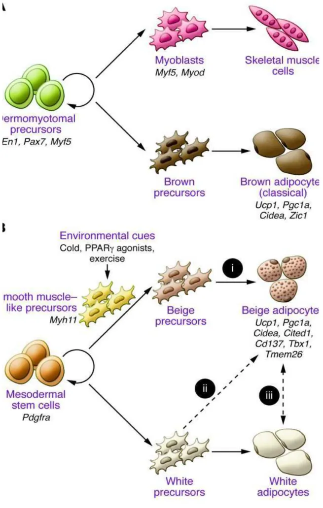
FIGURE 1. DEVELOPMENTAL ORIGINS OF BROWN AND BEIGE ADIPOCYTES (SIDOSSIS L AND KAJIMURA S., 2015)
photoperiodism, electromagnetic nature factors, severe aerodynamic regime, climate change) causes changes in regulatory and homeostatic systems that determine the characteristics of the formation and course of pathology. Hypoxia, hypoglycemia, hypovitaminosis and other abnormalities are typical for the northerners (Kaznacheev V.P., Kulikov V.Yu., Panin L.E., 1980; Deryapa N.R., Ryabinin I.F., 1977; Avtsyn A.P., Zhavoronkov A.A., Marachev A.G., 1985, Panin L.E., 1978; Panin L.E., 1980; Boyko E.R., 1996; Boyko E.R., 2005; Boyko E.R., Bichkayeva F.A., 1997; Kochan T.I., 2008). Adaptive alteration of the human body in conditions of the North is characterized by staging with the development of adaptive stress of the body with the activation of specific metabolic response complexes, which in the indigenous population are due to the appearance of “systemic structural trace of adaptation” and are genetically fixed during the evolution process.
2.3. Brown adipose tissue in human
Brown adipose tissue (BAT), a specialized fat that dissipates energy to produce heat, plays an important role in the regulation of energy balance. Two types of thermogenic adipocytes with distinct developmental and anatomical features exist in rodents and humans: classical brown adipocytes and beige (also referred to as brite) adipocytes. While classical brown adipocytes are located mainly in dedicated BAT depots of rodents and infants, beige adipocytes sporadically reside with white adipocytes and emerge in response to certain environmental cues, such as chronic cold exposure, a process often referred to as “browning” of white adipose tissue. Recent studies indicate the existence of beige adipocytes in adult humans, making this cell type an attractive therapeutic target for obesity and obesity-related diseases, including type 2 diabetes.
2.4. Anatomical features of brown and beige adipocytes
Based on the developmental and anatomical features, at least two types of thermogenic adipocytes exist in mammals: classical brown adipocytes and beige adipocytes (also known as brite adipocytes).
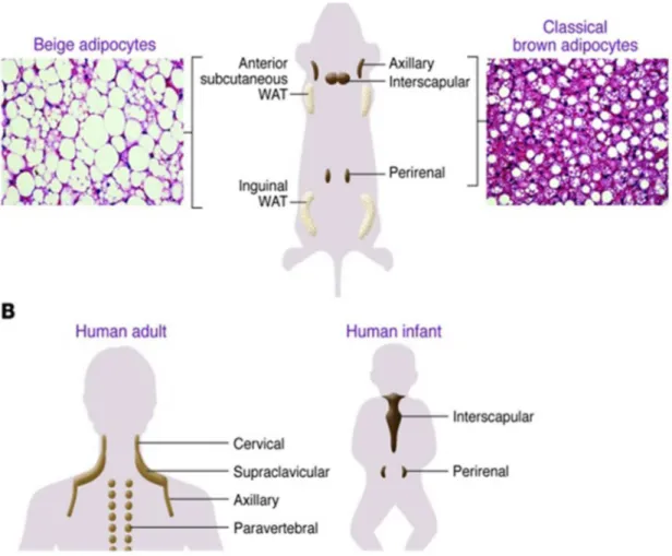
As summarized in Figure 2, classical brown adipocytes are found in the major dedicated BAT depots of rodents, such as in the interscapular, perirenal, and periaortic regions.
FIGURE 2. ANATOMICAL LOCATIONS OF THERMOGENIC ADIPOCYTES IN MICE AND HUMANS (SIDOSSIS L., KAJIMURA S., 2015)
Since BAT-mediated thermogenesis is crucial for maintaining body temperature in infants, formation of the BAT depots (i.e., classical brown adipocyte development) is largely completed during the prenatal stage in order to produce heat right after birth through nonshivering thermogenesis. On the other hand, beige adipocytes exist mainly in subcutaneous white adipose tissue (WAT) of rodents. Beige adipocytes sporadically reside within WAT, and their development is dramatically induced in response to certain external cues, such as chronic cold exposure, exercise, and long-term treatment with PPARγ agonists (e.g., rosiglitazone). This phenomenon, which occurs in the postnatal stages, is often called “browning” of WAT (Kajimura S, Saito M., 2014; Harms M, Seale P., 2013; Wu J, Cohen P, Spiegelman BM., 2013). When stimulated by such external cues, beige adipocytes express UCP1 protein at a similar level to classical brown adipocytes and exhibit UCP1-dependent thermogenic capacity (Shabalina IG. et al., 2013; Wu J, et al., 2012). In humans, classical brown adipocytes exist in the interscapular BAT of infants (Lidell ME, et al., 2013). While BAT depots of rodents remain throughout life, human brown adipocytes in the interscapular region gradually disappear with age (Heaton J.M., 1972). In adults, 18F-FDG-PET/CT scans have identified active BAT depots in the cervical, supraclavicular, axillary, and paravertebral regions. From an evolutional point of view, BAT around the neck of adult humans and nonhuman primates may evolve to protect the brain by warming up the blood supplied to the brain.
The two types of thermogenic adipocytes are also distinct in their cellular heterogeneity within adipose tissues. The interscapular and perirenal BAT depots are largely composed of classical brown adipocytes with multilocular lipid droplets and separated from WAT by a layer of connective tissue (Cinti S., 1999). In contrast, beige adipocytes in WAT exist together with unilocular white adipocytes, without any separation by a distinct later of connective tissue.
Under cold stimulation, clusters of beige adipocytes emerge in the area in which tyrosine hydroxylase–expressing (noradrenergic) nerve fibers are densely
innervated (Cinti S., 1999; Murano I. et al, 2009), as norepinephrine released from the sympathetic nerves is a powerful stimulator of WAT browning. Intriguingly, adult human brown adipocytes are also sporadically found together with white adipocytes, and the density is well correlated with the number of tyrosine hydroxylase–positive noradrenergic nerves (Lidell ME, et al., 2013; Zingaretti MC., et al., 2009).
2.5. Developmental origins and plasticity of brown and beige adipocytes
Brown and beige adipocytes share many biochemical characteristics, including enriched mitochondria, multilocular lipid droplets, and expression of UCP1. However, they originate from distinct cellular lineages. As shown in Figure 2A, classical brown adipocytes originate from a subpopulation of dermomyotomes that can also give rise to skeletal muscle. A lineage-tracing study found that engrailed-1–expressing (En1-expressing) cells in the central dermomyotome differentiate to BAT, skeletal muscle, and dermis (Atit R. et al., 2006). Nearly the
entire population of classical brown adipocytes in the interscapular BAT arises from precursors that express Myf5, a gene known to be expressed in committed skeletal muscle precursors (Seale P. et al., 2008). A pulse-chase tracing study used another myogenic marker, Pax7, and showed that the divergence of myoblasts and brown adipocyte progenitors occurred between embryonic days 9.5 and 11.5 in mice (Lepper C., Fan C.M., 2010). As an independent line of evidence, the transcriptional profile of classical brown adipocytes resembles that of skeletal muscle cells (Walden T.B. et al., 2009; Timmons J.A. et al., 2007). Similarly, the mitochondrial proteomic profile of BAT is more related to that of skeletal muscle than that of WAT (Forner F. et al., 2009).The cellular origin of beige adipocytes has not been completely elucidated. UCP1-expressing beige adipocytes in the epididymal WAT arise through the proliferation and differentiation of precursors that express platelet-derived growth factor receptor α (PDGFRα), CD34, and spinocerebellar ataxia type 1 (SCA1) (Lee Y.H. et al., 2012). Beige adipocytes in the inguinal WAT were
reported to derive from Myf5-negative precursors (Seale P. et al., 2008), whereas other studies showed that a subset of beige adipocytes in multiple WAT depots, such as anterior subcutaneous and perigonadal depots, can originate from Myf5-positive cells (Sanchez-Gurmaches J. et al., 2008). Furthermore, a recent paper by Spiegelman’s group showed that a subset (10%–15%) of UCP1-positive beige adipocytes, but not classical brown adipocytes, arises from precursors that express Myh11, one of the most selective markers for smooth muscle cells (Long J.Z. et al., 2014). Collectively, these results indicate that beige adipocytes have distinct cellular origins from classical brown adipocytes and are composed of heterogeneous cell populations.
Another intriguing yet enigmatic issue is the cellular plasticity of beige adipocytes. Several studies indicate that mature white adipocytes may directly convert into brown adipocytes (i.e., transdifferentiation) in vivo (Himms-Hagen et al Am J Physiol 2000), supporting the idea that beige adipocytes could correspond to intermediate forms. For example, adipocytes at an intermediate state between white and brown contain “paucilocular” lipid droplets and express the brown marker UCP1 during the process of cold adaptation (Barbatelli G. et al., 2009, Reviewed in Cinti S, Comprehensive Physiology 2018). A large part of the newly formed brown and beige adipocytes in subcutaneous WAT is negative for BrdU or Ki67, such that mitotic proliferation is not required for the cold-induced brown-beige adipocyte differentiation (Lee Y.H.et al., 2012; Himms-Hagen J, Melnyk A, Zingaretti MC, Ceresi E, Barbatelli G., 2000; Frontini A, et al., 2013). Furthermore, a recent genetic lineage–tracing study showed that beige adipocytes converted into mature white adipocytes after 5 weeks of warm adaptation. These white adipocytes can reconvert into beige adipocytes after additional cold stimulation (Rosenwald M, Perdikari A, Rülicke T, Wolfrum C., 2013). On the other hand, Scherer’s group performed pulse-chase fate-mapping experiments of mature adipocytes and found that the majority of newly developed beige adipocytes stem from de novo differentiation of precursors in subcutaneous WAT (Sharp LZ, et al. 2012). Hence, the
transdifferentiation hypothesis still needs to be critically tested by monitoring the life cycle of beige adipocytes at a single-cell resolution.
While BAT has long been appreciated as an important player in cold defense in human infants, BAT thermogenesis was thought to be completely blunted during adulthood.
Previous literature has indicated that BAT may have a potential role in regulation of not only body temperature during cold exposure but also body weight and lipid metabolism. The physiology and activity of BAT have been scientifically investigated, mainly in rodents, in numerous studies in recent decades. These studies have confirmed the presence of BAT and shown the benefits of activating BAT for regulation of body weight. Various studies underline that “cafeteria-fed” animals had increased mass and function of BAT, while surgical excision of BAT in mice resulted in increased body weight. Moreover, overexpression of UCP1 in transgenic mice protected against obesity, and adrenergic stimulation of the beta3-adrenoreceptor resulted in appearance of BAT in WAT regions, accompanied by increased expression of UCP1 and reduction in body weight (Kozak LP.et al., 2010). BAT was initially noticed in humans several decades ago. The function of BAT in infants was described in the 1960s, and the question of the clinical significance of BAT in adults was then raised in the 1970s (Himms-Hagen J., 1979; Heaton JM., 1972). Interestingly, a highly quoted work from Finland in 1981 demonstrated that outdoor workers had BAT surrounding their neck arteries much more often than indoor workers, suggesting that working in the cold can retain BAT (Huttennen P., et al., 1981). Pheochromocytoma, a catecholamine-secreting tumor of the adrenal medulla, was shown to be associated with activation of BAT in the 1980s; this finding suggested that human BAT has the potential for thermogenic activity and may contribute to weight loss through the high level of catecholamines activating BAT (Lean ME. et al., 1986). Catecholamines can induce weight loss through different pathways. However, it has been shown that FDG-PET uptake in BAT was no longer apparent after resection of a pheochromocytoma tumor (Kuji I. et al., 2008). Indeed, a recent study correlated elevated plasma levels of
metanephrine and BAT activity, providing evidence that catecholamines stimulate BAT thermogenesis in the presence of an adrenal tumor and are negatively associated with adiposity (Wang Q. et al., 2011). However, BAT has been generally considered nonfunctional with no significant physiological relevance in healthy adult humans until recent years. The renewed interest in BAT occurred as a result of an incidental finding. In the 1990s, radiologists performed FDG-PET scans to detect increased glucose uptake in tumors and identify metastatic progression. During these scans in patients with cancer, radiologists noticed a repeated pattern of bilateral symmetrical glucose uptake in the upper chest and neck regions, initially ascribed to artefacts or muscle uptake because of their symmetrical distribution (Engel H.et al., 1996). After combining FDG tomography with CT, it became possible to determine the composition of this symmetrical uptake and this mass was identified as adipose tissue. In 2002, using hybrid PET/CT imaging, Hany and colleagues elegantly suggested that the symmetrical FDG uptake was related to adipose tissue and probably represented activated BAT during the increased sympathetic activity induced by the cold stress in the imaging room (Hany TF. et al., 2002). Several other reports discussing FDG-PET uptake in BAT in adult humans were published soon after, mainly in nuclear medicine journals (Cohade C. et al., 2003; Yeung HW., et al., 2003). Surprisingly, it took several years until Nedergaard and colleagues raised awareness of the “unexpected evidence for active BAT in adult humans” and its potential to be of metabolic significance in human physiology and in the efforts to combat obesity (Nedergaard J., Bengtsson T., Cannon B., 2007). Still, because of the retrospective nature of the FDG-PET studies, the presence of active BAT in humans was believed to be apparent in only a small percentage of adults. This notion changed after the results of parallel prospective studies from several independent working groups were presented in 2009. Cypess and colleagues retrospectively analyzed FDG-PET/CT scans performed in 1972 patients for various diagnostic reasons (Cypress AM. et al., 2009). Concentrations of depots of BAT were identified in regions of the anterior neck and thorax. Positive scans were seen in 7.5 % of women and 3 % of men (this
low percentage was because the studies were performed in a nonstimulated state and PET/CT identifies uptake mainly in tissues with increased metabolic activity). The probability of detection of BAT was inversely correlated with age, outdoor temperature, use of a beta-blocker, and body mass index (BMI) in the older patients. Additionally, UCP1 activity was identified in 33 biopsy specimens from the same cervical and supraclavicular regions in which BAT was observed on PET/CT. At the same time, prospective studies were conducted to intentionally examine BAT activity in cold stressed subjects. Lichtenbelt et al. found BAT activity using FDG-PET/CT in 23 of 24 healthy men during mild exposure to cold (15°C for 2h, ensuring that there was no cold-induced shivering thermogenesis by muscles) but not under thermoneutral conditions. This activity had negative correlation with BMI, suggesting that the reduced BAT activity in obese individuals may make it a target for the treatment of obesity (Van Marken Lichtenbelt WD. et al., 2009). Saito and colleagues also demonstrated that cold-induced uptake was observed on FDG-PET/ CT in 27 of 32 young healthy volunteers but in only 2 of 24 old-erly subjects, with no detectable uptake in warm conditions. The BAT activity was inversely correlated to BMI and was higher during the winter months (Saito et al., 2009). Another study performed at the same time examined histologic samples of adipose tissue from the neck of patients undergoing thyroid surgery (Zingaretti MC., Cinti S., 2009). UCP1 activity was found in one-third of the 35 patients, with evidence of BAT precursors. BAT had high sympathetic innervation and capillary density. Virtanen et al. additionally showed that expo- sure to cold increased FDG-PET/CT glucose uptake 15-fold in the supraclavicular area of 5 healthy volunteers (Virtanen KA. et al., 2009). Three of their subjects underwent tissue biopsies from areas corresponding to the glucose uptake, showing that these tissues express messenger RNA for markers of BAT and substantial levels of UCP1 protein and cytochrome c (a mitochondrial marker that is abundant in BAT). Collectively, these studies in humans demonstrated the presence of BAT mass and activity in adults based on cold-induced glucose uptake tests and biopsies, showing variability (30–100%) in study results based on age, BMI values, gender, and ambient temperature. However,
these values were much greater than those of the earlier retrospective PET/CT studies, which reported a low prevalence of BAT in adult humans, varying from 2.5% to 8.5% of tested individuals because of thermoneutral noncold-induced conditions. The next step was to confirm that BAT is indeed metabolically active in humans and contributes to cold-induced nonshivering thermogenesis. Researchers recently measured blood perfusion in BAT using a technique of intravenous injection of [15O] H2O and a dynamic PET/CT emission scan. They
found that cold activation of BAT leads to a greater than 2-fold increase in the per- fusion rate of the tissue in parallel to elevated glucose uptake, reflecting the dense vascularity of this tissue and the increased oxygen requirement. Moreover, increased blood flow was associated with whole-body energy expenditure during cold exposure, indicating active thermogenesis (Orava J. et al., 2011). Others have also shown that increased energy expenditure after exposure to cold in healthy volunteers is correlated to BAT activity quantified from FDG uptake (Yoneshiro T. et al., 2011). Recently, Quellet and colleagues found that cold activation of BAT is associated with not only increased blood flow, but also higher oxidative metabolism in the tissue (Quellet V. et al., 2012). This finding was confirmed by exposing healthy subjects to controlled cold and injecting a bolus of labeled radioactive acetate into their blood. The acetate is distributed to tissues according to their proportional blood flow. In this technique, loss of radioactivity from a tissue is an indication of active oxidative metabolism, and the radioactivity disappeared from BAT with a half-life of minutes in cold but not in warm subjects. Furthermore, nonesterified fatty acid uptake was quantified, showing increased uptake in cold- activated supraclavicular BAT in comparison to subcutaneous adipose tissue and resting skeletal muscles; these results suggest increased utilization of triglycerides as a source of energy for BAT thermogenesis.
A recent study using oxygen-15 PET imaging that evaluated the relationship between BAT oxidative metabolism and FDG uptake reported that despite elevated glucose uptake in BAT of adult humans, activated BAT did not contribute much to total energy expenditure (Muzik O. et al., 2012). The low activity of BAT depots in
human subjects may reflect the low density of the brown adipocytes, and thus, the abundance of these cells would need to be increased in order to affect energy expenditure. FDG-PET/CT is currently the imaging modality of choice for investigating BAT activity non-invasively. Recent developments in magnetic resonance imaging (MRI) may aid in the evaluation of BAT activity without exposure to ionizing radiation, with a better reproducibility and reduced cost. Recent studies have demonstrated the feasibility of measuring the volume and function of BAT in vivo using routine MRI sequences (Chen Y.I. et al., 2012). Other novel imaging techniques, including infrared thermography and new imaging tracers such as 4-18F-fluorobenzyltriphenylphos-phonium (18F-FBnTP), are promising noninvasive tools for detection and monitoring of BAT in humans (Lee P. et al., 2011: Madar I. et al., 2011). In this study we compared anatomical areas that may contain BAT in autoptic biopsies from outdoor and indoor living Siberian people. Furthermore, we also studied the gene expression of typical BAT genes in mononucleated cells of the blood extracted from outdoor workers in the north area of Siberia. This last study is based on the observation that BAT genes can respond to cold exposure also in mononuclear blood cells in rodents (Reynés et al., 2015).
3. Material and methods
3.1 Gene expression
3.1.1. Experimental design
Peripheral blood mononuclear cells (PBMCs) constitute an easily obtainable fraction of blood cell consisting basically in lymphocytes and monocytes. All samples from blood were obtained from 219 volunteers - healthy men aged 25 to 56 years.
The cold exposed group included 180 diamond miners who were engaged in open-pit diamond mining during the winter period at Verchoyansky district of Yakutia that is located above the Polar circle. The average time they spent in the cold due to their duties is about 6-8 hours a day. Blood samples were collected during February in Anabarsky district of Yakutia (the average temperature during that period was about -45/-52°C).
The control group consisted of 29 healthy male volunteers, the blood was taken during the summer (August), and the subjects were not exposed to the cold. The average age and other parameters are shown in table 1.
Table 1 Cohorts Cold exposure (h) Mean age (y) Height (cm) Weight (cm) Body mass index Waist (cm) Hips (cm) W/H Control group n=29 0 34,07±5 ,49 174,55±4,68 78,21±7,4 9 25,73± 2,55 90,31±9,4 7 100,45±3 ,98 0.90±0,08 Diamond miners n=180 6-8 35,06±1 0,02 171,04±5,92 74,11±11, 62 25,22± 3,61 88,06±10, 81 97,23±6, 63 0,90±0,06
3.1.2. RNA isolation
Total RNA was isolated from whole blood samples that were collected into EDTA coated vacutainers and immediately transported to the laboratory for further processing. Blood samples were kept at -70°C before RNA isolation.
Total RNA was isolated as previously described. Briefly, 8 ml of human peripheral blood were transferred into a 50 ml polypropylene conical centrifuge tube. Then RBC Lysis Buffer was added to a final volume of 45 ml with subsequent incubation at room temperature for 10 minutes. Cells were pelleted by centrifugation at 600 x g (approx. 1,400 rpm) for 10 minutes. The supernatant was decanted and the pellet was resuspended in 1 ml of RBC Lysis Buffer and transferred to a 1.5 ml microcentrifuge tube. Afterwards cells were pelleted for 2 minutes by centrifuging in a microfuge at room temperature at 3000 rpm and subsequently resuspended in 1 ml DPBS and pelleted again by centrifugation. The pellet was resuspended in 1200 µl of TRIzol solution and 0.2 ml of Chloroform and vortexed for 15 seconds. Samples were centrifuged at 13,000 rpm for 10 minutes at 4°C. The upper phase was transfered to a clean microcentrifuge tubes and an equal volume of cold isopropanol was added to the mixture. Tubes were inverter several times and placed in a -20°C freezer to precipitate. Samples were centrifuged at 13,000 rpm for 10 minutes at 4°C. After centrifugation the supernatant was carefully removed, and the pellet was rinsed with 0.5 ml of ice-cold 75% ethanol. After rinsing, samples were centrifuged at 13,000 rpm for 10 minutes at 4°C after which all the supernatant was removed and samples were let dry at room temperature for 10 minutes. The RNA pellet was dissolved in 20 µl of RNAse-free water. The RNA quality was assessed with IMPLEN P-300 nanophotometer. After quantitation RNA samples were stored at -80 before use.
3.1.3. PCR analysis
Gene expression in PBMCs was determined by real-time RT-PCR SFX96 Real-Time System. Fifty nanograms of total RNA from PBMCs was reverse transcribed to cDNA
by using iScript cDNA synthesis kit (Bio-Rad). The reaction conditions were as follows: 25°C for 5 min, 42°C for 30 min, and 85°C for 5 min. The reverse transcription was performed in T-100 Thermal Cycler (Bio-Rad). Each PCR reaction mix included diluted (1:5) cDNA template, forward and reverse primers (1µM), SYBR Green PCR Master Mix (Bio-Rad) and nuclease free water to a total volume of 20 µl. PCR reaction conditions were as follows: 15 s at 95°C, 1 min at 60°C, and 15 s at 95°C. To verify the purity of the products, a melting curve was produced after each run according to the manufactures instructions. All data are expressed as means ± standard deviation (SD).
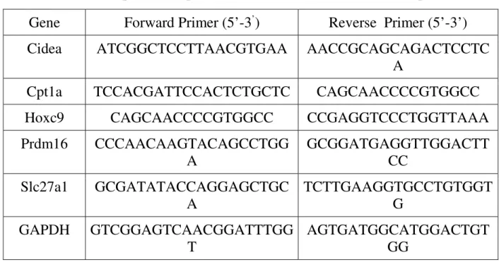
Primers that have been used in experiments are shown in table 2.
Table 2. Nucleotide sequences of primers used for real-time RT-PCR amplification
Gene Forward Primer (5’-3’) Reverse Primer (5’-3’)
Cidea ATCGGCTCCTTAACGTGAA AACCGCAGCAGACTCCTC
A
Cpt1a TCCACGATTCCACTCTGCTC CAGCAACCCCGTGGCC
Hoxc9 CAGCAACCCCGTGGCC CCGAGGTCCCTGGTTAAA
Prdm16 CCCAACAAGTACAGCCTGG A GCGGATGAGGTTGGACTT CC Slc27a1 GCGATATACCAGGAGCTGC A TCTTGAAGGTGCCTGTGGT G GAPDH GTCGGAGTCAACGGATTTGG T AGTGATGGCATGGACTGT GG
Cidea, cell death-inducing DNA fragmentation factor-α-like effector A; Cpt1a, carnitine palmitoyl transferase 1a; Hoxc9, homeo box C9; Prdm16, PR domain containing protein-16; Slc27a1, solute carrier family 27.
3.2. Morphology
3.2.1. Subjects (human adipose tissue)
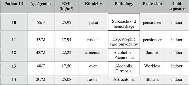
Fat biopsies were obtained from male (n=9) and female (n=5) patients aged 43,38±9,80 (mean±SD) with body mass index (BMI) from 15 to 48. Adipose tissue samples from periaortic and perirenal depots were taken posthumously in the Pathology Anatomical Department of the National Center of Medicine (Yakutsk, Russian Federation). Some patient included in this research had work or life style outdoor.
The clinical details of the subjects are summarized in Table 3.
Patient ID Age/gender BMI
(kg/m2)
Ethnicity Pathology Profession Cold
exposure
1 -/M - yakut Trauma Homeless outdoor
2 56/M -russian Myocardial infarction, atherosclerosis Homeless outdoor 3 46/M -uzbek Subarachnoid
hemorrhage. Trader indoor
4 64/M - russian Myocardial infarction pensioneer indoor 5 -/M - russian Alcoholism. Alcoholic cardiomyopathy Homeless outdoor 6 30/M 28,24 yakut Pancreatic
necrosis Farmer outdoor
7 46/F 15,44 russian
Total pancreosclerosis
.
Workless indoor
8 61/F 29,32 yakut DIC-Syndrome pensioneer indoor
9 65/F 48,94 russian Diabetes, II
type. Phlegmon of hip. Sepsis.
Patient ID Age/gender BMI (kg/m2)
Ethnicity Pathology Profession Cold
exposure
10 55/F 25,92 yakut Subarachnoid
hemorrhage pensioneer indoor
11 53/M 27,94 russian Hypertrophic cardiomyopathy pensioneer indoor 12 43/M 22,22 armenian Alcoholism. Pneumonia Janitor indoor 13 48/F 17,50 even Alcoholis. Cirrhosis. Workless indoor
14 20/M 25,08 russian Astrocitoma Student indoor
Table 3. Clinical data of subjects included in the study
3.2.2. Light microscopy
Fat samples were fixed by immersion in 4% paraformaldehyde in 0.1 M phosphate buffer, pH 7.4, overnight at 4 0C. After a thorough rinse in phosphate buffer, they were dehydrated in a graded series of ethanol, cleared in xylene and paraffin-embedded. Serial paraffin sections 3-4 µm in thickness were obtained from each samples. Some were stained with hematoxylin and eosin to assess morphology. All observations were performed with a Nikon Eclipse 800 light microscope (Nikon, Tokyo, Japan) equipped with a CCD camera.
3.2.3. Immunohistochemistry
UCP1 or tyrosine hydroxylase (TH) immunoreactivity was examined as follows. Sections were incubated with rabbit anti-UCP1 or sheep anti-TH, according to the avidin-biotin complex (ABC) method as follows: 1) endogenous peroxidase blocking with 3% hydrogen peroxide in distilled water; 2) normal serum (2% in PBS) for 20
minutes to reduce nonspecific background; 3) incubation with primary antibodies against UCP1 or TH (Tab. 2) overnight at 4°C; 4) after a thorough rinse in PBS, incubation in 1:200 v/v biotinylated secondary antibody solution (Vector Labs, Burlingame, CA, USA); 5) ABC kit (Vector Labs); 6) enzymatic reaction to reveal peroxidase with Sigma Fast 3,3’-diaminobenzidine (Sigma-Aldrich, St. Louis, MO, USA) as substrate. Finally sections were counterstained with hematoxylin and mounted in Eukitt (Fluka, Deisenhofen, Germany). To assess the specificity of the antibodies, negative controls were obtained in each instance by omitting the primary antibody.
The primary antibodies used in the study are shown in Table 2:
Primary antibody Working dilution Host/Isotype Washing solution Company anti-TH polyclonal
anti-sheep
1:400 Rabbit IgG PBS AB1542, Merck Millipore
anti-UCP1 polyclonal
anti-rabbit
1:500 Sheep IgG PBS ab10983, abcam
Tab.2. Primary antibodies used for immunohistochemistry (IHC)
3.2.4. Morphometry
TH-positive parenchymal nerve fibers (i.e fibers closely associated to adipocytes in the depots) were counted; perivascular fibers (those in contact with arterioles and venules) were not considered in the quantitative analysis. Ten random pictures for each depots were studied using a Nikon Eclipse E800 light microscopy with a 40X objective. The density of TH-immunoreactive fibers was calculated as the number of fibers per 100 adipocytes.
Sections stained for UCP1 were used for adipocytes counting: ten random pictures for each depots were studied at 20X. Adipocyte numbers were measured using Lucia Image software (version 4.82, Nikon Instruments, Florence, Italy). Results are given as percentage UCP1-immunoreactive multilocular adipocytes of the total number of counted adipocytes.
3.2.5. Statistical analysis
Results are given as mean ± SD. Significance was analysed using an unpaired t-test (In STAT, GraphPad, San Diego, CA, USA). Differences between groups were considered significant when P ≤ 0.05. Linear correlations were calculated by a nonparametric test (Spearman) using GRAPHPAD PRISM, version 3.00 for Windows.
3.2.6. Ethics statement
All experimental procedure followed in this study were reviewed and approved by the Ethical Committee of the Yakut Scientific Center of complex medical problems (Yakutsk, Russian Federation). All subjects provided written informed consent (Permission n 209).
4. Results
4.1. Gene expression of peripheral blood mononuclear cells is affected by cold.
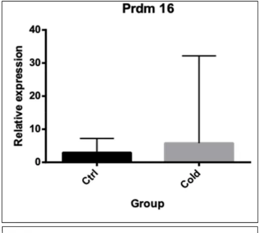
We studied the gene expression of typical BAT genes, such as PRDM16, Slc27a, Cpt1a, Hoxc9 and Cidea, of peripheral blood mononuclear cells (PBMC) extracted from outdoor workers in the north area of Siberia. This study is based on the observation that BAT genes can respond to cold exposure also in mononuclear blood cells in rodents (Reines B. et al., 2015).
PR domain containing 16, also known as PRDM16, is a protein that in humans is encoded by the PRDM 16 gene. PRDM 16 acts as a transcription coregulator that controls the development of brown adipocytes in brown adipose tissue. The expression of PRDM 16 gene was observed in mononuclear cells of the peripheral blood in the two study groups. Expression of the gene PRDM 16 in the outdoor workers group was increased compared with control group. (Fig.3).
FIGURE 3. GENE EXPRESSION OF PRDM 16 MEASURED IN PBMC. CTRL - CONTROL GROUP, COLD - COLD EXPOSED GROUP
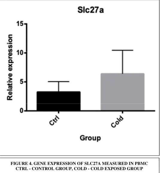
Еxpression of integral transmembral protein Slc27a, the main function of which enhances the process of absorption of fatty acids in cell, was increased in group of outdoor workers ( Fig. 4), but the increase was not significant.
FIGURE 4. GENE EXPRESSION OF SLC27A MEASURED IN PBMC CTRL - CONTROL GROUP, COLD - COLD EXPOSED GROUP
The Cpt1a gene promotes the synthesis of the enzyme carnitine palmitoyltransferase 1a, which is found in the liver. This enzyme is involved in the oxidation of fatty acids, in this multistep process is the conversion of fatty acids into ATP. The expression of Cpt1a gene was increased in cold exposed group in comparison with the control group (Fig.5).
FIGURE 5. GENE EXPRESSION OF CPT1A MEASURED IN PBMC CTRL - CONTROL GROUP, COLD - COLD EXPOSED GROUP
The same happened for Hoxc9, beige adipocyte marker, whose expression was also elevated in workers with long exposure to cold, but at very low levels (Fig.6).
FIGURE 6. GENE EXPRESSION OF HOXC9 MEASURED IN PBMC. CTRL - CONTROL GROUP, COLD - COLD EXPOSED GROUP
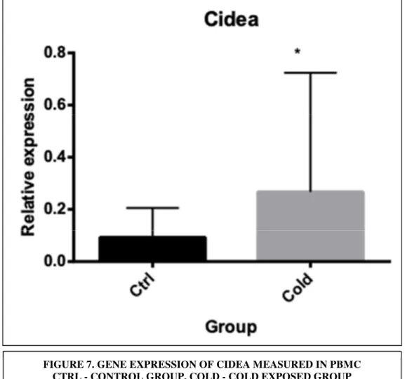
Expression of Cidea, a well-known brown adipocyte marker, was significantly increase in response to cold (Fig.7).
Our results showed that peripheral blood cells could reflect gene expression of markers of BAT in response to cold.
FIGURE 7. GENE EXPRESSION OF CIDEA MEASURED IN PBMC CTRL - CONTROL GROUP, COLD - COLD EXPOSED GROUP
4.2. Direct evidence of brown adipose tissue in adult humans from Siberia
We examined the tissues for the presence of UCP1 immunoreactive cells and analyzed the tissues for the BAT characteristics of dense innervation. We observed 2 subjects groups: one group of patients worked outdoors in cold condition, while the second group had an inside job. Adipose tissue samples were taken posthumously: for each subject, we analyzed periaortic and perirenal depots. Most of the periaortic and perirenal depots observed, clearly showed UCP1-positive islands.
Comparing the two groups, we can see that outdoor workers had much more brown adipose tissue and more intensely stained for the functional protein UCP1 than indoor workers (Fig.8).
FIGURE 8. PRESENCE OF BROWN ADIPOSE ISLANDS IN HUMAN ADIPOSE TISSUE. IMMUNOHYSTOCHEMICAL EXAMINATION OF UCP1 POSITIVITY: A — PERIRENAL ADIPOSE TISSUE OF AN INDOOR PATIENT, B — PERIRENAL ADIPOSE TISSUE OF AN OUTDOOR PATIENT.
We compared the percentage of multilocular adipocytes in the adipose tissue of different localization, periaortic and perirenal, in indoor and outdoor patients. In the periaortic adipose tissue we do not observe differences between the two groups of patients, while as regards perirenal depots, the percentage of multilocular adipocytes in outdoor patients is higher than in indoor patients, even if the increase is not statistically significant (Fig. 9).
One characteristic of classic BAT is its dense innervation by the sympathetic nervous system; this innervation enables it to respond rapidly to external stimuli (cold, food) with increased activity (Cannon B. and Nedergaard J., 2004). To examine whether this characteristic was also present in the brown fat areas in the periaortic and perirenal adipose tissues, we examined tissue sections for the presence of the adrenergic nerve marker tyrosine hydroxylase (TH) (Cinti et al., 2001).
As seen in Fig. 10, TH density seemed to increase in adipose tissue of outdoor patients compared with TH density in indoor patients.
PERIRENAL ADIPOSE TISSUE PERIAORTIC ADIPOSE TISSUE
FIGURE 9. PERCENTAGE OF MULTILOCULAR ADIPOCYTES IN PERIAORTIC AND PERIRENAL ADIPOSE TISSUE. DIFFERENCE BETWEEN OUTDOOR AND INDOOR PATIENTS.
We also analyzed the relationship between of TH-positive fibers and the percentage of multilocular adipocytes in different fat depots in outdoor and indoor patients.
The results showed that there is a positive correlation between the presence of multilocular adipocytes and the density of positive TH fibers. In periaortic adipose tissue this correlation is statistically significant (Fig. 11).
FIGURE 10. DENSE INNERVATION IN BROWN-FAT ISLANDS.
IMMUNOHISTOCHEMICAL EXAMINATION OF TH POSITIVITY: A — PERIAORTIC OF AN INDOOR PATIENT; B — PERIAORTIC OF AN OUTDOOR PATIENT.
FIGURE 11. THE RELATIONSHIP BETWEEN THE NUMBER OF TH-POSITIVE FIBERS AND PERCENTAGE OF MULTILOCULAR ADIPOCYTES.
Perirenal adipose tissue
Periaortic adipose tissue
5. Discussion
After the re-discovery of brown adipose tissue (BAT) in humans, there is increasing interest in the study of induction of this thermogenic tissue as a basis to combat obesity and related complications (Cinti S., 2018). Cold exposure is one of the strongest stimuli able to activate BAT and to induce the appearance of brown-like adipocytes in white fat depots (browning process). We analyzed the potential of peripheral blood mononuclear cells (PBMCs) to reflect BAT activity based on previous studies that gave promising results in mice.
BAT studies in humans require invasive techniques such as biopsies of adipose tissue or the use of techniques such as positron emission tomography, which implies the use of radioactive isotopes. Thus, it would be of interest to have a readily available source of biomarkers in mononuclear cells of peripheral blood as it was shown in experimental procedures with mice (Garcia-Ruiz et al., 2009; Díaz-Rúa et al., 2015).
Here we aim to establish the usefulness of mononuclear cells of peripheral blood as a tool to perform studies related to BAT activation and WAT browning by analyzing the expression of key brown adipocyte markers in response to the main thermogenic stimulus - cold acclimation in a population of Siberian individuals chronically exposed to low temperature for climatic reasons.
Our data have shown that gene expression in a subset of blood cells, PBMCs reflect certain features of brown adipose tissue response to cold exposure.
It is well known that adrenergic stimulation, such as cold exposure, induces an increase of fatty acid catabolism, especially in WAT, while it decreases fatty acid synthesis (Caimari A. et al., 2012; Jankovic A, et al., 2013). According to this, we observed increased expression of a key gene involved in fatty acid β-oxidation, Cpt1a. Caimari and colleagues described that PBMCs can reflect lipid metabolism gene expression that occur in adipose tissue in response to different stimulus, such as feeding/fasting conditions (Caimari A. et al., 2010). Here we show how PBMCs also reflected increased Cpt1a expression in response to cold exposure, which occurs in adipose tissue.
Another key regulator of BAT is Prdm16, which codes for a key transcription factor involved in UCP1 expression (Reviewed in: Ishibashi J and Seale Temperature, 2015). We observed increased Prdm16 expression in PBMCs of Siberian outdoor workers. We also analyzed mRNA expression of other brown adipocyte markers, such as Cidea, which code for a multifunctional protein that is highly expressed in BAT mitochondria (Barneda et al., 2013; Wu et al., 2012), and the expression of Slc27a1, which codes for fatty acid transport protein 1, and is considered a brown marker (Wu et al., 2012) and, as expected, its expression increased in response to cold exposure in blood PBMCs (Hondares et al., 2011).
Hoxc9 has been previously described in fat of outdoor workers after chronic cold exposure (Waldén et al., 2012).
PBMCs are able to express 80% of the genome, including tissue-specific transcripts (Liew et al., 2006). Although, these cells did not express the key BAT thermogenic gene Ucp1, they were able to express other brown markers such as Cidea, Hoxc9, Prdm16, Cpt1a and Slc27a1 in peripheral blood of Siberian people. To our knowledge, there are no previous reports on the study of browning effects of cold exposure on PBMC gene expression. The data presented here demonstrate that these cells can reflect some of the cold response changes in gene expression usually found in fat. The most relevant data was the significant increase in Cidea mRNA expression observed in cold-exposed Siberian group.
The possibility of using an easily obtainable biological material, such as PBMCs, to perform BAT studies opens new and interesting possibilities for analyzing the relevance of this tissue and of WAT browning on energy homeostasis and body weight control in humans by using non-invasive approaches.
Classically, adult humans have been considered not to possess active brown adipose tissue (BAT). This has meant, for instance, that variations in the amount and activity of BAT between individuals have not been considered to contribute to the development of obesity, and that stimulation of BAT amount and activity has generally been disregarded as an avenue for obesity therapy.
However, in recent years unexpected evidence for the presence of active BAT in adult humans has serendipitously appeared (Nedergaard J., Bengtsson T., and Cannon B., 2007). This has been the outcome of positron emission tomography (PET) examination of 18-fluorodeoxyglucose (FDG) uptake in cancer patients. In some patients a FDG uptake pattern may be seen that has a distribution such that it is reasonable to ascribe it to uptake in BAT in these patients. As glucose uptake is associated with metabolically active tissue, the implication is that this uptake pattern represents metabolically active BAT in adult humans. There is principal ample evidence to support this contention (Nedergaard J., Bengtsson T., and Cannon B., 2007). However, the critical issue allowing us to conclude that these uptake patterns really represent BAT is the presence of the brown-fat-specific protein uncoupling protein-1 (UCP1) in the adipose depots corresponding to the glucose uptake areas. UCP1 is the protein that uncouples mitochondrial substrate combustion from ATP synthesis and thus transfers food energy directly into heat (Nedergaard J. et al., 2001; Cannon B. and Nedergaard J. et al., 2004).
The establishment of its presence in these depots is essential for concluding that these depots really would be able to fulfill the functional roles of BAT.
In this work, we observed adipose tissue of patients from Siberia on the presence of the UCP1. Our results showed that most of the periaortic and perirenal depots observed, clearly showed UCP1-positive islands. There have been observations demonstrating the presence of UCP1 in adult humans (Lean M. E. J. and James W. P. T. 1986; Lean M. E. J., 1989). Comparing the two groups, we can see that outdoor workers had much more brown adipose tissue and more intensely stained for the functional protein UCP1 than indoor workers.
Under cold stimulation, clusters of brown adipocytes emerge in the area in which tyrosine hydroxylase–expressing (noradrenergic) nerve fibers are densely innervated (Cinti S., 1999; Murano I. et al., 2009), as norepinephrine released from the sympathetic nerves is a powerful stimulator of WAT browning. Intriguingly, adult human brown adipocytes are also sporadically found together with white adipocytes,
and the density is well correlated with the number of tyrosine hydroxylase–positive noradrenergic nerves (Lidell ME, et al., 2013; Zingaretti MC., et al., 2009).
We analyzed the relationship between of TH-positive fibers and the percentage of multilocular adipocytes in different fat depots in outdoor and indoor patients. The results showed that there is a positive correlation between the presence of multilocular adipocytes and the density of positive TH fibers. In periaortic adipose tissue, this correlation is statistically significant.
6. References
Adler A. I., Boyko E. J., Schraer C. D. Daily consumption of seal oil or salmon associated with lower risk of noninsulin dependent diabetes mellitus in Eskimo and Athabaskan Indians of Alaska // Arctic Medical. res. 1994. Vol. 53 (2). P. 271 – 275. Aghajanyan N. Ecological portrait of a person in the North / N. A. Aghajanyan, N. V. Ermakova. - M: KRUK, 1997. - 207 p. Alekseeva T.I. Human adaptation in various ecological niches of the earth: Biological aspects. Publishing house MNEPU, 1998. - 279с.
Atit R, et al. Beta-catenin activation is necessary and sufficient to specify the dorsal
dermal fate in the mouse. Dev Biol. 2006; 296(1):164–176. doi:
10.1016/j.ydbio.2006.04.449.
Barbatelli G, et al. The emergence of cold-induced brown adipocytes in mouse white fat depots is determined predominantly by white to brown adipocyte transdifferentiation. Am J Physiol Endocrinol Metab. 2010; 298(6):E1244–E1253. doi: 10.1152/ajpendo.00600.2009.
Barneda D , Frontini A , Cinti S , Christian M .Dynamic changes in lipid droplet-associated proteins in the “browning” of white adipose tissues.Biochim Biophys Acta 1831: 924-933, 2013.
Bobrov N.I. Physiological and hygienic aspects of human acclimatization in the North / N.I. Bobrov, O.I. Lomov, V.P.Tikhomirov. - L .: Medicine, 1979. - 184 p.
Boyko E.R. Patterns of metabolism in humans in the Far North / E.R. Boyko, F.A. Bichkayeva // Physiological patterns of hormonal, metabolic, immunological changes in the human body in the European North. - Syktyvkar, 1997. - p. 34. - 37.
Boyko E.R. Physiological and biochemical basis of human activity in the North / E.R. Smartly - Ekaterinburg: Ural Branch of the Russian Academy of Sciences, 2005. –189 p.
Boyko E.R. Some regularities of metabolic changes in humans in the Far North // Human Physiology. - 1996. - Vol. 22., №4. - pp. 122 - 128.
Boyko E.R., Tkachev A.V. Characteristics of lipid metabolism in permanent residents of the North // Human Physiology. - 1994. - V. 2., №2. - p. 136 –140.
Caimari A , Oliver P , Keijer J , Palou A. Peripheral blood mononuclear cells as a model to study the response of energy homeostasis-related genes to acute changes in feeding conditions.OMICS 14: 129-141, 2010.
Caimari A , Oliver P , Palou A .Adipose triglyceride lipase expression and fasting regulation are differently affected by cold exposure in adipose tissues of lean and obese Zucker rats. J. Nutr Biochem 23: 1041-1050, 2012.
Caimari A , Oliver P , Rodenburg W , Keijer J , Palou A. Feeding conditions control the expression of genes involved in sterol metabolism in peripheral blood mononuclear cells of normal weight and diet-induced (cafeteria) obese rats.J Nutr Biochem 21: 1127-1133, 2010.
Caimari A , Oliver P , Rodenburg W , Keijer J , Palou A .Slc27a2 expression in peripheral blood mononuclear cells as a molecular marker for overweight development.Int J Obes (Lond) 34: 831-839, 2010.
Cannon, B., and Nedergaard, J. Brown adipose tissue: function and physiological significance. Physiol. Rev. 84, 277–359, 2004.
Cinti S, Zingaretti MC, Cancello R, Ceresi E, Ferrara P. Morphologic techniques for the study of brown adipose tissue and white adipose tissue. Methods Mol Biol. 2001;155:21-51.
Cinti S. Adipose Organ Development and Remodeling. Compr Physiol. 2018, 8:1357-1431.
Cinti S. The Adipose Organ. Milano, Italy: Editrice Kurtis; 1999.
Deryapa N.R. Human Adaptation in the Polar Regions of the Earth. L. Medicine, 1977. –296с.
Forner F, Kumar C, Luber CA, Fromme T, Klingenspor M, Mann M. Proteome differences between brown and white fat mitochondria reveal specialized metabolic functions. Cell Metab. 2009;10(4):324–335. doi: 10.1016/j.cmet.2009.08.014.
Frontini A, et al. White-to-brown transdifferentiation of omental adipocytes in patients affected by pheochromocytoma. Biochim Biophys Acta. 2013;1831(5):950–959. García-Ruiz E , Keijer J., Caimari A, van Scholhorst E M., Palou A, Oliver P. Peripheral blood mononuclear cells as a source to detect markers of homeostatic alterations caused by the intake of diets with an unbalanced macronutrient composition. J. Nutr. Biochem. 2015 Apr;26(4):398-407.
Harms M, Seale P. Brown and beige fat: development, function and therapeutic potential. Nat Med. 2013;19(10):1252–1263. doi: 10.1038/nm.3361.
Himms-Hagen J, Melnyk A, Zingaretti MC, Ceresi E, Barbatelli G. Multilocular fat cells in WAT of CL-316243-treated rats derive directly from white adipocytes. Am J Physiol Cell Physiol. 2000;279(3):C670–C681.
Hondares E , Iglesias R , Giralt A , Gonzalez FJ , Giralt M , Mampel T , Villarroya F. Thermogenic activation induces FGF21 expression and release in brown adipose tissue.J Biol Chem 286: 12983-12990, 2011.
Isaakyan L.A. Metabolic structure of temperature adaptations / L.A. Isahakian. - L., 1972.
Ishibashi J., Seale Medicine P .Beige can be slimming.Science 328: 1113-1114, 2010. Ishibashi J and Seale P. Functions of Prdm16 in thermogenic fat cells. Temperature (Austin); 2 (1); 65 - 72, 2015.
Ivanov KI Features of the actual nutrition of the population of the Republic of Sakha (Yakutia).Far Eastern Medical Journal. - 2005. - № 2. - p. 72 - 74.
Izyumenko S.A. The climate of Yakutsk / S.A. Izyumenko. - L.: Gidrometeoizdat, 1982. - 246p.
Jul E., Mulvad G., Pedersen N. S. The Relationship between a low rate of ischaemie heart disease and the traditional Greenlandic diet with high amounts of monounsaturated and N-3 poliunsaturated fatti acids // Arctic. Med. Res. – Vol. 53 (2). – P. 282 – 284.
Kajimura S, Saito M. A new era in brown adipose tissue biology: molecular control of brown fat development and energy homeostasis. Annu Rev Physiol. 2014;76:225–249. doi: 10.1146/annurev-physiol-021113-170252.
Kaznacheev V.P. Some features of human adaptation in high latitudes. Human Physiology. - 1979. - Vol.5, №2. - p. 286 –293.
Kennedy AR, Pissios P, Otu H, Roberson R, Xue B. A high-fat, ketogenic diet induces a unique metabolic state in mice .Am J Physiol Endocrinol Metab 292: E1724 –E1739, 2007.
Khaltaev N.G., klochkova E.V., Tikhonov A.V. Nutrition and risk factors for coronary heart disease in men of the Chukotka Autonomous Region. Cardiology. - 1984. - № 4. - p. 62 - 64.
Khamnagadaev II, Polikarpov L.S., Gankin M.I. Nutrition of the indigenous rural population of Yakutia. Therapeutic Archive. - 2003. - № 1. - p. 34 - 37.
Khasnulin V.I., Khasnulina A.V., Chechetkina I.I. Northern stress, the formation of hypertension in the North, approaches to prevention and treatment. Human Ecology. - 2009. - №6. - P.26 - 30.
Kochan T.I. Comprehensive assessment of the influence of the conditions of the North on metabolism, the physiological and psycho-emotional state of a person. Human Physiology. - 2008. - Vol. 34, № 3. - P. 106 - 113.
Kondror I.S. Essays on human physiology and hygiene in the Far North. Medicine, 1968. – 280p.